Calcium in PDB 1bj3: Crystal Structure of Coagulation Factor IX-Binding Protein (IX-Bp) From Venom of Habu Snake with A Heterodimer of C-Type Lectin Domains (original) (raw)
Protein crystallography data
The structure of Crystal Structure of Coagulation Factor IX-Binding Protein (IX-Bp) From Venom of Habu Snake with A Heterodimer of C-Type Lectin Domains, PDB code: 1bj3was solved by H.Mizuno, Z.Fujimoto, M.Koizumi, H.Kano, H.Atoda, T.Morita, with X-Ray Crystallography technique. A brief refinement statistics is given in the table below:
| Resolution Low / High (Å) | 6.00 / 2.60 |
|---|---|
| Space group | P 1 21 1 |
| Cell size a, b, c (Å), α, β, γ (°) | 58.569, 56.794, 39.616, 90.00, 95.80, 90.00 |
| R / Rfree (%) | 18.2 / 27.1 |
Calcium Binding Sites:
The binding sites of Calcium atom in the Crystal Structure of Coagulation Factor IX-Binding Protein (IX-Bp) From Venom of Habu Snake with A Heterodimer of C-Type Lectin Domains (pdb code 1bj3). This binding sites where shown within 5.0 Angstroms radius around Calcium atom.
In total 2 binding sites of Calcium where determined in the Crystal Structure of Coagulation Factor IX-Binding Protein (IX-Bp) From Venom of Habu Snake with A Heterodimer of C-Type Lectin Domains, PDB code: 1bj3:
Jump to Calcium binding site number: 1; 2;
Calcium binding site 1 out of 2 in 1bj3
Go back to  Calcium Binding Sites List in 1bj3
Calcium binding site 1 out of 2 in the Crystal Structure of Coagulation Factor IX-Binding Protein (IX-Bp) From Venom of Habu Snake with A Heterodimer of C-Type Lectin Domains
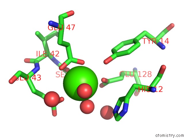
Mono view
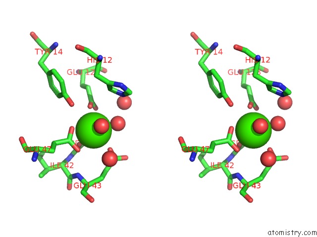
Stereo pair view
| | A full contact list of Calcium with other atoms in the Ca binding site number 1 of Crystal Structure of Coagulation Factor IX-Binding Protein (IX-Bp) From Venom of Habu Snake with A Heterodimer of C-Type Lectin Domains within 5.0Å range: probe atom residue distance (Å) B Occ A:Ca130 b:15.6 occ:1.00 O A:HOH170 2.2 22.1 1.0 OE2 A:GLU47 2.3 10.6 1.0 OE2 A:GLU128 2.4 14.3 1.0 O A:SER41 2.5 11.9 1.0 OE1 A:GLU43 2.5 21.9 1.0 O A:HOH171 2.5 23.3 1.0 OG A:SER41 2.6 10.9 1.0 OE1 A:GLU47 2.8 13.0 1.0 CD A:GLU47 2.9 13.3 1.0 CD A:GLU128 3.2 11.1 1.0 OE1 A:GLU128 3.3 17.1 1.0 CD A:GLU43 3.5 19.7 1.0 C A:SER41 3.6 12.0 1.0 OH A:TYR14 3.8 11.9 1.0 CB A:SER41 3.9 8.4 1.0 CA A:SER41 4.2 11.7 1.0 O A:HOH142 4.2 27.4 1.0 N A:SER41 4.3 12.4 1.0 CG A:GLU43 4.3 18.5 1.0 CG A:GLU47 4.3 10.7 1.0 OE2 A:GLU43 4.3 15.3 1.0 CD2 A:HIS12 4.5 12.7 1.0 O A:HOH152 4.5 23.9 1.0 NE2 A:HIS12 4.6 11.7 1.0 CG A:GLU128 4.6 11.0 1.0 N A:GLU43 4.7 16.7 1.0 N A:ILE42 4.7 12.9 1.0 CZ A:TYR14 4.7 11.9 1.0 CB A:GLU43 4.8 14.4 1.0 CE2 A:TYR14 4.8 10.1 1.0 CA A:ILE42 5.0 12.8 1.0 | | --------------------------------------------------------------------------------------------------------------------------------------------------------------------------------------------------------------------------------------------------------------------------------------------------------------------------------------------------------------------------------------------------------------------------------------------------------------------------------------------------------------------------------------------------------------------------------------------------------------------------------------------------------------------------------------------------------------------------------------------------------------------------------------------------------------------------------------------------------------------------------------------------------------------------------------------------------------------------------------------------------------------------------------------------------------------------------------------------------------------------------------------------------------------------------------------------------------------------------------------------------------------------------------------------------------------------------------------------------------------------------------------------------- |
Calcium binding site 2 out of 2 in 1bj3
Go back to Calcium Binding Sites List in 1bj3
Calcium binding site 2 out of 2 in the Crystal Structure of Coagulation Factor IX-Binding Protein (IX-Bp) From Venom of Habu Snake with A Heterodimer of C-Type Lectin Domains
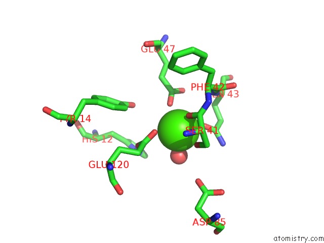
Mono view
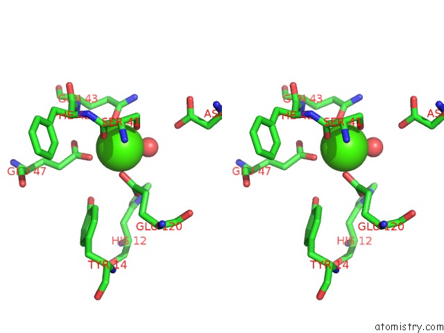
Stereo pair view
| | A full contact list of Calcium with other atoms in the Ca binding site number 2 of Crystal Structure of Coagulation Factor IX-Binding Protein (IX-Bp) From Venom of Habu Snake with A Heterodimer of C-Type Lectin Domains within 5.0Å range: probe atom residue distance (Å) B Occ B:Ca124 b:21.9 occ:1.00 O B:HOH162 2.2 26.6 1.0 O B:SER41 2.3 15.2 1.0 O B:HOH157 2.3 26.3 1.0 OE1 B:GLN43 2.3 15.5 1.0 OE2 B:GLU47 2.4 14.8 1.0 OG B:SER41 2.4 11.6 1.0 OE2 B:GLU120 2.6 15.9 1.0 C B:SER41 3.4 14.8 1.0 CD B:GLU120 3.4 14.0 1.0 CD B:GLN43 3.4 13.1 1.0 CD B:GLU47 3.4 14.1 1.0 OE1 B:GLU120 3.7 15.1 1.0 CB B:SER41 3.7 14.1 1.0 OE1 B:GLU47 3.8 12.2 1.0 CA B:SER41 4.0 14.5 1.0 NE2 B:GLN43 4.1 10.3 1.0 N B:SER41 4.3 13.6 1.0 N B:GLN43 4.4 16.9 1.0 N B:PHE42 4.5 14.4 1.0 CG B:GLN43 4.5 14.5 1.0 OH B:TYR14 4.6 10.4 1.0 CB B:GLN43 4.6 12.8 1.0 OD2 A:ASP85 4.6 16.2 1.0 CG B:GLU120 4.7 14.3 1.0 ND1 B:HIS12 4.7 21.8 1.0 CG B:GLU47 4.8 15.2 1.0 CA B:PHE42 4.8 14.1 1.0 CE1 B:HIS12 4.9 23.4 1.0 | | ------------------------------------------------------------------------------------------------------------------------------------------------------------------------------------------------------------------------------------------------------------------------------------------------------------------------------------------------------------------------------------------------------------------------------------------------------------------------------------------------------------------------------------------------------------------------------------------------------------------------------------------------------------------------------------------------------------------------------------------------------------------------------------------------------------------------------------------------------------------------------------------------------------------------------------------------------------------------------------------------------------------------------------------------------------------------------------------------------------------------------------------------------------------------------------------------------------------------------------------------------------------------------------------------------- |
Reference:
H.Mizuno, Z.Fujimoto, M.Koizumi, H.Kano, H.Atoda, T.Morita. Crystal Structure of Coagulation Factor IX-Binding Protein From Habu Snake Venom at 2.6 A: Implication of Central Loop Swapping Based on Deletion in the Linker Region. J.Mol.Biol. V. 289 103 1999.
ISSN: ISSN 0022-2836
PubMed: 10339409
DOI: 10.1006/JMBI.1999.2756
Page generated: Thu Jul 11 06:26:21 2024