dermoscopy (original) (raw)
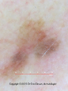
Eccentric pigmentation
36-year-old woman with an atypical pigmented lesion on her left leg
Demoscopy revealed:
- an eccentric hyperpigmentation
- an atypical reticular network
- pseudopods
Pathology revealed a superficial spreading melanoma with Breslow index at 0.25 mm
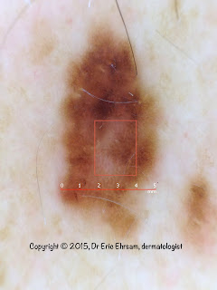
A whitish lesion
A 48-year-old woman consulted for a whitish scar-like lesion on her back.
Clinical picture: whitish scar-like lesion with a pigmented border. Patient has a background of former heavy sun exposure.
At dermoscopy:
- no signs in favor of a melanocytic lesion
- a structureless scar-like white pattern
- pinkish areas without any vascular structures
- a peripheral pigmentation (red square) in favor of leaf-like areas
Pathology revealed a superficial basal cell carcinoma
A temporal lesion
A 60-year-old woman consulted for this temporal pigmented lesion
Dermoscopy revealed:
- blue-gray dots / globules
- leaf-like areas
Pathology was in favor of a basal cell carcinoma
Brachial melanoma
A 54-year-old man had a rapidly growing lesion on his right back for 6 months.
Dermoscopy revealed:
- a vascular pattern with some polymorphous and atypical vessels
- peripheral pigmentation
- irregular globules
- eccentric blotches
Pathology revealed a superficial spreading melanoma with Breslow index at 1.53 mm
A pigmented lesion on a shoulder
A 48-year-old man consulted for this pigmented lesion on his right shoulder.
Dermoscopy revealed:
- an atypical reticular pattern
- structureless areas with regression
- a retiform depigmentation (negative pigment network)
Pathology was in favor of a melanoma in situ.
Small, beware !
A 42-year-old man consulted for this small (4mm) pigmented lesion on his left calf.
Dermoscoy revealed:
- asymmetry of colors and structures
- some irregular blotches
- an atypical pigment network
- a radial streaming
Lesion was excised and pathology was in favor or a melanoma in situ.
A pigmented lesion
A 32-year-old man had his moles checked up.
Dermoscopy revealed:
- whitish globules / white cobblestone pattern
- Light-brown peripheral network
In favor of an accessory nipple
Central white cobblestone pattern
Reference:
Dermoscopy of accessory nipples in authors’ own study
G. Kamińska-Winciorek, J. Szymszal, W. Silny
Postep Derm Alergol 2014; XXXI, 3: 127–133
A pigmented labial lesion
A 24-year-old woman consulted for a pigmented labial lesion.
Dermoscopy revealed multiple curves of semicircle, U or V mimicking the scales of a fish.
This fish scale-like pattern was in favor of a mucosal melanotic macule.
Fish scale-like pattern
Fish scale-like pattern is a new pattern described in mucosal melanotic macules.
Reference:
Dermoscopy of pigmented lesion on mucocutaneous junction and mucous membrane.
Lin J, Koga H, Takata M, Saida T.
BJD 161 (6); 1255-61
A yellowish lesion
A 70-year-old woman consulted for this small lesion on her right upper lid.
Dermoscopy revealed:
- a yellow clod pattern
- vessels arranged in a radial arrangement
typical of a sebaceous hyperplasia
Red lesions
A 62-year-old woman consulted for an acquired red tumor on her left ankle.
Dermoscopy revealed:
- regular red dots
- in a stringlike distribution
in favor of a clear cell acanthoma
Black lesion.
A 25-year-old woman consulted for a dark pigmented lesion on her abdomen
Dermoscopy revealed:
- asymmetry of form
- a central blotch
- regular pseudopods
Pathology was in favor of melanocytic Spitz nevus
Cheek lesion.
A 55-year-old man consulted for a pigmented lesion on his left cheek
Dermoscopy revealed:
- an irregular broadened network
- rhomboidal structures
Pathology was in favor of a lentigo maligna melanoma in situ.
A small pigmented lesion on a leg
A 29-year-old woman consulted for a small pigmented lesion on her right leg.
Dermoscopy revealed
- spoke wheel areas
Pathology revealed a superficial basal cell carcinoma
Regression.
A 21-year-man consulted for this lesion on his arm
Dermoscopy revealed:
- a whitish - pinkish structureless area
- with gray dots
in favor of a regressive lesion.
Pathology was in favor of a fully regressive melanocytic lesion. A fully regressive melanoma cannot be ruled out.
Pigmented lesion on neck.
A 57 year-old-woman consulted for a pigmented lesion on her neck.
Dermoscopy revealed:
- no melanocytic structures
- a structureless pattern
- irregular blue gray dots/globules
Pathology was in favor of a Bowen's disease.
Another back lesion.
A 34-year-old man consulted for a changing pigmented lesion on his back.
Dermoscopy revealed:
- asymmetry
- an atypical reticular pattern
- a regression area
Pathology revealed a melanoma in situ.
Back lesion
A 65-year-old man with many pigmented lesions on his back. One of this lesion was atypical.
Dermoscopy revealed:
- multiple colors
- an atypical reticular network
- regression
- a reticular depigmentation (rectangle)
Pathology revealed a melanoma in situ.
Nodular lesion
A 57-year-old woman consulted for this nodular lesion on her right chest.
Dermoscopy revealed:
- a non pigmented lesion
- remnant of pigmentation (red circles)
- a pink veil
- atypical and polymorphous vessels
Pathology revealed a nodular melanoma (Breslow index 1.6mm)
Thoracic pigmented lesion
Man 52 years old; Acquired pigmented lesion on chest.
Dermoscopy revealed:
- multiple colors
- asymmetry of form and structures
- an atypical network
- a blue white veil
- irregular dots and globules
Pathology revealed a melanoma in situ.
Starburst pattern
A 43-year-old man consulted for this acquired pigmented lesion on his left leg.
Dermoscopy revealed:
- a central blotch
- a regular starburst pattern
Two diagnoses were evoked: Reed nevus or melanoma. Regular blotch and symmetrical starburst pattern favored Reed nevus.
Lesion was excised and pathology revealed a superficial spreading melanoma with a Breslow index at 0.28mm.
An ugly duckling
A 26-year-old woman consulted for many moles. One of them was considered "an ugly duckling".
Dermoscopy revealed:
- an irregular blotch
- asymmetry of form
- chrysalis-like structures
Pathology revealed a superficial spreading melanoma (SSM) with a Breslow index at 0,6mm


