Magnesium in PDB 6mxg: Crystal Structure of Trypanosoma Brucei Hypoxanthine-Guanine Phosphoribosyltranferase in Complex with Xmp (original) (raw)
Enzymatic activity of Crystal Structure of Trypanosoma Brucei Hypoxanthine-Guanine Phosphoribosyltranferase in Complex with Xmp
All present enzymatic activity of Crystal Structure of Trypanosoma Brucei Hypoxanthine-Guanine Phosphoribosyltranferase in Complex with Xmp:
2.4.2.8;
Protein crystallography data
The structure of Crystal Structure of Trypanosoma Brucei Hypoxanthine-Guanine Phosphoribosyltranferase in Complex with Xmp, PDB code: 6mxgwas solved by D.Teran, L.Guddat, with X-Ray Crystallography technique. A brief refinement statistics is given in the table below:
| Resolution Low / High (Å) | 47.19 / 2.39 |
|---|---|
| Space group | C 1 2 1 |
| Cell size a, b, c (Å), α, β, γ (°) | 117.570, 94.370, 45.000, 90.00, 111.97, 90.00 |
| R / Rfree (%) | 25.7 / 28.5 |
Magnesium Binding Sites:
The binding sites of Magnesium atom in the Crystal Structure of Trypanosoma Brucei Hypoxanthine-Guanine Phosphoribosyltranferase in Complex with Xmp (pdb code 6mxg). This binding sites where shown within 5.0 Angstroms radius around Magnesium atom.
In total 4 binding sites of Magnesium where determined in the Crystal Structure of Trypanosoma Brucei Hypoxanthine-Guanine Phosphoribosyltranferase in Complex with Xmp, PDB code: 6mxg:
Jump to Magnesium binding site number: 1; 2; 3; 4;
Magnesium binding site 1 out of 4 in 6mxg
Go back to  Magnesium Binding Sites List in 6mxg
Magnesium binding site 1 out of 4 in the Crystal Structure of Trypanosoma Brucei Hypoxanthine-Guanine Phosphoribosyltranferase in Complex with Xmp
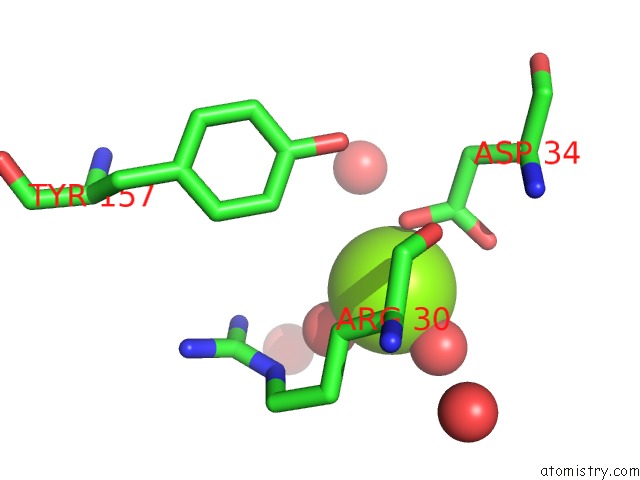
Mono view
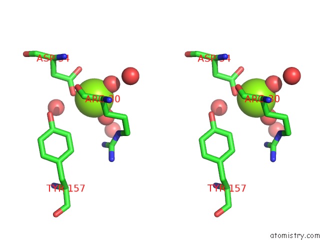
Stereo pair view
| | A full contact list of Magnesium with other atoms in the Mg binding site number 1 of Crystal Structure of Trypanosoma Brucei Hypoxanthine-Guanine Phosphoribosyltranferase in Complex with Xmp within 5.0Å range: probe atom residue distance (Å) B Occ A:Mg302 b:23.0 occ:1.00 O A:HOH420 2.0 15.0 1.0 O A:HOH412 2.0 32.8 1.0 OD2 A:ASP34 2.0 49.3 1.0 CG A:ASP34 2.8 32.7 1.0 OD1 A:ASP34 2.9 32.8 1.0 O A:HOH416 3.8 21.0 1.0 NE A:ARG30 4.1 39.1 1.0 O A:ARG30 4.1 28.7 1.0 CB A:ARG30 4.1 22.1 1.0 O A:HOH410 4.1 15.6 1.0 NH1 A:ARG30 4.2 35.5 1.0 CB A:ASP34 4.3 23.6 1.0 OH A:TYR157 4.5 38.8 1.0 CE1 A:TYR157 4.5 25.7 1.0 CZ A:ARG30 4.6 31.9 1.0 CA A:ARG30 4.6 34.9 1.0 C A:ARG30 4.7 29.6 1.0 CG A:ARG30 4.8 19.6 1.0 O A:HOH427 4.9 38.9 1.0 CZ A:TYR157 5.0 25.9 1.0 | | ------------------------------------------------------------------------------------------------------------------------------------------------------------------------------------------------------------------------------------------------------------------------------------------------------------------------------------------------------------------------------------------------------------------------------------------------------------------------------------------------------------------------------------------------------------------------------------------------------------------------------------------------------------------------------------------------------------------------------------------------------------------------------------------------------------------------------------------------------------------------------------------------------------------------------------------------------------------------------------------------ |
Magnesium binding site 2 out of 4 in 6mxg
Go back to  Magnesium Binding Sites List in 6mxg
Magnesium binding site 2 out of 4 in the Crystal Structure of Trypanosoma Brucei Hypoxanthine-Guanine Phosphoribosyltranferase in Complex with Xmp
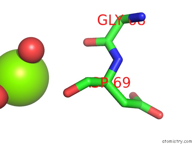
Mono view
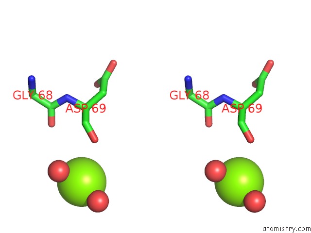
Stereo pair view
| | A full contact list of Magnesium with other atoms in the Mg binding site number 2 of Crystal Structure of Trypanosoma Brucei Hypoxanthine-Guanine Phosphoribosyltranferase in Complex with Xmp within 5.0Å range: probe atom residue distance (Å) B Occ A:Mg303 b:33.1 occ:1.00 O A:HOH428 2.1 11.1 1.0 O A:HOH422 2.1 23.5 1.0 O A:ASP69 4.1 44.0 1.0 O A:GLY68 4.6 27.5 1.0 C A:ASP69 5.0 37.2 1.0 | | ------------------------------------------------------------------------------------------------------------------------------------------------------------------------------------------------------------------------------------------------------------------------------------------------------------------------------------------------------------------------------------------------------------------------------------------------------------------------- |
Magnesium binding site 3 out of 4 in 6mxg
Go back to  Magnesium Binding Sites List in 6mxg
Magnesium binding site 3 out of 4 in the Crystal Structure of Trypanosoma Brucei Hypoxanthine-Guanine Phosphoribosyltranferase in Complex with Xmp
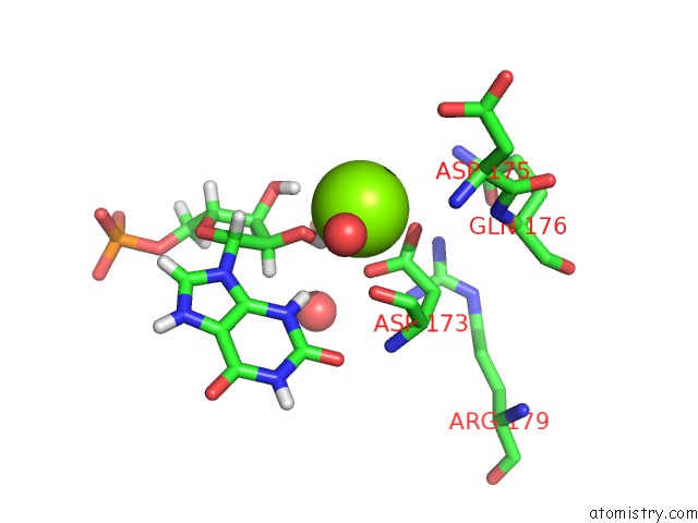
Mono view
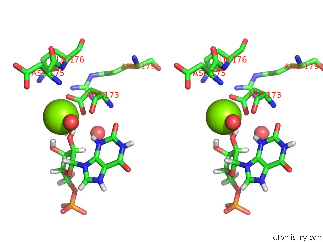
Stereo pair view
| | A full contact list of Magnesium with other atoms in the Mg binding site number 3 of Crystal Structure of Trypanosoma Brucei Hypoxanthine-Guanine Phosphoribosyltranferase in Complex with Xmp within 5.0Å range: probe atom residue distance (Å) B Occ B:Mg302 b:32.6 occ:1.00 H2O1 B:XMP301 1.8 48.9 1.0 O B:HOH402 2.2 38.4 1.0 OD2 B:ASP173 2.2 35.1 1.0 O2' B:XMP301 2.2 40.8 1.0 OD1 B:ASP173 2.3 41.9 1.0 CG B:ASP173 2.5 33.4 1.0 H3O1 B:XMP301 2.7 37.7 1.0 O4 B:SO4304 3.0 32.6 1.0 C2' B:XMP301 3.5 49.3 1.0 O3' B:XMP301 3.6 31.4 1.0 O B:ASP173 3.7 38.8 1.0 O1 B:SO4304 3.8 40.0 1.0 H3 B:XMP301 3.9 39.8 1.0 S B:SO4304 3.9 23.2 1.0 CB B:ASP173 3.9 36.4 1.0 H2' B:XMP301 3.9 59.1 1.0 C3' B:XMP301 4.1 39.0 1.0 C B:ASP173 4.2 36.2 1.0 H1' B:XMP301 4.4 44.6 1.0 NH2 B:ARG179 4.4 33.6 1.0 H3' B:XMP301 4.5 46.9 1.0 O3 B:SO4304 4.5 36.7 1.0 N B:ASP175 4.5 48.1 1.0 C1' B:XMP301 4.6 37.2 1.0 CA B:ASP173 4.6 34.6 1.0 O B:HOH419 4.7 27.2 1.0 N3 B:XMP301 4.8 33.1 1.0 N B:GLN176 4.8 49.4 1.0 NE2 B:GLN176 4.9 34.4 1.0 N B:ASP173 4.9 28.2 1.0 | | --------------------------------------------------------------------------------------------------------------------------------------------------------------------------------------------------------------------------------------------------------------------------------------------------------------------------------------------------------------------------------------------------------------------------------------------------------------------------------------------------------------------------------------------------------------------------------------------------------------------------------------------------------------------------------------------------------------------------------------------------------------------------------------------------------------------------------------------------------------------------------------------------------------------------------------------------------------------------------------------------------------------------------------------------------------------------------------------------------------------------------------------------------------------------------------------------------------------------------------------------------------------------------------------------------------------------------------------------------------------------- |
Magnesium binding site 4 out of 4 in 6mxg
Go back to  Magnesium Binding Sites List in 6mxg
Magnesium binding site 4 out of 4 in the Crystal Structure of Trypanosoma Brucei Hypoxanthine-Guanine Phosphoribosyltranferase in Complex with Xmp
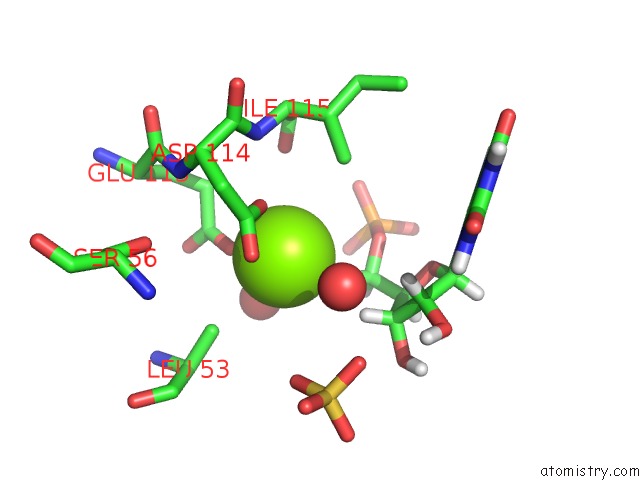
Mono view
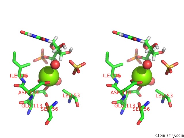
Stereo pair view
| | A full contact list of Magnesium with other atoms in the Mg binding site number 4 of Crystal Structure of Trypanosoma Brucei Hypoxanthine-Guanine Phosphoribosyltranferase in Complex with Xmp within 5.0Å range: probe atom residue distance (Å) B Occ B:Mg303 b:27.9 occ:1.00 OE2 B:GLU113 2.1 29.8 1.0 OD1 B:ASP114 2.2 33.1 1.0 H5'1 B:XMP301 2.7 57.1 1.0 CG B:ASP114 2.9 26.3 1.0 OD2 B:ASP114 2.9 29.1 1.0 O B:HOH416 3.2 22.2 1.0 CD B:GLU113 3.3 30.2 1.0 H3' B:XMP301 3.4 46.9 1.0 CB B:LEU53 3.5 44.3 1.0 C5' B:XMP301 3.6 47.6 1.0 CG2 B:ILE115 3.9 23.0 1.0 CG B:GLU113 4.0 41.3 1.0 O5' B:XMP301 4.1 58.0 1.0 H5'2 B:XMP301 4.1 57.1 1.0 O B:HOH419 4.1 27.2 1.0 O B:ILE115 4.2 21.1 1.0 OE1 B:GLU113 4.3 28.9 1.0 C3' B:XMP301 4.3 39.0 1.0 N B:ILE115 4.3 17.4 1.0 O2 B:SO4304 4.3 20.1 1.0 CB B:ASP114 4.4 28.2 1.0 H2' B:XMP301 4.5 59.1 1.0 C4' B:XMP301 4.5 44.8 1.0 N B:ASP114 4.6 24.3 1.0 OG B:SER56 4.8 47.8 1.0 O4 B:SO4304 4.8 32.6 1.0 CA B:ASP114 4.9 23.3 1.0 C2' B:XMP301 5.0 49.3 1.0 CA B:LEU53 5.0 41.9 1.0 CA B:ILE115 5.0 19.0 1.0 CB B:ILE115 5.0 15.0 1.0 | | ------------------------------------------------------------------------------------------------------------------------------------------------------------------------------------------------------------------------------------------------------------------------------------------------------------------------------------------------------------------------------------------------------------------------------------------------------------------------------------------------------------------------------------------------------------------------------------------------------------------------------------------------------------------------------------------------------------------------------------------------------------------------------------------------------------------------------------------------------------------------------------------------------------------------------------------------------------------------------------------------------------------------------------------------------------------------------------------------------------------------------------------------------------------------------------------------------------------------------------------------------------------------------------------------------------------------------------------------------------------------------------------------------------- |
Reference:
D.Teran, E.Dolezelova, D.T.Keough, D.Hockova, A.Zikova, L.W.Guddat. Crystal Structures of Trypanosoma Brucei Hypoxanthine - Guanine - Xanthine Phosphoribosyltransferase in Complex with Imp, Gmp and Xmp. Febs J. 2019.
ISSN: ISSN 1742-464X
PubMed: 31287615
DOI: 10.1111/FEBS.14987
Page generated: Tue Oct 1 12:22:14 2024