Ophthalmologic Manifestations of Atopic Dermatitis: Background, Pathophysiology, Epidemiology (original) (raw)
Overview
Background
Atopy is the hereditary predisposition to allergy or hypersensitivity. [1] Symptoms may present as a dermatitis, hay fever, or asthma. [1] According to Rapoza, Besnier first characterized atopic dermatitis, and many Europeans still use his name to describe the disease (prurigo Besnier). [1] This disease was labeled eczema for many years in the United States until Coca and Cooke coined the term atopy as a skin hypersensitivity seen in patients with hereditary allergies. Wise and Sulzberger have been credited with the term atopic dermatitis to describe a group of diseases associated with atopic conditions that may be seen in all age groups. [2]
Atopic dermatitis (AD) is a chronic and progressive skin disorder that includes an acute stage, characterized by erythema with edema and vesicles, and a chronic stage with skin lichenification. [3] The SCORAD (Severity Scoring of Atopic Dermatitis) Index characterizes the extent and severity of atopic dermatitis based on surface area involved, intensity of erythema, edema, oozing/crusting, excoriation, lichenification and dryness, as well as subjective symptoms of pruritis and sleep loss. [4] The purpose of this article is to describe the ophthalmologic complications of atopic dermatitis, specifically keratoconus, atopic keratoconjunctivitis (AKC), vernal keratoconjunctivitis (VKC), bacterial blepharoconjunctivitis, cicatricial entropion, cicatricial ectropion, retinal detachment, cataract and glaucoma. [3] These ophthalmologic complications can occur with or without other systemic complications including skin infections, gastrointestinal diseases, renal diseases, autoimmune diseases and psychological and psychiatric diseases. [3]
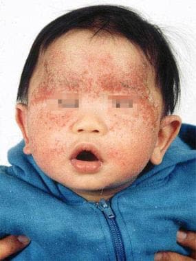
Typical atopic dermatitis on the face of an infant.
Keratoconus (KC) is a non-inflammatory, progressive corneal thinning disorder that leads to corneal protrusion, irregular astigmatism, and corneal scarring in the final stages, that eventually leads to distorted and impaired vision. [3] The incidence of keratoconus is reported as approximately 0.005% in the general population, but from 0.5% to 39% in those with AD. [3] One of the symptoms of AD is pruritis, which stimulates eye rubbing, and many studies have demonstrated a link between eye rubbing and keratoconus. [5, 6, 7, 8] Lastly, there have been reports that individuals with KC are more likely to have atopic tendencies than the healthy control group (35% vs 12%), most commonly allergic rhinitis, but also asthma and AD. [9]
Atopic keratoconjunctivitis (AKC) is a chronic inflammatory allergic disease, with clinical characteristics including conjunctivitis, corneal ulceration, superficial punctate keratitis and corneal neovascularization. [3]
Vernal keratoconjunctivitis (VKC) is typically a self-limiting, atopic disease of the conjunctiva, limbus and/or cornea, with usual onset prior to age 10 years, in individuals with a personal or family history of atopy. [10]
The incidence of bacterial conjunctivitis and blepharitis in AD patients has reportedly been higher than those without AD (86% vs 25%), with S. Aureus identified as the most common pathogen amongst patients with AD (67%). [3] As a result, patients with AD may benefit from pre- and post-operative antibiotic regimens against S. Aureus to prevent infection and its sequelae.
Cicatricial entropion describes inward turning of the eyelid margin and its appendages secondary to chronic inflammatory changes and subsequent fibrosis, scarring and shortening of the posterior lamellae (e.g. conjunctiva, tarsus), and can lead to lash-cornea touch that may lead to corneal abrasion, scarring, thinning, neovascularization, or even corneal ulceration and perforation in severe cases. [11]
Cicatricial ectropion, in contrast, is caused by shortening of the anterior lamella (i.e. skin and orbicularis muscle), and is also secondary to chronic inflammatory changes including fibrosis and scarring. Similar to other types of ectropion, cicatricial ectropion can lead to exposure keratopathy, corneal abrasion, ulceration, and even thinning and perforation. [12]
Another serious ophthalmologic complication of AD includes retinal detachment, which may be due to eye rubbing as a result of atopic dermatitis-associated pruritis. A review of case studies and another retrospective study [13] , demonstrated that retinal breaks in patients with AD are most likely found in the peripheral fundus, near the ora serrata, which are similar to those caused by trauma (e.g. eye rubbing). [13, 14, 15] Serum IgE levels have not been reported to have an impact on RD. [14]
In patients with AD, cataracts are usually bilateral, symmetric, and occur in the anterior and/or posterior subcapsular regions. [3] Their progression has been linked to eye rubbing and severity of facial skin lesions, which are likely a sign of increased facial pruritis. [16] Some articles have not demonstrated a relationship with serum IgE levels, the duration of systemic or facial topical corticosteroid use and cataract development, [14] whereas other studies have proved an association between high serum IgE levels and cataract development in those with AD. [17]
The relationship with glaucoma and AD is rarely described. Some researchers have denied a correlation between the two conditions, [18] while Takakuwa et al. have focused on patients with severe AD and advanced glaucoma in order to propose a new clinical entity, "atopic glaucoma", that is thought to arise secondary to increased intraocular inflammatory responses, such as higher IL8 and CCL2 expression in the aqueous humor of these patients. [19] Takakuwa et al. excluded those cases showing an apparent relation between glucocorticoid use and IOP in order to propose this new clinical diagnosis. [19] These researchers believe that atopic glaucoma is associated with proinflammatory factors, is defined by vertical cup-disc ratio >0.7 and/or neuroretinal rim notching, compatible visual field loss and IOP >21 mmHg. [19]
![]()
Pathophysiology
Atopic dermatitis is primarily caused by cellular immune deficiency and elevated immunoglobulin E (IgE). The pathogenesis can be traced to a genetically inherited, bone marrow–derived cell associated with chromosome 11q. [20] Abnormal skin reactivity also plays a major role in the development of the disease. Irritants to the skin are believed to predispose an individual to develop dermatitis more often than simply exposure to an allergenic trigger. [20] Nonetheless, patients frequently have a history of food or inhalant allergies or eventually develop them. [20]
One of the symptoms of AD is pruritis, which is associated with keratoconus, though the exact mechanism remains unknown. Potential mechanisms of eye rubbing leading to KC include increased corneal temperature, released inflammatory mediators in the tear film and increased enzymatic activity, increased intraocular and hydrostatic tissue pressure, change in keratocytes and a decrease in corneal shear strength. [3]
![]()
Epidemiology
Frequency
United States
Atopic dermatitis affects approximately 17% of children and 5% of adults in the United States. [21]
Race
No clear racial predisposition appears to exist. [21]
Sex
Males appear to be affected more frequently by AKC and especially VKC than females, though this difference decreases with increasing age. [22]
Age
Children most commonly are affected, with 80% developing the disease before age 7 years. Less than 2% will have an onset after age 20 years. Most sources agree that persistence after age 20 years is uncommon. Only an estimated 10% of patients older than 20 years continue to be symptomatic. [23]
Specific ophthalmologic complications of AD occur at different ages. For example, the peak incidence of AKC is estimated between 30-50 years old, whereas the majority of VKC cases first occur between 10-12 years old. [10, 24]
However, in general, ophthalmologic complications of AD were far more frequent for adults than for children. Specifically, more severe complications leading to ocular morbidity or visual impairment were rare in younger children with mild atopic dermatitis. [25, 26]
Environmental
The highest incidence of atopic dermatitis is in urban areas. It seems there may be environmental factors in urban areas that predisposes the population to atopic dermatitis as compared to rural settings, but further evaluation of this relationship is needed for the exact cause. [27]
Specifically, VKC is primarily seen in hot and dry climates, such as West Africa, Middle East, Japan, India and South America, which is though to be secondary to a higher level of pollution by pollens and various other allergens in these areas. [10]
![]()
Prognosis
The prognosis is good if the inflammation can be kept under control with therapy. [3] Unfortunately, atopic disease can be controlled but not cured, and management focuses on improvement on symptoms, and prevention of complications and sequelae that can lead to vision loss, while limiting side effects of treatment. [3] Unfortunately, some cases are so severe that treatment will not prevent vision loss and other debilitating complications.
![]()
Patient Education
For excellent patient education resources, visit eMedicineHealth's Skin Conditions and Beauty Center. Also, see eMedicineHealth's patient education article Eczema.
![]()
- Rapoza PA, Chandler JW. Atopic dermatitis. Weingeist T, Gould D, eds. The Eye in Systemic Disease. Philadelphia: Lippincott; 1990. 606-609.
- Shen CP, Xing H, Ma L. [Research advances in atopic dermatitis]. Zhongguo Yi Xue Ke Xue Yuan Xue Bao. 2009 Feb. 31(1):103-6. [QxMD MEDLINE Link].
- Marta Pietruszyńska, Anna Zawadzka-Krajewska, Patrycja Duda, Monika Rogowska, Iwona Grabska-Liberek, and Marek Kulus. Ophthalmic manifestations of atopic dermatitis. Postepy Dermatol Alergol. 2020 Apr. 37(2):174-179. [QxMD MEDLINE Link]. [Full Text].
- Consensus Report of the European Task Force on Atopic Dermatitis. Severity scoring of atopic dermatitis: the SCORAD index. Consensus Report of the European Task Force on Atopic Dermatitis. Dermatology. 1993. 186:23-31. [QxMD MEDLINE Link]. [Full Text].
- Harrison RJ, Klouda PT, Easty DL, Manku M, Charles J, Stewart CM. Association between keratoconus and atopy. Br J Ophthalmol. 1989 Oct. 73(10):816-22. [QxMD MEDLINE Link].
- Bawazeer A, Hodge W, Lorimer B. Atopy and keratoconus: a multivariate analysis. r J Ophthalmol. 2000. 84:834–6. [QxMD MEDLINE Link].
- Sugar J, Macsai MS. What causes keratoconus?. Cornea. 2012. 31:716-9. [QxMD MEDLINE Link].
- Mashor RS, Kumar NL, Ritenour RJ, Rootman DS. Keratoconus caused by eye rubbing in patients with Tourette syndrome. Can J Ophthalmol. 2011. 46:83-6. [QxMD MEDLINE Link].
- Rahi A, Davies P, Ruben M, Lobascher D, Menon J. Keratoconus and coexisting atopic disease. Br J Ophthalmol. 1977. 61:761-4. [QxMD MEDLINE Link].
- De Smedt S, Wildner G, Kestelyn P. Vernal keratoconjunctivitis: an update. Br J Ophthalmol. 2013 Jan. 97(1):9-14. [QxMD MEDLINE Link].
- Weber AC, Chundury RV, Perry JD, Sundar G. Entropion. AAO EyeWiki. Available at https://eyewiki.aao.org/Entropion. 2021 Feb 13; Accessed: July 10 2021.
- Phelps PO, Belliveau MJ, Vargason CW, Goel S, Yen MT, Marcet MM, et al. Ectropion. AAO EyeWiki. Available at https://eyewiki.aao.org/Ectropion. 2020 Nov 24; Accessed: July 10, 2021.
- Sasoh M, Mizutani H, Matsubara H, Furuta M, Matsui Y, Yamanaka K, et al. Incidence of retinal detachment associated with atopic dermatitis in Japan: review of cases from 1992 to 2011. Clin Ophthalmol. 2015 Jun 23. 9:1129-34. [QxMD MEDLINE Link].
- Taniguchi H, Ohki O, Yokozeki H, Katayama I, Tanaka A, Kiyosawa M, et al. Cataract and retinal detachment in patients with severe atopic dermatitis who were withdrawn from the use of topical corticosteroid. J Dermatol. 1999 Oct. 26(10):658-65. [QxMD MEDLINE Link].
- Hida T, Tano Y, Okinami S, Ogino N, Inoue M. Multicenter retrospective study of retinal detachment associated with atopic dermatitis. Jpn J Ophthalmol. 2000 Jul-Aug. 44(4):407-18. [QxMD MEDLINE Link].
- Nagaki Y, Hayasaka S, Kadoi C. Cataract progression in patients with atopic dermatitis. J Cataract Refract Surg. 1999 Jan. 25(1):96-9. [QxMD MEDLINE Link].
- Nippon Ganka Gakkai Zasshi. Sasabe T, Suwa Y, Kawamura T, Aoki T. [Cataracts occur in patients with atopic dermatitis when the serum IgE increases]. 1997 May. 101(5):389-92. [QxMD MEDLINE Link].
- Thyssen JP, Toft PB, Halling-Overgaard AS, Gislason GH, Skov L, Egeberg A. Incidence, prevalence, and risk of selected ocular disease in adults with atopic dermatitis. J Am Acad Dermatol. 2017 Aug. 77(2):280-286. [QxMD MEDLINE Link].
- Takakuwa K, Hamanaka T, Mori K, Chin S, Shinmei Y, Funaki T, et al. Atopic Glaucoma: Clinical and Pathophysiological Analysis. J Glaucoma. 2015 Dec. 24(9):662-8. [QxMD MEDLINE Link].
- Jung T, Stingl G. Atopic dermatitis: therapeutic concepts evolving from new pathophysiologic insights. J Allergy Clin Immunol. 2008 Dec. 122(6):1074-81. [QxMD MEDLINE Link].
- Beck KM, Seitzman GD, Yang EJ, Sanchez IM, Liao W. Ocular Co-Morbidities of Atopic Dermatitis. Part I: Associated Ocular Diseases. Am J Clin Dermatol. 2019 Dec. 20 (6):797-805. [QxMD MEDLINE Link].
- Bezan DJ. Eye itch. Bezan DJ, Larussa FP, Nishimoto JH, et al, eds. Differential Diagnosis in Primary Eye Care. Boston: Butterworth-Heinemann; 1999. 67-71.
- Brenninkmeijer EE, Legierse CM, Sillevis Smitt JH, Last BF, Grootenhuis MA, Bos JD. The course of life of patients with childhood atopic dermatitis. Pediatr Dermatol. 2009 Jan-Feb. 26(1):14-22. [QxMD MEDLINE Link].
- Nutten S. Atopic dermatitis: global epidemiology and risk factors. Ann Nutr Metab. 2015. 66 Suppl 1:8-16. [QxMD MEDLINE Link].
- Carmi E, Defossez-Tribout C, Ganry O, Cene S, Tramier B, Milazzo S, et al. Ocular complications of atopic dermatitis in children. Acta Derm Venereol. 2006. 86 (6):515-7. [QxMD MEDLINE Link].
- Kaujalgi R, Handa S, Jain A, Kanwar AJ. Ocular abnormalities in atopic dermatitis in Indian patients. Indian J Dermatol Venereol Leprol. 2009 Mar-Apr. 75 (2):148-51. [QxMD MEDLINE Link].
- Silverberg JI, Barbarot S, Gadkari A, Simpson EL, Weidinger S, Mina-Osorio P, et al. Atopic dermatitis in the pediatric population: A cross-sectional, international epidemiologic study. Ann Allergy Asthma Immunol. 2021 Apr. 126 (4):417-428.e2. [QxMD MEDLINE Link].
- Bagheri N, Wajda B, Calvo C, Durrani A. The Will's Eye Manual. 7th edition. Lippincott Williams and Wilkins; 2016.
- Hanifin JM. Atopic dermatitis: broadening the perspective. J Am Acad Dermatol. 2004 Jul. 51(1 Suppl):S23-4. [QxMD MEDLINE Link].
- Bielory B, Bielory L. Atopic dermatitis and keratoconjunctivitis. Immunol Allergy Clin North Am. 2010 Aug. 30(3):323-36. [QxMD MEDLINE Link].
- Cornish KS, Gregory ME, Ramaesh K. Systemic cyclosporin A in severe atopic keratoconjunctivitis. Eur J Ophthalmol. 2010 Sep-Oct. 20(5):844-51. [QxMD MEDLINE Link].
- Hsu JI, Pflugfelder SC, Kim SJ. Ocular complications of atopic dermatitis. Cutis. 2019 Sep. 104 (3):189-193. [QxMD MEDLINE Link].
- Bunya VY, Garff K, Nallasamy N. Atopic Keratoconjunctivitis. EyeWiki. Available at https://eyewiki.aao.org/Atopic_Keratoconjunctivitis. 2021 March 29; Accessed: 2021 June 20.
- Clark RAF, Kristal L. Atopic dermatitis. Sams J, Lynch PJ, eds. Principles and Practice of Dermatology. 2nd ed. New York: Churchill Livingstone Inc; 1996. 403-418.
- Anderson PC, Dinulos JG. Atopic dermatitis and alternative management strategies. Curr Opin Pediatr. 2009 Feb. 21(1):131-8. [QxMD MEDLINE Link].
- Gagnon L. FDA Approves Verkazia for Vernal Keratoconjunctivitis, an Allergic Eye Disease. Medscape. Available at https://www.medscape.com/viewarticle/953874?src=WNL_mdpls_210706_mscpedit_opth&uac=193997DV&spon=36&impID=3489569&faf=1. 2021 June 28; Accessed: July 10, 2021.
- Maeng MM, De Moraes CG, Winn BJ, Dagi LRG. Effect of Topical Periocular Steroid Use on Intraocular Pressure: A Retrospective Analysis. Ophthalmic Plast Reconstr Surg. 2019 Feb 4. [QxMD MEDLINE Link]. [Full Text].
- Kaujalgi R, Handa S, Jain A, Kanwar AJ. Ocular abnormalities in atopic dermatitis in Indian patients. Indian J Dermatol Venereol Leprol. 2009 Mar-Apr. 75(2):148-51. [QxMD MEDLINE Link].
- Suda K, Akagi T, Ikeda HO, Kameda T, Hasegawa T, Miyake M, et al. Atopic dermatitis as a risk factor for severe visual field loss in youth-a retrospective cohort study of glaucoma under steroid treatment. Graefes Arch Clin Exp Ophthalmol. 2021 Jan. 259 (1):129-136. [QxMD MEDLINE Link].
- Beck KM, Seitzman GD, Yang EJ, Sanchez IM, Liao W. Ocular Co-Morbidities of Atopic Dermatitis. Part II: Ocular Disease Secondary to Treatments. Am J Clin Dermatol. 2019 Dec. 20 (6):807-815. [QxMD MEDLINE Link].
- Bansal, Ashish, Simpson, Eric L., Paller, Amy S., et al. Conjunctivitis in Dupilumab Clinical Trials for Adolescents with Atopic Dermatitis or Asthma. American Journal of Clinical Dermatology. 2021. 22(1):101-115. (15p).
- Yamane, Maya L. M., Belsito, Donald V., Glass, Lora R. Dagi. Two differing presentations of periocular dermatitis as a side effect of dupilumab for atopic dermatitis. Orbit. Oct2019. Vol. 38 Issue 5:p390-394. 5p.
- Typical atopic dermatitis on the face of an infant.

Author
Coauthor(s)
Sehwa Nick Kang MD Candidate, University of Texas Health Science Center at San Antonio, Long School of Medicine
Disclosure: Nothing to disclose.
Constance L Fry, MD Professor, Department of Ophthalmology, UT Health San Antonio Long School of Medicine; Associate Preceptor, American Society of Ophthalmic Plastic Surgery, Oculofacial Fellowship, Medical Director, MARC Ophthalmology Clinic, Director, Ophthalmic Plastic Surgery and Ophthalmic Oncology, UT Health San Antonio
Constance L Fry, MD is a member of the following medical societies: Academy of Master Teachers, UTMB Health, American Academy of Ophthalmology, American Association of Ophthalmic Oncologists and Pathologists, American Society of Ophthalmic Plastic and Reconstructive Surgery, Research to Prevent Blindness, San Antonio Society of Ophthalmology
Disclosure: Nothing to disclose.
Specialty Editor Board
Francisco Talavera, PharmD, PhD Adjunct Assistant Professor, University of Nebraska Medical Center College of Pharmacy; Editor-in-Chief, Medscape Drug Reference
Disclosure: Received salary from Medscape for employment. for: Medscape.
Christopher J Rapuano, MD Professor, Department of Ophthalmology, Sidney Kimmel Medical College of Thomas Jefferson University; Director of the Cornea Service, Wills Eye Hospital
Christopher J Rapuano, MD is a member of the following medical societies: American Academy of Ophthalmology, American Ophthalmological Society, American Society of Cataract and Refractive Surgery, Contact Lens Association of Ophthalmologists, Cornea Society, Eye Bank Association of America, International Society of Refractive Surgery
Disclosure: Serve(d) as a director, officer, partner, employee, advisor, consultant or trustee for: AAO; OMIC; American Society of Ophthalmic Trauma (ASOT); Avellino, Baxis; Bio-Tissue; Celularity; Dompe; Emmecell; Glaukos; Kala; Oyster Point; Tarsus; TearLab
Serve(d) as a speaker or a member of a speakers bureau for: Dompe
Received research grant from: Glaukos
Received income in an amount equal to or greater than $250 from: AAO; OMIC; Baxis; Bio-Tissue; Cellularity; Dompe; Glaukos; Kala; TearLab
stock options for: RPS, Fount Bio.
Chief Editor
Additional Contributors