MucilAir™ (MA) (original) (raw)
General features of MucilAir™
MucilAir™ is an in vitro tissue model of the human upper airway epithelium cultured at the air liquid interface (ALI). It is a powerful and predictive model for in vitro research and tests. MucilAir™ tissue is mainly composed of the following cell types:
- Basal cells (progenitor cells)
- Goblet cells (mucus producing cells)
- Ciliated cells (with active cilia)
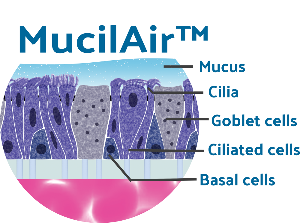
Figure 1: schematic representation of a MucilAir™ cross section
A meticulous tissue production
Since 2006 we produce MucilAir™ with high standards of quality and reproducibility. The tissue production is GIVIMP certified since 2023.
To ensure high predictability, MucilAir™ is solely reconstituted using low passage (P1), human primary cells. The primary cells are isolated from human biological Samples, under ethical approval and donor consent. Each batch of tissue is delivered with a certificate of Analysis (COA) with donor information and Quality Control (QC) results.
MucilAir™ could be reconstituted from cells isolated from different anatomical regions:
- Nasal (MucilAir™-Nasal)
- Tracheal (MucilAir™-Tracheal)
- Bronchial (MucilAir™-Bronchial)
Patients with several pathologies are available for MucilAir™ production
- Healthy (non-smoker)
- Healthy (smoker)
- COPD (Chronic Obstructive Pulmonary Disease)
- Asthmatic
- Cystic Fibrosis (several mutations available)
- Allergic Rhinitis
MucilAir™ recapitulates the function of the human upper airway epithelium
Our models mimic the characteristics of in vivo tissues:
Long shelf life
Once the production cycle is terminated, MucilAir™ is in a stable homeostatic state for several months (even up to up to a year) with a slow but physiological cell renewal. It maintains its functionality and cellular population during its entire life cycle.
Presence of the three main representative cellular types of the upper airway:
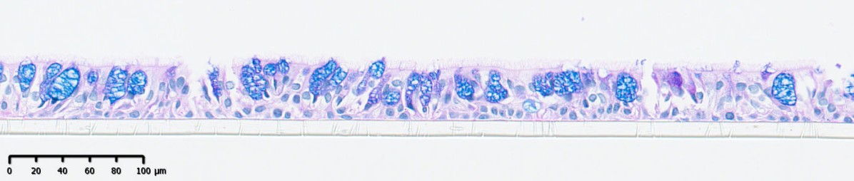
Figure 2: histological cross section of MucilAir™ with Alcian blue H/E staining of the tissue allowing to visualize the intrtissular Mucins, contained in the Goblet cells (dark blue).
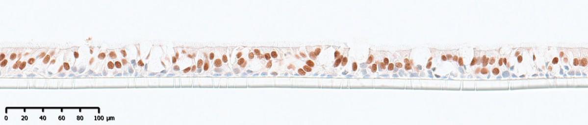
Figure 3: histological cross section of MucilAir™ with FoxJ1 staining allowing to visualize the nucleus of the ciliated cells (dark brown).
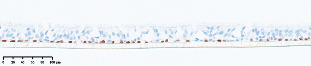
Figure 4: histological cross section of MucilAir™ with P63 staining allowing to visualize the nucleus of the ciliated cells (dark brown).
Functional cilia
As observed on the human tissue, a dense cilia layer is present on the surface of MucilAir™.
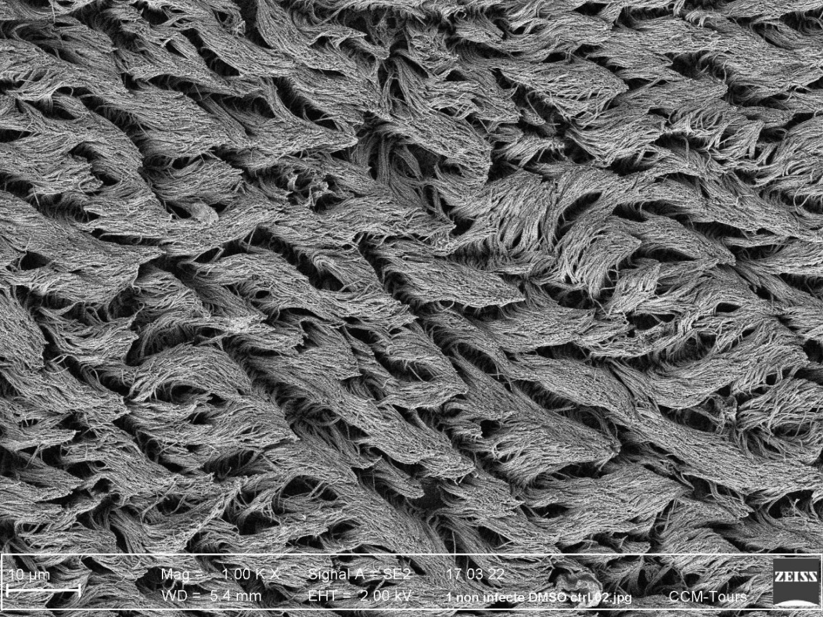
Figure 5: Electron Microscopy image of MucilAir™ top view (SEM x10K).
The Cilia Beating Frenquency (CBF) could be monitored with our tailored software (Cilia-X) and is part of the MucilAir™ QC.
Mucus production
Goblet cells produce and secrete mucus on the apical side of the tissue. The mucus layer is therefore produced by the tissue itself in a physiological, complex and dynamic manner.
Figure 6: Schematic animation of the mucus release on the apical side
Mucociliary clearance
Thanks to the presence of functional cilia beating cells and the mucus layer, a functional mucociliary clearance is present on top of MucilAir™.
Tight junctions
Being the first barrier in the airway the tissue is naturally tight thanks to the presence of tight junctions.
Barrier function could be quantified with a Trans Epithelia Electrical Resistance (TEER) device, and is part of the MucilAir™ QC.
Native cell surface receptor and ion channels
Using low passage cells allows to keep the native ion channel (CFTR, ENAC, etc…) allowing the use of the model in electrophysiology experiments.
Native cell surface receptors (TMPRSS2, ACE2, FCRn, etc.) are also present. This makes the model an efficient tool to study viral infections.
Release of soluble factors
MucilAir™ is also a very potent immune-regulator and secrete a wide panel of Cytokines, Chemokines and, Metalloproteinases that could be present at a constitutive level but also in stimulated condition.
An easy and ready-to-use model
We handle all the production and QC process to deliver MucilAir™ ready-to-use at your lab. No fancy equipment or cell culture skills are required for its maintenance.
Once received, transfer the tissues in fresh medium and in the incubator. Then change the medium twice a week, occasionally perform a mucus wash. That’s it.
MucilAir™ culture medium, dedicated, chemically defined and serum-free culture medium ensure a stable composition from batch to batch.
Need technical support for your experiment with MucilAir™? Contact us!
Easy shipping
We ship routinely our tissues to Europe, America and Asia. Our packaging method allows to deliver safely the tissues to these locations.
We can evaluate with you the delivery to your place.
MucilAir™ is widely referenced in the literature
More than +800 scientific publications using Epithelix tissues.
Check out our publication page!