BSCI 424 Pathogenic Microbiology -- Corynebacterium (original) (raw)
(1).gif)
BSCI 424 — PATHOGENIC MICROBIOLOGY — Fall 2000

Corynebacterium Summary
.gif)
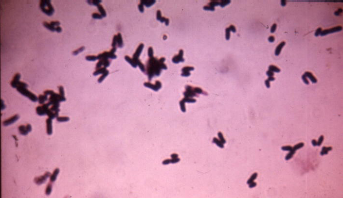
Gram stain of Corynebacterium spp. demonstrating "Chinese letters" formations
General Overview:
 C. diphtheriae and related organisms are collectively termed coryneforms or diphtheroids
Corynebacteria possess capsular (K) and somatic antigens (O)
Morphology & Physiology:
Small, nonmotile, irregularly staining pleomorphic Gram-positive rods with club-shaped swelled ends but no spores; may be straight or slightly curved (see WebLinked image; see WebLinked image)
Palisade arrangement of cells in short chains ("V" or "Y" configurations) or in clumps resembling "Chinese letters"
Cells tend to lie parallel to one another (palisades) or at acute angles (coryneforms), due to their snapping type of division
May also contain inclusion bodies, known as metachromatic granules, which are composed of inorganic polyphosphates (volutin) that serve as energy reserves and are not membrane bound
Aerobic or facultatively anaerobic
Fermentative metabolism (carbohydrates to lactic acid); form acid but not gas from certain carbohydrates
Fastidious; Slow growth on enriched medium
Catalase positive
Cell wall containing unusual lipids: meso-diaminopimelic acids; arabino-galactan polymers; short-chain mycolic acids (member of CMN (Corynebacterium, Mycobacterium, Nocardia) group)
Corynebacterium urealyticum strongly urease positive
Clinical Syndromes:
Determined by site of infection, host immunity, and virulence of the organism
Corynebacterium diptheriae: toxigenic strains cause diphtheria in humans
Initially: sore throat, low-grade fever; followed by adherent pseudomembrane on the tonsils and pharynx
Corynebacterium jeikeium: opportunistic infections (especially in immunocompromised patients)
Corynebacterium urealyticum: urinary tract infections (UTI�s); rare but important
Corynebacterium pseudotuberculosis: subacute relapsing lymphadenitis
Corynebacterium ulcerans: pharnygitis
Corynebacterium xerosis: bacteremia, skin infections, pneumonia in immunocompromised hosts (e.g., patients with blood disorders, bone marrow transplants, intravenous catheters) and pharyngitis
Corynebacterium pseudodiphtheriticum: endocarditis and lower-respiratory tract infections
Epidemiology:
Widely distributed in nature; worldwide in occurrence
Human is the only natural host
Corynebacterium diptheriae:
Corynebacterium jeikeium: carriage on skin of up to 40% of hospitalized patients (e.g., bone marrow transplants)
Several species form part of the common microbiota of the human respiratory tract and other mucous membranes, the conjunctiva, and the skin
Pathogenesis & Immunity:
Non-pathogenic species are called "diphtheroids"; two species commonly found in humans are Corynebacterium xerosis and Corynebacterium pseudodiphtheriticum
Pathogenic type species is Corynebacterium diphtheriae, which produces a potent exotoxin and causes diphtheria in humans
- B fragment binds to receptor sites on target cells and toxin is internalized by receptor-mediated endocytosis
- A fragment blocks protein synthesis by ADP-ribosylation of elongation factor-2 (EF-2)
Corynebacterium jeikeium: multiple antibiotic resistance important in opportunistic infections of immunocompromised patients
Corynebacterium urealyticum: urease hydrolyzes urea; release of NH4+, increase in pH, alkaline urine, renal stones
Laboratory Identification:
Microscopy
Culture
    1. **var. _gravis_:** large, flat, rough, dark-gray colonies; not hemolytic; very few small metachromatic granules; form a pellicle in broth
    2. **var. _mitis_:** smooth, convex, black, shiny, entire colonies; hemolytic; prominent metachromatic granules; diffuse turbidity in broth
    3. **var. _intermedius_:** dwarf, flat, umbilicate colony with a black center and slightly crenated periphery; not hemolytic; fine granular deposit in broth
Diphtheroids may be distinguished from C. diphtheriae by means of CTA sugar fermentation reactions and tests for toxigenicity
In vivo test:
In vitro test: Elek test (immunodiffusion)
Treatment, Prevention & Control:
Antitoxin
Antibiotics
Distinguishing Characteristics of Corynebacterium spp.
| ORGANISM | CELLULAR MORPHOLOGY | HEMOLYSIS | SUGAR FERMENTATION | TOXIN | |
|---|---|---|---|---|---|
| GLUCOSE | SUCROSE | ||||
| C. diphtheriae | Slender pleomorphic rods; often club-shaped; often banded or beaded with irregularly staining granules | + | + | - | + |
| C. pseudodiphtheriticum | Short rods; no granules; clubs rare | - | - | - | - |
| C. xerosis | Polar staining rods; few club forms | - | + | + | - |
(1).gif) Go to Pathogen List
Go to Pathogen List
BSCI 424 — Pathogenic Microbiology — BSCI 424 HomePage
Designed & Maintained by David M. Rollins
Copyright © 2000, D.M. Rollins and S.W. Joseph
Revised: August 2000
URL: http://life.umd.edu/classroom/bsci424