Differential Responses of Human Mononuclear Phagocytes to Mycobacterial Lipoarabinomannans: Role of CD14 and the Mannose Receptor (original) (raw)
Abstract
CD14 is a signaling receptor for both gram-negative bacterial lipopolysaccharide (LPS) and mycobacterial lipoarabinomannan (LAM) that lacks terminal mannosyl units (AraLAM). In contrast, terminally mannosylated LAM (ManLAM) binds the macrophage mannose receptor (MMRc), although the ability of the MMRc to serve as a signaling receptor has not been previously reported. We compared the abilities of AraLAM and ManLAM to induce distinct responses in two monocytic cell populations, freshly isolated human peripheral blood monocytes (PBM) and monocyte-derived macrophages (MDM). The responses examined were chemotaxis and transient changes in free cytosolic calcium ([Ca2+]in). We found that AraLAM but not ManLAM was chemotactic for both PBM and MDM. Migration of these cells in vitro to AraLAM was specifically blocked by an anti-CD14 monoclonal antibody, suggesting that CD14 mediates the chemotactic response to AraLAM. Subsequently, we found that AraLAM induced a transient rise in [Ca2+]in levels within a subpopulation of PBM but not MDM. This response was blocked by anti-CD14 antibodies. In contrast, ManLAM induced a transient rise in [Ca2+]in levels within a subpopulation of MDM but not PBM. This response was blocked by either anti-CD14 or anti-MMRc antibodies. These data suggest that the MMRc can serve as a signaling receptor and that coligation of both CD14 and the MMRc is required to elicit a specific response. Thus, one response to LAM (chemotaxis) can be elicited solely by engaging CD14, whereas a different response (changes in [Ca2+]in levels) depends on both the differentiation state of the cells and concomitant engagement of CD14 and the MMRc.
Uptake of Mycobacterium tuberculosis by mononuclear phagocytes is the first step leading to the development of tuberculosis infection. Following ingestion of the bacilli, the innate immune response against tuberculosis is predominantly directed by activated macrophages (reviewed in reference 17). The cell wall glycolipid lipoarabinomannan (LAM) is one of many mycobacterial products that can affect these immune responses. Vesicles containing LAM are released from phagosomes following macrophage ingestion of M. tuberculosis (36, 38), suggesting that transport of mycobacterial products out of infected macrophages is possible. Furthermore, the presence of anti-LAM antibodies in the sera of tuberculosis patients suggests that LAM is released from infected macrophages in vivo (29). LAM is comprised of a mannose-rich core polysaccharide, containing highly branched arabinofuranosyl side chains, linked via a phosphatidylinositol moiety at the reducing terminus to acyl groups consisting of palmitic and tuberculostearic acids. LAM isolated from pathogenic M. tuberculosis and M. bovis BCG is capped with mannose residues at the nonreducing arabinofuranosyl termini (ManLAM), whereas LAM isolated from rapidly growing avirulent mycobacteria lacks mannose caps at the arabinofuranosyl ends (AraLAM [10, 26]). The presence or absence of terminal mannose residues has been shown to affect the biological activity of LAM. For example, tumor necrosis factor (TNF) production can be induced in macrophages by purified LAM, although AraLAM is 100-fold more potent in this respect than ManLAM (11, 13). Similar results have been observed for interleukin-1 (IL-1) (41), IL-6 (13), chemokines (28, 40), and nitric oxide (28) production. In contrast, both AraLAM and ManLAM induce similar amounts of transforming growth factor β (TGF-β) production in human monocytes (13).
Two potential LAM receptors have been identified on monocytic cells. Zhang and colleagues first showed that the release of IL-1β and TNF by LAM-stimulated human blood mononuclear cells could be blocked by an anti-CD14 monoclonal antibody (MAb) (40). CD14 is a 55-kDa glycosylphosphatidylinositol-linked protein expressed on the surface of monocytes, macrophages, microglial cells, and polymorphonuclear leukocytes which serves as a receptor for gram-negative bacterial lipopolysaccharide (LPS) (reviewed in reference 42). Evidence that LAM can bind directly to CD14 was provided by the demonstration that AraLAM could compete for the binding of LPS to soluble CD14 in vitro (27). A role for CD14 in the receptor-mediated uptake of nonopsonized M. tuberculosis was suggested by studies which showed that both anti-CD14 MAbs and soluble CD14 could significantly block the uptake of M. tuberculosis by human microglial cells (25). In contrast, ManLAM has been shown to function as the ligand which is most likely to mediate uptake of M. tuberculosis via the macrophage mannose receptor (MMRc) on human blood monocyte-derived macrophages (MDM) (31, 32). The MMRc is a 162-kDa glycoprotein expressed in abundance on MDM and tissue macrophages but not on freshly isolated peripheral blood monocytes (PBM) (reviewed in reference 33). A role for ManLAM in the MMRc-mediated adherence of M. tuberculosis to MDM was suggested by the finding that an anti-LAM MAb blocked the binding of M. tuberculosis to MDM by up to 49% (31). A subsequent study revealed that differences in the ability of LAM from different strains of M. tuberculosis to mediate adherence to macrophages and to serve as ligands for the MMRc are not solely determined by the presence of terminal mannosyl units (32).
In this study, we compared the capacity of AraLAM and ManLAM to regulate different monocytic cell functions in vitro. We found that purified AraLAM, but not ManLAM, could induce a chemotactic response in human PBM and MDM. Antibody blocking and inhibitor data suggest that CD14 serves as a signaling receptor for AraLAM. This chemotactic response is distinct from the abilities of ManLAM and AraLAM to differentially induce a transient rise in free cytosolic calcium levels in the two cell populations. The capacity of PBM to generate a calcium response upon exposure to AraLAM appears to involve CD14, whereas the capacity of MDM to generate a calcium response following exposure to ManLAM requires engagement of both CD14 and the MMRc. Lastly, exposure of MDM to either AraLAM or ManLAM resulted in the selective down-regulation of the function of complement receptor CR3, although LAM treatment did not affect the level of surface CR3 expression.
MATERIALS AND METHODS
Antibodies and reagents.
ManLAM (purified from M. tuberculosis Erdman) and AraLAM (purified from a rapidly growing avirulent mycobacterium) were provided by John Belisle (Department of Microbiology, Colorado State University). The endotoxin contamination of these preparations was typically less than 3 ng per mg of LAM. The CS-35 anti-LAM MAb has been previously described (25) and was also provided by John Belisle. The anti-human CD14 MAb (TUK4) and anti-human CD11b MAb (2LPM19c) were purchased from Dako Corp. (Santa Barbara, Calif.). The anti-human CD14 MAb 3C10 was provided by Douglas Golenbock (Boston University School of Medicine). The anti-human MMRc MAb was provided by Philip Stahl (Washington University School of Medicine). LPS-free synthetic Rhodobacter sphaeroides lipid A (RSLA) was provided by Michael Lewis (Eisai Research Institute, Andover, Mass.). Formylmethionyl-leucyl-phenylalanine (fMLP) was purchased from Sigma (St. Louis, Mo.). RPMI 1640 tissue culture medium, medium supplements, and antibiotics were purchased from BioWhittaker (Walkersville, Md.). Fetal bovine serum (FBS) was purchased from HyClone Laboratories (Logan, Utah). LPS levels in medium components were less than 10 pg/ml (final concentration). For the complement receptor function experiments, C5-deficient serum was purchased from Sigma, washed sheep erythrocytes (SRBC) were purchased from Cappell (Durham, N.C.), and anti-SRBC immunoglobulin M was purchased from DiaMedix (Miami, Fla.).
Isolation of human PBM and preparation of MDM.
Blood was drawn from healthy young adult volunteers, with no known history of exposure to M. tuberculosis or BCG, into heparinized (100 U/ml) syringes. The blood was sedimented for 1 h at 37°C, and the leukocyte-rich plasma was washed, pelleted, and resuspended in RPMI 1640 plus 0.4% bovine serum albumin. These peripheral blood mononuclear cells were then layered on a Ficoll-Paque cushion and centrifuged at room temperature at 500 × g for 45 min. Cells (3 × 10⁶ cells/ml) were incubated in dishes containing RPMI 1640 supplemented with 25 mM HEPES, 2% penicillin and streptomycin, 1% l-glutamine, and 10% fetal bovine serum (complete medium) for 45 to 60 min at 37°C in a humidified 5% CO2 incubator. Dishes were then vigorously washed with warm complete medium to remove nonadherent cells. Adherent cells were ≥90% monocytes as determined by modified Wright-Giemsa staining. These adherent PBM were subsequently cultured for an additional 48 h to prepare MDM. For use in these experiments, cells were removed by gentle scraping with a rubber policeman, washed twice in complete medium, and resuspended in phosphate-buffered saline (PBS) for loading with the fluorogenic calcium indicator. Cell viability was consistently >91% as determined by trypan blue dye exclusion. The data presented are from one representative experiment using cells from a single donor. At least three experiments were performed for each condition, using cells from different donors.
In vitro migration assays.
The ability of LAM to affect PBM and MDM migration was measured by a modified Boyden chamber assay as previously described (4). Samples were placed in the lower wells of the chamber (Neuroprobe, Cabin John, Md.) and separated from target cells (PBM or MDM) by passage through a nitrocellulose filter with an average pore size of 5 μm. For checkerboard experiments, samples containing LAM were added to lower wells, upper wells, or both in various concentrations. Migration was carried out for 1.5 h at 37°C in a humidified 5% CO2 incubator. The filters were labeled, placed on glass slides, clarified, and stained with hematoxylin. Migration into the filters was quantitated via light microscopy by counting the number of cells which had migrated past a fixed depth into the filter (typically 50 to 70 μm), with migration expressed as a percent of unstimulated migration (migration index, control = 100%). All assays were performed in duplicate, and 10 high-power fields were counted for each sample. The mean values were compared to control by Student’s t test. Differences among sample means were compared using by analysis of variance. Differences were considered significant if the P value was less than 0.05. In blocking experiments, cells were pretreated with either anti-CD14 (1 μg/ml), anti-MMRc MAb (1 μg/ml), or RSLA (1 μg/ml) for 5 min at 4°C prior to stimulation with LAM. Data are expressed as migration index plus standard error of the mean.
Measurement of changes in [Ca2+]in.
The assays were performed as previously described (5, 14). Briefly, PBM and MDM were washed twice with PBS (125 mM NaCl, 2 mM NaH2PO4, 8 mM Na2HPO4, 5 mM KCl, 5 mM glucose [pH 7.4]) and then incubated with the fluorogenic calcium indicator Indo-1 AM (5 μM; Molecular Probes, Eugene, Oreg.) for 10 min at 37°C followed by 10 min at room temperature. The cells were then diluted with 5 volumes of PBS, pelleted by centrifugation, and resuspended at 107 cells/ml in fresh PBS. We have previously found that this procedure removes excess unhydrolyzed Indo-1 AM without perturbing the cells. Indo-1-loaded cells were analyzed for calcium responsiveness to AraLAM (1 μg/ml), ManLAM (1 μg/ml), or fMLP (10−7 M), using a FACS 440 flow cytometer (Beckton Dickinson, Mountain View, Calif.). An excitation wavelength of 355 nm was used, and emissions were monitored at both 405 and 485 nm. All PBM and MDM cultures were initially tested with fMLP to demonstrate responsiveness. Exposure of monocytic cells to fMLP leads to a rapid transient increase in intracellular calcium concentration, followed by a slower decrease as the cytosolic Ca2+ redistributes into other organelles, or into the extracellular milieu via specific channels. Because the final intracellular calcium concentration ([Ca2+]in) after this redistribution is always higher than that of the resting cells, it is possible to analyze each preparation of PBM for evidence of prestimulation. Extensively prestimulated cultures were discarded. Normally <6% of the cells were found to be prestimulated. In blocking experiments, cells were reacted with either an anti-CD14 MAb (1 μg/ml), an anti-MMRc MAb (1 μg/ml), an isotype control antibody (1 μg/ml), or RSLA (1 μg/ml) for 5 min at 4°C prior to treatment with LAM.
Surface phenotype and flow cytometry analysis.
Determinations of formyl peptide receptor levels on monocytes were made by using _N_-formyl-Norleu-Leu-Phe-Norleu-Tyr-Lys-fluorescein isothiocyanate (FITC) (Molecular Probes), prepared as previously described, and measured by fluorescence-activated cell sorting (FACS) (6). Histograms demonstrating MAb binding were gated as appropriate to illustrate phenotypic characteristics of responding cell subpopulations by using a Micro-VAX computer (Digital Equipment Corp., Maynard, Mass.) and Becton Dickinson Kinpro software. Surface expression of CD14 and CD11b on adherent PBM exposed to AraLAM (1 μg/ml) and ManLAM (1 μg/ml) for 48 h was determined by incubating the cells with either FITC-labeled anti-human CD14 or anti-human CD11b MAb (1 μg per 106 cells) at 4°C for 20 min prior to flow cytometry. Controls included 48-h MDM incubated in medium alone and then reacted with AraLAM or ManLAM 20 min prior to MAb staining.
Complement receptor function.
To examine the effect of LAM on complement receptor function, adherent mononuclear phagocytes were incubated in medium alone or in medium containing AraLAM or ManLAM (10 μg/ml) for 48 h. C3bi-coated sheep erythrocytes were prepared as previously described (37) and were added to the monocytes (10 SRBC per monocyte) for 60 min at 37°C. Unbound erythrocytes were removed by washing, 200 MDM were counted under a light microscope, and the percentage of MDM that bound erythrocytes was calculated. Only MDM that bound at least two erythrocytes were considered positive.
RESULTS
Purified AraLAM induces PBM and MDM migration in vitro.
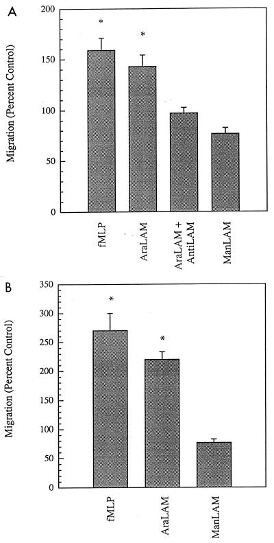
Because we had previously demonstrated that both AraLAM and ManLAM could induce T-cell migration in vitro (4), we sought to determine if these molecules could similarly induce the migration of monocytic cells. AraLAM and ManLAM were compared for the ability to induce the migration of MDM in vitro. We observed that AraLAM and fMLP, but not ManLAM, induced migration of these cells (Fig. 1A). Similar responses were observed when PBM were used in the migration assay; ManLAM failed to induce a migratory response in PBM and reproducibly inhibited random migration of these cells (Fig. 1B). The maximal migration index (percent migration relative to control cells) exhibited substantial donor variability and ranged between 139 and 289% of control (unstimulated) migration. Because these assays are performed under serum-free conditions, the migratory response is clearly serum independent. Migration to AraLAM was not due to contaminating LPS, because LPS alone failed to induce cell migration even at concentrations as high as 100 ng/ml (data not shown). The specificity of this migration-inducing activity in MDM was subsequently confirmed by using an anti-LAM MAb (CS-35) previously shown to bind both AraLAM and ManLAM with high affinity (26). We found that induction of MDM migration by AraLAM was completely blocked by coincubation with the anti-LAM MAb (Fig. 1A), whereas an isotype control antibody did not block migration (data not shown).
FIG. 1.
PBM and MDM chemotactic responses to AraLAM and ManLAM. Cell migration was measured in modified Boyden chambers, and data are expressed as a percent of unstimulated (control) migration in medium alone. (A) Forty-eight-hour MDM migration induced by fMLP (100 nM), AraLAM (1 μg/ml), AraLAM plus anti-LAM MAb CS-35 (1 μg/ml), and ManLAM (1 μg/ml). The anti-LAM MAb alone had no effect on cell migration (not shown; n = 4 separate experiments from different donors). (B) Fresh (0-h) PBM migration induced by fMLP (100 nM), AraLAM (1 μg/ml), and ManLAM (1 μg/ml). Asterisks denote statistically significant migration compared with controls (P < 0.05, n = 3).
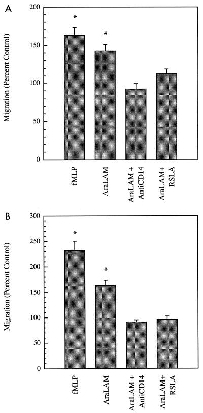
Zhang and colleagues previously reported that antibodies against the LPS receptor CD14 were capable of specifically blocking AraLAM-induced cytokine production (40). Therefore, we assessed the ability of an anti-human CD14 MAb (3C10) to block AraLAM-induced migration of MDM. As shown in Fig. 2A, we found that migration of MDM to AraLAM was completely blocked by the anti-CD14 MAb. In addition, we observed that AraLAM-induced migration could be blocked by the LPS antagonist RSLA (Fig. 2A). AraLAM-induced migration of PBM was also blocked by anti-CD14 MAb and RSLA (Fig. 2B). Together, these studies suggest that the migratory response of MDM to AraLAM is mediated by CD14 and is reminiscent of the capacity of AraLAM (but not ManLAM) to induce cytokine production in human and murine mononuclear phagocytes (11, 13, 28, 40, 41). We subsequently used checkerboard analysis to demonstrate that migration of MDM to AraLAM is chemotactic, not simply chemokinetic (data not shown). Because chemotactic responses are typically initiated via receptor-mediated mechanisms, our observations are consistent with role of CD14 as the receptor for AraLAM.
FIG. 2.
Blocking of MDM and PBM chemotactic responses to AraLAM by anti-CD14 and RSLA. MDM (A) and PBM (B) migration to AraLAM (1 μg/ml) was measured in Boyden chambers as described for Fig. 1. The abilities of the anti-CD14 MAb 3C10 (1 μg/ml) and the LPS antagonist RSLA (1 μg/ml) to inhibit this response were determined. Cells were preincubated with the anti-CD14 MAb or RSLA for 5 min at 4°C prior to LAM treatment. Anti-CD14 or RSLA alone had no effect on macrophage migration (not shown). Asterisks denote statistically significant migration compared with controls (P < 0.05, n = 3).
AraLAM and ManLAM differentially induce transient changes in [Ca2+]in in PBM and MDM.
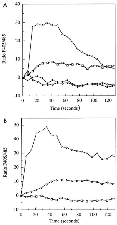
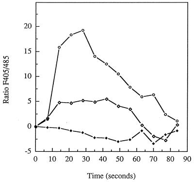
We examined the ability of LAM to induce an intracellular Ca2+ signal in PBM and MDM. For these experiments, cells were loaded with the intracellular fluorescent probe Indo-1 AM and then stimulated with either fMLP (100 nM), AraLAM (1 μg/ml), or ManLAM (1 μg/ml). Changes in [Ca2+]in were continuously recorded as the Indo-1 405 nm/485 nm fluorescence emission ratio (the F405/485 ratio, which is proportional to [Ca2+]in) by FACS as previously described (5, 34, 35). Both PBM (Fig. 3A) and MDM (Fig. 3B) responded to fMLP with a rapid [Ca2+]in flux, as previously reported. When stimulated with LAM, PBM responded only to AraLAM, demonstrating a slow [Ca2+]in response (Fig. 3A). In contrast, ManLAM failed to induce a [Ca2+]in flux in the PBM. Gating of responsive cell subpopulations demonstrated that this response was restricted to 12 to 20% of cells displaying a CD14hi phenotype (data not shown). We subsequently evaluated the response of MDM to LAM. In contrast to the PBM, MDM developed a [Ca2+]in flux only in response to ManLAM (Fig. 3B); this response was slower and smaller than the fMLP-induced transient. FACS analysis revealed that this response was also restricted to a small subpopulation of cells (<18% [not shown]). Lastly, the [Ca2+]in response of MDM to ManLAM, but not to fMLP, was completely blocked by coincubation of the cells with an anti-MMRc MAb prior to stimulation (Fig. 4). Treatment of MDM with the LPS antagonist RSLA also blocked the ManLAM-induced [Ca2+]in response (data not shown). In contrast, it has been previously reported that LPS fails to induce a rapid [Ca2+]in flux in PBM or MDM (19).
FIG. 3.
[Ca2+]in response to AraLAM or ManLAM in 0- and 48-h monocytes/macrophages. Cells were loaded with the fluorescent probe Indo-1 and then stimulated with either fMLP (100 nM; open circles), AraLAM (1 μg/ml; squares), AraLAM plus anti-CD14 (each at 1 μg/ml; closed circles), or ManLAM (1 μg/ml; diamonds). Changes in [Ca2+]in were continuously recorded as changes in Indo-1 F405/485 ratio (proportional to [Ca2+]in by FACS. Representative tracings from each series of experiments are illustrated. (A) Fresh (0-h) PBM demonstrated brisk responses to fMLP stimulation and to AraLAM; there was no response to ManLAM (n = 5). Gating of responsive cell subpopulations revealed that 13.6% of PBM responded to AraLAM (not shown). (B) With 48-h MDM, [Ca2+]in responses to fMLP were similar to those of PBM. ManLAM induced a slow [Ca2+]in signal, whereas there was no response to AraLAM (n = 5). Gating of responsive cell subpopulations revealed that 12.4% of MDM responded to ManLAM (not shown).
FIG. 4.
Inhibition of the MDM [Ca2+]in response to ManLAM by an anti-MMRc MAb. Indo-1-loaded MDM were stimulated with fMLP (circles) or ManLAM (open diamonds), and changes in [Ca2+]in (F405/485 ratio) were continuously recorded by FACS as described for Fig. 3. In addition, aliquots of cells were reacted with an anti-MMRc MAb (1 μg/ml) for 5 min on ice prior to warming (37°C) and ManLAM stimulation (closed diamonds). Data illustrated are representative of three separate experiments from different donors. Addition of the anti-MMRc MAb alone to MDM at 37°C had no effect on resting cells or on subsequent fMLP-induced changes in [Ca2+]in (not shown).
Determination of surface receptor expression in cells exposed to LAM.
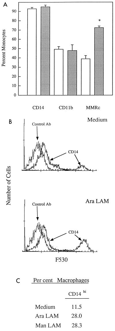
We used flow cytometry to determine if prolonged exposure of cells to LAM could alter the expression of selected cell surface molecules. Incubation of mononuclear phagocytes for 48 h in medium alone had no effect on surface expression of CD14 or CR3 (CD11b/CD18) compared with freshly isolated cells, whereas mannose receptor expression was up-regulated (Fig. 5A). The expression of total CD14, CR3, and formyl peptide receptors was then measured in cells treated with AraLAM (1 μg/ml) or ManLAM (1 μg/ml) for 48 h. All MDM stained for CD14, regardless of whether they were incubated in the presence or absence of AraLAM or ManLAM (Fig. 5B). Similarly, there was no change in CD11b or in formyl peptide receptor expression following incubation with LAM (data not shown). However, treatment of cells with LAM was consistently associated with an increase in a CD14hi subpopulation of MDM compared to cells incubated in medium alone (Fig. 5B), suggesting a divergence between this marker of cell maturation and function (6, 42). Cells incubated 48 h in medium alone and then exposed to AraLAM or ManLAM for 20 min prior to staining with anti-CD14 demonstrated no difference in surface CD14 expression from untreated cells, indicating that LAM did not interfere with the ability of the antibody to bind CD14 (data not shown). Binding of LAM into the plasma membrane of THP-1 cells was previously reported not to interfere with the expression of CD14 on these cells (20). These findings suggest that LAM signaling induced via CD14 involves engagement of epitopes distinct from those recognized by the anti-CD14 MAb.
FIG. 5.
Surface phenotypes of PBM and MDM incubated without and with LAM. (A) Surface phenotype of macrophages incubated in medium alone. Fresh PBM (0 h; open bars) and 48-h MDM (hatched bars) on ice were stained for CD14, CD11b, and MMRc and analyzed by FACS. The percentage of labeled cells is shown on the ordinate. The asterisk denotes statistically significant changes in receptor expression (P < 0.05, n = 4). (B) Effect of LAM on CD14 subpopulation phenotype. MDM incubated for 48 h (37°C, 5% CO2) in medium alone, AraLAM (1 μg/ml), or ManLAM (1 μg/ml) were stained with FITC-labeled anti-CD14 MAb TUK4 (0°C) and analyzed by FACS. Representative histograms obtained in assays using untreated (medium alone; top histogram) and AraLAM-treated (bottom histogram) MDM stained with the anti-CD14 MAb and with an isotype control MAb are illustrated. ManLAM incubation resulted in a CD14 staining pattern similar to that obtained after incubation with AraLAM (histogram not shown). (C) Percentage of cells expressing high levels of surface CD14 (CD14hi) following 48 h of treatment with AraLAM or ManLAM compared with cells incubated in medium (n = 4).
Down-regulation of fMLP responsiveness in cells exposed to LAM.
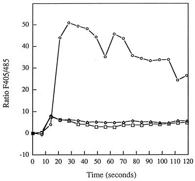
We previously noted that the responsiveness of MDM to fMLP increases with time in culture, as measured by increased [Ca2+]in (5). We therefore measured the effect of LAM on fMLP-induced [Ca2+]in responsiveness in MDM. Cells were treated for 48 h with LAM as described above and stimulated with fMLP, and [Ca2+]in fluxes were then measured. As shown in Fig. 6, fMLP responsiveness was markedly decreased in cells exposed to either AraLAM or ManLAM, whereas fMLP binding to these cells was not affected (data not shown). These studies indicate that LAM can selectively reduce fMLP responsiveness in MDM that otherwise develop enhanced fMLP responsiveness with time in culture.
FIG. 6.
Effect of LAM incubation on MDM [Ca2+]in response to fMLP. MDM were incubated for 48 h (37°C, 5% CO2) without (circles) or with AraLAM (1 μg/ml; squares) or ManLAM (1 μg/ml; diamonds) prior to determination of the intracellular [Ca2+] response to fMLP (100 nM). Changes in [Ca2+]in are shown as in Fig. 3 and 4. Data illustrated are representative of four separate experiments.
Down-regulation of complement receptor function in cells exposed to LAM.
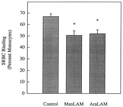
Our earlier findings showed that LAM did not affect CR3 levels, as measured by flow cytometry. The possibility remained that LAM affects complement receptor function. This possibility is predicated on the known ability of complement receptor function to be altered without affecting the expression of these receptors on the cell surface (16) and was assessed by measuring the effect of LAM treatment on the ability of MDM to bind opsonized SRBC. Mononuclear phagocytes were incubated for 48 h without (control), or in the presence of AraLAM (1 μg/ml) or ManLAM (1 μg/ml), and then assayed for the ability to bind opsonized SRBC. As shown in Fig. 7, incubation with either AraLAM or ManLAM reduced opsonized SRBC binding compared to control cells. This decreased binding was observed despite our earlier finding that LAM had no effect on CR3 receptor expression on these cells. This finding suggests that LAM exerts a posttranslational effect on the function of complement receptors (16).
FIG. 7.
Effect of LAM incubation on complement receptor CR3 function of MDM. MDM were incubated without (control) or with AraLAM or ManLAM (each at 1 μg/ml) for 48 h (37°C, 5% CO2) and were then assayed for binding of opsonized SRBC by light microscopy as described in Materials and Methods. Asterisks denote a statistically significant reduction in binding compared with control cells (P < 0.05, n = 4).
DISCUSSION
In this paper, we report that AraLAM and ManLAM can induce distinct CD14- and MMRc-dependent responses in human monocytic cells and that these responses are highly dependent on the differentiation state of the cells. Purified AraLAM, but not ManLAM, possesses chemoattractant activity for human monocytic cells under serum-free conditions in vitro, and this activity is specifically inhibited by an anti-CD14 MAb or by the LPS antagonist RSLA. This chemoattractant activity is dependent on the presence of a concentration gradient, demonstrating that AraLAM is chemotactic for these cells. Unlike the chemotactic response, AraLAM induces a transient rise in free cytosolic calcium levels in PBM but not MDM. This calcium flux could be blocked by either anti-CD14 antibodies or RSLA. In contrast, ManLAM induced a transient rise in free cytosolic calcium levels in MDM but not PBM. This response could be blocked by either anti-CD14 antibodies, RSLA, or anti-MMRc antibodies. This finding suggests functional ligation of these two receptors is needed to initiate a calcium response. ManLAM-responsive MDM were found to be a subpopulation of cells which progressively acquired high levels of CD14 expression on their surface when cultured ex vivo. Lastly, prolonged exposure of MDM to either AraLAM or ManLAM resulted in a selective decrease in fMLP responsiveness and CR3 function.
CD14 and the MMRc have been previously shown to participate in both the binding of M. tuberculosis to monocytic cells and the subsequent activation of these cells, by serving as receptors for LAM (27, 31). The differential activation of these cells by AraLAM and ManLAM suggests that LAM may be a virulence factor involved in the persistence of M. tuberculosis within macrophages (9, 28, 40, 41). Our data extend these previous findings by demonstrating that AraLAM is chemotactic for monocytic cells and that this response is mediated by CD14. Although LPS and AraLAM share the capacity to induce a variety of proinflammatory cytokines and chemokines, it is interesting that LPS does not share the chemotaxis-inducing activity of AraLAM. Furthermore, calcium fluxes could be induced in mononuclear phagocytes by AraLAM and ManLAM (depending on the differentiation state on the cell), or by cross-linking CD14 (3), but not by LPS alone. Thus, there appear to be some CD14-dependent responses that are shared by LAM and LPS and other CD14-dependent responses that are not shared. This possibility is supported by the recent demonstration that both LPS and AraLAM could induce TNF production by human neutrophils, although only LPS induced cell adherence (23). The mechanism by which engagement of CD14 leads to cellular activation in monocytic cells remains unclear. Numerous studies have suggested that CD14 does not signal directly but interacts with an additional protein(s) required to subsequently effect a signal transduction event (15, 21, 22). One possibility is that distinct CD14-associated signaling molecules are involved in different AraLAM-induced responses. We recently reported that a transfected human fibroblast line which constitutively expresses CD14 can be activated by LPS but not by AraLAM (30). These data suggest that the transfected fibroblasts possess a CD14-associated signal-transducing molecule required for LPS activation but lack a distinct molecule required for activation by AraLAM.
The ability of ManLAM to induce a calcium flux in MDM is one of the few indications that ManLAM can induce a specific response in these cells. Previous studies have shown that ManLAM is a weak inducer of most cytokines (11) and of NF-κB translocation (8). In contrast, ManLAM has been reported to block gamma interferon-induced macrophage microbicidal activity (1). Another study showed that ManLAM induced nuclear translocation of the transcription factor KBF-1 in murine macrophages (8). KBF-1, a homodimer of the NF-κB subunit protein p50, can function as a transcriptional repressor by blocking the binding of the NF-κB p50-p65 heterodimer to DNA (18). Lastly, both AraLAM and ManLAM have been recently reported to induce similar levels of TGF-β expression in human monocytes (13). Together these studies suggest that ManLAM is an effective inducer of the suppressive cytokine TGF-β, while it is a poor inducer of proinflammatory cytokines (e.g., IL-1 and TNF) and other antimicrobial effector molecules (e.g., nitric oxide).
The ability of an anti-MMRc MAb to block the ManLAM-induced calcium flux in MDM is not unexpected in light of the ability of the MMRc to serve as a receptor for ManLAM (31, 32). A recent study demonstrated that engagement of the MMRc induced selective cytokine expression in macrophages, suggesting that the MMRc is an important signaling receptor (39). Our data also support a role for the MMRc in macrophage signaling. The known up-regulation of MMRc expression during the differentiation of monocytes to macrophages is consistent with our finding that ManLAM-induced a calcium flux in MDM but not PBM. Unexpectedly, we observed that an anti-CD14 MAb and RSLA could also block the ManLAM-induced calcium flux in MDM. While there are several possible explanations for these findings, we propose a model in which LAM induces a calcium flux by coligating distinct receptors. Specifically, ManLAM may need to coligate both CD14 and the MMRc in order to generate a calcium signal. Furthermore, coligation of CD14 and the MMRc may induce a signal that inhibits the migratory response in both PBM and MDM that is independent of the Ca2+ response. Thus, antibodies against either CD14 or the MMRc could block the calcium response. Similarly, AraLAM may need to coligate both CD14 and a second, unidentified receptor in order to generate a calcium signal. In this model, we invoke a second receptor that is expressed on PBM, but not MDM, because AraLAM does not evoke a calcium response in MDM and yet is capable of inducing MDM migration in vitro. While the cross-linking of CD14 alone has been shown to induce a delayed calcium flux (3), the inability of LPS or AraLAM to evoke a rapid calcium response in CD14+ MDM further supports our proposal that coligation of an additional receptor is required to generate a calcium transient. This model is in agreement with a report that coligation of CD31 (PEACAM-1) and FcγRII could specifically transduce activation signals leading to cytokine production in human peripheral blood mononuclear cells (12).
We have also reported here that exposure of PBM to LAM for 48 h leads to selective changes in both the phenotype and the function of the cells. CR3 function (i.e., ability to bind C3bi-coated SRBC) and fMLP responsiveness, as measured by induction of a rapid calcium flux, were reduced following exposure to either AraLAM or ManLAM, although no changes in their surface receptor expression were observed. Incubation in LAM, however, increased the subpopulation of MDM that expressed the CD14hi phenotype. It remains to be determined if these functional and phenotypic changes are mediated through the binding of LAM to CD14. We have previously shown that both AraLAM and ManLAM are chemotactic for human peripheral blood T cells, cells that lack both CD14 and the MMRc (4). This finding suggests that LAM can induce cellular responses through additional receptors. We have also reported that fMLP-induced [Ca2+]in responsiveness of MDM is restricted to the CD14hi subpopulation induced by adherence to plastic (6). Although the functional consequences of the phenotypic and functional changes on monocytic cells reported here are currently unknown, these findings allow us to distinguish between cell responses that are predominantly directed by AraLAM (e.g., production of proinflammatory cytokines and chemotaxis) and responses that are equally directed by AraLAM and ManLAM (e.g., alteration of CD14 expression, suppression of fMLP responsiveness, and CR3 function). In the former case, responses may depend on the ability of LAM to coligate CD14 and an additional receptor whose expression is dependent on the differentiation state of the cells.
It should be noted that M. tuberculosis strains that differ in their degrees of virulence all possess LAM which is terminally mannosylated (i.e., ManLAM), although other subtle structural alterations appear to vary between strains of M. tuberculosis (32). While the role of ManLAM in the pathogenesis of tuberculosis remains unclear, data from several laboratories support the possibility that ManLAM contributes to mycobacterial virulence by promoting bacterial adhesion to mononuclear phagocytes (32) while failing to induce significant cytokine or nitric oxide production (2, 28). This may serve to minimize the induction of microbicidal activities in infected macrophages via autocrine and/or paracrine cytokine production. The inability of ManLAM, unlike AraLAM, to induce the chemotaxis of monocytic cells may benefit the pathogen in vivo by minimizing the recruitment of additional alveolar macrophages or peripheral blood monocytes to the site of infection. Other studies concluded that the mechanisms by which M. tuberculosis evades killing by macrophages are independent of the receptor-mediated route of entry (24, 43). Thus, mycobacterial virulence is clearly multifactorial in nature and remains a central question in the study of tuberculosis.
ACKNOWLEDGMENTS
This work was supported by grants from the National Institutes of Health to J.B. (HL-46563 and HL-03035), E.R.S. (DK-31056 and DK-51478), and M.J.F. (HL-55681). ManLAM and AraLAM were provided by John Belisle under NIH contract NO1 AI-25147.
REFERENCES
- 1.Adams L B, Fukutomi Y, Krahenbuhl J L. Regulation of murine macrophage effector functions by lipoarabinomannan from mycobacterial strains with different degrees of virulence. Infect Immun. 1993;61:4173–4181. doi: 10.1128/iai.61.10.4173-4181.1993. [DOI] [PMC free article] [PubMed] [Google Scholar]
- 2.Barnes P F, Chatterjee D, Abrams J S, Lu S, Wang E, Yamamura M, Brennan P J, Modlin R L. Cytokine production induced by Mycobacterium tuberculosis lipoarabinomannan. Relationship to chemical structure. J Immunol. 1992;149:541–547. [PubMed] [Google Scholar]
- 3.Beekhuizen H, Blokl I, van Furth R. Cross-linking of CD14 molecules on monocytes results in a CD11/CD18- and ICAM-1-dependent adherence to cytokine-stimulated human endothelial cells. J Immunol. 1993;150:950–959. [PubMed] [Google Scholar]
- 4.Berman J S, Blumenthal R L, Kornfeld H, Cook J A, Cruikshank W W, Vermeulen M W, Chatterjee D, Belisle J T, Fenton M J. Chemotactic activity of mycobacterial lipoarabinomannans for human blood T lymphocytes in vitro. J Immunol. 1996;156:3828–3835. [PubMed] [Google Scholar]
- 5.Bernardo J, Brink H, Simons E R. Time dependence of transmembrane potential changes and intracellular calcium fluxes in stimulated human monocytes. J Cell Physiol. 1988;134:131–136. doi: 10.1002/jcp.1041340116. [DOI] [PubMed] [Google Scholar]
- 6.Bernardo, J., A. Billingslea, K. F. Seetoo, J. Macauley, and E. R. Simons. Adherence-dependent calcium signaling in monocytes: induction of a CD14-high phenotype subpopulation. J. Immunol. Methods, in press. [DOI] [PubMed]
- 7.Bernardo J, Newburger P E, Brennan L, Brink H F, Bresnick S, Weil G, Simons E R. Simultaneous flow cytometric measurements of cytoplasmic and membrane potential changes upon fMLP exposure as HL-60 cells mature into granulocytes: using [Ca2+]i as an indicator of granulocyte maturity. J Leukocyte Biol. 1990;47:265–274. doi: 10.1002/jlb.47.3.265. [DOI] [PubMed] [Google Scholar]
- 8.Brown M C, Taffet S M. Lipoarabinomannans derived from different strains of Mycobacterium tuberculosis differentially stimulate the activation of NF-κB and KBF-1 in murine macrophages. Infect Immun. 1995;63:1960–1968. doi: 10.1128/iai.63.5.1960-1968.1995. [DOI] [PMC free article] [PubMed] [Google Scholar]
- 9.Chan J, Fan X D, Hunter S W, Brennan P J, Bloom B R. Lipoarabinomannan, a possible virulence factor involved in persistence of Mycobacterium tuberculosis within macrophages. Infect Immun. 1991;59:1755–1761. doi: 10.1128/iai.59.5.1755-1761.1991. [DOI] [PMC free article] [PubMed] [Google Scholar]
- 10.Chatterjee D, Lowell K, Rivoire B, McNeil M, Brennan P J. Lipoarabinomannan of Mycobacterium tuberculosis. Capping with mannosyl residues in some strains. J Biol Chem. 1992;267:6234–6239. [PubMed] [Google Scholar]
- 11.Chatterjee D, Roberts A D, Lowell K, Brennan P J, Orme I M. Structural basis of capacity of lipoarabinomannan to induce secretion of tumor necrosis factor. Infect Immun. 1992;60:1249–1253. doi: 10.1128/iai.60.3.1249-1253.1992. [DOI] [PMC free article] [PubMed] [Google Scholar]
- 12.Chen W, Knapp W, Majdic O, Stockinger H, Bohmig G A, Zlabinger G. Co-ligation of CD31 and Fc gamma RII induces cytokine production in human monocytes. J Immunol. 1994;152:3991–3997. [PubMed] [Google Scholar]
- 13.Dahl K E, Shiratsuchi H, Hamilton B D, Ellner J J, Toossi Z. Selective induction of transforming growth factor β in human monocytes by lipoarabinomannan of Mycobacterium tuberculosis. Infect Immun. 1996;64:399–405. doi: 10.1128/iai.64.2.399-405.1996. [DOI] [PMC free article] [PubMed] [Google Scholar]
- 14.Davies T, Bernardo J, Lazzari K, Brennan L, Simons E R. Cytosolic calcium determination: a fluorometric technique. Methods Nutr Biochem. 1991;2:102–106. [Google Scholar]
- 15.Delude R, Savedra R, Zhao H, Thieringer R, Yamamoto S, Fenton M J, Golenbock D T. CD14 potentiates cellular responses to endotoxin without imparting ligand-specific recognition. Proc Natl Acad Sci USA. 1995;92:9288–9292. doi: 10.1073/pnas.92.20.9288. [DOI] [PMC free article] [PubMed] [Google Scholar]
- 16.Diamond M, Springer T. The dynamic regulation of integrin adhesiveness. Curr Biol. 1994;4:506–517. doi: 10.1016/s0960-9822(00)00111-1. [DOI] [PubMed] [Google Scholar]
- 17.Fenton M J, Vermeulen M W. Immunopathology of tuberculosis: roles of macrophages and monocytes. Infect Immun. 1996;64:683–690. doi: 10.1128/iai.64.3.683-690.1996. [DOI] [PMC free article] [PubMed] [Google Scholar]
- 18.Grimm S, Baeuerle P A. The inducible transcription factor NF-κB: structure-function relationship of its protein subunits. Biochem J. 1993;290:297–308. doi: 10.1042/bj2900297. [DOI] [PMC free article] [PubMed] [Google Scholar]
- 19.Hurme M, Viherluoto J, Nordstrom T. The effect of calcium mobilization on LPS-induced IL-1β production depends on the differentiation stage of the monocytes/macrophages. Scand J Immunol. 1992;36:506–511. doi: 10.1111/j.1365-3083.1992.tb02966.x. [DOI] [PubMed] [Google Scholar]
- 20.Ilangumaran S, Arni S, Poincelet M, Theler J-M, Brennan P J, ud-Din N, Hoessli D. Integration of mycobacterial lipoarabinomannans into GPI-rich domains of lymphomonocytic cell plasma membranes. J Immunol. 1995;155:1334–1342. [PubMed] [Google Scholar]
- 21.Kitchens R L, Ulevitch R J, Munford R S. Lipopolysaccharide (LPS) partial structures inhibit responses to LPS in a human macrophage cell line without inhibiting LPS uptake by a CD14-mediated pathway. J Exp Med. 1992;176:485–494. doi: 10.1084/jem.176.2.485. [DOI] [PMC free article] [PubMed] [Google Scholar]
- 22.Lee J-D, Kravchenko V, Kirkland T, Han J, Mackman N, Moriarty A, Leturcq D, Tobias P, Ulevitch R. GPI-anchored or integral membrane forms of CD14 mediate identical cellular responses to endotoxin. Proc Natl Acad Sci USA. 1993;90:9930–9934. doi: 10.1073/pnas.90.21.9930. [DOI] [PMC free article] [PubMed] [Google Scholar]
- 23.Nick J A, Brown K, Worthen G S. Functional responses and intracellular signaling of human neutrophils by lipoarabinomannan. Am J Respir Crit Care Med. 1997;155:A336. [Google Scholar]
- 24.Ortalo-Magne A, Andersen A B, Daffe M. The outermost capsular arabinomannans and other mannoconjugates of virulent and avirulent tubercle bacilli. Microbiology. 1996;142:927–935. doi: 10.1099/00221287-142-4-927. [DOI] [PubMed] [Google Scholar]
- 25.Peterson P, Gekker G, Hu S, Sheng W, Anderson W, Ulevitch R, Tobias P, Gustafson K, Molitor T, Chao C. CD14 receptor-mediated uptake of nonopsonized Mycobacterium tuberculosis by human microglia. Infect Immun. 1995;63:1598–1602. doi: 10.1128/iai.63.4.1598-1602.1995. [DOI] [PMC free article] [PubMed] [Google Scholar]
- 26.Prinzis S, Chatterjee D, Brennan P J. Structure and antigenicity of lipoarabinomannan from Mycobacterium bovis BCG. J Gen Microbiol. 1993;139:2649–2658. doi: 10.1099/00221287-139-11-2649. [DOI] [PubMed] [Google Scholar]
- 27.Pugin J, Heumann D, Tomasz A, Kravchenko V, Akamatsu Y, Nishijima M, Glauser M, Tobias P, Ulevitch R. CD14 is a pattern recognition receptor. Immunity. 1994;1:509–516. doi: 10.1016/1074-7613(94)90093-0. [DOI] [PubMed] [Google Scholar]
- 28.Roach T I, Barton C H, Chatterjee D, Blackwell J M. Macrophage activation: lipoarabinomannan from avirulent and virulent strains of Mycobacterium tuberculosis differentially induces the early genes c-fos, KC, JE, and tumor necrosis factor-alpha. J Immunol. 1993;150:1886–1896. [PubMed] [Google Scholar]
- 29.Sada E, Brennan P J, Herrera T, Torres M. Evaluation of lipoarabinomannan for the serological diagnosis of tuberculosis. J Clin Microbiol. 1990;2:2587–2590. doi: 10.1128/jcm.28.12.2587-2590.1990. [DOI] [PMC free article] [PubMed] [Google Scholar]
- 30.Savedra R, Delude R L, Ingalls R R, Fenton M J, Golenbock D T. Mycobacterial lipoarabinomannan recognition requires a receptor that shares components of the endotoxin signaling system. J Immunol. 1996;157:2549–2554. [PubMed] [Google Scholar]
- 31.Schlesinger L S, Hull S R, Kaufman T M. Binding of the terminal mannosyl units of lipoarabinomannan from a virulent strain of Mycobacterium tuberculosis to human macrophages. J Immunol. 1994;152:4070–4079. [PubMed] [Google Scholar]
- 32.Schlesinger L S, Kaufman T M, Iyer S, Hull S R, Marchiando L K. Differences in mannose receptor-mediated uptake of lipoarabinomannan from virulent and attenuated strains of Mycobacterium tuberculosis by human macrophages. J Immunol. 1996;157:4568–4575. [PubMed] [Google Scholar]
- 33.Stahl P. The macrophage mannose receptor: current status. Am J Respir Cell Mol Biol. 1990;2:317–319. doi: 10.1165/ajrcmb/2.4.317. [DOI] [PubMed] [Google Scholar]
- 34.Strohmeier G R, Brunkhorst B A, Seetoo K F, Bernardo J, Simons E R. Neutrophil functional responses depend upon immune complex valency. J Leukocyte Biol. 1995;58:403–414. doi: 10.1002/jlb.58.4.403. [DOI] [PubMed] [Google Scholar]
- 35.Strohmeier G R, Brunkhorst B A, Seetoo K F, Meshulam T, Bernardo J, Simons E R. The role of Fc-gamma R subclasses, Fc-gamma RII and RIII, in the activation of human neutrophils by low and high valence immune complexes. J Leukocyte Biol. 1995;58:415–422. doi: 10.1002/jlb.58.4.415. [DOI] [PubMed] [Google Scholar]
- 36.Sturgill-Koszycki S, Schlesinger P H, Chakraborty P, Haddix P L, Collins H L, Fok A K, Allen R D, Gluck S L, Heuser J, Russell D G. Lack of acidification in Mycobacterium phagosomes produced by exclusion of the vesicular proton-ATPase. Science. 1994;263:678–681. doi: 10.1126/science.8303277. [DOI] [PubMed] [Google Scholar]
- 37.VanVliet K E, DeGraaf-Miltenburg L A M, Verhoef J, VanStrijp J A G. A flow cytometric rosetting assay for the analysis of Fc receptors and C3 receptors on HSV-infected cells. J Immunol Methods. 1993;157:57–64. doi: 10.1016/0022-1759(93)90070-n. [DOI] [PubMed] [Google Scholar]
- 38.Xu S, Cooper A, Sturgill-Koszycki S, van Heyningen T, Chatterjee D, Orme I M, Allen P, Russell D. Intracellular trafficking in Mycobacterium tuberculosis and Mycobacterium avium-infected macrophages. J Immunol. 1994;153:2568–2578. [PubMed] [Google Scholar]
- 39.Yamamoto Y, Klein T W, Friedman H. Involvement of the mannose receptor in cytokine interleukin-1β (IL-1β), IL-6, granulocyte-macrophage colony stimulating factor responses, but not in chemokine macrophage inflammatory protein 1β (MIP-1β), MIP-2, and KC responses, caused by attachment of Candida albicans to macrophages. Infect Immun. 1997;65:1077–1082. doi: 10.1128/iai.65.3.1077-1082.1997. [DOI] [PMC free article] [PubMed] [Google Scholar]
- 40.Zhang Y, Broser M, Cohen H, Bodkin M, Law K, Reibman J, Rom W N. Enhanced interleukin-8 release and gene expression in macrophages after exposure to Mycobacterium tuberculosis and its components. J Clin Invest. 1995;95:586–592. doi: 10.1172/JCI117702. [DOI] [PMC free article] [PubMed] [Google Scholar]
- 41.Zhang Y, Doerfler M, Lee T C, Guillemin B, Rom W N. Mechanisms of stimulation of interleukin-1β and tumor necrosis factor-α by Mycobacterium tuberculosis components. J Clin Invest. 1993;91:2076–2083. doi: 10.1172/JCI116430. [DOI] [PMC free article] [PubMed] [Google Scholar]
- 42.Ziegler-Heitbrock H W L, Ulevitch R J. CD14: cell surface receptor and differentiation marker. Immunol Today. 1993;14:121–125. doi: 10.1016/0167-5699(93)90212-4. [DOI] [PubMed] [Google Scholar]
- 43.Zimmerli S, Edwards S, Ernst J D. Selective receptor blockage during phagocytosis does not alter the survival and growth of Mycobacterium tuberculosis in human macrophages. Am J Respir Cell Mol Biol. 1996;15:760–770. doi: 10.1165/ajrcmb.15.6.8969271. [DOI] [PubMed] [Google Scholar]
