Targeted disruption of transcriptional regulatory function of p53 by a novel efficient method for introducing a decoy oligonucleotide into nuclei (original) (raw)
Abstract
Decoy oligonucleotides have been used for functional sequestering of transcription factors. Efficient introduction into cells is a prerequisite for the oligonucleotides to exert their blocking function. Lipofection is the most widely used technique for that purpose because of its convenience and relatively high efficiency. However, the transduction efficiency of lipofection largely depends on cell types and experimental conditions and the introduced nucleotides are not specifically directed to nuclei where they exert their major function. In the present study, we designed a new system for transporting oligonucleotides into cell nuclei. The vehicle is composed of glutathione-_S_-transferase, 7 arginine residues, the DNA-binding domain of GAL4 and a nuclear localization signal, which are linked with flexible glycine stretches. The p53-responsive element linked to the GAL4 upstream activating sequence was efficiently transferred by the vehicle protein into nuclei of primary cultures of neuronal cells, embryonic stem cells and various human normal cells. Transcriptional activation of p21WAF1/CIP1 and Bax by p53 on exposure to cisplatin was completely blocked by introducing the p53 decoy oligonucleotide. Thus, the system developed in the present study can be a convenient and powerful tool for specifically disrupting the function of DNA-binding proteins in culture.
INTRODUCTION
Functional inactivation of specific gene products is a powerful and promising tool not only for studying biological relevance of a given gene but also for developing target gene-based therapeutic measures. The ‘loss-of-function’ of a gene can beachieved by a number of different approaches, including the expression of a dominant negative form of proteins, intracellular introduction of specific antibodies, antisense oligonucleotides, siRNA and decoy oligonucleotides for DNA-binding proteins (1–4). Among those approaches, the decoy oligonucleotide method is advantageous in terms of promptness of effects, simplicity and easiness in handling (5,6).
In cells, gene expression is principally regulated at the transcriptional level by DNA-binding proteins known as transcription factors. Introduction of an excess amount of a DNA segment harboring a _cis_-element into cells results in functional sequestering of the counterpart-transacting factor from acting on the endogenous target promoters. Thus, decoy oligonucleotides, including those for NF-κB, E2F and AP-1, have been successfully used in vivo as well as in vitro (7–13).
Many attempts have been made to improve the efficiency of the decoy oligonucleotide method. Phosphothioation was shown to stabilize decoy oligonucleotides introduced into cells (7,14). Also, oligonucleotides became more stable when both ends were locked by adding an extra 2′-O, 4′-C-methylene bridge to the ribose ring (15). Ahn et al. (16) showed that circular dumbbell oligonucleotides were more resistant to exonuclease, exhibiting more prolonged effects. Another problem to be overcome is how to efficiently introduce decoy oligonucleotides into cells. The most frequently used method to introduce oligonucleotides into cells is liposome-mediated transfection and its utility has been shown in many biological contexts. Recently, however, Bene et al. (17) showed that an AP-1 decoy oligonucleotide remained mainly in the perinuclear cytoplasm and exhibited a marginal biological effect when transfected using a liposome-mediated method. A similar phenomenon was observed in vivo by Griesenbach et al. (18). To address this problem, we designed a new vehicle protein to facilitate the efficient introduction of decoy oligonucleotides into cell nuclei. Here, we show that the vehicle protein efficiently transported a decoy oligonucleotide for p53 into nuclei of various cell types, including primary neuronal cells and mouse embryonic stem (ES) cells, resulting in complete suppression of transcriptional activity of p53.
MATERIALS AND METHODS
Cell culture
A p53-proficient human colon cancer cell line, HCT116, was a kind gift from Dr B. Vogelstein (Johns Hopkins University), and a p53-deficient human prostate cancer cell line, PC3, was provided by ATCC (Rockville, MD). A normal human fibroblast strain, OUMS-36, was provided by Dr M. Namba (Niimi College). DMEM (Nissui, Tokyo, Japan) and HAM'S F-12 K medium were used for HCT116 and OUMS-36 cells and for PC3 cells, respectively, with a supplement of 10% fetal bovine serum (FBS). Normal neonatal human epidermal keratinocytes (NHKs) (Cascade Biologics, Portland, OR) were cultured in an animal-product free medium, EpiLife, with a growth supplement, V2. The mouse ES cell line CGR8 (19) was a kind gift from Dr H. Tanba (Osaka University), and the cells were cultured in DMEM supplemented with 15% FBS, 1% non-essential amino acids solution (Invitrogen, Carlsbad, CA), nucleotide stock solution (30 mM each of adenosine, guanosine, cytidine and uridine, and 10 mM thymidine), 110 μM 2-mercapto ethanol and 1000 U/ml mouse leukocyte inhibitory factor (Sigma). Primary cultures of adult rat hepatocytes and embryonic mouse and rat brain cells were performed as described previously (20,21). DMEM supplemented with 10 μM dexamethasone and 10 μg/ml insulin and DMEM with 10 mM dibutyryl cAMP (Sigma) and 1 mM theophylline (Sigma) were used for the hepatocytes and brain cells, respectively. Human leukocytes were prepared from peripheral blood using a leukocyte purification kit (LeucoSep; Greiner Bio-one, Tokyo, Japan).
Plasmid constructs
A DNA fragment encoding 7 glycine residues (7G) and 7 arginine residues (7R) was synthesized and integrated into a downstream site of glutathione-_S_-transferase (GST) of the vector pGEX6P1 (Amersham Biosciences). The resulting plasmid was designated as pGEX6P1-7R. A PCR was performed with a forward primer encoding 7G and a 5′-region of the DNA-binding domain (DB) of GAL4 (22) (5′-ccggaattccggggaggaggaggaggaggaggaatgaagctactgtcttctat-3′) and a reverse primer encoding a 3′-region of GAL4 DB, 3G, and the nuclear localization signal of SV40 [nuclear localization signal (NLS): PKKKRKV (23)] (5′-tcccccgggggacacctttcgcttcttctttggtcctcctccaatcgatacagtcaactgtc-3′) using a plasmid harboring GAL4 as a template. The amplified fragment was inserted into pGEX6P1 and pGEX6P1-7R to construct pGEX6P1-GAL4-NLS and pGEX6P1-7R-GAL4-NLS, respectively.
To make a decoy oligonucleotide for p53, a DNA fragment encoding the GAL4 upstream activating sequence [UAS; cggagtactgtcctccg; (24)] and two copies of a p53-responsive element (5′-tacagaacatgtctaagcatgctgggg-3′) in tandem with EcoRI linkers at both end (altogether 95 bp in length) was synthesized and integrated into the vector pDNR-CMV (Clontech). To prepare a mutant oligonucleotide, 5′-catg-3′ shown above in bold was changed to 5′-tcgc-3′.
Nucleotide sequences of these constructs were verified by sequencing.
Purification of recombinant proteins
pGEX6P1, pGEX6P1-7R, pGEX6P1-GAL4-NLS and pGEX6P1-7R-GAL4-NLS were transfected into Escherichia coli [BL21-Codon Plus-(DE3)-RIL; Stratagene] to produce proteins composed of GST only, GST-7G-7R, GST-7G-GAL4-3G-NLS and GST-7G-7R-7G-GAL4-3G-NLS, respectively. For purification of nucleic acid-free recombinant proteins, the respective E.coli was frozen and thawed twice in a buffer (20% sucrose, 0.6 M NaCl, 1 mM phenylmethlysulfonyl fluoride, 10 mM Tris–HCl, pH 7.5) followed by sonication for 10 min (Sonifier 250, Bransonn). Contaminated nucleic acid was degradated by incubating with 30 μg/ml DNase I (Sigma), 30 μg/ml RNase A (Sigma) and 10 mM MgCl2. After sonicating again, the lysates were centrifuged for removing insoluble debris and each protein was purified from the supernatants using a Sephadex 4B column (Amersham Biosciences). For the preparation of a protein composed of 7G-7R-7G-GAL4-3G-NLS, GST-7G-7R-7G-GAL4-3G-NLS was cleaved with PreScission protease (Amersham Biosciences) and the GST was removed using the Sephadex 4B column.
Introduction of oligonucleotides into cells
Oligonucleotides were introduced into cells by applying in a mixture with the vehicle proteins in serum-free media for 1 h. Lipofection was performed using Lipofectamine 2000 (Invitrogen).
Labeling and monitoring the fate of the decoy oligonucleotides
The wild-type or mutant decoy oligonucleotide for p53 was prepared by restricting plasmid DNAs and purified by an agarose electrophoresis. Labeling of the decoy oligonucleotides was performed using [γ-32P]ATP (HAS, Budapest, Hungary) and T4 polynucleotide kinase (New England BioLabs, Beverly, MA). For monitoring the fate of the labeled decoy DNA in cells, the introduced oligonucleotides were recovered by extracting cells with a hypotonic buffer (10 mM KCl, 0.1% Triton X-100, 5 mM EDTA, 10 mM Tris–HCl, pH 8.0) followed by another extraction with a buffer (500 mM KCl, 1.0% Triton X-100, 5 mM EDTA, 10 mM Tris–HCl, pH 8.0). The combined extracts were subjected to a conventional phenol–chloroform method.
Intracellular localization of the vehicle protein and oligonucleotides
The purified recombinant proteins and decoy DNA were labeled with Cy3 and Alexa Fluor 488 using a Cy3 Antibody Labeling Kit (Amersham Biosciences) and a ULYSIS Alexa Fluor 488 Nucleic Acid Labeling Kit (Molecular Probes, Eugene, OR), respectively. Intracellular localization of the introduced molecules in living and fixed cells were observed using a fluorescent microscope (IX71-22FL/PH; CCD camera, DP50; objective lens, LCPlan F1 40×; Olympus) and a laser-scanning microscope (Axioplan 2; objective lens, Plan-Apocgomat 63× 1.4 oil DC; Carl Zeiss MicroImaging), respectively.
Western blot analysis
Immunoblotting was performed using a rabbit anti-GST antibody (Amersham Biosciences), a mouse anti-GAL4 (DB) antibody (Santa Cruz Biotechnology, Santa Cruz, CA), a mouse anti-human p53 antibody (Santa Cruz Biotechnology), a rabbit anti-human p21WAF1/CIP1 antibody (Santa Cruz Biotechnology), a rabbit anti-human Bax antibody (Upstate Biotechnology, Lake Placid, NY) or a mouse anti-human tubulin antibody (Sigma), followed by the application of a horseradish peroxidase-conjugated anti-mouse or anti-rabbit IgG antibody (Cell Signaling Technology Inc., Beverly, MA). Positive signals were visualized using a chemiluminescence system (ECL plus, Amersham Biosciences).
Northern blot analysis
Total RNA was isolated by the acid guanidinium thiocyanate/phenol–chloroform method. Northern blot analysis was performed under conventional conditions. Briefly, 20 μg RNA of each sample was fractionated in a 1.0% agarose gel and transferred to a Nytran Plus nylon membrane (Amersham Biosciences). Probes synthesized from entire cDNAs of human p21WAF1/CIP1 and GAPDH were used.
Electrophoresis mobility shift assay
Electrophoresis mobility shift assay (EMSA) was performed under conditions similar to those described by Nakano et al. (25). Briefly, 32P-labeled decoy DNA probes (0.5 μg) were incubated with 0.1–2 μg of crude nuclear extracts of HCT116 or PC3 cells in a reaction mixture (20 μl) containing 60 mM KCl, 5 mM MgCl2, 0.1 mM EDTA, 1 μg poly(dI–dC), 1 mM DTT, 5% glycerol and 20 mM HEPES, pH 7.9. For a supershift assay, 2 μg of purified vehicle proteins and/or 0.5–2 μg of a mouse anti-human p53 antibody (Santa Cruz Biotechnology) were added to the reaction mixture. DNA–protein complexes were then separated by electrophoresis in a 5% polyacrylamide gel under non-denaturing conditions.
Chromatin immunoprecipitation assay
Chromatin immunoprecipitation was performed according to a method described previously (26). In brief, HCT116 cells were cross-linked by adding formaldehyde to 1% final concentration, and p53–DNA complex was immunoprecipitated with a mouse anti-human p53 antibody (Santa Cruz Biotechnology). A p53-responsive region in the p21WAF1/CIP1 promoter was amplified using a forward primer, 5′-gtgcagacagcagtggggcttagag-3′ and a reverse primer, 5′-cagaacccaggcttggagcagctac-3′.
RESULTS
Design of experiments
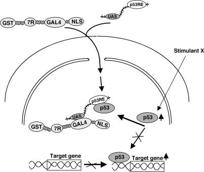
Figure 1 shows the design of a method to efficiently introduce a decoy oligonucleotide into nuclei. Two tandem p53-responsive elements (p53RE) for sequestering p53 protein were linked to the yeast UAS sequence, which binds to GAL4 in the vehicle protein. The vehicle protein (GST-7GR-Ga-NLS; 45.8 kDa) is composed of GST, 7G residues, 7 arginine residues, 7 glycine residues, the DB of GAL4, 3 glycine residues and the NLS from the N-terminal side. The poly-arginine and NLS facilitate the incorporation of proteins into cells and nuclei, respectively (23,27,28). GST is used for the purification of the recombinant proteins and also for promoting cellular incorporation of proteins (Figure 4B). The glycine stretches provide flexible linkages between each domain. The oligonucleotide was expected to be efficiently transferred to nuclei by the help of the vehicle protein, resulting in the inhibition of p53 function by sequestering it.
Figure 1.
A schematic diagram of the method to introduce a decoy oligonucleotide into nuclei. The oligonucleotide is composed of UAS and two p53-responsive elements in tandem (p53RE). The vehicle protein (GST-7GR-Ga-NLS) is composed of GST, 7 glycine residues, 7 arginine residues (7R), 7 glycine residues, the DNA-binding domain (DB) of GAL4, 3 glycine residues and an NLS. UAS is for binding to GAL4 in the vehicle and p53RE sequesters p53 protein. 7R and GST promote the incorporation of the protein into cells. GST is also used for purification of the protein.
Figure 4.
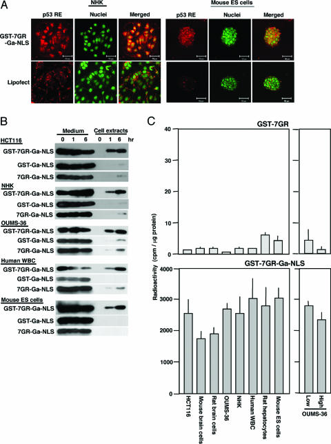
Efficient introduction of the decoy oligonucleotide into diverse types of cells. (A) The decoy oligonucleotide labeled with Alexa 546 (in red) was applied with the vehicle protein or by lipofection onto normal human keratinocytes (NHKs) or mouse ES cells and observed by a confocal laser microscope. Nuclei were stained with SYBR Green (in green). (B) Proteins of different structural compositions were applied to various types of cells, and the media and cell extracts were analyzed by western blotting using anti-GAL4 antibody. GST-7GR-Ga-NLS, the complete vehicle protein; GST-Ga-NLS, a vehicle protein lacking 7 arginine residues; 7GR-Ga-NLS, a vehicle protein lacking GST. (C) Radiolabeled decoy oligonucleotide was applied to various types of cells with the complete vehicle protein (GST-7GR-Ga-NLS) or a protein lacking GAL4 and NLS (GST-7GR), and radioactivity in cell extracts was determined. Low and high indicate low-density and high-density cultures of OUMS-36, human normal diploid fibroblasts.
Specific binding of the decoy oligonucleotide to p53
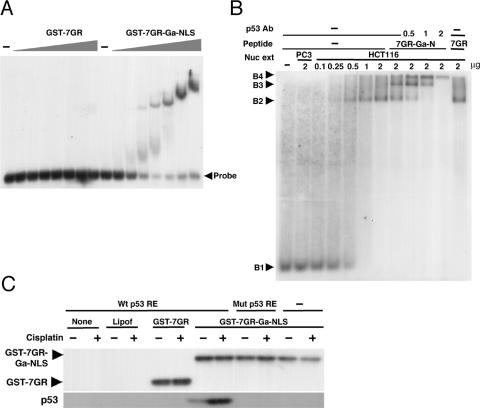
We first examined whether the decoy oligonucleotide binds to the vehicle protein and p53. When the labeled oligonucleotide was incubated with the vehicle protein GST-7GR-Ga-NLS in vitro, it dose dependently bound to the protein as demonstrated by EMSA, whereas it showed no binding activity to the protein lacking GAL4 and NLS domains (GST-7GR) (Figure 2A). The oligonucleotide could simultaneously bind to p53 protein in a nuclear extract prepared from HCT116 cells as well as to the vehicle protein (Figure 2B). Specificity of the binding was confirmed by supershift of the band observed when an antibody against p53 was added to the reaction mixture and also by the absence of the band shift when incubated with a nuclear extract from p53-deficient PC3 cells. To examine whether the oligonucleotide bound to p53 protein within cells, the oligonucleotide was introduced into HCT116 cells by means of lipofection, GST-7GR, or GST-7GR-Ga-NLS, and the vehicle proteins recovered using glutathione beads from cell extracts were analyzed by western blotting (Figure 2C). p53 protein was detected only when the oligonucleotides containing the wild-type p53RE were applied with GST-7GR-Ga-NLS. These results indicate that the oligonucleotide designed in the present study can simultaneously bind to p53 as well as the vehicle GST-7GR-Ga-NLS in vitro and in cells.
Figure 2.
Binding of the decoy oligonucleotide to the vehicle protein. (A) Binding of the decoy oligonucleotide was assessed by EMSA. Labeled decoy nucleotide was incubated with increasing amounts of the vehicle peptide described in the legend to Figure 1 (GST-7GR-Ga-NLS) or that lacking GAL4 and NLS (GST-7GR) and analyzed by electrophoresis. (B) Binding of the decoy nucleotide not only with the vehicle protein but also with p53 protein in a nuclear extract. Labeled decoy nucleotide was incubated with indicated proteins and increasing amounts of nuclear extract prepared from p53-proficient HCT116 cells. In some tubes, an antibody against p53 was added. Arrowheads indicate the following positions: B1, the probe only; B2, the probe bound to p53; B3, the probe bound to p53 and the vehicle protein; and B4, the probe bound to p53, the vehicle protein and p53 antibody. 7GR-Ga-N and 7GR correspond to GST-7GR-Ga-NLS and GST-7GR, respectively. (C) Binding of the decoy oligonucleotide with p53 in cells. The decoy oligonucleotide (wild-type or mutated) was applied by means of lipofection (Lipof), GST-7GR or GST-7GR-Ga-NLS to HCT116 cells treated with cisplatin (10 μg/ml, 6 h) or not treated. Introduced proteins were recovered from cell extracts using glutathione beads and analyzed by western blotting using an antibody against GST (upper panel) or an antibody against p53 (lower panel).
Efficient introduction of the decoy oligonucleotide into different types of cells, particularly into nuclei
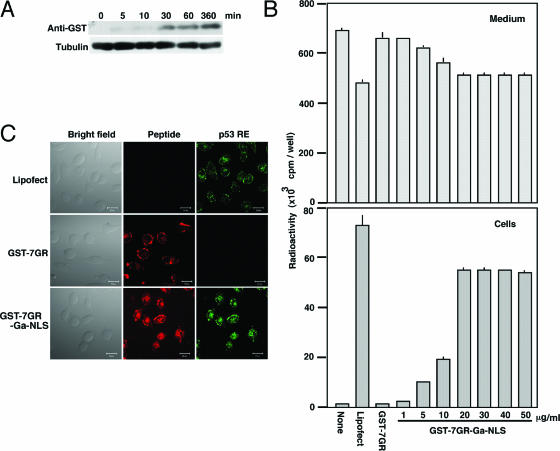
When GST-7GR-Ga-NLS was applied to HCT116 cells, the vehicle protein was first detected in the cells at 30 min and the amount slightly increased up to 360 min after the application (Figure 3A). Incorporation of the labeled oligonucleotide by GST-7GR-Ga-NLS depends on the dose of the vehicle protein as assayed at 7 h after application (Figure 3B). Twenty micrograms of GST-7GR-Ga-NLS was sufficient to attain the maximum introduction to 104 HCT116 cells cultured in 1 ml of medium. Under these conditions, ∼10% of the 200 ng oligonucleotide added to the medium was incorporated into the cells. Although lipofection could also efficiently introduce the oligonucleotide into the cells, most of the incorporated oligonucleotides were found in the perinuclear cytoplasm (Figure 3C, top panels) in accordance with the report by Bene et al. (17). In contrast, the oligonucleotide was detected mostly in nuclei when introduced using GST-7GR-Ga-NLS (Figure 3C, bottom panels). The preferential introduction of the decoy oligonucleotide into nuclei by GST-7GR-Ga-NLS in comparison with lipofection was also observed in NHK cells and mouse ES cells (Figure 4A). Applicability of the present method to a wide variety of cells was confirmed by western blotting (Figure 4B) and by the determination of radioactivity after the application of the labeled oligonucleotide (Figure 4C). GST-7GR-Ga-NLS facilitated the incorporation of the oligonucleotide in primary cultures of mouse and rat brain neuronal cells and rat hepatocytes, in normal human keratinocytes, fibroblasts (OUMS-36) and leukocytes, and in mouse ES cells. Efficiency of the cellular incorporation was not affected by cell density in OUMS-36 (Figure 4C, lower right panel). Either GST or 7-arginine stretch alone could not promote the protein transduction (Figure 4B). GST-7GR-Ga, a vehicle protein lacking NLS, hardly transported oligonucleotides into nuclei (data not shown).
Figure 3.
Introduction of the decoy nucleotide into cells by the vehicle protein. (A) Time course of cellular uptake of the vehicle protein. GST-7GR-Ga-NLS (20 μg) was applied to HCT116 cells and was recovered from cell extracts using glutathione beads at various times after the application. (B) Introduction of the decoy oligonucleotide into HCT116 cells by the vehicle proteins. Radioactivity in cells and medium was determined 6 h after the application of labeled decoy oligonucleotide to HCT116 cells by lipofection (Lipofect), GST-7GR (50 μg/ml) or indicated amounts of GST-7GR-Ga-NLS. (C) Introduction of the decoy oligonucleotide into cell nuclei. The decoy oligonucleotide labeled with Alexa Fluor 488 (in green) was applied to HCT116 cells with the vehicle proteins labeled with Cy3 (in red) and observed 6 h after application. The cells were observed by a confocal laser microscope at a plane horizontally cutting in the middle part of cells.
Inhibition of the function of p53
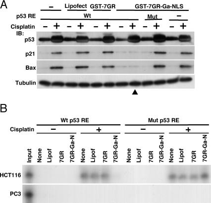
Biological relevance of the decoy oligonucleotide introduction was studied by examining the activity of p53 in transcriptional regulation. The wild-type or mutated decoy oligonucleotide was applied to HCT116 cells with GST-7GR-Ga-NLS. One hour later, cisplatin (10 μg/ml) was added to the medium and the cells were harvested for analysis 6 h after the oligonucleotide application. By treatment with cisplatin, the protein level of p53 was elevated and this was not affected by any treatments for the introduction of oligonucleotides (Figure 5A). In accordance with the reported findings that the p21WAF1/CIP1 and Bax promoters were activated on exposure to cisplatin (29,30), we detected increased amounts of p21WAF1/CIP1 and Bax proteins in the cells treated with cisplatin. The induction of both proteins was completely suppressed when the cells were pretreated with the wild-type oligonucleotide and GST-7GR-Ga-NLS (Figure 5A). To obtain some mechanistic insights, we examined binding of p53 to the p21WAF1/CIP1 promoter by a chromatin immunoprecipitation assay. p53 protein was activated to bind to the p21WAF1/CIP1 promoter by treatment with cisplatin and the binding was completely suppressed when the oligonucleotide harboring the wild-type p53-responsive element, but not that having the mutated element, was introduced by GST-7GR-Ga-NLS (Figure 5B). Although lipofection could introduce the oligonucleotide with efficiency comparable with that by GST-7GR-Ga-NLS (Figure 3B), the induction of p21WAF1/CIP1 and Bax protein (Figure 5A) and the binding p53 to the p21WAF1/CIP1 promoter (Figure 5B) were not appreciably affected. This indicates that transportation of the decoy oligonucleotide to the nuclei and not to the cytoplasm is crucial for modulating function of transacting factors in cells.
Figure 5.
Biological effects of introduction of the decoy oligonucleotide. (A) A decoy oligonucleotide carrying wild-type p53RE or mutated p53RE was introduced into HCT116 cells with or without cisplatin treatment. Cell extracts prepared 6 h after the application were analyzed for indicated proteins by western blotting. Arrowhead indicates the critical lane showing suppression of the function of p53. (B) Chromatin immunoprecipitation assay for p53 protein bound to the p21WAF1/CIP1 promoter. The cells were treated under conditions similar to those described in (A) and immunoprecipitates with anti-p53 antibody were subjected to PCR amplification using primers covering p53 RE in the p21WAF1/CIP1 promoter. PC3 was used as a negative control because of its deficiency of p53. 7GR-Ga-N and 7GR correspond to GST-7GR-Ga-NLS and GST-7GR, respectively.
Stability of the decoy oligonucleotide in cells
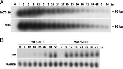
Finally, we examined stability of the incorporated oligonucleotide and duration of the functional sequestering of p53 protein. Labeled decoy oligonucleotide was introduced into HCT116 cells or NHK cells, and the radioactivity recovered from the cells was monitored by autoradiography after electrophoresis. As shown in Figure 6A, the intracellular oligonucleotide levels remained essentially unchanged until 12 h, gradually declined afterwards reaching a level of about half around 24 h, and decreased to an undetectable level around 48 h after the application in both cell types. Duration of the biological effects of the introduced oligonucleotide was studied in NHK cells because HCT116 cells underwent apoptotic cell death by treatment with cisplatin. NHK cells were treated with cisplatin 1 h after the introduction of the wild-type or mutated decoy oligonucleotide into the cells, and then p21WAF1/CIP1 mRNA level was monitored (Figure 6B). In the cells exposed to the mutated p53 decoy oligonucleotide, p21WAF1/CIP1 mRNA was induced and remained at similar levels during the observation period of 72 h. Introduction of the wild-type p53 decoy oligonucleotide completely suppressed the induction of p21WAF1/CIP1 mRNA up to 24 h. A low level of p21WAF1/CIP1 mRNA was detected at 36 h and the amount had increased to a level comparable with that of untreated cells at 48 h. This kinetics accorded well with the amount the decoy oligonucleotide in the cells (Figure 6A).
Figure 6.
Stability of the decoy oligonucleotide in cells. (A) Radiolabeled decoy oligonucleotide was applied with GST-7GR-Ga-NLS to HCT116 or NHK cells and amounts of the intact oligonucleotide (95 bp in length) recovered from the cells was monitored by electrophoresis and autoradiography. (B) Northern blot analysis for p21WAF1/CIP1 mRNA after the introduction of the oligonucleotides. NHK cells were subjected to application of the wild-type or mutated decoy oligonucleotide and GST-7GR-Ga-NLS at the time point 0, exposed to cisplatin (10 μg/ml) from 1 h later and harvested at indicated time points. GAPDH was used as a control for applied amounts of RNA.
DISCUSSION
In the present study, we designed a vehicle protein for efficient introduction of decoy nucleotides into cell nuclei. It is composed of GST, 7 arginine residues, the DB of GAL4 and the NLS of SV40 large T antigen, which are linked with flexible glycine stretches. The present system has the following advantages:
- It is applicable for a broad variety of cells, including primary cultured cells and ES cells (Figure 4B and C).
- It is highly efficient in terms of transduced cell rate (practically 100%) (Figures 3C and 4A).
- It is efficient in terms of targeting the oligonucleotide for cell nuclei (Figure 3C), which is a prerequisite for interfering with transcriptional activation (Figure 5A and B).
- It is prompt and efficient in exhibiting the interfering function (Figures 3A and 5A).
- It is applicable for other DNA-binding proteins whose target elements are known.
- It is easy to use and economical. The recombinant vehicle protein can be produced in a large amount and easily purified and stocked.
In most studies on the effect of decoy oligonucleotides, lipofection has been used for transducing oligonucleotides into cells. Efficiency of transfection by lipofection is high compared with the efficiencies of other DNA transfection methods. However, the efficiency largely depends on cell types. Another potential shortcoming of lipofection is that it does not directly help nuclear translocation of the oligonucleotide, which is critical for interfering with the function of transcription factors (17). In the present study, we attempted to actively transport oligonucleotide into nuclei. As shown in Figure 3C, nearly half of the incorporated oligonucleotide was found in the nuclei, a much larger proportion than that observed in cells transduced by lipofection.
The GST and 7 arginine residues cooperatively facilitate to introduce the protein into cells (Figure 4B). Namiki et al. (31) reported that the fluorescent-dye-labeled GST from Schistosoma japonicum penetrated into mammalian cells even when conjugated to a short-peptide load. In the present system, GST derived from yeast alone could not promote the protein transduction (Figure 4B). A number of different molecules are known to transduce proteins into cells. TAT domain derived from human immunodeficiency virus (32) and 11-arginine stretch (27,28) are widely used for protein transduction. Covalent conjugation of proteins with polyethylenimine is another potent measure for protein transduction (33,34). The strong cation charge of polyethylenimine, however, results in non-specific binding to DNA and is inappropriate for the present purpose.
p53 is known to exert its transactivation capacity through forming a homotetramer (35). At present, it is not clear whether p53 protein formed a tetramer complex in the presence of the decoy oligonucleotides under the present conditions. Since the DNA-binding domain resides in the central part of p53 protein and is apart from the C-terminal tetramerization domain, the p53 protein bound with the oligonucleotides could still form homotetramers. However, the vehicle protein that most likely remains combined with the oligonucleotides may interfere with the association of p53 protein. Chan et al. (36) showed that a mutant p53 defective in DNA binding, unlike p53 mutated in the transactivation domain, was inefficient in exhibiting the dominant negative effect. This indicates inefficiency in inhibiting the transactivation capacity of p53 by blocking its DNA-binding activity. The number of p53 molecules per cell was estimated to be several hundred thousand at most (37). In the present study, ∼10% of the applied amount of the oligonucleotide was incorporated into cells (Figure 3). At the lowest estimate, about one hundred excess molecules of the oligonucleotide were present in cell nuclei in comparison with that of p53 protein, being sufficient to completely sequester the transacting factor.
The present method can be used not only for studying the functions of transacting factors but also the function of any proteins that exert their functions through binding to a DNA region in a sequence-specific manner. It can also be used for the determination of DNA elements relevant to transcriptional regulation among candidate segments in the promoter region. With the features described, the present method could certainly be a convenient and powerful tool for studying the functions of DNA-binding proteins in culture, though not suitable for using in vivo.
Acknowledgments
This work was supported by grants-in-aid from the Ministry of Education, Science and Culture of Japan (14370260 to N.H.), the Cosmetology Research Promotion Fund (J-03-20 to N.H.) and the Okayama Medical Foundation (to M.S.). This work was performed as a part of a research and development project of the Industrial Science and Technology Program supported by the New Energy and Industrial Technology Development Organization (NEDO). Funding to pay the Open Access publication charges for this article was provided by the Japan Society for the Promotion of Science (17790-150 to M.S.).
Conflict of interest statement. None declared.
REFERENCES
- 1.Wall N.R., Shi Y. Small RNA: can RNA interference be exploited for therapy. Lancet. 2003;362:1401–1403. doi: 10.1016/S0140-6736(03)14637-5. [DOI] [PubMed] [Google Scholar]
- 2.Scanlon K.J. Anti-genes: siRNA, ribozymes and antisense. Curr. Pharm. Biotechnol. 2004;5:415–420. doi: 10.2174/1389201043376689. [DOI] [PubMed] [Google Scholar]
- 3.Mann M.J., Dzau V.J. Therapeutic applications of transcription factor decoy oligonucleotides. J. Clin. Invest. 2000;106:1071–1075. doi: 10.1172/JCI11459. [DOI] [PMC free article] [PubMed] [Google Scholar]
- 4.Morishita R., Tomita N., Kaneda Y., Ogihara T. Molecular therapy to inhibit NFkappaB activation by transcription factor decoy oligonucleotides. Curr. Opin. Pharmacol. 2004;4:139–146. doi: 10.1016/j.coph.2003.10.008. [DOI] [PubMed] [Google Scholar]
- 5.Isner J.M. Oligonucleotide therapeutics—novel cardiovascular targets. Nature Med. 1997;3:834–835. doi: 10.1038/nm0897-834. [DOI] [PubMed] [Google Scholar]
- 6.Cho-Chung Y.S., Park Y.G., Lee Y.N. Oligonucleotides as transcription factor decoys. Curr. Opin. Mol. Ther. 1999;1:386–392. [PubMed] [Google Scholar]
- 7.Bielinska A., Shivdasani R.A., Zhang L.Q., Nabel G.J. Regulation of gene expression with double-stranded phosphorothioate oligonucleotides. Science. 1990;250:997–1000. doi: 10.1126/science.2237444. [DOI] [PubMed] [Google Scholar]
- 8.Sullenger B.A., Gallardo H.F., Ungers G.E., Gilboa E. Overexpression of TAR sequences renders cells resistant to human immunodeficiency virus replication. Cell. 1990;63:601–608. doi: 10.1016/0092-8674(90)90455-n. [DOI] [PubMed] [Google Scholar]
- 9.Yamada T., Horiuchi M., Morishita R., Zhang L., Pratt R.E., Dzau V.J. In vivo identification of a negative regulatory element in the mouse renin gene using direct gene transfer. J. Clin. Invest. 1995;96:1230–1237. doi: 10.1172/JCI118156. [DOI] [PMC free article] [PubMed] [Google Scholar]
- 10.Morishita R., Gibbons G.H., Horiuchi M., Ellison K.E., Nakama M., Zhang L., Kaneda Y., Ogihara T., Dzau V.J. A gene therapy strategy using a transcription factor decoy of the E2F binding site inhibits smooth muscle proliferation in vivo. Proc. Natl Acad. Sci. USA. 1995;92:5855–5859. doi: 10.1073/pnas.92.13.5855. [DOI] [PMC free article] [PubMed] [Google Scholar]
- 11.Morishita R., Sugimoto T., Aoki M., Kida I., Tomita N., Moriguchi A., Maeda K., Sawa Y., Kaneda Y., Higaki J., et al. In vivo transfection of cis element ‘decoy’ against nuclear factor-kappaB binding site prevents myocardial infarction. Nature Med. 1997;3:894–899. doi: 10.1038/nm0897-894. [DOI] [PubMed] [Google Scholar]
- 12.Morishita R., Gibbons G.H., Horiuchi M., Kaneda Y., Ogihara T., Dzau V.J. Role of AP-1 complex in angiotensin II-mediated transforming growth factor-beta expression and growth of smooth muscle cells: using decoy approach against AP-1 binding site. Biochem. Biophys. Res. Commun. 1998;243:361–367. doi: 10.1006/bbrc.1997.8012. [DOI] [PubMed] [Google Scholar]
- 13.Park Y.G., Nesterova M., Agrawal S., Cho-Chung Y.S. Dual blockade of cyclic AMP response element- (CRE) and AP-1-directed transcription by CRE-transcription factor decoy oligonucleotide. Gene-specific inhibition of tumor growth. J. Biol. Chem. 1999;274:1573–1580. doi: 10.1074/jbc.274.3.1573. [DOI] [PubMed] [Google Scholar]
- 14.Khaled A.R., Butfiloski E.J., Sobel E.S., Schiffenbauer J. Use of phosphorothioate-modified oligodeoxynucleotides to inhibit NF-kappaB expression and lymphocyte function. Clin. Immunol. Immunopathol. 1998;86:170–179. doi: 10.1006/clin.1997.4486. [DOI] [PubMed] [Google Scholar]
- 15.Crinelli R., Bianchi M., Gentilini L., Magnani M. Design and characterization of decoy oligonucleotides containing locked nucleic acids. Nucleic Acids Res. 2002;30:2435–2443. doi: 10.1093/nar/30.11.2435. [DOI] [PMC free article] [PubMed] [Google Scholar]
- 16.Ahn J.D., Morishita R., Kaneda Y., Lee S.J., Kwon K.Y., Choi S.Y., Lee K.U., Park J.Y., Moon I.J., Park J.G., et al. Inhibitory effects of novel AP-1 decoy oligodeoxynucleotides on vascular smooth muscle cell proliferation in vitro and neointimal formation in vivo. Circ. Res. 2002;90:1325–1332. doi: 10.1161/01.res.0000023200.19316.d5. [DOI] [PubMed] [Google Scholar]
- 17.Bene A., Kurten R.C., Chambers T.C. Subcellular localization as a limiting factor for utilization of decoy oligonucleotides. Nucleic Acids Res. 2004;32:e142. doi: 10.1093/nar/gnh139. [DOI] [PMC free article] [PubMed] [Google Scholar]
- 18.Griesenbach U., Cassady R.L., Cain R.J., du Bois R.M., Geddes D.M., Alton E.W. Cytoplasmic deposition of NFkappaB decoy oligonucleotides is insufficient to inhibit bleomycin-induced pulmonary inflammation. Gene Ther. 2002;9:1109–1115. doi: 10.1038/sj.gt.3301776. [DOI] [PubMed] [Google Scholar]
- 19.Meyer N., Jaconi M., Landopoulou A., Fort P., Puceat M. A fluorescent reporter gene as a marker for ventricular specification in ES-derived cardiac cells. FEBS Lett. 2000;478:151–158. doi: 10.1016/s0014-5793(00)01839-1. [DOI] [PubMed] [Google Scholar]
- 20.Miyazaki M., Handa Y., Oda M., Yabe T., Miyano K., Sato J. Long-term survival of functional hepatocytes from adult rat in the presence of phenobarbital in primary culture. Exp. Cell. Res. 1985;159:176–190. doi: 10.1016/s0014-4827(85)80047-1. [DOI] [PubMed] [Google Scholar]
- 21.Takei N., Endo Y. Ca2+ ionophore-induced apoptosis on cultured embryonic rat cortical neurons. Brain Res. 1994;652:65–70. doi: 10.1016/0006-8993(94)90317-4. [DOI] [PubMed] [Google Scholar]
- 22.Sadowski I., Bell B., Broad P., Hollis M. GAL4 fusion vectors for expression in yeast or mammalian cells. Gene. 1992;118:137–141. doi: 10.1016/0378-1119(92)90261-m. [DOI] [PubMed] [Google Scholar]
- 23.Yoneda Y., Semba T., Kaneda Y., Noble R.L., Matsuoka Y., Kurihara T., Okada Y., Imamoto N. A long synthetic peptide containing a nuclear localization signal and its flanking sequences of SV40 T-antigen directs the transport of IgM into the nucleus efficiently. Exp. Cell. Res. 1992;201:313–320. doi: 10.1016/0014-4827(92)90279-h. [DOI] [PubMed] [Google Scholar]
- 24.Giniger E., Varnum S.M., Ptashne M. Specific DNA binding of GAL4, a positive regulatory protein of yeast. Cell. 1985;40:767–774. doi: 10.1016/0092-8674(85)90336-8. [DOI] [PubMed] [Google Scholar]
- 25.Nakano K., Mizuno T., Sowa Y., Orita T., Yoshino T., Okuyama Y., Fujita T., Ohtani-Fujita N., Matsukawa Y., Tokino T., et al. Butyrate activates the WAF1/Cip1 gene promoter through Sp1 sites in a p53-negative human colon cancer cell line. J. Biol. Chem. 1997;272:22199–22206. doi: 10.1074/jbc.272.35.22199. [DOI] [PubMed] [Google Scholar]
- 26.Takahashi Y., Rayman J.B., Dynlacht B.D. Analysis of promoter binding by the E2F and pRB families in vivo: distinct E2F proteins mediate activation and repression. Genes Dev. 2000;14:804–816. [PMC free article] [PubMed] [Google Scholar]
- 27.Mitchell D.J., Kim D.T., Steinman L., Fathman C.G., Rothbard J.B. Polyarginine enters cells more efficiently than other polycationic homopolymers. J. Pept. Res. 2000;56:318–325. doi: 10.1034/j.1399-3011.2000.00723.x. [DOI] [PubMed] [Google Scholar]
- 28.Matsushita M., Tomizawa K., Moriwaki A., Li S.T., Terada H., Matsui H. A high-efficiency protein transduction system demonstrating the role of PKA in long-lasting long-term potentiation. J. Neurosci. 2001;21:6000–6007. doi: 10.1523/JNEUROSCI.21-16-06000.2001. [DOI] [PMC free article] [PubMed] [Google Scholar]
- 29.Fan S., el-Deiry W.S., Bae I., Freeman J., Jondle D., Bhatia K., Fornace A.J., Jr, Magrath I., Kohn K.W., O'Connor P.M. p53 gene mutations are associated with decreased sensitivity of human lymphoma cells to DNA damaging agents. Cancer Res. 1994;54:5824–5830. [PubMed] [Google Scholar]
- 30.Perego P., Giarola M., Righetti S.C., Supino R., Caserini C., Delia D., Pierotti M.A., Miyashita T., Reed J.C., Zunino F. Association between cisplatin resistance and mutation of p53 gene and reduced bax expression in ovarian carcinoma cell systems. Cancer Res. 1996;56:556–562. [PubMed] [Google Scholar]
- 31.Namiki S., Tomida T., Tanabe M., Iino M., Hirose K. Intracellular delivery of glutathione S-transferase into mammalian cells. Biochem. Biophys. Res. Commun. 2003;305:592–597. doi: 10.1016/s0006-291x(03)00807-6. [DOI] [PubMed] [Google Scholar]
- 32.Nagahara H., Vocero-Akbani A.M., Snyder E.L., Ho A., Latham D.G., Lissy N.A., Becker-Hapak M., Ezhevsky S.A., Dowdy S.F. Transduction of full-length TAT fusion proteins into mammalian cells: TAT-p27Kip1 induces cell migration. Nature Med. 1998;4:1449–1452. doi: 10.1038/4042. [DOI] [PubMed] [Google Scholar]
- 33.Sakaguchi M., Miyazaki M., Takaishi M., Sakaguchi Y., Makino E., Kataoka N., Yamada H., Namba M., Huh N.H. S100C/A11 is a key mediator of Ca(2+)-induced growth inhibition of human epidermal keratinocytes. J. Cell. Biol. 2003;163:825–835. doi: 10.1083/jcb.200304017. [DOI] [PMC free article] [PubMed] [Google Scholar]
- 34.Futami J., Kitazoe M., Maeda T., Nukui E., Sakaguchi M., Kosaka J., Miyazaki M., Kosaka M., Tada H., Seno M., et al. Intracellular delivery of proteins into mammalian living cells by polyethylenimine-cationization. J. Biosci. Bioeng. 2005;99:95–103. doi: 10.1263/jbb.99.95. [DOI] [PubMed] [Google Scholar]
- 35.Friedman P.N., Chen X., Bargonetti J., Prives C. The p53 protein is an unusually shaped tetramer that binds directly to DNA. Proc. Natl Acad. Sci. USA. 1993;90:3319–3323. doi: 10.1073/pnas.90.8.3319. [DOI] [PMC free article] [PubMed] [Google Scholar]
- 36.Chan W.M., Siu W.Y., Lau A., Poon R.Y. How many mutant p53 molecules are needed to inactivate a tetramer? Mol. Cell. Biol. 2004;24:3536–3551. doi: 10.1128/MCB.24.8.3536-3551.2004. [DOI] [PMC free article] [PubMed] [Google Scholar]
- 37.Filippini G., Griffin S., Uhr M., Eppenberger H., Bonilla J., Cavalli F., Soldati G. A novel flow cytometric method for the quantification of p53 gene expression. Cytometry. 1998;31:180–186. [PubMed] [Google Scholar]
