Cell Surface Heparan Sulfate Promotes Replication of Toxoplasma gondii (original) (raw)
Abstract
Previous work suggests that cell surface heparan sulfate acts as a receptor for the Apicomplexan parasite Toxoplasma gondii. Using Chinese hamster ovary cell mutants defective in heparan sulfate biosynthesis, we show that heparan sulfate is necessary and sufficient for infectivity. Further, we demonstrate that the parasite requires N sulfation of heparan sulfate initiated by _N_-deacetylase/_N_-sulfotransferase-1, but 2-O sulfation and 6-O sulfation appear to be dispensable. In order to study the role of heparan sulfate in other cell types, we created a conditional allele for _N_-deacetylase/_N_-sulfotransferase-1 by using Cre-loxP technology. Mammary tumor cells lacking _N_-deacetylase/_N_-sulfotransferase-1 exhibited reduced toxoplasma infectivity like Chinese hamster ovary cell mutants. Surprisingly, heparin, chemically modified heparinoids, and monoclonal antibodies to heparan sulfate had no effect on toxoplasma infection. T. gondii attachment and invasion were unchanged in _N_-deacetylase/_N_-sulfotransferase-1-inactivated cells as well, but replication was reduced. Thus, heparan sulfate does not appear to function as a receptor for T. gondii but instead facilitates parasite replication postinvasion.
Heparan sulfate is a glycosaminoglycan polymerized from _N_-acetylglucosamine (GlcNAc) and glucuronic acid (GlcA) attached to a protein core of a proteoglycan (reviewed in references 15, 24, and 27). Heparan sulfate proteoglycans can occur as transmembrane, while glycosylphosphatidylinositol (GPI)-linked, or secreted proteins. In the Golgi, the chains undergo a series of modifications of the sugar backbone, including N deacetylation/N sulfation of GlcNAc residues, epimerization of d-GlcA to l-iduronic acid (IdoA), and O sulfation. N deacetylation and N sulfation of GlcNAc residues by the enzyme _N_-deacetylase/_N_-sulfotransferase (NDST) is the initiating step in heparan sulfate modification. Subsequent sulfation occurs in stretches near N-sulfated glucosamine residues, resulting in clusters of sulfated sugar units. Mammalian cells express four NDSTs (20). NDST1 and NDST2 are ubiquitously expressed, whereas NDST3 and NDST4 are expressed only during development and in the adult brain (1, 2). NDST1 and NDST2 appear to contribute almost equally to N sulfation based on composition studies of cells derived from mutant mice (23, 43a).
Viruses, bacteria, and parasites utilize heparan sulfate as receptors for infection, invasion, and colonization (37, 42). Heparan sulfate may act as a coreceptor, in which the initial interaction with heparan sulfate facilitates a second interaction with a protein receptor or glycolipid. For example, herpes simplex virus interacts with heparan sulfate via glycoprotein C, followed by binding to one or more Hve receptors (39). Alternatively, heparan sulfate could act as a primary receptor, since cells continuously endocytose heparan sulfate proteoglycans (45). Thus, some parasites might “piggy-back” into cells bound to heparan sulfate proteoglycans.
Toxoplasma gondii is a member of the Apicomplexa family that includes Plasmodium, Cryptosporidium, Eimeria, and Sarcocystis (10, 40). Toxoplasma, an obligate intracellular parasite with a complex life cycle, is one the most widespread parasites infecting humans and animals. Almost all infected individuals are asymptomatic; however, the disease can be serious in immunocompromised patients and in infants from congenital infections (43, 44). The tachyzoite form of T. gondii, a rapidly growing form of the parasite found in acute infections, can efficiently invade and rapidly multiply in almost every type of nucleated mammalian cell, suggesting that a common invasion mechanism may exist (40).
Recently, T. gondii has been suggested to utilize heparan sulfate for attachment to mammalian cells (12, 33). Specifically, Chinese hamster ovary (CHO) cells lacking heparan sulfate were >60% reduced for T. gondii infection (33). Furthermore, the CHO cell mutant pgsE-606, deficient in NDST1, had similarly reduced infectivity, implying that sulfation of the chain played a role in toxoplasma attachment. In another study, soluble glycosaminoglycans exhibited a dose-dependent inhibition of gliding, suggesting a role for heparan sulfate in gliding motility and toxoplasma migration across extracellular matrix (12). In the present study, we have further analyzed how the structure of heparan sulfate affects T. gondii infection in cultured cells, using a more complete set of mutant cell lines and mammary epithelial cells derived from mutant mice. In contrast to previous findings, we report that the role of heparan sulfate in infection is not related to attachment but rather to replication in the parasitophorous vacuole.
MATERIALS AND METHODS
Cell lines and culture.
Wild-type CHO, pgsG-224 (defective in all glycosaminoglycans due to a deficiency in glucuronosyltransferase I [GlcATI]), pgsE-606 (defective in GlcNAc N sulfation due to a mutation in GlcNAc _N_-deacetylase/_N_-sulfotransferase [NDST1]), pgsA-677 (defective in heparan sulfate due to a mutation in EXT1), pgsF-17 (defective in uronyl 2-O sulfation), and HS-6OST (60% reduced in 6-O sulfation) cells were cultured in Ham F-12 medium supplemented with 10% fetal bovine serum (FBS; HyClone), penicillin G (100 U/ml), and streptomycin sulfate (100 μg/ml) (3-5, 29, 46). All cells were cultured at 37°C in an atmosphere of 5% CO2-95% air and 100% humidity. All cell lines were free of mycoplasma based on a PCR test (Stratagene).
Generation of NDST1-inactivated cell lines.
Transgenic C57BL/6 mice bearing loxP sites inserted around exon 2 of NDST1 (NDST1 f/f) were crossed to mice bearing Polyoma middle T antigen driven from the long terminal repeat sequences in mouse mammary tumor virus (MMTV) to initiate tumor formation in the mammary gland (22). Mammary tumor cells (MTCs) were purified from tumors according to the protocol of Pullan et al. (36). MTCs were cultured by serial passage in Dulbecco modified Eagle medium (DMEM) supplemented with 10% FBS, penicillin G (100 U/ml), and streptomycin sulfate (100 μg/ml). In order to inactivate NDST1, cells were infected with a Cre recombinase-expressing adenovirus according to the method of Li et al. (28). E1 and E3 defective adenovirus vector Ad-CMV-Cre (expressing Cre gene from hCMV promoter) was provided by the UCSD Program in Human Gene Therapy. Cells were treated twice over 4 days for 90 min in medium with 108 PFU/ml. PCR analysis verified the deletion of 99+% of the _loxP_-flanked exon of NDST1. The resulting cell line, MTC-NDST1−/−, was cultured in DMEM as described above.
cDNA rescue of GlcATI and NDST1 mutants.
Rescue of pgsG-224 mutants with GlcATI cDNA was described previously (4). An NDST1 cDNA expression vector was prepared as described by Grobe and Esko (18). MTC-_NDST1_−/− cells were transfected with NDST1 cDNA using Lipofectamine by standard methods (Invitrogen). Clones were isolated and determined to have regained NDST1 activity by restoration of wild-type levels of FGF2 binding (4).
Toxoplasma culture and preparation.
Toxoplasma strain RH (ATCC 50174) was cultured by serial passage on human foreskin fibroblast monolayers (HFF; ATCC CRL-1634) maintained in DMEM supplemented with 10% FBS. Tachyzoites were isolated from HFFs by passing infected cells through a 25-gauge needle, filtering the lysates through a 3-μm-pore-size filter, and centrifugation. The cells were washed twice in DMEM with 1% FBS (invasion media) at 4°C and used immediately.
Toxoplasma infectivity assay.
Toxoplasma infectivity was determined by the method of Pfefferkorn and Pfefferkorn (34). Briefly, host cells were plated on 24-well plates at a density of 105 per well. The next day the cells were washed twice with phosphate-buffered saline (PBS), and 4 × 10⁵ tachyzoites were added in 250 μl of invasion medium. After 1 h, the monolayers were washed three times with PBS and incubated at 37°C in DMEM with 10% FBS (growth medium) and 2.5 μCi of [5,6-3H]uracil per well (NEN; 30 to 50 Ci [1.1 to 1.8 TBq]/mmol). At the indicated times, monolayers were washed three times with PBS, lysed with 0.1% sodium dodecyl sulfate, and counted for radioactivity by liquid scintillation spectrometry. All assays were done in triplicate and repeated multiple times.
Chemical and enzymatic modification of heparan sulfate.
To reduce pharmacologically N sulfation of heparan sulfate, monolayers of MTCs or CHO cells were grown in 24-well plates and treated overnight with 30 mM sodium chlorate at 37°C in normal growth medium. The chlorate-containing medium was removed, and infectivity assays were performed as described above. Heparan sulfate was removed from live cells by digestion twice with 1 mU each of heparin lyases I, II, and III (Calbiochem) for 4 h in growth medium. To determine the extent of removal of heparan sulfate, the cells were fixed with 3.7% formaldehyde in growth medium for 10 min at room temperature and stained with MAb 10E4 (1:200) in PBS with 1% FBS for 1 h at room temperature (13). Antibody binding was detected by immunofluorescence with Alexa 488-labeled donkey anti-mouse antibodies (Molecular Probes).
Assays with metabolically labeled T. gondii.
Tachyzoites were cultured on HFF monolayers in medium supplemented with 5 μCi of [3H]uracil/ml for 2 days. Parasites were purified and incubated on monolayers of wild-type CHO, mutant pgsE-606, MTCs, or MTC-NDST1−/− at a 10:1 ratio of parasites to cells for the times indicated. Monolayers were washed three times with ice-cold PBS to remove unassociated parasites, lysed in 0.1% sodium dodecyl sulfate, and counted by liquid scintillation. Under these washing conditions, >90% of the parasites associated with cells were intracellular.
Immunofluorescence detection of T. gondii binding or invasion.
Tachyzoite binding or invasion was determined by differential antibody staining by a slight modification of previously described methods (14). Rabbit polyclonal anti-SAG1 and mouse MAb to SAG-1 (DG52) were kindly provided by John Boothroyd (Stanford University). Host cells were grown on glass coverslips to confluency (∼5 × 10⁶ cells). Tachyzoites were added at a ratio of 4:1 parasites to cells. After 30 or 60 min, cells were washed three times with ice-cold PBS and fixed with 3.7% formaldehyde for 10 min at room temperature. After washing and blocking in PBS containing 5% FBS, the coverslips were stained with rabbit anti-SAG1 antibodies labeled with Alexa 488 (green) at a 1:2,000 dilution for 1 h in PBS containing 1% FBS. Coverslips were then washed in PBS, and cells were permeabilized by incubation with acetone for 5 min at −20°C. After washing and blocking as before, coverslips were stained with MAb DG52 labeled with Alexa 544 (red) (1:1,000) in PBS containing 1% FBS. Finally, the coverslips were stained with DAPI (4′,6′-diamidino-2-phenylindole; Molecular Probes), mounted on slides, and viewed by epifluorescence microscopy. Green parasites were scored as extracellular, and those that were red, but not green were scored as intracellular. Fewer than 10% of the tachyzoites were extracellular. Experiments were done in triplicate and repeated at least twice.
Tachyzoite replication was measured after 4 and 16 h by using the immunofluorescence assay described above except the cells were permeabilized immediately and only MAb DG52 labeled with Alexa 488 was used to stain intracellular parasites.
RESULTS
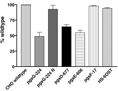
T. gondii infects virtually every known avian and mammalian cell type, suggesting a common mechanism of infection. In the present study, we define infection as a three-step process: (i) adhesion of the parasite to the host cell; (ii) invasion, in which the parasite physically enters the cells and forms a parasitophorous vacuole; and (iii) replication of the parasite in the vacuole. Previously published data has suggested that heparan sulfate might be a receptor for T. gondii adhesion (12, 33). In order to examine the specificity of this interaction to a greater extent, we analyzed the overall process of tachyzoite infection in several CHO cell heparan sulfate-deficient mutants by using an assay in which [3H]uracil incorporation into replicating tachyzoites was measured (Fig. 1). This assay depends on all three steps as defined above. pgsD-677 cells lack heparan sulfate due to a mutation in the copolymerase EXT1, and pgsG-224 cells lack all glycosaminoglycans due to a mutation in the initiating enzyme glucuronosyltransferase I (GlcATI). The incorporation of [3H]uracil was less effective in both mutants compared to wild-type cells (reduced ∼50%), and restoration of heparan sulfate synthesis in pgsG-224 cells by transfection with a vector containing GlcATI cDNA restored incorporation to normal levels (Fig. 1) (4). Thus, the decrease in infection specifically depended on heparan sulfate. The biology of heparan sulfate depends on modifications of the carbohydrate backbone, including N deacetylation/N sulfation of subsets of GlcNAc residues, 6-O sulfation of glucosamine residues, and 2-O-sulfation of uronic acids. To determine which modifications might be required for toxoplasma infection, we tested various CHO cell mutants defective in the sulfotransferase enzymes. _T. gondii_-infected mutants with reduced 6-O sulfation (HS-6OST) or entirely lacking 2-O sulfation (pgsF-17) to the same extent as wild-type cells (Fig. 1). Infection of NDST1-deficient cells (pgsE-606) was reduced to the same extent as mutants entirely lacking heparan sulfate compared to wild type, supporting previous results (33). These findings suggest that T. gondii requires only the action of NDST1 for efficient infection.
FIG. 1.
Heparan sulfate is required for efficient infection of CHO cells by T. gondii. Various CHO mutants defective in heparan sulfate biosynthesis were tested for the ability to support tachyzoite infection by the selective incorporation of [3H]uracil. pgsG-224 lacks all glycosaminoglycans, pgsD-677 lacks heparan sulfate, pgsE-606 lacks NDST1 and makes undersulfated heparan sulfate, pgsF-17 lacks uronic acid 2-O sulfation, and HS-6OST demonstrates 60% less GlcNAc 6-O sulfation. pgsG-224R contains the cDNA for GlcATI and makes glycosaminoglycans normally. Each bar represents the average of triplicate determinations ± the standard deviation and is representative of multiple experiments.
N sulfation is required for infectivity in epithelial cells.
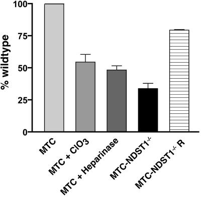
In order to examine whether T. gondii infection in other cell types depends on NDST1, we generated mice in which the NDST1 gene could be inactivated in a tissue-specific manner by using Cre-loxP recombination (NDST1 f/f) (19). These animals were then crossed to mice harboring the polyoma middle T-antigen under the control of MMTV transcriptional elements. Mammary epithelial tumor cells (i.e., MTCs) were isolated from tumors arising in NDST1 f/f MMTV-T-antigen mice. First, NDST1 f/f MTCs were treated with heparin lyases I, II, and III to remove heparan sulfate, and loss of heparan sulfate was determined by the measuring the decline of 10E4 reactivity, an MAb that binds to a common heparan sulfate epitope. Both 10E4 reactivity (data not shown) and T. gondii infection were reduced by ∼50% in heparinase-treated MTCs (Fig. 2). Pharmacological reduction of sulfation by treatment with sodium chlorate also blocked T. gondii infection (Fig. 2), as seen previously in HFF cells (33).
FIG. 2.
N sulfation of heparan sulfate is required for efficient infection of mouse mammary tumor cells by T. gondii. MTCs were purified from _NDST1_f/f mice. Cells were treated with either 30 mM sodium chlorate (ClO3) to inhibit sulfation; 0.1 mU heparinases I, II, and III to remove heparan sulfate; or adenovirus expressing Cre-recombinase to inactivate NDST1 (MTC-NDST1−/−). Tachyzoite infection was measured by the selective incorporation of [3H]uracil. [3H]uracil incorporation was rescued in MTC-NDST1−/− by stable transfection of NDST1 cDNA (MTC-NDST1−/− R). Each bar represents the average of triplicate determinations ± the standard deviation and is representative of multiple experiments.
NDST1 was inactivated in NDST1 f/f MTCs by infection with adenovirus expressing Cre-recombinase (28). NDST1-deficient cells (MTC-NDST1−/−) exhibited ∼50% reduction in N sulfation, a value consistent with that seen in pgsE-606 cells which also lack NDST1 (5). Inactivation of NDST1 also reduced infection by T. gondii by 50 to 70% (Fig. 2). Introduction of an expression vector containing NDST1 cDNA restored infection nearly to that seen in the wild type. Similar results were obtained in hepatocytes lacking NDST1 (9), demonstrating that N sulfation of heparan sulfate by NDST1 is necessary and sufficient for T. gondii infection in multiple cell types.
Heparin and heparan sulfate blocking antibodies have no affect on T. gondii infection.
Since sulfation of heparan sulfate appeared to be required for T. gondii infectivity, we hypothesized that heparan sulfate served as a receptor for parasite adhesion. Thus, we predicted that attachment would be disrupted by soluble heparin, a highly sulfated form of heparan sulfate. Heparin can block many protein-heparan sulfate interactions when added at low concentrations (<10 μg/ml) (8). However, adding up to 1 mg of heparin/ml did not affect infection of CHO or MTC cells, nor did lower doses restore infectivity to NDST1−/− cells (data not shown). Chemically modified forms of heparin lacking 2-O-sulfate, 6-O-sulfate, or N-sulfate groups also did not block infection (data not shown). Heparin preparations such as these will decrease invasion of hepatocytes by malaria sporozoites, but only under conditions that mimic blood flow (35). Since T. gondii failed to invade cells under flow conditions, we could not test this possibility. Finally, MAb 10E4, specific to heparan sulfate, did not inhibit toxoplasma infection of MTCs (data not shown). This antibody binds to a common epitope on most heparan sulfate chains and bound well to MTCs (13).
Loss of NDST1 does not affect attachment and invasion of T. gondii.
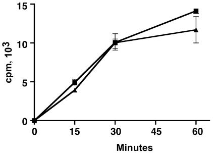
The lack of effect by heparin and MAb 10E4 suggested that cell surface heparan sulfate might not affect adhesion per se but rather a later step in infection such as invasion or replication. To examine invasion, we incubated metabolically labeled tachyzoites with CHO and MTC cells for 15, 30, or 60 min and measured counts associated with the cells. Surprisingly, tachyzoites were able to attach and invade NDST1−/− cells at levels comparable to those of wild-type cells (Fig. 3).
FIG. 3.
N-sulfation of heparan sulfate does not affect attachment/invasion of T. gondii. [3H]uracil-labeled T. gondii was incubated with CHO (v) or pgsE-606 cells (σ) for the times indicated. The cells were washed extensively with ice-cold PBS to remove unassociated parasites. The amount of radioactivity associated with the cells was measured by liquid scintillation counting. Each point represents the average of triplicate determinations ± the standard deviation and is representative of multiple experiments.
To confirm this result, we infected cells for 15 or 30 min with T. gondii and then directly visualized intracellular parasites with antibodies to surface antigen 1 (SAG1) of T. gondii (Table 1). No difference in the number of intracellular parasites was observed 0.25 and 0.5 h postinfection. To ensure that the maximum extent of invasion occurred, toxoplasma was added to host cells for 0.5 h and then removed, and the cells were incubated for another 4 h. Again, no difference was observed in the number of infected cells 4 h postinvasion.
TABLE 1.
T. gondii invade N-sulfation mutants at the same rate as the wild typea
| Cell type | Time (h) | Mean no. of tachyzoites/field ± SD |
|---|---|---|
| CHO wild type | 0.25 | 7 ± 2 |
| 0.5 | 16 ± 4 | |
| 0.5/4 | 20 ± 4 | |
| pgsE-606 | 0.25 | 8 ± 2 |
| 0.5 | 16 ± 2 | |
| 0.5/4 | 22 ± 5 | |
| MTC wild type | 0.25 | 9 ± 1 |
| 0.5 | 21 ± 4 | |
| 0.5/4 | 18 ± 8 | |
| MTC-NDST1−/− | 0.25 | 9 ± 2 |
| 0.5 | 21 ± 5 | |
| 0.5/4 | 16 ± 7 |
Heparan sulfate affects the rate of replication for intracellular tachyzoites.
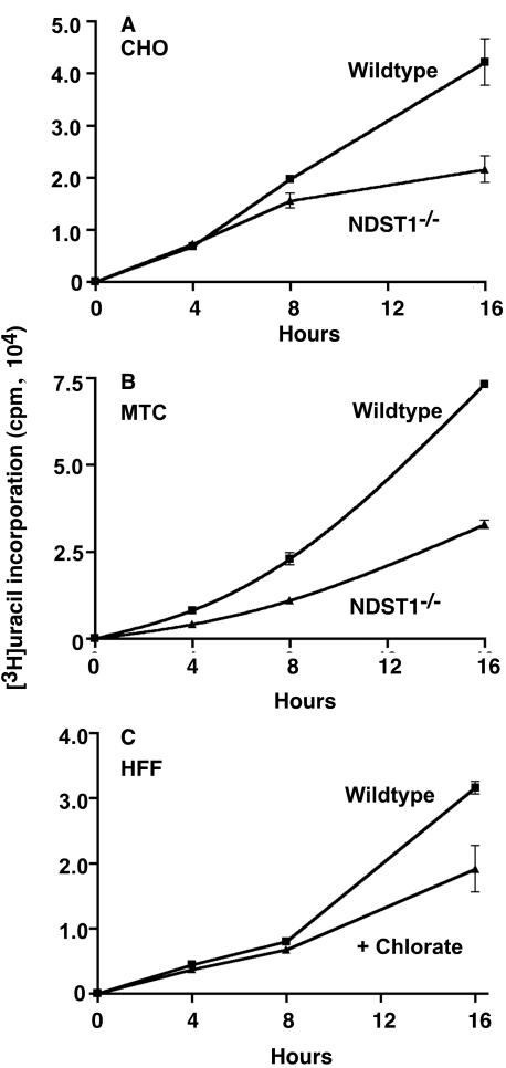
Taken together, these data suggested that heparan sulfate does not act as a receptor for T. gondii or affect invasion but instead affects parasite survival or replication. To test this idea, we cultured RH tachyzoites by serial passage on pgsE-606 or MTC-NDST1−/− cells. Parasites survived through multiple passages but seemed to lyse mutant cells at a slower rate than that for wild-type cells (∼72 h versus ∼48 h, respectively). This finding suggested that lack of NDST1 might affect the rate of tachyzoite replication. Since incorporation of uracil by intracellular parasites reflects their rate of division, we measured [3H]uracil incorporation at various times postinvasion. No significant differences in 3H counts were observed in wild-type cells, NDST1−/− CHO cells, and NDST1−/− MTCs 4 h postinvasion (Fig. 4A and B). However, after 8 h the level of incorporation of [3H]uracil was reduced in the mutants, resulting in a twofold difference after 16 h. Similar results were obtained with HFF with or without chlorate treatment (Fig. 4C).
FIG. 4.
Replication of tachyzoites in cells with altered heparan sulfate. CHO wild-type and pgsE-606 (A), MTCs and MTC-NDST1−/− (B) or HFF, wild-type, and chlorate treated (C) were incubated with tachyzoites for 1 h at 37°C. The cells were washed extensively and incubated at 37°C in the presence of [3H]uracil for the times indicated. Each point represents the average of triplicate determinations ± the standard deviation and is representative of multiple experiments.
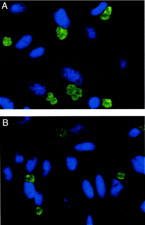
These results suggested that T. gondii might divide faster in cells with fully sulfated heparan sulfate. Tachyzoites do not divide for several hours after invasion. Thus, at 4 h mutant and wild-type cells had ∼20 intracellular parasites/field (Table 1). After 18 h, the average number of intracellular parasites/field increased due to replication, and the increase was greater in wild-type cells compared to NDST1−/− cells (Table 2, 115 versus 81, respectively). Representative fields from wild-type and knockout cells are shown in Fig. 5. Interestingly, the number of parasitophorous vacuoles was not altered, but the number of vacuoles containing eight parasites (three divisions, 23) versus four parasites (two divisions, 22) was greater in wild-type cells compared to the mutant (Table 2). Similar results were observed with MTCs even though T. gondii did not replicate as well in MTCs compared to CHO cells (Table 2).
TABLE 2.
Sulfation by NDST1 stimulates T. gondii replicationa
| Cell type | Total no. of tachyzoites/field ± SD | No. of vacuoles ± SD containing: | |||
|---|---|---|---|---|---|
| One tachyzoite | Two tachyzoites | Four tachyzoites | Eight tachyzoites | ||
| CHO wild type | 115 ± 11 | 3* | 2 ± 2 | 7 ± 4 | 10 ± 3 |
| pgsE-606 | 81 ± 10 | 1* | 3 ± 3 | 9 ± 5 | 5 ± 3 |
| MTC wild type | 40 ± 4 | 1 ± 3 | 3 ± 2 | 3 ± 2 | 3 ± 1 |
| MTC-NDST1−/− | 29 ± 5 | 6 ± 2 | 5 ± 1 | 3 ± 2 | 1* |
FIG. 5.
T. gondii divide slower in cells with reduced sulfation of heparan sulfate. Cells were infected with tachyzoites for 60 min, washed extensively, and incubated for 18 h at 37°C. The cells were stained with MAb DG52 (green, tachyzoites) and DAPI (nuclei) showing that toxoplasma replicated to a lesser extent in cells lacking NDST1. (A) Wild-type CHO cells; (B) pgsE-606 cells.
DISCUSSION
T. gondii invades a wide variety of mammalian species and cell types, suggesting a common mechanism of entry. The ubiquitous nature of the cell surface glycan, heparan sulfate, led others to hypothesize it as a receptor for T. gondii. Indeed, several studies have demonstrated that altering heparan sulfate alters infection (12, 33). In this report, we have qualified and extended these studies to other cell types by using both genetic and pharmacological approaches to alter heparan sulfate structure. Our major findings show that (i) heparan sulfate is both necessary and sufficient for robust infection of several cell types, (ii) efficient infection depends on N sulfation of the chains, and (iii) heparan sulfate facilitates replication of T. gondii in the parasitophorous vacuole. Importantly, our data show that heparan sulfate does not serve as a receptor on the cell surface but instead facilitates parasite replication.
Numerous biological activities have been ascribed to heparan sulfate chains. In general, these activities depend on the arrangement of variously modified sugar residues that make up specific binding sites for proteins. One of the best-studied examples is the growth factor FGF-2, which binds to a pentasaccharide containing N-sulfated glucosamine (GlcNS) residues and at least one 2-O-sulfated IdoA. The FGF receptor binds to a sequence rich in GlcNS, 6-O-sulfated glucosamine residues, and 2-O-sulfated IdoA (21, 30). As expected, altering 2-O sulfation or 6-O sulfation in CHO cells diminishes FGF-2 binding and signaling (4, 46). In contrast these changes had no effect on T. gondii infection, but partial reduction of N sulfation diminished infectivity by ∼50%. In studies of cells lacking all N sulfation due to combined deficiency in NDST1 and NDST2, no further diminution of T. gondii infection has been observed (data not shown). These results show that NDST1 generates a specific pattern of sulfation required for efficient infection. Additional studies are needed to define the nature of the oligosaccharide sequence active in T. gondii infection.
T. gondii expresses a surface antigen (SAG3) that binds to heparan sulfate, which originally suggested that it might act as a ligand involved in heparan sulfate-dependent attachment (25). Mutants lacking SAG3 exhibited reduced infectivity, a finding consistent with this idea. However, our data indicate that changes in heparan sulfate structure do not affect attachment of the parasite to host cells. Monteiro et al. have reported that terminal sialic acid residues facilitate infection (31). Perhaps SAG3 binds to sialylated glycoconjugates, which are as abundant on the cell surface as heparan sulfate.
Our data show that cell surface heparan sulfate does not act as a receptor for attachment or invasion. Consistent with this finding, antibodies to heparan sulfate and exogenous heparin did not block attachment or infection (33). By radiolabeling studies and by immunofluorescence we showed that T. gondii bound and invaded wild-type and NDST1−/− cells to the same extent. Thus, the difference in infection relates to later steps in infection. Based on uracil incorporation and direct counting of cells in parasitophorous vacuoles, we concluded that parasites replicated slower in cells with altered heparan sulfate. Several possibilities exist to explain this finding. First, toxoplasma may take up heparan sulfate during invasion and use it as a nutrient. Thus, the change in heparan sulfate structure in the mutants could alter its nutritional value. In mammalian cells internalized heparan sulfate is degraded in lysosomes and the component sugars and sulfate are salvaged (32). Exogenous heparin is taken up by T. gondii, possibly by way of SAG3 (11, 12, 16). However, the fate of heparin internalized by toxoplasma is unknown. Second, heparan sulfate could be involved in the uptake of nutrients required by toxoplasma. For example, toxoplasma requires polyamines for proper growth, and recent studies indicate that heparan sulfate facilitates salvage of extracellular polyamines (6, 7, 38). However, attempts to restore growth by the addition of polyamines to the growth medium were unsuccessful. Third, heparan sulfate proteoglycans might be internalized and play a protective role in the parasitophorous vacuole. Efforts to visualize heparan sulfate in vacuoles by antibody staining (10E4 MAb) and deconvolution microscopy were unsuccessful. However, we cannot exclude the possibility that a small subpopulation of heparan sulfate proteoglycans were incorporated into the parasitophorous vacuole. Finally, heparan sulfate could be involved in signaling events required by T. gondii for proliferation. Erk, SAP/JNK, and p38 are phosphorylated upon T. gondii attachment (26, 41). Heparan sulfate facilitates signaling via these pathways, and altering its structure could affect the response to various growth factors or change the resting state of phosphorylation of these kinases.
Several molecules have surfaced as potential receptors or coreceptors for toxoplasma, including glycoconjugates terminating in sialic acid (31). Furtado et al. showed that T. gondii bound to cell surface β1 integrins and their ligands, laminin and type IV collagen, enhanced attachment (16). Grimwood et al. reported that parasite binding could be greatly increased as host cells proceeded from G1 phase to the mid-S phase, potentially by upregulation of a cell surface receptor (17). Conceivably, T. gondii may exploit these and other receptors for attachment, invasion, and replication dependent on the host cell. Although heparan sulfate does not appear to serve as a receptor, it clearly plays a role in its life cycle. Further studies are needed to examine this process in greater detail.
Acknowledgments
We thank David Sibley and Antonio Barragan for advice and help on this project. We thank Robert Rosenberg and Lijuan Zhang for providing the 6-O-sulfotransferase-deficient CHO cells and John Boothroyd for anti-SAG1 antibodies.
J.R.B. was supported by a Ruth L. Kirschstein NRSA Fellowship 05292-02. This study was supported by National Institutes of Health grants HL57345 and GM33063 to J.D.E.
REFERENCES
- 1.Aikawa, J., and J. D. Esko. 1999. Molecular cloning and expression of a third member of the heparan sulfate/heparin GlcNAc _N_-deacetylase/_N_-sulfotransferase family. J. Biol. Chem. 274**:**2690-2695. [DOI] [PubMed] [Google Scholar]
- 2.Aikawa, J., K. Grobe, M. Tsujimoto, and J. D. Esko. 2001. Multiple isozymes of heparan sulfate/heparin GlcNAc _N_-deacetylase/_N-_sulfotransferase: structure and activity of the fourth member, NDST4. J. Biol. Chem. 276**:**5876-5882. [DOI] [PubMed] [Google Scholar]
- 3.Bai, X. M., and J. D. Esko. 1996. An animal cell mutant defective in heparan sulfate hexuronic acid 2-O-sulfation. J. Biol. Chem. 271**:**17711-17717. [DOI] [PubMed] [Google Scholar]
- 4.Bai, X. M., G. Wei, A. Sinha, and J. D. Esko. 1999. Chinese hamster ovary cell mutants defective in glycosaminoglycan assembly and glucuronosyltransferase I. J. Biol. Chem. 274**:**13017-13024. [DOI] [PubMed] [Google Scholar]
- 5.Bame, K. J., and J. D. Esko. 1989. Undersulfated heparan sulfate in a Chinese hamster ovary cell mutant defective in heparan sulfate _N_-sulfotransferase. J. Biol. Chem. 264**:**8059-8065. [PubMed] [Google Scholar]
- 6.Belting, M., L. Borsig, M. M. Fuster, J. R. Brown, L. Persson, L. Å. Fransson, and J. D. Esko. 2002. Tumor attenuation by combined heparan sulfate and polyamine depletion. Proc. Natl. Acad. Sci. USA 99**:**371-376. [DOI] [PMC free article] [PubMed] [Google Scholar]
- 7.Belting, M., S. Persson, and L. Å. Fransson. 1999. Proteoglycan involvement in polyamine uptake. Biochem. J. 338**:**317-323. [PMC free article] [PubMed] [Google Scholar]
- 8.Bernfield, M., M. Götte, P. W. Park, O. Reizes, M. L. Fitzgerald, J. Lincecum, and M. Zako. 1999. Functions of cell surface heparan sulfate proteoglycans. Annu. Rev. Biochem. 68**:**729-777. [DOI] [PubMed] [Google Scholar]
- 9.Bishop, J. R., and J. D. Esko. 2005. The elusive role of heparan sulfate in Toxoplasma gondii infection. Mol. Biochem. Parasitol. 139**:**267-269. [DOI] [PubMed]
- 10.Black, M. W., and J. C. Boothroyd. 2000. Lytic cycle of Toxoplasma gondii. Microbiol. Mol. Biol. Rev. 64**:**607-623. [DOI] [PMC free article] [PubMed] [Google Scholar]
- 11.Botero-Kleiven, S., V. Fernandez, J. Lindh, A. Richter-Dahlfors, A. von Euler, and M. Wahlgren. 2001. Receptor-mediated endocytosis in an apicomplexan parasite (Toxoplasma gondii). Exp. Parasitol. 98**:**134-144. [DOI] [PubMed] [Google Scholar]
- 12.Carruthers, V. B., S. Håkansson, O. K. Giddings, and L. D. Sibley. 2000. Toxoplasma gondii uses sulfated proteoglycans for substrate and host cell attachment. Infect. Immun. 68**:**4005-4011. [DOI] [PMC free article] [PubMed] [Google Scholar]
- 13.David, G., X. M. Bai, B. Van der Schueren, J. J. Cassiman, and H. Van den Berghe. 1992. Developmental changes in heparan sulfate expression: in situ detection with MAbs. J. Cell Biol. 119**:**961-975. [DOI] [PMC free article] [PubMed] [Google Scholar]
- 14.Dobrowolski, J. M., and L. D. Sibley. 1996. Toxoplasma invasion of mammalian cells is powered by the actin cytoskeleton of the parasite. Cell 84**:**933-939. [DOI] [PubMed] [Google Scholar]
- 15.Esko, J. D., and S. B. Selleck. 2002. Order out of chaos: assembly of ligand binding sites in heparan sulfate. Annu. Rev. Biochem. 71**:**435-471. [DOI] [PubMed] [Google Scholar]
- 16.Furtado, G. C., Y. Cao, and K. A. Joiner. 1992. Laminin on Toxoplasma gondii mediates parasite binding to the β1 integrin receptor α6β1 on human foreskin fibroblasts and Chinese hamster ovary cells. Infect. Immun. 60**:**4925-4931. [DOI] [PMC free article] [PubMed] [Google Scholar]
- 17.Grimwood, J., J. R. Mineo, and L. H. Kasper. 1996. Attachment of Toxoplasma gondii to host cells is host cell cycle dependent. Infect. Immun. 64**:**4099-4104. [DOI] [PMC free article] [PubMed] [Google Scholar]
- 18.Grobe, K., and J. D. Esko. 2002. Regulated translation of heparan sulfate _N_-acetylglucosamine _N_-deacetylase/_N_-sulfotransferase isozymes by structured 5′-untranslated regions and internal ribosome entry sites. J. Biol. Chem. 277**:**30699-30706. [DOI] [PubMed] [Google Scholar]
- 19.Grobe, K., M. Inatani, S. Pallerla, J. Castagnola, Y. Yamaguchi, and J. D. Esko. Cerebral hypoplasma and craniofacial defects in mice lacking heparan sulfate NDST1 gene expression. Development, in press. [DOI] [PMC free article] [PubMed]
- 20.Grobe, K., J. Ledin, M. Ringvall, K. Holmborn, E. Forsberg, J. D. Esko, and L. Kjellén. 2002. Heparan sulfate and development: differential roles of the _N_-acetylglucosamine _N_-deacetylase/_N_-sulfotransferase isozymes. Biochim. Biophys. Acta Gen. Subj. 1573**:**209-215. [DOI] [PubMed] [Google Scholar]
- 21.Guimond, S., M. Maccarana, B. B. Olwin, U. Lindahl, and A. C. Rapraeger. 1993. Activating and inhibitory heparin sequences for FGF-2 (basic FGF). Distinct requirements for FGF-1, FGF-2, and FGF-4. J. Biol. Chem. 268**:**23906-23914. [PubMed] [Google Scholar]
- 22.Guy, C. T., R. D. Cardiff, and W. J. Muller. 1992. Induction of mammary tumors by expression of polyomavirus middle T oncogene: a transgenic mouse model for metastatic disease. Mol. Cell. Biol. 12**:**954-961. [DOI] [PMC free article] [PubMed] [Google Scholar]
- 23.Holmborn, K., J. Ledin, E. Smeds, I. Eriksson, M. Kusche-Gullberg, and L. Kjellen. 2004. Heparan sulfate synthesized by mouse embryonic stem cells deficient in NDST1 and NDST2 is 6-O-sulfated but contains no N-sulfate groups. J. Biol. Chem. 279**:**42355-42358. [DOI] [PubMed] [Google Scholar]
- 24.Iozzo, R. V. 1998. Matrix proteoglycans: from molecular design to cellular function. Annu. Rev. Biochem. 67**:**609-652. [DOI] [PubMed] [Google Scholar]
- 25.Jacquet, A., L. Coulon, J. De Nève, V. Daminet, M. Haumont, L. Garcia, A. Bollen, M. Jurado, and R. Biemans. 2001. The surface antigen SAG3 mediates the attachment of Toxoplasma gondii to cell-surface proteoglycans. Mol. Biochem. Parasitol. 116**:**35-44. [DOI] [PubMed] [Google Scholar]
- 26.Kim, L., B. A. Butcher, and E. Y. Denkers. 2004. Toxoplasma gondii interferes with lipopolysaccharide-induced mitogen-activated protein kinase activation by Mechanisms distinct from endotoxin tolerance. J. Immunol. 172**:**3003-3010. [DOI] [PubMed] [Google Scholar]
- 27.Kjellen, L., and U. Lindahl. 1991. Proteoglycans: structures and interactions. Annu. Rev. Biochem. 60**:**443-475. [DOI] [PubMed] [Google Scholar]
- 28.Li, Z. W., G. Stark, J. Gotz, T. Rulicke, M. Gschwind, G. Huber, U. Muller, and C. Weissmann. 1996. Generation of mice with a 200-kb amyloid precursor protein gene deletion by Cre recombinase-mediated site-specific recombination in embryonic stem cells. Proc. Natl. Acad. Sci. USA 93**:**6158-6162. [DOI] [PMC free article] [PubMed] [Google Scholar]
- 29.Lidholt, K., J. L. Weinke, C. S. Kiser, F. N. Lugemwa, K. J. Bame, S. Cheifetz, J. Massagué, U. Lindahl, and J. D. Esko. 1992. A single mutation affects both _N_-acetylglucosaminyltransferase and glucuronosyltransferase activities in a Chinese hamster ovary cell mutant defective in heparan sulfate biosynthesis. Proc. Natl. Acad. Sci. USA 89**:**2267-2271. [DOI] [PMC free article] [PubMed] [Google Scholar]
- 30.Maccarana, M., B. Casu, and U. Lindahl. 1993. Minimal sequence in heparin/heparan sulfate required for binding of basic fibroblast growth factor. J. Biol. Chem. 268**:**23898-23905. [PubMed] [Google Scholar]
- 31.Monteiro, V. G., C. P. Soares, and W. de Souza. 1998. Host cell surface sialic acid residues are involved on the process of penetration of Toxoplasma gondii into mammalian cells. FEMS Microbiol. Lett. 164**:**323-327. [DOI] [PubMed] [Google Scholar]
- 32.Neufeld, E. F., T. W. Lim, and L. J. Shapiro. 1975. Inherited disorders of lysosomal metabolism. Annu. Rev. Biochem. 44**:**357-376. [DOI] [PubMed] [Google Scholar]
- 33.Ortega-Barria, E., and J. C. Boothroyd. 1999. A Toxoplasma lectin-like activity specific for sulfated involved in host cell infection. J. Biol. Chem. 274**:**1267-1276. [DOI] [PubMed] [Google Scholar]
- 34.Pfefferkorn, E. R., and L. C. Pfefferkorn. 1977. Specific labeling of intracellular Toxoplasma gondii with uracil. J. Protozool. 24**:**449-453. [DOI] [PubMed] [Google Scholar]
- 35.Pinzon-Ortiz, C., J. Friedman, J. Esko, and P. Sinnis. 2001. The binding of the circumsporozoite protein to cell surface heparan sulfate proteoglycans is required for Plasmodium sporozoite attachment to target cells. J. Biol. Chem. 276**:**26784-26791. [DOI] [PMC free article] [PubMed] [Google Scholar]
- 36.Pullan, S., J. Wilson, A. Metcalfe, G. M. Edwards, N. Goberdhan, J. Tilly, J. A. Hickman, C. Dive, and C. H. Streuli. 1996. Requirement of basement membrane for the suppression of programmed cell death in mammary epithelium. J. Cell. Sci. 109(Pt. 3)**:**631-642. [DOI] [PubMed] [Google Scholar]
- 37.Rostand, K. S., and J. D. Esko. 1997. Microbial adherence to and invasion through proteoglycans. Infect. Immun. 65**:**1-8. [DOI] [PMC free article] [PubMed] [Google Scholar]
- 38.Seabra, S. H., R. A. DaMatta, F. G. de Mello, and W. de Souza. 2004. Endogenous polyamine levels in macrophages is sufficient to support growth of Toxoplasma gondii. J. Parasitol. 90**:**455-460. [DOI] [PubMed] [Google Scholar]
- 39.Shukla, D., and P. G. Spear. 2001. Herpesviruses and heparan sulfate: an intimate relationship in aid of viral entry. J. Clin. Investig. 108**:**503-510. [DOI] [PMC free article] [PubMed] [Google Scholar]
- 40.Sibley, L. D., and N. W. Andrews. 2000. Cell invasion by un-palatable parasites. Traffic 1**:**100-106. [DOI] [PubMed] [Google Scholar]
- 41.Valere, A., R. Garnotel, I. Villena, M. Guenounou, J. M. Pinon, and D. Aubert. 2003. Activation of the cellular mitogen-activated protein kinase pathways ERK, P38 and JNK during Toxoplasma gondii invasion. Parasite 10**:**59-64. [DOI] [PubMed] [Google Scholar]
- 42.Wadström, T., and Å. Ljungh. 1999. Glycosaminoglycan-binding microbial proteins in tissue adhesion and invasion: key events in microbial pathogenicity. J. Med. Microbiol. 48**:**223-233. [DOI] [PubMed] [Google Scholar]
- 43.Wilson, C. B., J. S. Remington, S. Stagno, and D. W. Reynolds. 1980. Development of adverse sequelae in children born with subclinical congenital toxoplasma infection. Pediatrics 66**:**767-774. [PubMed] [Google Scholar]
- 43a.Wang, L., M. M. Fuster, P. Sriramarao, and J. Esko. Endothelial deficiency of heparin sulfate impairs L-selectin and chemokine mediated neutrophil trafficking during inflammatory responses. Nat. Immunol., in press. [DOI] [PubMed]
- 44.Wong, S. Y., and J. S. Remington. 1993. Biology of Toxoplasma gondii. AIDS 7**:**299-316. [DOI] [PubMed] [Google Scholar]
- 45.Yanagishita, M., and V. Hascall. 1992. Cell surface heparan sulfate proteoglycans. J. Biol. Chem. 267**:**9451-9454. [PubMed] [Google Scholar]
- 46.Zhang, L. J., D. L. Beeler, R. Lawrence, M. Lech, J. Liu, J. C. Davis, Z. Shriver, R. Sasisekharan, and R. D. Rosenberg. 2001. 6-_O_-sulfotransferase-1 represents a critical enzyme in the anticoagulant heparan sulfate biosynthetic pathway. J. Biol. Chem. 276**:**42311-42321. [DOI] [PubMed] [Google Scholar]
