Lymphatic endothelial reprogramming of vascular endothelial cells by the Prox-1 homeobox transcription factor (original) (raw)
Abstract
Lymphatic vessels are essential for fluid homeostasis, immune surveillance and fat adsorption, and also serve as a major route for tumor metastasis in many types of cancer. We found that isolated human primary lymphatic and blood vascular endothelial cells (LECs and BECs, respectively) show interesting differences in gene expression relevant for their distinct functions in vivo. Although these phenotypes are stable in vitro and in vivo, overexpression of the homeobox transcription factor Prox-1 in the BECs was capable of inducing LEC-specific gene transcription in the BECs, and, surprisingly, Prox-1 suppressed the expression of ∼40% of the BEC-specific genes. Prox-1 did not have global effects on the expression of LEC-specific genes in other cell types, except that it up-regulated cyclin E1 and E2 mRNAs and activated the cyclin e promoter in various cell types. These data suggest that Prox-1 acts as a cell proliferation inducer and a fate determination factor for the LECs. Furthermore, the data provide insights into the phenotypic diversity of endothelial cells and into the possibility of transcriptional reprogramming of differentiated endothelial cells.
Keywords: gene profiling/lymphatic endothelium/transcription factor Prox-1
Introduction
A major role of the lymphatic vasculature is to remove an excess of the protein-rich interstitial fluid that continuously escapes from the blood capillaries, and to return it to the blood circulation. In addition, the lymphatic system provides constant immune surveillance by filtering lymph and its antigens through the chain of lymph nodes, and also serves as one of the major routes for absorption of fat from the gut (Witte et al., 2001). It has been known for a long time that in many types of cancer the lymphatic vessels provide a major pathway for tumor metastasis, and regional lymph node dissemination correlates with the progression of the disease (Karpanen and Alitalo, 2001). Hereditary lymphedema, post-surgical secondary lymphedema and lymphatic obstruction in filariasis are all characterized by disabling and disfiguring swelling of the affected tissues, linked to the insufficiency or obstruction of the lymphatics (Witte et al., 2001).
In spite of the importance of lymphatic vessels in medicine, the cell biology of this part of the vascular system has received little attention until recently. Studies during the past 5 years have revealed the existence of the lymphatic-specific vascular endothelial growth factors VEGF-C and VEGF-D, which serve as ligands for the receptor tyrosine kinase VEGFR-3, and demonstrated their importance for the normal development of the lymphatic vessels (Jeltsch et al., 1997; Mäkinen et al., 2001a; Veikkola et al., 2001). These molecules also appear to be involved in the development of lymphedema and lymphatic metastasis (for a review, see Karkkainen et al., 2001; Karpanen and Alitalo, 2001).
When compared with the blood vascular endothelium, the lymphatic endothelium shows specific morphological and molecular characteristics. For example, the lymphatic capillaries are larger than the blood capillaries, they have an irregular or collapsed lumen with no red blood cells, a discontinuous basal lamina, overlapping intercellular junctional complexes and anchoring filaments that connect the lymphatic endothelial cells to the extracellular matrix (Witte et al., 2001). Unlike the blood capillaries, the lymphatic capillaries lack pericyte coverage. Recent studies have demonstrated that the blood vascular and lymphatic endothelial cells (BECs and LECs, respectively) represent differentiated cell lineages without evidence of any interconversion between the distinguishing phenotypic properties (Kriehuber et al., 2001; Mäkinen et al., 2001b). However, the mechanisms responsible for the lymphatic differentiation program are still unknown.
We wanted to find out whether the homeodomain transcription factor Prox-1 could regulate the differentiated LEC and BEC phenotypes, because targeted disruption of Prox-1 in mice was reported to result in the arrest of lymphatic vessel development (Wigle and Oliver, 1999). To address this question, we identified genes that are differentially expressed between primary BECs and LECs, and analyzed the changes in gene expression induced by overexpression of Prox-1 in the BECs.
Results
Identification of differentially expressed genes
We isolated BECs and LECs from cultures of human dermal microvascular endothelial cells (HDMECs) using magnetic microbeads and antibodies against the lymphatic endothelial cell surface marker podoplanin (Breiteneder-Geleff et al., 1999; Mäkinen et al., 2001b). The purities of the isolated BEC and LEC populations were confirmed to be >99%, as assessed by immunofluorescence using antibodies against VEGFR-3 and podoplanin (data not shown). The isolated cells were cultured for a couple of passages, and RNA was extracted, labeled and used for hybridization with oligonucleotide microarrays containing sequences from ∼12 000 known genes, i.e. ∼1/3 of the total number of all predicted human transcripts.
Consistent with their known lymphatic-specific gene expression patterns in vivo and in vitro, podoplanin, desmoplakin I/II and the macrophage mannose receptor I were found specifically in the LECs (Breiteneder-Geleff et al., 1999; Ebata et al., 2001; Irjala et al., 2001); therefore, further characterization of the gene expression profiles was carried out. About 300 genes were found to be differentially expressed between the LECs and BECs, when using a reproducible signal log2 ratio of 1.0 (2-fold difference) in the replicate analyses (most of these genes are also expressed in other cell types). The microarray data were validated by northern blotting for 29 of the selected genes (see Figure 1A for examples; a full list of the differentially expressed genes is available as Supple mentary data at The EMBO Journal Online).
Fig. 1. Examples of differentially expressed genes in LECs and BECs. (A) Northern blotting and hybridization for the indicated transcripts. Equal loading was verified by probing with GAPDH. For the microarray analyses, RNA was extracted from LECs that were cultured in the presence of VEGF-C (+C). When validating the array results, RNA was also extracted as a control from cultures of LECs in which VEGF-C was not added (–C). STAT6 is expressed specifically in BECs but not in LECs. Western blotting for STAT6 and STAT5b (B), and immunofluorescence double-staining for STAT6 (red) and LEC- specific marker podoplanin (green) (C). The nuclei were stained with Hoechst fluorochrome (blue).
We detected the most striking differences between the BECs and LECs in the expression of pro-inflammatory cytokines and chemokines [interleukin (IL)-8, IL-6, mono cyte chemotactic protein-1] and receptors (UFO/axl, CXCR4, IL-4R) as well as genes involved in cytoskeletal and cell–cell or cell–matrix interactions (see Table I for other examples). The expression of pro-inflammatory cytokines predominantly in the BECs can be explained, at least partially, by the fact that the STAT6 transcription factor, which is activated in response to IL-4 (Ihle, 2001), is expressed specifically in the BECs, as detected by western blotting and immunofluorescence (Figure 1B and C).
Table I. BECs and LECs express distinct sets of pro-inflammatory cytokines, chemokines and receptors, as well as distinct cell adhesion molecules and cytoskeletal proteins.
| | BEC | LEC | | | ------------------------------------- | ------------------------------------ | -------------------------- | | Adhesion molecules | Integrin α5 | Integrin α9* | | | Integrin β5 | Integrin α1 | | | | ICAM-1*, ICAM-2 | Macrophage mannose receptor I* | | | | N-cadherin* | | | | | Selectin P, selectin E* | | | | | CD44* | | | | Cytoskeletal proteins | Vinculin | Desmoplakin I and II* | | | Claudin 7* | Adducin γ | | | | Actin, α2 | Plakoglobin | | | | Profilin 2 | α-actinin-2 associated LIM protein* | | | ECM proteins | Collagens 8A1*, 6A1*, 1A2* | Matrix Gla protein* | | | Laminin, γ2*, α5 | | | | | Versican* | | | | | Proteoglycan 1 | | | | ECM modulation | MMP-1, MMP-14 | TIMP-3 | | | uPA* | | | | | Plasminogen activator inhibitor I | | | | | Cathepsin C | | | | Cytokines, chemokines and receptors | IL-8*, IL-6* | IL-7* | | | Monocyte chemoattractant protein 1 | SDF-1b* | | | | UFO/axl* | | | | | CXCR4 | | | | | CCRL2/CKRX* | | | | | IL-4 receptor | | | | Total | 167 genes | 135 genes |
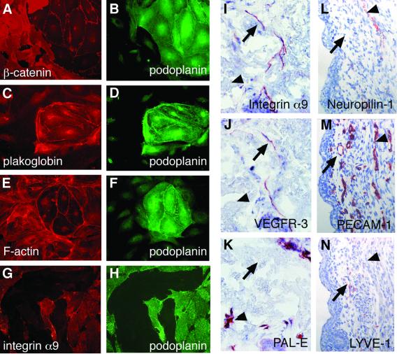
Cadherins are a family of membrane receptors that mediate the formation of stable cell–cell junctions via homophilic cell adhesion. The cytoplasmic domains of cadherins interact with β-catenin, plakoglobin (γ-catenin) and p120_ctn_, which link them to the actin cytoskeleton via α-actinin, vinculin, ZO-1, ZO-2 and spectrin (Provost and Rimm, 1999). We found that BECs expressed significantly higher levels of β-catenin (Figure 2A and B) and vinculin (data not shown), whereas plakoglobin was present mostly on the LECs (Figure 2C and D). Staining of LECs and BECs also revealed striking differences in the organization of the actin cytoskeleton. BECs displayed numerous stress fibers, which in LECs were almost totally absent, and instead a cortical distribution of actin was observed (Figure 2E and F).
Fig. 2. Cytoskeletal structures, integrin α9 and NRP-1 expression in BECs and LECs. Mixed cultures of LECs and BECs were double-stained for β-catenin (A), plakoglobin (C), F-actin (E) and integrin α9 (G), and for the LEC-specific marker podoplanin (green; B, D, F and H). Expression of integrin α9 in the lymphatic (arrow) but not in blood vessel endothelia (arrowhead). Adjacent sections of human skin were stained with antibodies against integrin α9 (I), VEGFR-3 (J) or blood vessel endothelial antigen PAL-E (K). Expression of NRP-1 in a subset of blood vessels (arrowhead) but not lymphatic vessels (arrow). Adjacent sections of mouse skin were stained with antibodies against NRP-1 (L), the pan-endothelial marker PECAM-1 (M) or lymphatic endothelial marker LYVE-1 (N).
Integrins, which are important mediators of cell adhesion (Giancotti and Ruoslahti, 1999), consist of α and β subunits, which bind extracellular matrix proteins, while the cytoplasmic domains interact with the cytoskeleton and with proteins involved in signal transduction. Integrin α5, which acts as a subunit of the fibronectin receptor, was mainly expressed in the BECs. In contrast, integrins α1 and α9, which provide subunits for the receptors for laminin and collagen and for osteopontin and tenascin, respectively, were expressed in the LECs (Figures 1A, 2G and H). In human skin, antibodies against integrin α9 stained specifically lymphatic capillaries, while blood vessel endothelia were negative (Figure 2I–K). In addition, integrin α9 was detected in arterial smooth muscle cells, as reported previously (Palmer et al., 1993; data not shown). Interestingly, mice lacking integrin α9β1 were reported to develop respiratory failure due to the accumulation of a milky pleural (presumably lymphatic) effusion and to die within 6–12 days after birth (Huang et al., 2000).
Prox-1 expression reprograms the BEC transcriptional profile
In the microarray analysis, the Prox-1 homeobox transcription factor was found to be expressed specifically in the LECs, and this result was confirmed by northern blot analysis and immunostaining (Figure 3A–C). Despite the fact that the Prox-1 gene was discovered nearly 10 years ago, Prox-1 target genes have not been identified so far. We used adenoviral gene transfer of Prox-1 in primary endothelial cells to address the question of Prox-1-induced gene expression. In order to eliminate the gene expression changes caused by the adenoviral infection, we used AdLacZ (encoding β-galactosidase) to infect control cells.

Fig. 3. AdProx-1 expression induces LEC-specific transcripts in the BECs. (A) Prox-1 mRNA is expressed in LECs but not BECs. LECs were cultured in the absence (–C) or in the presence (+C) of VEGF-C. (B and C) Human dermal microvascular endothelial cells were double-stained with monoclonal 2E11D11 (anti-VEGFR-3) and polyclonal Prox-1 antiserum. Note nuclear staining for Prox-1 (green) only in the VEGFR-3-positive LECs. (D) Examples of Prox-1-regulated genes. Northern blotting and hybridization of RNA from AdLacZ- or AdProx-1-infected BECs and LECs for the indicated transcripts. APC and GAPDH were used as controls for equal loading.
Preliminary titration experiments showed that infection of human microvascular endothelial cells with AdProx-1 or AdLacZ led to nuclear expression of the adenovirus-encoded protein in >90% of the cells by 24 h (data not shown). To investigate the changes in gene expression induced by Prox-1, we first used human cDNA filter arrays, which contain ∼1000 genes known to be important for general cellular pathways as well as genes specifically implicated in the regulation of cardiovascular function or hematopoiesis. AdProx-1 regulated the expression of 50 of these genes (see Supplementary data I available at The EMBO Journal Online), which was confirmed by northern blotting for 10 out of 11 selected genes. When compared with genes expressed differentially in LECs and BECs, 15 genes (i.e. ∼30%) modulated by Prox-1 were found to be differentially expressed between cultured LECs and BECs, suggesting that Prox-1 is a major regulator of the lymphatic endothelial cell identity.
We next asked whether the introduction of Prox-1 into BECs (where it is absent) can modify the transcriptional program of these cells towards the lymphatic endothelial phenotype. AdLacZ did not significantly alter the expression of BEC- or LEC-specific transcripts in oligonucleotide microarray analysis. In contrast, AdProx-1 increased the expression of many LEC-specific mRNAs, such as VEGFR-3, p57Kip2, desmoplakin I/II and α-actinin- associated LIM protein (Figure 3D; Table II). Surpris ingly, Prox-1 also suppressed the expression of ∼40% of genes characteristic for the BECs, such as the transcrip tion factor STAT6, the UFO/axl receptor tyrosine kinase, neuropilin-1 (NRP-1), monocyte chemoattractant protein-1 (MCP-1) and integrin α5 (Figure 3D; data not shown; see Table II and Supplementary data II for other examples). These results on gene expression analysis are in agreement with the in vivo studies of lymphatic vessels. For example, VEGFR-3 and desmoplakin I/II are found in the lymphatic endothelium (Kaipainen et al., 1995; Ebata et al., 2001), and we found that the VEGF co-receptor NRP-1, which was suppressed by Prox-1 in the BECs, is expressed in a subset of blood vessels, but not in lymphatic vessels in mouse skin (Figure 2L–N).
Table II. Examples of LEC- and BEC-specific genes regulated by Prox-1.
| | LEC-specific, upregulated | BEC-specific, downregulated | | | ------------------------------------- | ----------------------------------- | ---------------- | | Adhesion molecules | | Integrin α5 | | | | ICAM-2 | | | | | CD44 | | | | | Nr-CAM | | | | | Selectin-P | | | Cytoskeletal proteins | Desmoplakin I and II | Leupaxin | | | α-actinin-2 associated LIM protein | | | | ECM proteins | | Versican | | | | Proteoglycan 1 | | | ECM modulation | | MMP-14 | | | | uPA | | | | | Plasminogen activator inhibitor I | | | Receptor tyrosine kinases | VEGFR-3 | UFO/axl | | Transcription factors | CREM | STAT6 | | | ear-3 | TFEC | | | Cytokines, chemokines and receptors | | IL-6 | | | | MCP-1 | | | Cell cycle control | p57Kip2 | | | | Cyclin E2 | | | | Other | Cholesterol 25-hydroxylase | Neuropilin-1 | | | Thromboxane A2 receptor | Endothelial cell protein C receptor | | | Total | 28 genes | 63 genes | | | (19% of LEC-specific genes) | (38% of BEC-specific genes) | |
Prox-1 induces the expression of cyclin E1 and cyclin E2 in various cell types
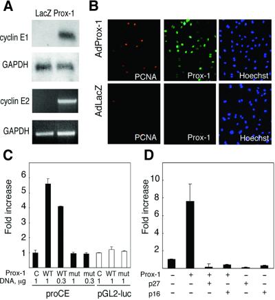
In addition to suppressing BEC-specific genes, overexpression of Prox-1 in BECs resulted in the upregulation of a group of genes linked to cell cycle S-phase pro gression, such as cyclin E1 and cyclin E2, histone H2B, proliferating cell nuclear antigen (PCNA) and dehydrofolate reductase (Figure 4A; data not shown). Increased levels of PCNA were also observed in AdProx-1-infected BECs by immunofluorescence (Figure 4B). In transient transfection experiments, Prox-1, but not a Prox-1 mutant containing two amino acid substitutions in its DNA-binding domain, stimulated the activity of the cyclin e promoter, whereas the activity of the control construct was not modified (Figure 4C). Because transcription factors of the E2F family are major regulators of S-phase progression and the cyclin e promoter contains an E2F binding site (Ohtani et al., 1995; Botz et al., 1996), we also tested whether Prox-1 can transactivate a synthetic reporter construct 6xE2F-luc, which contains six consensus E2F binding sites. As with the results obtained with the cyclin e promoter, Prox-1 strongly transactivated 6xE2F-luc reporter (Figure 4D). Co-transfection of cyclin inhibitors p16INK4a and p27Kip1a, which act by increasing the concentration of transcriptionally inactive retinoblastoma (Rb)–E2F complexes, completely abolished transactivation of 6xE2F-luc reporter by Prox-1, further confirming the specificity of the Prox-1 effect.
Fig. 4. Prox-1 induces expression of cyclins E1 and E2, and transactivates the cyclin e promoter. (A) Total RNA was extracted from AdLacZ- or AdProx-1-infected BECs and used for northern blotting (cyclin E1) or RT–PCR (cyclin E2). (B) BECs infected with AdLacZ or AdProx-1 were stained for PCNA (red), Prox-1 (green) or DNA (blue). (C) Prox-1 transactivates the cyclin e promoter. Promoter activity is presented as fold induction of luciferase activity (mean ± SD) in cells transfected with the expression plasmid encoding wild-type or mutant Prox-1 and with the promoter-luc constructs. C, empty expression vector pAMC; WT, mycProx-1/pAMC; mut, mycProx- 1mut/pAMC. pGL2-luc was used as a reporter control. (D) Prox-1-dependent transactivation of 6xE2F-luc reporter is abolished by co-expression of p27/Kip1a and p16.
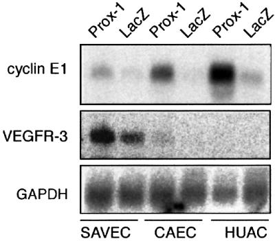
In order to study whether the Prox-1-induced changes of gene expression are cell type specific, we analyzed changes in gene expression after AdProx-1 or AdLacZ infection of two additional endothelial cell types, namely human coronary artery endothelial cells (CAECs) and saphenous vein endothelial cells (SAVECs), as well as human amniotic epithelial cells (HUACs) as an example of a non-endothelial cell type. In all these cell types, AdProx-1 strongly upregulated levels of cyclins E1 and E2, histone H2B and PCNA. However, AdProx-1 induced VEGFR-3 only in CAECs and SAVECs, and not in HUACs (Figure 5; data not shown). These data suggest that the capacity of Prox-1 to induce cell proliferation is distinct from its role as a regulator of the lymphatic endothelial phenotype, as the latter most likely requires the presence of other endothelial-specific transcriptional co-activators.

Fig. 5. Prox-1 induces expression of cyclin E1 in different cell types, while the induction of VEGFR-3 is endothelial cell specific. Total RNA was extracted from AdProx-1- or AdLacZ-infected primary SAVECs, CAECs and HUACs, and used for northern blotting and hybridization with the indicated probes. Equal loading was verified by probing with GAPDH.
Discussion
Molecular discrimination of the blood vessels and lymphatic vessels is essential in studies of diseases involving the vascular system and in the targeted treatment of such diseases. In the present study, we addressed the question of the genetic program controlling the identity of lymphatic capillary endothelial cells as opposed to blood endothelial cells using a gene profiling approach. We show that the two cell lineages express highly distinct sets of genes, which are involved in the regulation of multiple endothelial cell functions such as cell–cell interaction and production of pro-inflammatory cytokines. Furthermore, our data show that the Prox-1 homeobox transcription factor can convert the transcriptional program of cultured microvascular endothelium towards the lymphatic endothelial phenotype and that it is capable of inducing cell proliferation-associated changes in different cell types, such as upregulation of cyclin E1/E2 expression via an E2F-dependent pathway. Our results explain in part the reported lack of lymphatic differentiation in Prox-1-deficient embryos: the first lymphatic sprouts that normally bud out from the anterior cardinal vein at E10.5 are arrested in _Prox-1_–/– embryos at E11.5 and fail to develop further (Wigle and Oliver, 1999). Our results also suggest that the distinct phenotypes of cells in the adult vascular endothelium are plastic and sensitive to transcriptional reprogramming, which could be useful for the various future therapeutic applications of endothelial cells.
Interestingly, the expression of Prox-1 in primary endothelial cells leads to upregulation of VEGFR-3, a receptor tyrosine kinase that is specific for the lymphatic endothelium after mid-gestation and essential for proper lymphatic growth and function (Karkkainen and Petrova, 2000). For example, overexpression of the VEGFR-3 ligand VEGF-C in the skin of transgenic mice results in the selective over-proliferation of lymphatic vessels, whereas inactivating mutations of VEGFR-3 in humans and mice leads to lymphatic hypoplasia and lymphedema (Jeltsch et al., 1997; Irrthum et al., 2000; Karkkainen et al., 2000, 2001). Our data therefore suggest that the upregulation of VEGFR-3 expression by Prox-1 is one of the key pathways involved in the establishment of lymphatic endothelial cell identity. Prox-1 also upregulates the levels of p57Kip2, another molecule found to be selectively expressed in the LECs. Although a lymphatic phenotype has not been reported in p57Kip2 deleted mice, it is of interest that the lens phenotype is reminiscent of that of the Prox-1-deficient embryos (Yan et al., 1997; Zhang et al., 1997). The p57Kip2 levels were strongly decreased in the _Prox-1_–/– lens fiber cells, suggesting that Prox-1 acts upstream of p57Kip2 and that both molecules are part of a common transcriptional program (Wigle et al., 1999).
Unexpectedly, in addition to inducing some LEC-specific genes, Prox-1 also suppressed a large number of BEC-specific transcripts. Consistent with our data and while this manuscript was being prepared, Wigle et al. (2002) reported that in Prox-1-deficient embryos, endothelial cells budding from the veins show misexpression of certain blood vascular and lymphatic endothelial markers including VEGFR-3. Suppression of some BEC-specific molecules such as MCP-1, IL-6 and P-selectin may be a consequence of downregulation of the transcription factor STAT6, which has been shown to be important for the regulation of the expression of pro-inflammatory cytokines and chemokines (Goebeler et al., 1997; Khew-Goodall et al., 1999; Kriebel et al., 1999).
In conclusion, our genomic-scale expression analysis suggests that the default endothelial cell phenotype in the absence of Prox-1 corresponds to the blood vascular phenotype and that Prox-1 is a key player in specifying the lymphatic endothelial cell phenotype. Furthermore, we have identified here a large number of important endothelial genes that are directly or indirectly regulated by this transcription factor. The differential expression of some of these genes can be used to explain some known biological differences between the blood vessels and lymphatic vessels. Some of the other genes identified will provide exciting new avenues of research into the biology of the lymphatic vessels.
Materials and methods
Antibodies
We used monoclonal mouse anti-human VEGFR-3 (clone 2E11D11), PAL-E (Monosan), STAT6 (Transduction Laboratories), β-catenin (BD Biosciences) and plakoglobin (MAb 11E4; Sacco et al., 1995), and polyclonal rabbit anti-human podoplanin (Breiteneder-Geleff et al., 1999), STAT5b (Santa Cruz Biotechnology) and biotinylated mouse anti-PCNA (Zymed). Rabbit anti-human Prox-1 was produced against a GST–Prox-1 fusion protein; an aliquot was also kindly provided by Dr Jörg Wilting (Anatomisches Institut der Albert-Ludwigs-Universität, Freiburg, Germany). Mouse anti-human integrin α9 was a generous gift from Dr Dean Sheppard (University of California at San Francisco, San Francisco, CA) and Dr Curzio Rüegg (University of Lausanne Medical School, Lausanne, Switzerland). Rabbit anti-mouse NRP-1 was a gift from Dr Hajime Fujisawa (Division of Biological Science, Nagoya University Graduate School of Science, Japan) and rabbit anti-mouse LYVE-1 was kindly provided by Dr Erkki Ruoslahti (The Burnham Institute, La Jolla, CA). The fluorochrome-conjugated secondary antibodies were obtained from Jackson ImmunoResearch.
Cell culture and transfection
HUACs were cultured in Med199 medium in the presence of 5% FCS. HDMECs, CAECs and SAVECs were from PromoCell, and were used at passage 3–7. Podoplanin antibodies, MACS colloidal super-paramagnetic MicroBeads conjugated to goat anti-rabbit IgG antibodies, LD and MS separation columns and Midi/MiniMACS separators (Miltenyi Biotech) were used for cell sorting according to the manufacturer’s instructions. BECs (CD31+/podoplanin–) were isolated from HDMECs using LD-negative selection columns and a pure population of LECs was subsequently obtained using MS-positive selection columns. The isolated cells were cultured on fibronectin-coated plates for 1–2 passages, as described, prior to the analysis (Mäkinen et al., 2001b).
For luciferase assays, U2OS cells were transfected by the calcium phosphate method and luciferase assays were carried out 36 h post-transfection according to the Dual-Luciferase® Reporter assay instructions (Promega). Firefly luciferase activity was normalized to Renilla luciferase levels. Cyclin e promoter construct proCE was provided by Dr C.Sardet (Institut de Genetique moleculaire de Monpellier, Montpellier, France).
RNA isolation, northern blotting and microarray analyses
Total RNA was isolated and DNase I treated in RNeasy columns (Qiagen). Northern blots were hybridized with 32P-labeled probe fragments produced by RT–PCR using HDMEC RNA, except that the p57_kip2_ probe was amplified from skeletal muscle RNA. The primers were designed to amplify 300–700 bp of the coding sequence, and all PCR fragments were sequenced to confirm their identity.
32P-labeled probes for hybridization with the Atlas filters (Clontech) were prepared using 2–5 µg of total RNA according to the manufacturer’s instructions, with the exception that the probe was purified using Nick-25 columns (Pharmacia Biotech, Belgium). Following hybridizations and washes, the membranes were analyzed using a Fuji BAS 100 phosphoimager. For the Affymetrix® microarray analysis, four independent BEC and LEC sample preparations and hybridizations were carried out using RNA extracted from four lots of cells isolated from different individuals. Total RNA (5 µg) was used for the synthesis of double-stranded (ds) cDNA using the Custom SuperScript ds-cDNA Synthesis Kit (Invitrogen). Biotin-labeled cRNA was prepared using the Enzo BioArray™ HighYield™RNA Transcript Labeling Kit (Affymetrix), and the unincorporated nucleotides were removed using RNeasy columns (Qiagen). The hybridization, washing and staining of Human Genome 95Av2 microarrays were carried out according to the manufacturer’s instructions (Affymetrix, GeneChip® Expression Analysis Technical Manual). The probe arrays were scanned at 570 nm using an Agilent GeneArray® Scanner and the readings from the quantitative scanning were analyzed by the Affymetrix® Microarray Suite version 5.0 and Data Mining Tool version 3.0. For the comparisons, the hybridization intensities were calculated using a global scaling intensity of 100.
Construction, site-directed mutagenesis and use of AdProx-1
Prox-1 cDNA was amplified by RT–PCR using total RNA from human endothelial cells and the primers 5′-GCCATCTAGACTACTCATGAAGCAGCT-3′ and 5′-GCGCAGAATTCGGCCCTGACCATGACAGCACA-3′, and cloned into the pAMC expression vector, producing N-terminally Myc-tagged Prox-1. The construct was then subcloned into pAdCMV for adenovirus production. AdProx-1 and AdLacZ virus stocks were produced as described elsewhere (Laitinen et al., 1998). Adenovirally produced Prox-1 migrated with a molecular weight of ∼85 kDa and it was also recognized by antibodies against a Prox-1 C-terminal peptide. Mutant Prox-1 N625A/R627A was made using a QuikChange site-directed mutagenesis kit (Stratagene) and the primers 5′-CTCATCAAGTGGTTTAGCGCTTTCCGTAGTTTTACTAC-3′ and 5′-GTAGTAAAACTCACGGAAGCGCTAAACCACTTGATGAG-3′.
HDMECs, HUACs, CAECs, SAVECs, BECs and LECs were plated 24 h before the adenoviral infection at a density of 8000 cells/cm2 and infected for 1 or 6 h (HUACs) in serum-free medium at 50–100 p.f.u./cell. At the end of the incubation period, the cells were washed and then cultured in complete medium for 20–24 h.
Immunofluorescence and immunohistochemistry
The cells were cultured on coverslips, fixed with MetOH and stained with the primary antibodies and fluorochrome-conjugated secondary antibody or Cy3-conjugated streptavidin. F-actin was stained using Texas Red-conjugated phalloidin (Molecular Probes). Cells were counterstained with Hoechst 33258 fluorochrome (Sigma) and viewed in a Zeiss Axioplan 2 fluorescent microscope.
For integrin α9 staining, normal human skin obtained after surgical removal was embedded in Tissue-Tek® (Sakura), frozen and sectioned. The sections (6 µm) were fixed in cold acetone for 10 min and stained with the primary antibodies, followed by peroxidase staining using the Vectastain Elite ABC kit (Vector Laboratories) and 3-amino-9-ethyl carbazole (Sigma). For NRP-1 staining, E15.5 mouse embryo was fixed with 4% paraformaldehyde, dehydrated and embedded in paraffin. Sections (5 µm) were stained with antibodies against NRP-1, lymphatic vessel endothelial hyaluronan receptor-1 (LYVE-1) and platelet endothelial cell adhesion molecule-1 (PECAM-1) (PharMingen), using a Tyramide Signal Amplification kit (TSA, NEN).
Supplementary data
Supplementary data are available at The EMBO Journal Online.
Acknowledgments
Acknowledgements
We thank Dr Outi Monni for help with the microarray experiments and Dr Olli Saksela for the human skin sections. Tapio Tainola, Alun Parsons, Mari Helanterä, Sanna Karttunen, Paula Hyvärinen and Miina Miller are acknowledged for expert technical assistance. This study was supported by grants from the Finnish Cancer Organizations and the Ida Montini, Emil Aaltonen and Sigrid Juselius Foundations, Human Frontier Science Program and National Institutes of Health.
References
- Botz J., Zerfass-Thome,K., Spitkovsky,D., Delius,H., Vogt,B., Eilers,M., Hatzigeorgiou,A. and Jansen-Durr,P. (1996) Cell cycle regulation of the murine cyclin E gene depends on an E2F binding site in the promoter. Mol. Cell. Biol., 16, 3401–3409. [DOI] [PMC free article] [PubMed] [Google Scholar]
- Breiteneder-Geleff S. et al. (1999) Angiosarcomas express mixed endo thelial phenotypes of blood and lymphatic capillaries: podoplanin as a specific marker for lymphatic endothelium. Am. J. Pathol., 154, 385–394. [DOI] [PMC free article] [PubMed] [Google Scholar]
- Ebata N., Nodasaka,Y., Sawa,Y., Yamaoka,Y., Makino,S., Totsuka,Y. and Yoshida,S. (2001) Desmoplakin as a specific marker of lymphatic vessels. Microvasc. Res., 61, 40–48. [DOI] [PubMed] [Google Scholar]
- Giancotti F.G. and Ruoslahti,E. (1999) Integrin signaling. Science, 285, 1028–1032. [DOI] [PubMed] [Google Scholar]
- Goebeler M., Schnarr,B., Toksoy,A., Kunz,M., Brocker,E.B., Duschl,A. and Gillitzer,R. (1997) Interleukin-13 selectively induces monocyte chemoattractant protein-1 synthesis and secretion by human endothelial cells. Involvement of IL-4Rα and Stat6 phosphorylation. Immunology, 91, 450–457. [DOI] [PMC free article] [PubMed] [Google Scholar]
- Huang X.Z., Wu,J.F., Ferrando,R., Lee,J.H., Wang,Y.L., Farese,R.V.,Jr and Sheppard,D. (2000) Fatal bilateral chylothorax in mice lacking the integrin α9β1. Mol. Cell. Biol., 20, 5208–5215. [DOI] [PMC free article] [PubMed] [Google Scholar]
- Ihle J.N. (2001) The Stat family in cytokine signaling. Curr. Opin. Cell Biol., 13, 211–217. [DOI] [PubMed] [Google Scholar]
- Irjala H., Johansson,E.L., Grenman,R., Alanen,K., Salmi,M. and Jalkanen,S. (2001) Mannose receptor is a novel ligand for l-selectin and mediates lymphocyte binding to lymphatic endothelium. J. Exp. Med., 194, 1033–1041. [DOI] [PMC free article] [PubMed] [Google Scholar]
- Irrthum A., Karkkainen,M.J., Devriendt,K., Alitalo,K. and Vikkula,M. (2000) Congenital hereditary lymphedema caused by a mutation that inactivates VEGFR3 tyrosine kinase. Am. J. Hum. Genet., 67, 295–301. [DOI] [PMC free article] [PubMed] [Google Scholar]
- Jeltsch M. et al. (1997) Hyperplasia of lymphatic vessels in VEGF-C transgenic mice. Science, 276, 1423–1425. [DOI] [PubMed] [Google Scholar]
- Kaipainen A., Korhonen,J., Mustonen,T., van Hinsbergh,V.M., Fang,G.-H., Dumont,D., Breitman,M. and Alitalo,K. (1995) Expression of the _fms_-like tyrosine kinase FLT4 gene becomes restricted to lymphatic endothelium during development. Proc. Natl Acad. Sci. USA, 92, 3566–3570. [DOI] [PMC free article] [PubMed] [Google Scholar]
- Karkkainen M.J. and Petrova,T.V. (2000) Vascular endothelial growth factor receptors in the regulation of angiogenesis and lymph angiogenesis. Oncogene, 19, 5598–5605. [DOI] [PubMed] [Google Scholar]
- Karkkainen M.J., Ferrell,R.E., Lawrence,E.C., Kimak,M.A., Levinson,K.L., Alitalo,K. and Finegold,D.N. (2000) Missense mutations interfere with VEGFR-3 signaling in primary lymphoedema. Nat. Genet., 25, 153–159. [DOI] [PubMed] [Google Scholar]
- Karkkainen M.J. et al. (2001) A model for gene therapy of human hereditary lymphedema. Proc. Natl Acad. Sci. USA, 98, 12677–12682. [DOI] [PMC free article] [PubMed] [Google Scholar]
- Karpanen T. and Alitalo,K. (2001) Lymphatic vessels as targets of tumor therapy? J. Exp. Med., 194, F37–F42. [DOI] [PMC free article] [PubMed] [Google Scholar]
- Khew-Goodall Y., Wadham,C., Stein,B.N., Gamble,J.R. and Vadas,M.A. (1999) Stat6 activation is essential for interleukin-4 induction of P-selectin transcription in human umbilical vein endothelial cells. Arterioscler. Thromb. Vasc. Biol., 19, 1421–1429. [DOI] [PubMed] [Google Scholar]
- Kriebel P., Patel,B.K., Nelson,S.A., Grusby,M.J. and LaRochelle,W.J. (1999) Consequences of Stat6 deletion on Sis/PDGF- and IL-4-induced proliferation and transcriptional activation in murine fibroblasts. Oncogene, 18, 7294–7302. [DOI] [PubMed] [Google Scholar]
- Kriehuber E., Breiteneder-Geleff,S., Groeger,M., Soleiman,A., Schoppmann,S.F., Stingl,G., Kerjaschki,D. and Maurer,D. (2001) Isolation and characterization of dermal lymphatic and blood endothelial cells reveal stable and functionally specialized cell lineages. J. Exp. Med., 194, 797–808. [DOI] [PMC free article] [PubMed] [Google Scholar]
- Laitinen M. et al. (1998) Adenovirus-mediated gene transfer to lower limb artery of patients with chronic critical leg ischemia. Hum. Gene Ther., 9, 1481–1486. [DOI] [PubMed] [Google Scholar]
- Mäkinen T. et al. (2001a) Inhibition of lymphangiogenesis with resulting lymphedema in transgenic mice expressing soluble VEGF receptor-3. Nat. Med., 7, 199–205. [DOI] [PubMed] [Google Scholar]
- Mäkinen T. et al. (2001b) Isolated lymphatic endothelial cells transduce growth, survival and migratory signals via the VEGF-C/D receptor VEGFR-3. EMBO J., 20, 4762–4773. [DOI] [PMC free article] [PubMed] [Google Scholar]
- Ohtani K., DeGregori,J. and Nevins,J.R. (1995) Regulation of the cyclin E gene by transcription factor E2F1. Proc. Natl Acad. Sci. USA, 92, 12146–12150. [DOI] [PMC free article] [PubMed] [Google Scholar]
- Palmer E.L., Ruegg,C., Ferrando,R., Pytela,R. and Sheppard,D. (1993) Sequence and tissue distribution of the integrin α9 subunit, a novel partner of β1 that is widely distributed in epithelia and muscle. J. Cell Biol., 123, 1289–1297. [DOI] [PMC free article] [PubMed] [Google Scholar]
- Provost E. and Rimm,D.L. (1999) Controversies at the cytoplasmic face of the cadherin-based adhesion complex. Curr. Opin. Cell Biol., 11, 567–572. [DOI] [PubMed] [Google Scholar]
- Sacco P.A., McGranahan,T.M., Wheelock,M.J. and Johnson,K.R. (1995) Identification of plakoglobin domains required for association with N-cadherin and α-catenin. J. Biol. Chem., 270, 20201–20206. [DOI] [PubMed] [Google Scholar]
- Veikkola T. et al. (2001) Signalling via vascular endothelial growth factor receptor-3 is sufficient for lymphangiogenesis in transgenic mice. EMBO J., 20, 1223–1231. [DOI] [PMC free article] [PubMed] [Google Scholar]
- Wigle J.T. and Oliver,G. (1999) Prox1 function is required for the development of the murine lymphatic system. Cell, 98, 769–778. [DOI] [PubMed] [Google Scholar]
- Wigle J.T., Chowdhury,K., Gruss,P. and Oliver,G. (1999) Prox1 function is crucial for mouse lens-fibre elongation. Nat. Genet., 21, 318–322. [DOI] [PubMed] [Google Scholar]
- Wigle J.T., Harvey,N., Detmar,M., Lagutina,I., Grosveld,G., Gunn,M.D., Jackson,D.G. and Oliver,G. (2002) An essential role for Prox1 in the induction of the lymphatic endothelial cell phenotype. EMBO J., 21, 1505–1513. [DOI] [PMC free article] [PubMed] [Google Scholar]
- Witte M.H., Bernas,M.J., Martin,C.P. and Witte,C.L. (2001) Lymph angiogenesis and lymphangiodysplasia: from molecular to clinical lymphology. Microsc. Res. Tech., 55, 122–145. [DOI] [PubMed] [Google Scholar]
- Yan Y., Frisen,J., Lee,M.H., Massagué,J. and Barbacid,M. (1997) Ablation of the CDK inhibitor p57Kip2 results in increased apoptosis and delayed differentiation during mouse development. Genes Dev., 11, 973–983. [DOI] [PubMed] [Google Scholar]
- Zhang P. et al. (1997) Altered cell differentiation and proliferation in mice lacking p57KIP2 indicates a role in Beckwith–Wiedemann syndrome. Nature, 387, 151–158. [DOI] [PubMed] [Google Scholar]


