σB Modulates Virulence Determinant Expression and Stress Resistance: Characterization of a Functional rsbU Strain Derived from Staphylococcus aureus 8325-4 (original) (raw)
Abstract
The accessory sigma factor σB controls a general stress response that is thought to be important for Staphylococcus aureus survival and may contribute to virulence. The strain of choice for genetic studies, 8325-4, carries a small deletion in rsbU, which encodes a positive regulator of σB activity. Consequently, to enable the role of σB in virulence to be addressed, we constructed an rsbU+ derivative, SH1000, using a method that does not leave behind an antibiotic resistance marker. The phenotypic properties of SH1000 (8325-4 rsbU+) were characterized and compared to those of 8325-4, the rsbU mutant, parent strain. A recognition site for σB was located in the promoter region of katA, the gene encoding the sole catalase of S. aureus, by primer extension analysis. However, catalase expression and activity were similar in SH1000 (8325-4 rsbU+), suggesting that this promoter may have a minor role in catalase expression under normal conditions. Restoration of σB activity in SH1000 (8325-4 rsbU+) resulted in a marked decrease in the levels of the exoproteins SspA and Hla, and this is likely to be mediated by reduced expression of agr in this strain. By using Western blotting and a sarA-lacZ reporter assay, the levels of SarA were found to be similar in strains 8325-4 and SH1000 (8325-4 rsbU+) and sigB mutant derivatives of these strains. This finding contrasts with previous reports that suggested that SarA expression levels are altered when they are measured transcriptionally. Inactivation of sarA in each of these strains resulted in an expected decrease in agr expression; however, the relative level of agr in SH1000 (8325-4 rsbU+) remained less than the relative levels in 8325-4 and the sigB mutant derivatives. We suggest that SarA is not likely to be the effector in the overall σB-mediated effect on agr expression.
The pathogenic bacterium Staphylococcus aureus has the ability to cause a wide variety of human diseases ranging from superficial abscesses and wound infections to deep and systemic infections, such as osteomyelitis, endocarditis, and septicemia. This ability has been attributed to the large repertoire of toxins, exoenzymes, adhesins, and immune-modulating proteins that it produces (37, 42). These proposed virulence determinants are believed to be temporally and environmentally regulated in response to the requirements of the organism during growth in vivo (42). Environmental regulation of virulence determinant expression is pertinent to the biology of S. aureus since this organism is commonly isolated from the anterior nares, where it lives as a harmless commensal (37).
Two major regulatory genetic determinants, agr (accessory gene regulator) (1, 32, 42, 43) and sar (staphylococcal accessory regulator) (11, 13, 16, 17, 47), mediate control of virulence determinant expression. Completion of the S. aureus genome has revealed a multitude of potential sarA product homologues, and some of these, including SarH1 (16, 51), SarT (50), and Rot (39), have been shown to have an impact on the expression of determinants previously found to be Agr and/or SarA regulated. The accessory sigma factor σB has been the subject of much interest in S. aureus (4, 10, 24, 25, 26, 34, 35). A number of virulence-associated loci, including coa, sarA, sarH1, and clfA (2, 21, 51), are transcriptionally regulated by σB. In addition, the S. aureus σB regulon, like that of Bacillus subtilis (22, 44, 45), contains many components that are perceived to be important for protecting the cell from various environmental stresses (24). The production of biofilms by S. aureus is controlled, possibly indirectly, by σB (46), and the ability to form an adherent biofilm has been implicated in the virulence of Staphylococcus epidermidis (33). Determining the exact role of σB in S. aureus has been impeded, however, by the presence of an rsbU mutation in the genetic lineage used most frequently for molecular and physiological analyses, 8325-4 (RN6390). This mutation, in a positive regulator of σB function, produces a strong defect in σB activity (26). The contribution of σB to virulence, when σB was inactivated in an alternative genetic background with an intact rsbU locus, was tested in a variety of animal models in which the sarA and agr loci are required for virulence (40). This study convincingly demonstrated that inactivation of sigB resulted in no significant reduction in virulence. Despite the failure to demonstrate attenuation of a sigB mutant in any animal model tested to date, the presence of σB promoters in the upstream regulatory regions of many virulence-associated loci demands that the role of this sigma factor be investigated further.
In this paper, we describe the construction and phenotype of an 8325-4-derived, functional rsbU strain, designated SH1000 (8325-4 rsbU+). This strain is an important prerequisite for effective study of the role of S. aureus σB in a well-characterized genetic background. We also compare our data with the data obtained for a functional rsbU strain described recently.
MATERIALS AND METHODS
Bacterial strains, plasmids, and growth conditions.
S. aureus and Escherichia coli strains and plasmids are listed in Table 1. E. coli was grown in Luria-Bertani medium at 37°C. S. aureus was grown at 37°C with shaking at 250 rpm in 100 ml of brain heart infusion (BHI) broth (culture/flask volume ratio, 1:2.5; Oxoid) by using defined conditions described previously (8), unless indicated otherwise. When included, antibiotics were added at the following concentrations: ampicillin, 100 mg liter−1; kanamycin, 50 mg liter−1; neomycin, 50 mg liter−1; tetracycline, 5 mg liter−1; and erythromycin and lincomycin, 5 and 25 mg liter−1, respectively.
TABLE 1.
Strains, plasmids, and primers used
| Strain, plasmid, or primer | Genotype or description | Reference(s) or source |
|---|---|---|
| E. coli DH5α | φ80 Δ_(lacZ)M15_ Δ_(argF-lac)U169 endA1 recA1 hsdR17_ (rK− mK+) deoR thi-1 supE44 gyrA96 relA1 | 48 |
| S. aureus strains | ||
| 8325-4 | Wild-type strain cured of prophages | Lab stock |
| RN4220 | Restriction-deficient transformation recipient | Lab stock |
| MJH499 | rsbU rsbV rsbW sigB::pMAL30 integrant in RN4220 | This study |
| MJH500 | rsbU rsbV rsbW sigB::pMAL30 integrant in 8325-4 | This study |
| MJH501 | Functional rsbU derivative of RN4220 rsbU+ | This study |
| MJH502 | SH1000 rsbU+ sigB::tet | |
| SH1000 | Functional rsbU derivative of 8325-4 rsbU+ | This study |
| SH1001 | SH1000 agr::tet | This study |
| SH1002 | SH1000 sarA::kan | This study |
| PC6911 | agr::tet | 9 |
| PC1839 | sarA::kan | 9 |
| PC400 | sigB::tet | 10 |
| MJH006 | 8325-4 katA::pAZ106 katA+ | 30, 31 |
| MJH506 | SH1000 katA::pAZ106 katA+ | This study |
| MJH606 | SH1000 katA::pAZ106 katA+ sigB::tet | This study |
| LES07 | 8325-4 sspA::pAZ106 sspA+ | This study |
| LES08 | SH1000 sspA::pAZ106 sspA+ | This study |
| PC161 | 8325-4 sarA::pAZ106 sarA+ | 9 |
| JLA311 | SH1000 sarA::pAZ106 sarA+ | This study |
| PC4030 | 8325-4 sarA::pAZ106 sarA+ sigB::tet | 10 |
| JLA313 | SH1000 sarA::pAZ106 sarA+ sigB::tet | This study |
| SH101F7 | 8325-4 agr (RNA III)::pAZ106 agr+ | 23 |
| JLA341 | SH1000 agr (RNA III)::pAZ106 agr+ | This study |
| PC604 | 8325-4 agr (RNA III)::pAZ106 agr+ sigB::tet | This study |
| JLA343 | SH1000 agr (RNA III)::pAZ106 agr+ sigB::tet | This study |
| PC600 | 8325-4 agr (RNA III)::pAZ106 agr+ sarA::kan | This study |
| JLA345 | SH1000 agr (RNA III)::pAZ106 agr+ sarA::kan | This study |
| PC602 | 8325-4 agr (RNA III)::pAZ106 agr+ sigB::tet sarA::kan | This study |
| JLA347 | SH1000 agr (RNA III)::pAZ106 agr+ sigB::tet sarA::kan | This study |
| PC322 | 8325-4 hla::pAZ106 hla+ | 9 |
| JLA371 | SH1000 hla::pAZ106 hla+ | This study |
| PC4044 | 8325-4 hla::pAZ106 hla+ sigB::tet | This study |
| JLA373 | SH1000 hla::pAZ106 hla+ sigB::tet | This study |
| PC3221 | 8325-4 hla::pAZ106 hla+ sarA::kan | This study |
| JLA375 | SH1000 hla::pAZ106 hla+ sarA::kan | This study |
| JLA376 | 8325-4 hla::pAZ106 hla+ sigB::tet sarA::kan | This study |
| JLA377 | SH1000 hla::pAZ106 hla+ sigB::tet sarA::kan | This study |
| Plasmids | ||
| pAZ106 | Promoterless lacZ erm insertion vector | 30 |
| pMAL30 | 3-kb OL-80-OL-81 rsbU rsbV rsbW sigB PCR fragment in pAZ106 | This study |
| pLES2 | 1.15-kb OG-15-OG-16 sspA PCR fragment in pAZ106 | This study |
| Primersa | ||
| OL-15 | AATTGGATCCGACCACAATGCCCAATACAACC | |
| OL-78 | TATCTACCAATCTTTGATAATCTCGATAAC | |
| OL-79 | GCTCTAGAGTTCAAGACATTAGATG | |
| OL-80 | GTGAGGATCCGAAGCTTTTCCGATAGAGTGTGAAG | |
| OL-81 | GCTTGAATTCATACGTCTCGGAACATGTACACTCC | |
| OL-177 | ACACTGCAGGAATGGTAACATGGTAATAAT | |
| OG-15 | CCGTCTAGAGTGCCAATGTTCCAGCTCAAATAGC | |
| OG-16 | CCGGGATCCGAATCTTAGGTGTTTGCTGTTTGC |
Construction of functional rsbU strain SH1000 (8325-4 rsbU+).
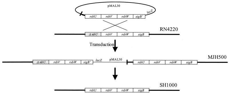
A 3-kb fragment encompassing the complete _rsbU_-_rsbV_-rsbW region and part of sigB from S. aureus strain Newman was amplified by using Pwo DNA polymerase (Roche) with primers OL-80 and OL-81 (Table 1). Following purification, the PCR product was digested with _Bam_HI and _Eco_RI and cloned into pAZ106 (30) by using standard cloning techniques (48). The resulting plasmid, pMAL30 (Table 1), was used to transform electrocompetent S. aureus RN4220 by the method of Schenk and Ladagga (49). The plasmid was integrated into the chromosome through homology with the parental copy by a Campbell type of event to produce a _sigB_-lacZ fusion. The unresolved locus was transferred into recipient 8325-4 cells by phage transduction (41) by using φ11 as the transducing phage. The rsb-sigB locus, which contained a duplication of the rsb genes, was resolved by overnight growth in BHI medium with no antibiotic to enable loss of the integrated plasmid, pMAL30, by homologous recombination between the duplicated sets of rsb genes (Fig. 1). Plating cells on plates containing X-Gal (5-bromo-4-chloro-3-indolyl-β-d-galactopyranoside) (50 mg liter−1) allowed clones to be isolated that were not blue and thus no longer contained the pMAL30-generated _sigB_-lacZ fusion; these clones were tested for erythromycin sensitivity. The following two methods were used to confirm replacement of the 8325-4 rsbU gene with the gene from strain Newman: a PCR performed with primers OL-78 and OL-79 (34) and genomic DNA sequencing (30) performed with the same primers. In addition, the integrity of the _rsb_-sigB locus and its immediate vicinity was verified by Southern blotting (data not shown).
FIG. 1.
Schematic diagram illustrating the method used for construction of SH1000 (8325-4 rsbU+). A single crossover of pMAL30 into the chromosome of RN4220 produced a sigB_-lacZ fusion, which was transferred to 8325-4 by transduction. The rsb-sigB locus was resolved by overnight growth in antibiotic-free medium followed by selection for white clones (loss of plasmid) and pigment formation (functional rsbU). Δ_rsbU is the defective gene containing an 11-bp deletion.
sigB::tet transductions.
The sigB::tet mutation was present in 8325-4, which has an rsbU deletion mutation. To introduce the sigB::tet mutation into derivatives of SH1000 without cotransducing the rsbU mutation, we screened transductants using the PCR method of Kullik and Giachino (34). Despite the fact that the rsbU and sigB mutations in these genes are separated by less than 3 kb, cotransduction was found to be only 80%, which facilitated isolation of sigB::tet rsbU+ derivatives.
Construction of an sspA-lacZ reporter.
The promoter region of sspA was amplified as a 1.15-kb, PCR-generated DNA fragment (position −1,000 to position 150 bp relative to the translational start site) by using OG-15 and OG-16 (Table 1). The purified DNA was digested with _Xba_I and _Bam_HI and cloned into similarly digested pAZ106. S. aureus RN4220 was transformed with the resulting plasmid, pLES2, and an integrant confirmed by Southern blotting was transduced into appropriate backgrounds by using φ11.
Enzyme assays.
Levels of β-galactosidase activity were measured as described previously (30). Fluorescence was measured by using a Victor plate reader (Wallac) with a 0.1-s count time and was calibrated with standard concentrations of 4-methyl-umbelliferone. One unit of β-galactosidase activity was defined as the amount of enzyme that catalyzed the production of 1 pmol of 4-methyl-umbelliferone per min per unit of optical density at 600 nm (OD600). Assays were performed with duplicate samples, and the values were averaged. The results presented here are representative of the results of three independent experiments that showed less than 10% variability.
The levels of alpha toxin (Hla) in culture supernatants were determined as described previously (9). One hemolytic unit was defined as the reciprocal value of the dilution that resulted in 50% lysis of rabbit erythrocytes per OD600 unit.
Protein samples, prepared as described previously (9), were resolved on a precast 12% (wt/vol) acrylamide gel containing Zymogram Ready Gel containing gelatin (Bio-Rad). Renaturation and visualization were performed according to the manufacturer's instructions.
Northern hybridization.
S. aureus strains were grown in 25 ml of BHI medium (culture/flask volume ratio, 1:10) at 37°C with shaking at 250 rpm. RNA was extracted from harvested cells as described previously (30). Ten micrograms of total RNA, which had been separated on a 1% (wt/vol) agarose gel and vacuum blotted onto a nylon membrane (Roche), was probed and washed under high- stringency conditions (0.1× SSC-1% sodium dodecyl sulfate [1× SSC is 0.15 M NaCl plus 0.015 M sodium citrate], 68°C) by using standard conditions (48). The concentration of RNA was measured at 260 nm, and equivalent loading on agarose gels was confirmed by ethidium bromide staining and UV visualization. The katA probe was a 32P-labeled _Bam_HI-_Hin_dIII fragment (position −159 to position 943 relative to the translational start point of katA) from a digest of PCR DNA amplified by using primers OL-15 and OL-177. Densitometric analysis of autoradiographs with different exposure times was performed by using ImageMaster 3.01 software (Amersham-Pharmacia).
Catalase assays, H2O2 challenge, and starvation survival.
Catalase activity was assayed spectrophotometrically at 240 nm (ɛ = 43.6 M liter−1 cm−1) as described by Beers and Sizer (3) by using 50 mM potassium phosphate buffer (pH 7.0) with 19.6 mM hydrogen peroxide. Hydrogen peroxide resistance assays were carried out as described previously (30, 52). Comparative starvation survival experiments were performed in amino acid-limiting CDM medium incubated statically at 25°C as described previously (52).
Western blotting.
Proteins were blotted onto a polyvinylidene difluoride membrane (Bio-Rad) and were detected by using antisera raised against Hla (36), staphylococcal serine protease (SspA) (36), and SarA (5) (1:6,000, 1:2,500, and 1:400 dilutions of the antibodies, respectively) and standard methods (48). Alkaline phosphatase-conjugated goat anti-rabbit secondary antibody (diluted 1:30,000) was used to detect Hla and SspA colorimetrically, and horseradish peroxidase-conjugated antibody was used to detect SarA with the enhanced chemiluminescence system (Amersham-Pharmacia). Pig serum (20%, vol/vol; Sigma) was included during blocking to improve detection of SarA. Densitometric analysis of autoradiographs with different exposure times was performed by using ImageMaster 3.01 software (Amersham-Pharmacia).
Virulence testing of strains in a murine skin abscess model.
S. aureus strains were grown to the stationary phase in BHI medium (15 h) and then harvested by centrifugation and washed twice in phosphate-buffered saline (PBS). The cell concentrations were adjusted to 5 × 10⁸ CFU ml−1, and then 200-μl portions of a cell suspension were injected subcutaneously into female 6- to 8-week-old BALB/c mice. After 7 days the mice were euthanatized with CO2, and skin lesions were aseptically removed and stored frozen in liquid nitrogen. The lesions were weighed, chopped, and homogenized in a mini-blender in 2.5 ml of ice-cold PBS. After 1 h of incubation on ice, the lesions were homogenized again before serial dilution of the suspension, and the total number of bacteria was counted by growth on BHI agar. The statistical significance of the recovery of strains was evaluated by using the Student t test with a 5% confidence limit.
RESULTS
Construction of SH1000 (8325-4 rsbU+).
The defective copy of rsbU in S. aureus 8325-4 was replaced with a copy of the intact gene from S. aureus Newman, without leaving behind an antibiotic resistance marker. To achieve this, a multiple-step approach was used (Fig. 1). First, the complete rsbU, rsbV, and rsbW genes together with part of the sigB gene were amplified as a 3-kb DNA fragment by PCR and cloned into pAZ106. The resulting plasmid, pMAL30, was used to transform S. aureus RN4220, and clones were obtained with duplicate copies of the rsbU, rsbV, and rsbW genes. Integration produced a sigB-lacZ fusion, and a selected clone, MJH499, was blue on X-Gal plates. The duplicated rsb-sigB locus was transferred to S. aureus 8325-4 by using φ11-mediated transduction to generate strain MJH500. Finally, to restore the rsb-sigB locus, MJH500 was grown overnight in the absence of antibiotics, and the culture was diluted and plated on X-Gal plates. Clones were then picked that were white, indicating loss of the plasmid, and they were screened for the formation of yellow pigment, indicating that a functional rsbU gene was present, by overnight growth on BHI agar. The frequency of loss of the plasmid was between 10−4 and 10−5.
A number of pigmented clones were isolated and screened for the presence of a functional rsbU locus by using the PCR method described by Kullik and Giachino (34). In addition, the integrity of rsbU at the site of the previous deletion was verified by genomic DNA sequencing (30). One clone, SH1000 (8325-4 rsbU+), was selected for use.
SH1000 (8325-4 rsbU+) was characterized phenotypically to compare it with a previously described functional rsbU strain of S. aureus BB255, which contained a tetracycline resistance marker (26). Expression of the cytosolic, σB-regulated protein Asp23 (N-terminal sequence, VDNNXAXQAYDXQ), production of the orange-yellow pigment staphyloxanthin, and a fourfold-greater minimum bactericidal concentration but not MIC of hydrogen peroxide were observed (data not shown), demonstrating that the phenotypic properties of SH1000 were those of a functional rsbU strain.
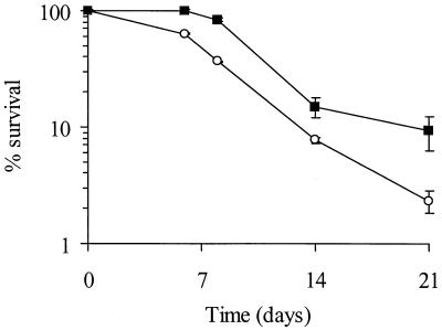
A number of additional phenotypic differences between SH1000 (8325-4 rsbU+) and 8325-4 were seen. SH1000 (8325-4 rsbU+) exhibited a decreased lag phase (15 to 20 min) before exponential growth began after dilution of an overnight culture into fresh medium (data not shown). Similar to the findings of Giachino et al. (26), we consistently observed a slightly increased growth yield for SH1000 (8325-4 rsbU+). The starvation survival capability of SH1000 (8325-4 rsbU+) in amino acid-limiting CDM medium measured for 21 days was greater than that of 8325-4 (Fig. 2). Starvation in this CDM medium results in a requirement for a number of oxidative stress resistance components (52).
FIG. 2.
Starvation survival kinetics of SH1000 (8325-4 rsbU+) (▪) and 8325-4 (○) during prolonged incubation at 25°C in amino acid-limiting CDM medium. The values are representative of the results of three separate experiments, and the error bars indicate the mean errors.
Catalase expression in SH1000.
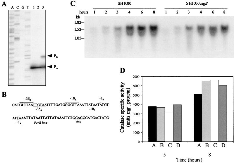
The increased resistance of SH1000 (8325-4 rsbU+) to hydrogen peroxide was hypothesized to be catalase mediated, by virtue of a putative consensus σB promoter element in the katA promoter region. We have previously shown that the katA promoter in 8325-4 contains the −35 and −10 elements of a σA promoter-binding site and a binding site for PerR, the peroxide regulon repressor (30, 31). Primer extension of RNA isolated from post-exponential-phase cultures (OD600, 8) of 8325-4, SH1000 (8325-4 rsbU+), and PC400 (8325-4 sigB) revealed a σB promoter, PB, upstream of katA in SH1000 (8325-4 rsbU+) (Fig. 3A). This σB-regulated transcript was absent in 8325-4 and PC400 (8325-4 sigB).
FIG. 3.
(A) Mapping of the 5′ ends of katA transcripts by primer extension analysis. One-hundred-microgram portions of RNA from post-exponential-phase cultures was used in reactions for 8325-4 (lane 1), PC400 (8325-4 sigB) (lane 2), and SH1000 (8325-4 rsbU+) (lane 3). Lanes A, C, G, and T show the dideoxy sequencing ladder obtained by using the same primer that was used for primer extension. (B) Potential −35 and −10 regions and transcriptional start sites (+1) for σA and σB are indicated by the subscripts A and B, respectively. The PerR box is indicated by boldface type, and rbs indicates the translational recognition sequence upstream of the translational start (underlined). (C) Transcript levels of katA during growth in 25 ml of BHI medium (medium/flask volume ratio, 1:10) at 37°C with shaking at 250 rpm. Samples were removed at the times indicated at the top, and the extracted RNA was probed by using a radiolabeled katA fragment. (D) Assay of catalase activity during growth in 25 ml of BHI medium (medium/flask volume ratio, 1:10) at 37°C with shaking at 250 rpm. Samples removed at different times were washed in PBS and then assayed. Bars A, SH1000 (8325-4 rsbU+); bars B, 8325-4; bars C, PC400 (8325-4 sigB); bars D, MJH502 (SH1000 rsbU+ sigB). Assays were performed in triplicate, and the means are shown. The results are representative of the results of two independent experiments.
To assess the contribution of the PB promoter to the regulation of katA expression, we probed RNA isolated from SH1000 (8325-4 rsbU+) and MJH502 (SH1000 rsbU+ sigB) throughout growth (Fig. 3B) by Northern blotting. As measured by densitometry, the levels of katA transcript on the blot were between 1.2 and 1.5 times greater in SH1000 (8325-4 rsbU+) than in MJH502 (SH1000 rsbU+ sigB), suggesting that PB has only a minor role under these conditions. When catalase was assayed during the post exponential and stationary phases of growth (after 5 and 8 h), similar levels of activity were observed (Fig. 3D). In contrast, when expression was measured by using a katA-lacZ transcriptional fusion described previously (30, 31), katA transcription was four- to sixfold lower during post-exponential-phase growth of MJH506 (SH1000 katA-lacZ) than during post-exponential-phase growth of MJH006 (8325-4 katA-lacZ) (data not shown). The disparity between the lacZ data and the data from the other assays may reflect increased turnover of the lacZ transcript via an unknown mechanism. We hypothesize that σB has little or no significance in the overall control of catalase expression under the conditions tested.
Expression of Hla and SspA.
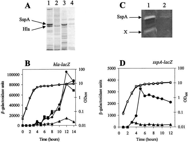
The exoproteins of S. aureus SH1000 (8325-4 rsbU+) and 8325-4 were precipitated from culture supernatants and visualized. The level of stationary-phase exoproteins was found to be much lower in SH1000 (8325-4 rsbU+) than in 8325-4, and the profiles revealed large reductions for several proteins, including Hla and SspA (Fig. 4A). The expression of Hla is known to be modulated indirectly by σB, since inactivation of sigB leads to hyperproduction of Hla (14). To quantify the observed difference in Hla expression, cultures were assayed for activity at 10 h, and this analysis showed that 8325-4 had 166.5 alpha toxin units (hemolytic units) and that SH1000 had 77.7 hemolytic units. Furthermore, to show that the reduction was controlled transcriptionally and was not due to alterations in protease activity, we assayed β-galactosidase expression using an hla-lacZ transcriptional reporter fusion. Expression of the hla-lacZ fusion was much reduced in SH1000 (8325-4 rsbU+), suggesting that the reduction in hla expression was primarily controlled at the transcriptional level (Fig. 4B).
FIG. 4.
(A) Exoproteins of 8325-4 (lane 1), SH1000 (8325-4 rsbU+) (lane 2), PC1839 (8325-4 sarA) (lane 3), and SH1002 (SH1000 sarA) (lane 4) purified from culture supernatants after 15 h of growth. The arrows indicate the positions of serine protease (SspA) and alpha toxin (Hla), as verified by Western blotting (data not shown). In PC1839 (8325-4 sarA) there was increased proteolysis of Hla, as shown by the number of smaller fragments. (B) Assay of transcription from an hla-lacZ fusion during growth. Expression of the reporter fusion in PC322 (8325-4 hla-lacZ) (• and ○), JLA371 (SH1000 hla-lacZ) (▴ and ▵), PC4044 (8325-4 sigB hla-lacZ) (▪ and □), and JLA373 (SH1000 sigB hla-lacZ) (♦ and ⋄) was measured at different times. •, ▴, ▪, and ♦, β-galactosidase activity; ○, ▵, □, and ⋄, bacterial growth (OD600). The SH1000 sigB mutant derivatives were confirmed to be rsbU+ by using the PCR method of Kullik and Giachino (34). (C) Protease activities of 8325-4 (lane 1) and SH1000 (8325-4 rsbU+) (lane 2) visualized by using a gelatin-containing zymogram. The arrow labeled X indicates the position of unknown protease activity; the position of SspA was verified by comparison with the position of purified protein (V8 protease; Sigma) (data not shown). (D) Assay of transcription from an sspA-lacZ fusion during growth. Expression of the reporter fusion in LES07 (8325-4 sspA-lacZ) (• and ○) and LES08 (SH1000 sspA-lacZ) (▴ and ▵) was measured at different times. • and ▴, β-galactosidase activity; ○ and ▵, bacterial growth (OD600).
The observation of reduced SspA in SH1000 (8325-4 rsbU+) (Fig. 4A) led us to examine the activities and amounts of other secreted proteases by using zymograms (Fig. 4C). The activity levels of SspA and a second unidentified protease were much reduced compared to the levels in 8325-4. As with hla, this reduction was controlled transcriptionally, since an assay of an sspA-lacZ transcriptional fusion determined that there was reduced β-galactosidase activity in SH1000 (8325-4 rsbU+) compared to the activity in 8325-4 (Fig. 3D). Inactivation of the agr and sarA genes in S. aureus is known to affect exoprotein production (9, 13, 32, 42), and consequently we examined the exoprotein profiles of SH1000 strains containing agr and sarA mutations. The agr mutant, SH1001, exhibited much reduced levels of exoproteins, such as Hla and SspA, as expected (data not shown). In contrast, SH1002 (SH1000 sarA) had increased exoprotein synthesis (Fig. 4A), and notably, the level of SspA was higher, which led to increased proteolysis of the other exoproteins, including Hla; this was confirmed by Western blotting by using antisera raised against Hla and SspA (data not shown).
Effect of σB on sarA and agr expression.
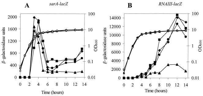
Expression of the genes encoding the virulence regulators SarA and Agr was measured in 8325-4 and SH1000 (8325-4 rsbU+) by using lacZ fusions constructed previously (9). A significant reduction in agr (RNA III) expression was observed during growth of SH1000 (8325-4 rsbU+) compared to the expression in 8325-4 (Fig. 5B). This finding matched the previous description of reduced agr (RNA III) expression by Giachino et al. (26). Since SarA can act as an activator of Agr expression (18), we examined whether the diminished level of agr (RNA III) was a consequence of reduced SarA expression. An assay of a sarA-lacZ reporter demonstrated that the overall levels of transcription of sarA in strains 8325-4 and SH1000 (8325-4 rsbU+) and sigB mutant derivatives of these strains were very similar (Fig. 5A). The σB-mediated reductions in expression of agr (RNA III), hla, and sspA are thus likely to be SarA independent.
FIG. 5.
Assay of transcription from a sarA-lacZ fusion and an agr (RNA III)-lacZ fusion during growth. (A) Expression of the reporter fusion in PC161 (8325-4 sarA-lacZ) (• and ○), JLA311 (SH1000 sarA-lacZ) (▴ and ▵), PC4030 (8325-4 sigB sarA-lacZ) (▪ and □), and JLA313 (SH1000 sigB sarA-lacZ) (♦ and ⋄) at different times. (B) Expression of the reporter fusion in SH101F7 (8325-4 agr [RNA III]-lacZ) (• and ○), JLA341 (SH1000 agr [RNA III]-lacZ) (▴ and ▵), PC604 (8325-4 sigB agr [RNA III]-lacZ) (▪ and □), and JLA343 (SH1000 sigB agr [RNA III]-lacZ) (♦ and ⋄) at different times. •, ▴, and ▪, β-galactosidase activity; ○, ▵, and □, bacterial growth (OD600).
The findings described above contrast with the report by Bischoff et al. (4) that sarA transcription is increased in a sigB+ strain compared to the sarA transcription in the rsbU deletion strain from which it was generated. Consequently, the SarA protein levels during growth of strains SH1000 (8325-4 rsbU+) and 8325-4 and their sigB mutant derivatives were determined by using cells grown under our standard defined growth conditions by Western blotting.
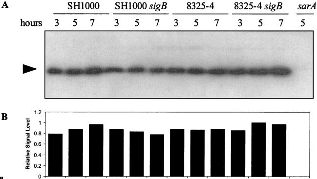
Samples removed at 3, 5, and 7 h were lysed, and total cellular proteins were probed by using anti-SarA antibodies (Fig. 6). At these times the levels of SarA were remarkably similar for each of the strains. Thus, when the lacZ reporter assay is used or when protein levels are detected by Western blotting, the levels of SarA and temporal regulation of SarA are very similar for strains 8325-4 and SH1000 (8325-4 rsbU+) and their sigB mutant derivatives.
FIG. 6.
(A) Equivalent amounts of total cellular proteins (OD600 of culture, 0.1), isolated after lysostaphin digestion from cultures grown for 3, 5, and 7 h, were blotted and probed with purified immunoglobulin G antibodies raised against SarA (5). (B) SarA signals quantified on the blot by densitometry. The relative signal level for each lane was compared to the maximum signal level obtained. The results are representative of the results of three independent experiments.
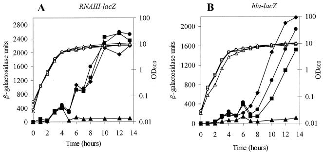
To confirm the apparent lack of a role for SarA in the overall negative effect of σB on the expression of agr (RNA III) and hla, we introduced a sarA mutation into agr (RNA III)-lacZ and hla-lacZ reporter strains by transduction. These reporters in strains 8325-4 and SH1000 (8325-4 rsbU+) and their sigB mutant derivatives were assayed throughout growth (Fig. 7). The sarA mutation decreased expression of each of the reporters, as expected (compare Fig. 7 with Fig. 4B and 5B). However, transcription of both agr (RNA III) and hla remained lower in SH1000 sarA than in 8325-4 sarA and the sigB derivatives (Fig. 7). The decrease in agr and hla expression was, therefore, due to a functional rsbU gene in SH1000 and not due to the activity of SarA.
FIG. 7.
Assay of an agr (RNA III)-lacZ fusion and an hla-lacZ fusion in a sarA mutant background during growth. (A) Expression of the reporter fusion in PC600 (8325-4 sarA agr [RNA III]-lacZ) (• and ○), JLA345 (SH1000 sarA agr [RNA III]-lacZ) (▴ and ▵), PC602 (8325-4 sarA sigB agr [RNA III]-lacZ) (▪ and □), and JLA347 (SH1000 sarA sigB agr [RNA III]-lacZ) (♦ and ⋄) at different times. (B) Expression of the reporter fusion in PC3221 (8325-4 sarA hla-lacZ) (• and ○), JLA375 (SH1000 sarA hla-lacZ) (▴ and ▵), JLA376 (8325-4 sarA sigB hla-lacZ) (▪ and □), and JLA377 (SH1000 sarA sigB hla-lacZ) (♦ and ⋄) at different times. •, ▴, and ▪, β-galactosidase activity; ○, ▵, and □, bacterial growth (OD600).
Comparison of virulence of 8325-4 and SH1000.
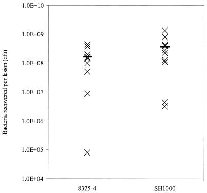
The virulence of SH1000 (8325-4 rsbU+) was determined by using an established murine subcutaneous skin abscess model of infection (9, 10, 20, 30, 31) and was compared to the virulence of 8325-4 (Fig. 8). When an inoculum of 108 CFU was used, the levels of recovery of SH1000 (8325-4 rsbU+) and 8325-4 were not significantly different (P = 0.157) (Fig. 8). The lesions produced were similar in size and appearance (data not shown).
FIG. 8.
Virulence of S. aureus strains in a murine skin abscess model of infection. Approximately 108 CFU of each strain was inoculated subcutaneously into 6- to 8-week-old BALB/c mice. Seven days after infection mice were euthanatized, lesions were removed and homogenized, and viable bacteria were counted after dilution and growth on BHI agar plates. The bar indicates the mean recovery for each strain.
DISCUSSION
In this study we constructed SH1000 (8325-4 rsbU+), a functional rsbU derivative of 8325-4. This is an important requirement for studying the control of virulence in S. aureus, since in most genetic studies the workers have used the 8325-4/RN6390 genetic lineage and the absence of antibiotic markers should facilitate future studies. The rsbU mutation that is present in the 8325-4 lineage is known to dramatically reduce σB activity and consequently affect expression of many virulence-associated loci. The phenotype of SH1000 (8325-4 rsbU+) was characterized, and differences between this strain and 8325-4 were observed. These differences included production of the orange pigment staphyloxanthin, decreased levels of alpha toxin, expression of asp23, and increased resistance to H2O2. While this work was being done, a tetracycline-resistant, functional rsbU derivative strain of BB255 was described (4, 26) which effectively has the same lineage, since it was derived from the same parent strain as 8325-4 and RN6390. In the study reported here, we found that in many cases the phenotype of SH1000 (8325-4 rsbU+) and the reported phenotype of the BB255 strain were similar; however, there were important differences that are described below.
The increase in hydrogen peroxide resistance was of interest as one of the major determinants of this characteristic in S. aureus is the very active sole catalase, KatA. Previous studies characterized the katA locus in 8325-4 and demonstrated the presence of a σA promoter, a PerR-binding site, and described positive regulation mediated via Fur (30, 31). Inspection of the identified promoter region of katA revealed a putative σB motif (Fig. 2A) with strong identity to the consensus sequence described by Gertz et al. (24). Primer extension of katA mRNA from SH1000 (8325-4 rsbU+), 8325-4, and PC400 (8325-4 sigB) revealed that the σB-dependent promoter, PB, was active in SH1000 (8325-4 rsbU+) but not in the other two strains. To assess the contribution of PB to the overall levels of the katA transcript, RNA from each of the strains was isolated throughout growth and probed for katA by Northern blotting. The amount of the katA transcript was found to be between 1.2 and 1.5 times greater in SH1000 (8325-4 rsbU+) than in MJH502 (SH1000 rsbU+ sigB) (Fig. 2B). Assays of catalase activity during growth of SH1000 (8325-4 rsbU+), MJH502 (SH1000 rsbU+ sigB), 8325-4, and PC400 (8325-4 sigB) revealed little difference among the strains (Fig. 2C), which is consistent with the similar levels of transcript observed in Northern blot and primer extension analyses. In contrast to these results, assays of a katA-lacZ reporter in SH1000 (8325-4 rsbU+) and 8325-4 revealed levels of β-galactosidase that were four- to sixfold lower in SH1000 (8325-4 rsbU+) than in 8325-4. The reason for this discrepancy is unclear, but this may reflect differences in turnover of the katA-lacZ message in these strains.
The similar transcription of katA and the similar expression of catalase activity in SH1000 (8325-4 rsbU+) and 8325-4 despite the presence of an active PB promoter in the former strain suggest that the overall contribution of this promoter is minor, at least under the conditions studied here. Previous work has demonstrated that katA is regulated by the PerR and Fur proteins, which control katA levels in response to various signals, including peroxide stress and the levels of manganese and iron in the cell (30, 31). The PB promoter may be important under some environmental conditions, such as alkaline and heat stress, under which expression of a number of σB-regulated genes is induced (24, 25, 34). Bischoff et al. (4) remarked that the levels of katA, when they were measured by Northern blotting, were equivalent in their marked functional rsbU+ strain and its rsbU mutant parent. We confirmed the similar transcript levels in SH1000 and 8325-4 and extended this observation to show similar levels of activity; however, importantly, there is potential for σB transcriptional control. The lack of an overall effect of σB on catalase levels may reflect an ability by the cell to maintain homeostasis via alternative regulatory mechanisms. H2O2 resistance is multifactorial, and the observed increase in the minimum bactericidal concentration of H2O2 may be due to other σB-regulated components that are expressed in SH1000 (8325-4 rsbU+) but not in 8325-4. A large regulon of σB-regulated genes has already been identified (25).
The most striking phenotype observed for SH1000 (8325-4 rsbU+) was the low level of exoprotein expression compared to the level of expression in 8325-4. This phenotype is similar to that observed for clinical strains of S. aureus described recently by Blevins et al. (6) and contrasts with that of 8325-4. A major reduction in protease expression, including sspA expression, was observed by using protease zymograms, Western blotting, and reporter assays performed with an sspA-lacZ fusion. The reduced level of Hla has been reported previously (26), and we have determined that this effect is transcriptional and not due to the changes in protease activity. The overall effect of an intact σB locus, therefore, is to reduce the level of expression of the sspA and hla genes. The mechanism for this is likely to involve the lower levels of agr in the cell, since we observed a large reduction in expression of an agr (RNA III)-lacZ fusion in SH1000 (8325-4 rsbU+). Bischoff et al. (4) similarly observed that the level of agr (RNA III) was reduced in their functional rsbU+ strain of BB255.
The most obvious candidate for mediating the reduction in agr levels was SarA, which is a known activator of agr expression (18, 19, 29). However, when a sarA-lacZ reporter was assayed or when the amount of SarA protein was determined by Western blotting in strains SH1000 (8325-4 rsbU+) and 8325-4 and their σB mutant derivatives, the levels were found to be very similar. Importantly, inactivation of sigB in each of the strains did not alter the level of SarA, suggesting that σB had little or no role under the conditions studied here. This is in accordance with the results of Blevins et al. (5), who reported that SarA is expressed constitutively. In their quantitative analysis of SarA during different stages of growth of RN6390 and clinical isolate UAMS-1, these authors observed no variation between the strains (6). We extended this observation to strains SH1000 (8325-4 rsbU+) and 8325-4 and their sigB mutant derivatives. However, this finding contrasts with the findings of Bischoff et al. (4). In the studies of these authors, expression of a sarA-lux fusion reporter strain was found to be greater during the stationary phase of growth in a marked rsbU+ strain than in its rsbU mutant parent. The reason for the difference in sarA expression between the two studies is unclear. However, since different strains, media, and growth conditions were used, we cannot exclude the possibility that genetic or environmental differences had an effect. Gertz et al. (25) showed that there was increased SarA expression in a sigB mutant of S. aureus COL by using two-dimensional electrophoresis; in this study minimal medium in which constitutive σB expression was observed was used. Thus, it is important to quantitatively study a number of environmental conditions to monitor the effect of σB on SarA expression. The σB-dependent promoter, _sarP_3 (2, 38), is most active during the exponential growth phase in strains with a wild-type rsbU locus (4). SarA represses its own transcription via the proximal _sarP_1 promoter but not via the more distal _sarP_3 promoter (7). Therefore, the failure to observe increased expression of SarA despite the presence of a σB promoter may be due to the autoregulatory capacity of SarA (5, 7) maintaining constant protein levels during growth. We hypothesize that in certain environmental conditions under which σB activity increases, which results in a concomitant reduction in SarA repression, SarA autoregulation increases and maintains the level of SarA. However, since we have found that in the conditions studied here the SarA protein level is relatively constant during growth irrespective of the rsbU or sigB genotype, the SarA level is unlikely to be the mediator for the reduction in expression of the virulence-associated loci seen in SH1000 (8325-4 rsbU+).
The properties of SH1000 (8325-4 rsbU+) mean that the 8325-4 lineage now behaves like clinical isolates which are typically low protease producers, have lower Hla levels of expression, and during growth in standard laboratory conditions have constant levels of SarA (6). The effects of σB on virulence determinant expression, although multiple and dramatic, did not alter the virulence of S. aureus in the mouse abscess model of infection. This result supports previous studies that showed that a sigB mutation in an rsbU+ parent strain did not produce attenuation in three separate animal models (40).
From the experiments reported here we conclude that mediation of the reduction in agr (RNA III) levels in SH1000 is independent of the level of SarA. Consequently, since the SarA level appears not to be the effector of the overall negative σB-mediated effect on virulence-associated loci, what then is the effector? The sequence of the S. aureus genome has revealed an impressive array of SarA homologues, including SarH1 (51), Rot (39), and SarT (50). These homologues have all been shown to repress exoprotein expression. A number of other candidate loci have an impact on exoprotein synthesis. These loci include 1E3 (12) and sae (27, 28), and when inactivated, the latter locus dramatically reduces expression of many exoproteins. To date, we have found that neither SarH1 nor Rot is the missing effector protein (J. L. Aish and S. J. Foster, unpublished data); we are currently attempting to identify this missing effector by epistasis. The σB-mediated modulation of expression of virulence-associated loci of S. aureus is intriguing, particularly since S. aureus colonizes the anterior nares, where it resides as a harmless commensal. It will be interesting to determine whether σB functions to discriminate between environments and to regulate exoprotein synthesis. SH1000 (8325-4 rsbU+) provides a useful genetic background to characterize the exact physiological role of σB in S. aureus and should facilitate comparisons with previous studies for this genetic lineage.
Acknowledgments
M.J.H. and J.L.A. contributed equally to this work.
We thank Mark Smeltzer for kindly providing SarA antibodies and Arthur Moir for N-terminal sequencing. We are grateful to Staffan Arvidson and Richard Proctor for providing strains. We acknowledge the S. aureus Genome Sequencing Project (strain 8325) and B. A. Roe, Y. Qian, A. Dorman, F. Z. Najar, S. Clifton, and J. Iandolo, who had funding from NIH and the Merck Genome Research Institute.
The BBSRC (M.J.H., J.L.A., I.J.W., L.S., and J.K.L.) and The Royal Society (S.J.F.) funded this research.
REFERENCES
- 1.Abdelnour, A., S. Arvidson, T. Bremell, C. Ryden, and A. Tarkowski. 1993. The accessory gene regulator (agr) controls Staphylococcus aureus virulence in a murine arthritis model. Infect. Immun. 61**:**3879-3885. [DOI] [PMC free article] [PubMed] [Google Scholar]
- 2.Bayer, M. G., J. H. Heinrichs, and A. L. Cheung. 1996. The molecular architecture of the sar locus in Staphylococcus aureus. J. Bacteriol. 178**:**4563-4570. [DOI] [PMC free article] [PubMed] [Google Scholar]
- 3.Beers, R. F., and I. W. Sizer. 1952. A spectrophotometric method for measuring the breakdown of hydrogen peroxide by catalase. J. Biol. Chem. 195**:**133-140. [PubMed] [Google Scholar]
- 4.Bischoff, M., J. M. Entenza, and P. Giachino. 2001. Influence of a functional sigB operon on the global regulators sar and agr in Staphylococcus aureus. J. Bacteriol. 183**:**5171-5179. [DOI] [PMC free article] [PubMed] [Google Scholar]
- 5.Blevins, J. S., A. F. Gillaspy, T. M. Rechtin, B. K. Hurlburt, and M. S. Smeltzer. 1999. The staphylococcal accessory regulator (sar) represses transcription of the Staphylococcus aureus collagen adhesin gene (cna) in an _agr_-independent manner. Mol. Microbiol. 33**:**317-326. [DOI] [PubMed] [Google Scholar]
- 6.Blevins, J. S., K. E. Beenken, M. O. Elasri, B. K. Hurlburt, and M. S. Smeltzer. 2002. Strain-dependent differences in the regulatory roles of sarA and agr in Staphylococcus aureus. Infect. Immun. 70**:**470-480. [DOI] [PMC free article] [PubMed] [Google Scholar]
- 7.Chackrabarti, S. W., and T. K. Misra. 2000. SarA represses agr operon expression in a purified in vitro Staphylococcus aureus transcription system. J. Bacteriol. 182**:**5893-5897. [DOI] [PMC free article] [PubMed] [Google Scholar]
- 8.Chan, P. F., and S. J. Foster. 1998. The role of environmental factors in the regulation of virulence-determinant expression in Staphylococcus aureus 8325-4. Microbiology 144**:**2469-2479 [DOI] [PubMed] [Google Scholar]
- 9.Chan, P. F., and S. J. Foster. 1998. Role of SarA in virulence determinant production and environmental signal transduction in Staphylococcus aureus. J. Bacteriol. 180**:**6232-6241. [DOI] [PMC free article] [PubMed] [Google Scholar]
- 10.Chan, P. F., S. J. Foster, E. Ingham, and M. O. Clements. 1998. The Staphylococcus aureus alternative sigma factor σB controls the environmental stress response but not starvation survival or pathogenicity in a mouse abscess model. J. Bacteriol. 180**:**6082-6089. [DOI] [PMC free article] [PubMed] [Google Scholar]
- 11.Cheung, A. L., J. M. Koomey, C. A. Butler, S. J. Projan, and V. A. Fischetti. 1992. Regulation of exoprotein expression in Staphylococcus aureus by a locus (sar) distinct from agr. Proc. Natl. Acad. Sci. USA 89**:**6462-6466. [DOI] [PMC free article] [PubMed] [Google Scholar]
- 12.Cheung, A. L., C. Wolz, M. R. Yeaman, and A. S. Bayer. 1995. Insertional inactivation of a chromosomal locus that modulates expression of potential virulence determinants in Staphylococcus aureus. J. Bacteriol. 177**:**3220-3226. [DOI] [PMC free article] [PubMed] [Google Scholar]
- 13.Cheung, A. L., M. G. Bayer, and J. H. Heinrichs. 1997. sar genetic determinants neces_sar_y for transcription of RNAII and RNAIII in the agr locus of Staphylococcus aureus. J. Bacteriol. 179**:**3963-3971. [DOI] [PMC free article] [PubMed] [Google Scholar]
- 14.Cheung, A. L., Y.-T. Chien, and A. S. Bayer. 1999. Hyperproduction of alpha-hemolysin in a sigB mutant is associated with elevated SarA expression in Staphylococcus aureus. Infect. Immun. 67**:**1331-1337. [DOI] [PMC free article] [PubMed] [Google Scholar]
- 15.Cheung, A. L., K. Schmidt, B. Bateman, and A. C. Manna. 2001. SarS, a SarA homolog repressible by agr, is an activator of protein A synthesis in Staphylococcus aureus. Infect. Immun. 69**:**2448-2455. [DOI] [PMC free article] [PubMed] [Google Scholar]
- 16.Cheung, A. L., and P. Ying. 1994. Regulation of alpha- and beta-hemolysins by the sar locus of Staphylococcus aureus. J. Bacteriol. 176**:**580-585. [DOI] [PMC free article] [PubMed] [Google Scholar]
- 17.Chien, Y., A. C. Manna, S. J. Projan, and A. L. Cheung. 1999. _sar_A, a global regulator of virulence determinants in Staphylococcus aureus, binds to a conserved motif essential for _sar_-dependent gene regulation. J. Biol. Chem. 274**:**37169-37176. [DOI] [PubMed] [Google Scholar]
- 18.Chien, Y. T., and A. L. Cheung. 1998. Molecular interactions between two global regulators, sar and agr, in Staphylococcus aureus. J. Biol. Chem. 273**:**2645-2652. [DOI] [PubMed] [Google Scholar]
- 19.Chien, Y. T., A. C. Manna, and A. L. Cheung. 1998. _sar_A level is a determinant of agr activation in Staphylococcus aureus. Mol. Microbiol. 30**:**991-1001. [DOI] [PubMed] [Google Scholar]
- 20.Clements, M. O., S. P. Watson, and S. J. Foster. 1999. Characterization of the major superoxide dismutase of Staphylococcus aureus and its role in starvation survival, stress resistance, and pathogenicity. J. Bacteriol. 181**:**3898-3903. [DOI] [PMC free article] [PubMed] [Google Scholar]
- 21.Deora, R., T. Tseng, and T. K. Misra. 1997. Alternative transcription factor σSB of Staphylococcus aureus: characterization and role in transcription of the global regulatory locus sar. J. Bacteriol. 179**:**6355-6359. [DOI] [PMC free article] [PubMed] [Google Scholar]
- 22.Engelmann, S., and M. Hecker. 1996. Impaired oxidative stress resistance of Bacillus subtilis sigB mutants and the role of katA and katE. FEMS Microbiol. Lett. 145**:**63-69. [DOI] [PubMed] [Google Scholar]
- 23.Fairhead, H. 1998. Ph.D. thesis. University of Sheffield, Sheffield, United Kingdom.
- 24.Gertz, S., S. Engelmann, R. Schmid, K. Ohlsen, J. Hacker, and M. Hecker. 2000. Regulation of sigmaB-dependent transcription of sigB and asp23 in two different Staphylococcus aureus strains. Mol. Gen. Genet. 261**:**558-566. [DOI] [PubMed] [Google Scholar]
- 25.Gertz, S., S. Engelmann, R. Schmid, A.-K. Ziebandt, K. Tischer, C. Scharf, J. Hacker, and M. Hecker. 2000. Characterization of the σB regulon in Staphylococcus aureus. J. Bacteriol. 182**:**6983-6991. [DOI] [PMC free article] [PubMed] [Google Scholar]
- 26.Giachino, P., S. Engelmann, and M. Bischoff. 2001. σB activity depends on RsbU in Staphylococcus aureus. J. Bacteriol. 183**:**1843-1852. [DOI] [PMC free article] [PubMed] [Google Scholar]
- 27.Giraudo, A. T., A. L. Cheung, and R. Nagel. 1997. The sae locus of Staphylococcus aureus controls exoprotein synthesis at the transcriptional level. Arch. Microbiol. 168**:**53-58. [DOI] [PubMed] [Google Scholar]
- 28.Giraudo, A. T., A. Calzolari, A. A. Cataldi, C. Bogni, and R. Nagel. 1999. The sae locus of Staphylococcus aureus encodes a two-component regulatory system. FEMS Microbiol. Lett. 177**:**15-22. [DOI] [PubMed] [Google Scholar]
- 29.Heinrichs, J. H., M. G. Bayer, and A. L. Cheung. 1996. Characterization of the sar locus and its interaction with agr in Staphylococcus aureus. J. Bacteriol. 178**:**418-423. [DOI] [PMC free article] [PubMed] [Google Scholar]
- 30.Horsburgh, M. J., M. O. Clements, H. Crossley, E. Ingham, and S. J. Foster. 2001. PerR controls oxidative stress resistance and iron storage proteins and is required for virulence in Staphylococcus aureus. Infect. Immun. 69**:**3744-3754. [DOI] [PMC free article] [PubMed] [Google Scholar]
- 31.Horsburgh, M. J., E. Ingham, and S. J. Foster. 2001. In Staphylococcus aureus, Fur is an interactive regulator with PerR, contributes to virulence, and is necessary for oxidative stress resistance through positive regulation of catalase and iron homeostasis. J. Bacteriol. 183**:**468-475. [DOI] [PMC free article] [PubMed] [Google Scholar]
- 32.Janzon, L., and S. Arvidson. 1990. The role of the delta-lysin gene (hld) in the regulation of virulence genes by the accessory gene regulator (agr) in Staphylococcus aureus. EMBO J. 9**:**1391-1399. [DOI] [PMC free article] [PubMed] [Google Scholar]
- 33.Knobloch, J. K., K. Bartscht, A. Sabottke, H. Rohde, H. Feucht, and D. Mack. 2001. Biofilm formation by Staphylococcus epidermidis depends on functional RsbU, an activator of the sigB operon: differential activation mechanisms due to ethanol and salt stress. J. Bacteriol. 183**:**2624-2633. [DOI] [PMC free article] [PubMed] [Google Scholar]
- 34.Kullik, I. I., and P. Giachino. 1997. The alternative sigma factor sigB in Staphylococcus aureus: regulation of the sigB operon in response to growth phase and heat shock. Arch. Microbiol. 167**:**151-159. [DOI] [PubMed] [Google Scholar]
- 35.Kullik, I., P. Giachino, and T. Fuchs. 1998. Deletion of the alternative sigma factor σB in Staphylococcus aureus reveals its function as a global regulator of virulence genes. J. Bacteriol. 180**:**4814-4820. [DOI] [PMC free article] [PubMed] [Google Scholar]
- 36.Lindsay, J. A., and S. J. Foster. 1999. Interactive regulatory pathways control virulence determinant production and stability in response to environmental conditions in Staphylococcus aureus. Mol. Gen. Genet. 262**:**323-331. [DOI] [PubMed] [Google Scholar]
- 37.Lowy, F. 1998. Staphylococcus aureus infections. N. Engl. J. Med. 339**:**520-532. [DOI] [PubMed] [Google Scholar]
- 38.Manna, A. C., M. G. Bayer, and A. L. Cheung. 1998. Transcriptional analysis of different promoters in the sar locus in Staphylococcus aureus. J. Bacteriol. 180**:**3828-3836. [DOI] [PMC free article] [PubMed] [Google Scholar]
- 39.McNamara, P. J., K. C. Milligan-Monroe, S. Khalili, and R. A. Proctor. 2000. Identification, cloning, and initial characterization of rot, a locus encoding a regulator of virulence factor expression in Staphylococcus aureus. J. Bacteriol. 182**:**3197-3203. [DOI] [PMC free article] [PubMed] [Google Scholar]
- 40.Nicholas, R. O., T. Li, D. McDevitt, A. Marra, S. Sucoloski, P. L. Demarsh, and D. R. Gentry. 1999. Isolation and characterization of a sigB deletion mutant of Staphylococcus aureus. Infect. Immun. 67**:**3667-3669. [DOI] [PMC free article] [PubMed] [Google Scholar]
- 41.Novick, R. P. 1991. Genetic systems in staphylococci. Methods Enzymol. 204**:**587-636. [DOI] [PubMed] [Google Scholar]
- 42.Novick, R. P. 2000. Pathogenicity factors and their regulation, p. 392-407. In V. A. Fischetti, R. P. Novick, J. J. Ferretti, D. A. Portnoy, and J. I. Rood (ed.), Gram-positive pathogens. ASM Press, Washington, D.C.
- 43.Novick, R. P., H. F. Ross, S. J. Projan, J. Kornblum, B. Kreiswirth, and S. Moghazeh. 1993. Synthesis of staphylococcal virulence factors is controlled by a regulatory RNA molecule. EMBO J. 12**:**3967-3975. [DOI] [PMC free article] [PubMed] [Google Scholar]
- 44.Price, C. W. 2000. Protective function and regulation of the general stress response in Bacillus subtilis and related gram-positive bacteria, p. 179-197. In G. Storz and R. Hengge-Aronis (ed.), Bacterial stress responses. ASM Press, Washington, D.C.
- 45.Price, C. W., P. Fawcett, H. Ceremonie, N. Su, C. K. Murphy, and P. Youngman. 2001. Genome-wide analysis of the general stress response in Bacillus subtilis. Mol. Microbiol. 41**:**757-774. [DOI] [PubMed] [Google Scholar]
- 46.Rachid, S., K. Ohlsen, U. Wallner, J. Hacker, M. Hecker, and W. Ziebuhr. 2000. Alternative transcription factor σB is involved in regulation of biofilm expression in a Staphylococcus aureus mucosal isolate. J. Bacteriol. 182**:**6824-6826. [DOI] [PMC free article] [PubMed] [Google Scholar]
- 47.Rechtin, T. M., A. F. Gillaspy, M. A. Schumacher, R. G. Brennan, M. S. Smeltzer, and B. K. Hurlburt. 1999. Characterization of the _sar_A virulence gene regulator of Staphylococcus aureus. Mol. Microbiol. 33**:**307-316. [DOI] [PubMed] [Google Scholar]
- 48.Sambrook, J., E. F. Fritsch, and T. Maniatis. 1989. Molecular cloning: a laboratory manual, 2nd ed. Cold Spring Laboratory Press, Cold Spring Harbor, N.Y.
- 49.Schenk, S., and R. A. Ladagga. 1992. Improved methods for electroporation of Staphylococcus aureus. FEMS Microbiol. Lett. 94**:**133-138. [DOI] [PubMed] [Google Scholar]
- 50.Schmidt, K. A., A. C. Manna, S. Gill, and A. L. Cheung. 2001. SarT, a repressor of α-hemolysin in Staphylococcus aureus. Infect. Immun. 69**:**4749-4758. [DOI] [PMC free article] [PubMed] [Google Scholar]
- 51.Tegmark, K., A. Karlsson, and S. Arvidson. 2000. Identification and characterization of SarH1, a new global regulator of virulence gene expression in Staphylococcus aureus. Mol. Microbiol. 37**:**398-409. [DOI] [PubMed] [Google Scholar]
- 52.Watson, S. P., M. O. Clements, and S. J. Foster. 1998. Characterization of the starvation-survival response of Staphylococcus aureus. J. Bacteriol. 180**:**1750-1758. [DOI] [PMC free article] [PubMed] [Google Scholar]
