BLNK: molecular scaffolding through ‘cis’-mediated organization of signaling proteins (original) (raw)
Abstract
Assembly of intracellular macromolecular complexes is thought to provide an important mechanism to coordinate the generation of second messengers upon receptor activation. We have previously identified a B cell linker protein, termed BLNK, which serves such a scaffolding function in B cells. We demonstrate here that phosphorylation of five tyrosine residues within human BLNK nucleates distinct signaling effectors following B cell antigen receptor activation. The phosphorylation of multiple tyrosine residues not only amplifies PLCγ-mediated signaling but also supports ‘_cis_’-mediated interaction between distinct signaling effectors within a large molecular complex. These data demonstrate the importance of coordinate phosphorylation of molecular scaffolds, and provide insights into how assembly of macromolecular complexes is required for normal receptor function.
Keywords: adaptor proteins/B cell antigen receptor/signal transduction
Introduction
The mechanisms by which receptor activation translates into cellular functions has been an area of intense investigation over the past three decades. A paradigm has evolved in which ligand binding to receptors activates multiple signaling pathways that coordinately generate second messengers important in cellular functions (reviewed in Jordan et al., 2000). In biological systems involving receptor tyrosine kinases, the receptor or its associated signaling subunits is typically phosphorylated on multiple tyrosine residues that, in turn, recruit effector molecules. The concept of molecular scaffolding suggests that signaling components are required to organize in a specific manner at a macromolecular level to mediate receptor functions (reviewed in Leo et al., 2002; Smith and Scott, 2002). However, the mechanism by which such a scaffold coordinates activation of downstream pathways is less well defined. More recently, the concept of cytoplasmic scaffolds has emerged. Molecules, such as IRS-1, Grb2 and Gab1, which typically reside in the cytosol but are recruited to the plasma membrane following receptor engagement, are also capable of binding multiple effector proteins. While a large body of biochemical data demonstrates the existence of such large molecular complexes, the concept of how these macromolecular complexes coordinate the generation of second messengers has been less well elucidated.
In B cells, binding of foreign antigens to the B cell antigen receptor (BCR) induces activation of three distinct families of cytoplasmic protein tyrosine kinases (PTKs), Src, Syk, and Tec, which are required for B cell proliferation, differentiation and apoptosis (reviewed in DeFranco, 1997; Benschop and Cambier, 1999; Kurosaki, 1999). All three PTK families phosphorylate adapter proteins, which, in turn, coordinate enzymes that generate second messengers including phosphoinositides and GTPases (Takata et al., 1994; Cox et al., 1996; Law et al., 1996; Fu et al., 1998; Okada et al., 2000). The requirement for all three PTK families in B cell function has been demonstrated by the B cell developmental and functional defects revealed in human and mice with mutations or deletions in these PTKs (Tsukada et al., 1993; Cheng et al., 1995; Hibbs et al., 1995; Kerner et al., 1995; Khan et al., 1995; Nishizumi et al., 1995; Turner et al., 1995; Hendricks et al., 1996).
The generation of second messengers requires the tyrosine phosphorylation of a number of effector molecules, which include phospholipase C (PLC) γ, the Vav Rho-GTPase guanine nucleotide exchange factor (GEF), the Shc adapter protein that regulates the Grb2/Son of Sevenless (SoS) Ras-GTPase GEF complex, and phosphoinositide 3 kinase (PI3K). Studies over the past four years have identified two B cell adapter molecules, BLNK for B cell linker protein (also known as SLP-65, BASH and BCA) and BCAP (for B cell adapter for PI3K), which link the cytoplasmic PTKs with the phosphorylation of downstream effector molecules (Fu et al., 1998; Gangi-Peterson et al., 1998; Goitsuka et al., 1998; Wienands et al., 1998; Okada et al., 2000). B cells lacking BLNK fail to elicit [Ca2+]i flux following BCR crosslinking and exhibit attenuated activation of all three families of MAPKs (Ishiai et al., 1999). DT40 B cells lacking BCAP demonstrate decreased activation of the PI3K pathway, and _bcap_–/– mice demonstrate defects in B cell development and function (Okada et al., 2000; Yamazaki et al., 2002).
As BLNK encodes no intrinsic enzymatic activity, its function is to serve as a scaffold by assembling macromolecular complexes that include enzymes (PLCγ, Vav and Btk) and additional linker proteins (Grb2 and Nck) (Fu and Chan, 1997; Fu et al., 1998; Wienands et al., 1998; Hashimoto et al., 1999; Jumaa et al., 2001). The functional significance for tyrosine phosphorylation of BLNK has been suggested by the inducible association of BLNK with a number of effector signaling molecules and the ability of a mutant BLNK molecule with decreased tyrosine phosphorylation to attenuate BCR-induced calcium-dependent responses (Fu et al., 1998). In this study, we analyzed roles for BLNK tyrosine phosphorylation and demonstrate that BLNK tyrosine phosphorylation is important for organizing _‘cis’_-mediated interactions of specific signaling components.
Results
In vivo phosphorylation of BLNK in B cells
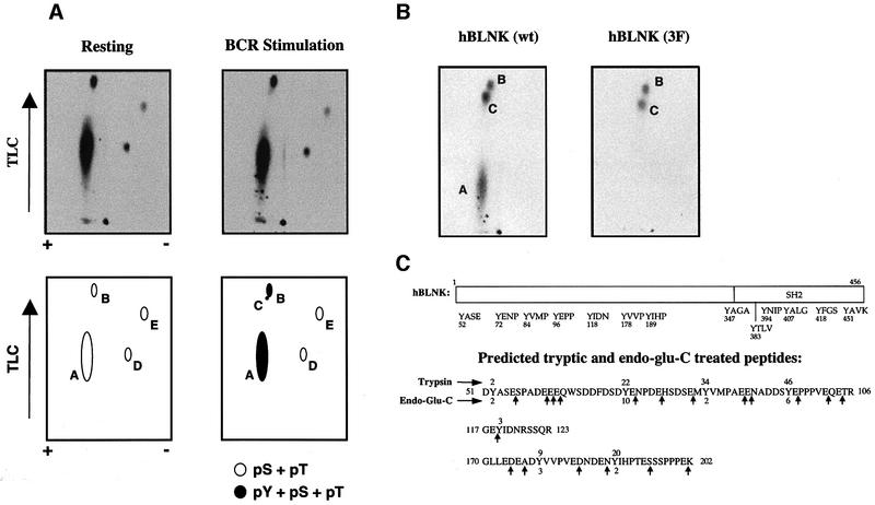
To understand how tyrosine phosphorylation of BLNK regulates BCR function, we analyzed the in vivo phosphorylation sites within human BLNK (hBLNK) in resting and activated B cells. Phosphorylated hBLNK from resting and BCR-activated Daudi B cells were digested with trypsin and peptides resolved by 2-dimensional TLC. Four major peptides, designated as A, B, D and E, containing both phosphoserine (pS) and phosphothreonine (pT), but not phosphotyrosine (pY) residues were isolated from resting cells (Figure 1A, left; data not shown). Following BCR activation, pY was detected in peptides A and B and an additional peptide (designated C) containing pY was also detected in BCR-activated cells (Figure 1A, right; data not shown). No pY residues were detected in peptides D and E isolated from resting or activated B cells. Hence, BCR activation results in tyrosine phosphorylation of peptides A, B and C.
Fig. 1. Identification of phosphorylated tyrosine sites in BLNK. (A) Phosphopeptide maps of in vivo labeled BLNK. Trypsin digested 32P-labeled hBLNK was isolated from resting (left) or BCR-activated (right) Daudi B cells and analyzed by 2-dimensional electrophoresis and TLC, as described in Materials and methods. The first dimension of electrophoretic separation is represented on the _x_-axis and ascending chromatography is represented on the _y_-axis. The bottom panel represents a schematic diagram of the resultant peptides. Each peptide was eluted from the TLC plate and further analyzed for phosphoamino acid content (data not shown). This data is summarized in the bottom panel with black spots representing peptides that contain pY, pS and pT, while the open spots represent peptides that contain only phosphoserine and phosphothreonine. These maps are representative of a minimum of five independent labeling experiments for each sample. (B) Phosphopeptide maps of in vitro labeled BLNK. Purified wild-type hBLNK (left) and hBLNK(Y3F) in which Ys 72, 84 and 96 are mutated to F (right) were phosphorylated by Syk in vitro in the presence of [γ32P]-ATP, digested with trypsin and analyzed by 2-dimensional TLC, as described in Materials and methods. These maps are representative of a minimum of six independent experiments for each sample. (C) Schematic diagram of predicted peptides. The three predicted tryptic peptides containing the seven N-terminal tyrosine residues of hBLNK are depicted. The first four tyrosine residues (Ys 52, 72, 84 and 96) are encoded within a 56 aa tryptic fragment. Y118 is encoded within a 7 aa peptide. Ys 178 and 189 are encoded within a 27 aa peptide. The cycle number of 32P released for the tryptic peptides by Edman degradation is depicted above each Y. The cycle number of 32P released for peptides additionally digested with endo-gluC is shown below each Y.
Identification of tyrosine phosphorylation sites within BLNK by peptide analysis
To facilitate the identification of the phosphorylated tyrosine residues, we analyzed the pattern of peptides derived from purified hBLNK, which was phosphorylated in vitro by its upstream PTK-Syk (Fu et al., 1998). The in vitro phosphorylated products demonstrated the presence of three major species that co-migrate with peptides A, B and C from the in vivo labeling experiments (Figure 1B, left; Supplementary figure 1A–F, available at The EMBO Journal Online).
Manual Edman sequencing of peptide A failed to release 32P within the first 13 cycles and suggested that this peptide may represent the tryptic peptide encoding amino acids (aas) 51–106 (Figure 1C). This peptide encompasses Y52 (cycle 2), Y72 (cycle 22), Y84 (cycle 34) and Y96 (cycle 46). Digestion with additional enzymes failed to cleave this peptide into smaller fragments and prohibited sequencing of smaller derivative peptides (data not shown). Hence, we examined the ability of Syk to phosphorylate a mutant hBLNK molecule, designated as hBLNK(3F) in which Ys 72, 84 and 96 (but not Y52 since cycle 2 was not phosphorylated) were mutated to phenylalanine (F). While hBLNK(3F) was phosphorylated on peptides B and C, no 32P was incorporated into peptide A (Figure 1B, right) and this is consistent with one or more of the three Y residues (Ys 72, 84 and 96) being phosphorylated by Syk (see below).
Edman sequencing of in vitro phosphorylated tryptic peptides B and C both released counts in cycle 9 (Supplementary figure 2A and B). Only Y178 of the 33 aa tryptic peptide encoding aas 170–202 would have released counts in this cycle (Figure 1C). Since this peptide also contains Y189 (cycle 20), which could not have been detected using manual Edman sequencing, we subjected peptides B and C to additional enzymatic digestion with endoproteinase-Glu C (endo-gluC). Manual sequencing revealed 32P in cycles 2 and 3 for both peptides B and C, which correspond to phosphorylation of Y178 (cycle 3) and Y189 (cycle 2) (Supplementary figure 2C and D). Consistent with this biochemical analysis, in vitro phosphorylation by Syk of a mutant hBLNK in which Y178 and Y189 were both mutated to Phe resulted in the loss of both peptides B and C without any effect on peptide A (data not shown). Together, these data are consistent with peptides B and C being derived from phosphorylation of Y178 and Y189, though the exact biochemical difference between peptides B and C remains unclear.
Confirmation of BLNK phosphorylation by immunologic analysis
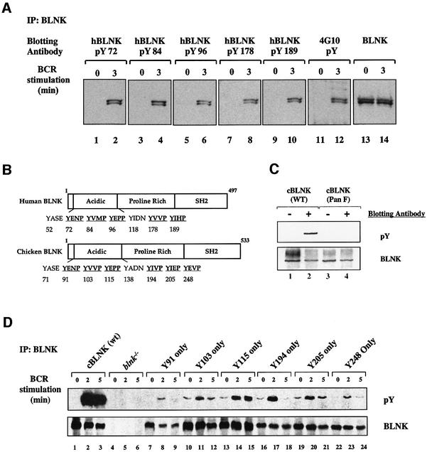
To confirm the identity of these phosphorylation sites, we generated a panel of phosphopeptide-specific antibodies specific for each site. Immunoblotting with each phosphospecific antibody demonstrated the absence of phosphorylation in resting Daudi cells and the inducible phosphorylation of all five Ys on both alternatively spliced hBLNK and hBLNK-s (short) molecules following BCR crosslinking (Figure 2A). No significant differences in the kinetics of phosphorylation were observed among the five Y sites (Supplementary figure 3).
Fig. 2. Generation of cells expressing wild-type and mutant BLNK molecules. (A) Analysis of the phosphorylation of hBLNK utilizing phosphospecific antibodies. hBLNK was immunoprecipitated from resting or BCR-activated cells and analyzed by immunoblotting with antiserum raised against each of the phosphorylated tyrosine residues in hBLNK (lanes 1–10), an anti-pY mAb (4G10, lanes 11–12), or an anti-BLNK antiserum (1761, lanes 13–14). The phosphospecific antibodies, as described in the Materials and methods, were used at 0.5 µg/ml in 0.5% BSA/TBST (0.05% Tween-20, 10 mM Tris pH 8.0 and 150 mM NaCl) and incubated for 1 h. The blots were washed three times for 15 min with TBST and incubated with HRP-conjugated anti-rabbit antisera (Pierce) diluted to 1:20 000 for 1 h. The blots were washed as before and developed by ECL according to manufacturer’s instructions (Pierce). (B) Comparison of hBLNK and cBLNK. Schematic diagrams of the N-terminal Ys are depicted. The Y residues that are within the conserved Syk phosphorylation sequence are underlined. (C) cBLNK(Pan F) is not phosphorylated on tyrosine residues following BCR crosslinking. Wild-type cBLNK or cBLNK(Pan F) were immunoprecipitated from resting or BCR-activated cells (M4, 4 µg/ml for 2 min at 37°C) and analyzed by immunoblotting with an anti-pY mAb (top) or an anti-BLNK antiserum (1761, bottom). (D) In vivo phosphorylation of cBLNK mutants expressing Y91, Y103, Y115, Y194 or Y205. Stable clones expressing wild-type or mutant cBLNK molecules were established as described in Materials and methods. CBLNK(wt) or the mutant BLNK molecules were immunoprecipitated from resting or BCR-activated cells (M4, 4 µg/ml for 2 or 5 min at 37°C) and analyzed by immunoblotting with an anti-pY mAb (top) or an anti-BLNK antiserum (1761, bottom).
To evaluate the roles of BLNK phosphorylation, we expressed the chicken BLNK (cBLNK) ortholog mutant in which the five conserved cBLNK Ys were mutated to F (Ys 91, 103, 115, 194 and 205 in cBLNK) in _blnk_–/– DT40 B cells (Figure 2B). However, this mutant cBLNK molecule [designated cBLNK(Y5F)] still demonstrated detectable tyrosine phosphorylation following BCR crosslinking (data not shown). A search of the cBLNK sequence revealed an additional tyrosine residue (Y248) that contains the preferred phosphorylation sequence (YE/DXP) for the Syk PTK. Additional mutation of Y248 to F [designated cBLNK(Pan F)] demonstrated no residual tyrosine phosphorylation following BCR crosslinking (Figure 2C). Since cBLNK(Pan F) still retained its ability to bind to Grb2, the overall structural integrity of cBLNK(Pan F) appears to be preserved as this interaction is mediated through SH3–proline interactions (data not shown).
To determine if BLNK phosphorylation sites occurred in a coordinated fashion, each of the six cBLNK Ys were re-introduced onto the cBLNK(Pan F) background. While no tyrosine phosphorylation was detected in the cBLNK(Pan F) mutant, each of the single Y reconstituted mutants was phosphorylated following BCR crosslinking (Figure 2D). Together, the biochemical and immunological studies indicate that Ys 72, 84, 96, 178 and 189 within hBLNK, and Ys 91, 103, 115, 194, 205 and 248 within cBLNK are phosphorylated following BCR activation.
Tyrosine phosphorylation of BLNK is required for [Ca2+]i and MAPK activation
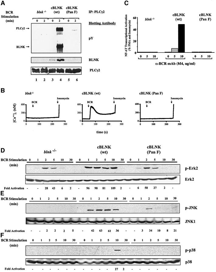
To understand the biological implications for tyrosine phosphorylation of BLNK, we analyzed the effects of the cBLNK(Pan F) mutation. Since BLNK associates with PLCγ following BCR crosslinking (Fu et al., 1998), we first analyzed the ability of cells expressing cBLNK(Pan F) to regulate the phosphoinositide-signaling pathway. No tyrosine phosphorylation of PLCγ2 was observed in cells expressing cBLNK(Pan F) (Figure 3A). In addition, cBLNK(Pan F) did not co-immunoprecipitate with PLCγ2. Correspondingly, cells expressing cBLNK(Pan F) failed to induce increases in [Ca2+]i or NF-AT transcriptional activation following BCR crosslinking (Figure 3B and C).
Fig. 3. Requirement for tyrosine phosphorylation of BLNK in [Ca2+]i and MAP activation. (A) Association and phosphorylation of PLCγ2 requires BLNK tyrosine phosphorylation. Transiently transfected PLCγ2 was immunoprecipitated from the _blnk_–/–, wild-type cBLNK or cBLNK(Pan F)-expressing cells from resting and BCR-activated cells (M4, 4 µg/ml for 2 min at 37°C) and analyzed by immunoblotting with anti-pY mAb (top), an anti-BLNK antiserum (1761, middle), or anti-PLCγ2 antiserum (bottom). (B) Absence of BCR induced [Ca2+]i in cells expressing cBLNK(Pan F). _blnk_–/– DT40 cells (left) or _blnk_–/– cells reconstituted with wild-type cBLNK (middle) or cBLNK(Pan F) (right) were analyzed for their ability to increase [Ca2+]i following BCR crosslinking (BCR arrow) or ionomycin (ionomycin arrow), as described in Materials and methods. (C) Transcriptional activation of an NF-AT/AP-1 reporter gene is dependent upon tyrosine phosphorylation of BLNK. _blnk_–/– DT40 cells (left) and cells reconstituted with wild-type cBLNK (middle) or cBLNK(Pan F) (right) were analyzed for their ability to activate an NF-AT/AP-1 responsive element. Reporter activity was analyzed for cells incubated with media alone, in media containing anti-BCR M4 mAb (5 or 10 µg/ml, shaded or filled bars, respectively), or media containing PMA and ionomycin. These data are representative of five independent experiments and of at least two independent clones. (D–F) Efficient activation of all three families of MAPKs is dependent on tyrosine phosphorylation of BLNK. _blnk_–/– DT40 cells (lanes 1–6) and cells reconstituted with wild-type cBLNK (lanes 7–12) or cBLNK(Pan F) (lanes 13–18) were analyzed for their ability to activate the Erk2 (panel D), JNK (panel E) and p38 (panel F) pathways. Cells were stimulated for the time periods denoted above each lane, lysed and immunoblotted with antibodies specific for the activated forms of each of the MAPKs. Equal loading of cell lysates was confirmed by blotting with antibodies for each of the MAPKs, as described in Materials and methods. Quantitation of bands was performed using UN-SCAN-IT software and the fold activation, as compared with resting cells (lanes 1, 7 and 13), is reported below each lane. This analysis is representative of a minimum of four independent experiments.
Since BLNK is also required for the efficient activation of the three families of MAPKs (Ishiai et al., 1999), we analyzed the requirements for the tyrosine phosphorylation of BLNK in MAPK activation. Expression of cBLNK(Pan F) failed to restore activation of Erk2, JNK, or p38 phosphorylation following BCR crosslinking (Figure 3D–F). Hence, tyrosine phosphorylation of BLNK is required for both [Ca2+]i and MAPK signaling pathways activated by the BCR.
Binding specificity of effector molecules to BLNK tyrosine phosphorylation sites
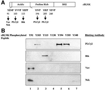
To assess binding specificity of the six phosphorylated tyrosine sites, we generated a panel of phosphorylated cBLNK peptides and assessed their binding to BLNK interacting proteins. Immunoblotting indicated that Ys 103, 194 and 205 of cBLNK preferentially bound PLCγ2, Y115 preferentially bound Btk, and Y91 preferentially bound to both Vav and Nck (Figure 4B).
Fig. 4. Binding specificity of effector molecules to BLNK tyrosine phosphorylation sites. (A) Schematic diagram of preferential binding sites of PLCγ2 Btk, Nck and Vav on cBLNK. (B) Binding specificity of PLCγ2, Btk, Vav and Nck to cBLNK tyrosine residues. Daudi lysates were incubated with each of the tyrosine phosphorylated peptides corresponding to the phosphorylated tyrosine residues of chicken BLNK and analyzed by immunoblotting with anti-PLCγ2 (top), anti-Btk (top middle), anti-Vav (bottom middle) and anti-Nck (bottom). A phosphorylated peptide corresponding to a Y residue that would not normally be tyrosine phosphorylated was used as a control (Y138).
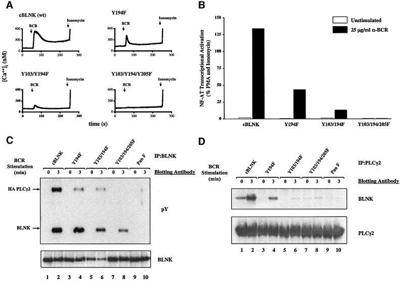
To evaluate the functional significance of these binding specificities, we generated a panel of mutants in which the three predicted PLCγ2 binding sites were mutated in various combinations (Y194F, Y103F/Y194F and Y103F/Y194F/Y205F, respectively). These cDNAs were expressed in _blnk_–/– DT40 cells and a minimum of two individual clones expressing comparable BLNK and BCR levels were analyzed (data not shown). While mutation of a single PLCγ2 binding site (Y194F) caused a significant reduction in [Ca2+]i, mutation of two PLCγ2 sites (Y103F/Y194F) resulted in a further reduction, and mutation of all three PLCγ2 binding sites abrogated [Ca2+]i mobilization (Figure 5A). Correspondingly, these mutant cells also demonstrated a graded reduction in transcriptional activation of an NF-AT responsive element, tyrosine phosphorylation of PLCγ2, and ability of PLCγ2 to bind BLNK (Figure 5B–D).
Fig. 5. Reduction of PLCγ-mediated signaling pathways with mutation of PLCγ binding sites. (A) Reduction in [Ca2+]i. _blnk_–/– DT40 cells expressing wild-type cBLNK, Y194F, Y103/194F or Y103/194/205F were analyzed for their ability to induce [Ca2+]i mobilization following BCR crosslinking or ionomycin. (B) Reduction in NF-AT transcriptional activation. _blnk_–/– DT40 cells expressing wild-type cBLNK, Y194F, Y103/194F or Y103/194/205F were analyzed for their ability to activate an NF-AT/AP-1 responsive element. Reporter activity was analyzed for cells incubated with media alone, in media containing anti-BCR mAb (M4, 25 µg/ml) or media containing PMA and ionomycin. (C) Reduced tyrosine phosphorylation and association of PLCγ2. BLNK was immunoprecipitated from PLCγ2 infected wild-type cBLNK-, Y194F-, Y103/194F- or Y103/194/205F-expressing cells from resting and BCR-activated cells (M4, 4 µg/ml for 3 min at 37°C) and analyzed by immunoblotting with anti-pY mAb (top) or an anti-BLNK antiserum (1761, bottom). Immunoprecipitation of PLCγ2 with an anti-HA mAb demonstrated similar graded reduction in PLCγ2 tyrosine phosphorylation (data not shown). (D) Reduced association of PLCγ2 with BLNK. Retrovirally infected PLCγ2 was immunoprecipitated from wild-type cBLNK-, Y194F-, Y103/194F- or Y103/194/205F-expressing cells from resting and BCR-activated cells (M4, 4 µg/ml for 3 min at 37°C) and analyzed by immunoblotting with an anti-BLNK antiserum (1761, top) or an anti-PLCγ2 antiserum (bottom).
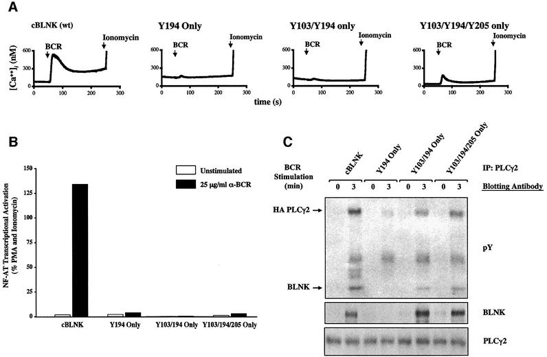
Reciprocally, we examined the effects of restoring the PLCγ2 binding sites in the cBLNK(Pan F) mutant molecule. Restoration of one (Y194 only) or two (Y103/Y194 only) of the PLCγ2 sites failed to induce any significant increases in [Ca2+]i or NF-AT transcriptional activation following BCR crosslinking (Figure 6A and B). Restoration of all three PLCγ2 binding sites (Y103/Y194/Y205 only) resulted in minimal BCR-induced [Ca2+]i and no significant NF-AT transcriptional activation. Despite the significant decrease in [Ca2+]i and NF-AT activation, the binding of PLCγ2 to cBLNK and the tyrosine phosphorylation of PLCγ2 was recapitulated in cells expressing cBLNK(Y103/194/205 only) and cBLNK (Y103/194 only) (Figure 6C). These data indicate that recruitment of PLCγ to cBLNK as well as tyrosine phosphorylation of PLCγ2 was insufficient to restore BLNK function, and an additional level of organization of signaling proteins on BLNK was required to mediate NF-AT transcriptional activation.
Fig. 6. Restoration of PLCγ binding sites does not reconstitute normal activation of PLCγ-mediated signaling pathways. (A) Expression of PLCγ-binding cBLNK does not restore normal calcium mobilization. _blnk_–/– DT40 cells expressing wild-type cBLNK, Y194 only, Y103/194 only, or Y103/194/205 only were analyzed for their ability to induce [Ca2+]i following BCR crosslinking or ionomycin. (B) Failure to restore NF-AT transcriptional activation. _blnk_–/– DT40 cells expressing wild-type cBLNK, Y194 only, Y103/194 only or Y103/194/205 only were analyzed for their ability to activate an NF-AT/AP-1 responsive element. Reporter activity was analyzed for cells incubated with media alone, in media containing anti-BCR mAb (M4, 25 µg/ml) or media containing PMA and ionomycin. (C) Reconstitution of BLNK-PLCγ2 interaction. PLCγ2 was immunoprecipitated from PLCγ2 infected wild-type cBLNK, _blnk_–/–, Y194 only, Y103/194 only, or Y103/194/205- expressing cells from resting and BCR-activated cells (M4, 4 µg/ml for 3 min at 37°C) and analyzed by immunoblotting with anti-pY mAb (top), anti-BLNK antiserum (1761, middle) or PLCγ2 (bottom).
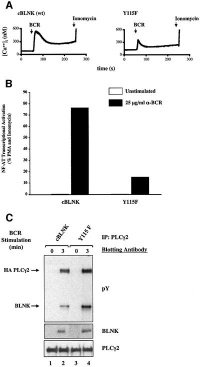
Because Btk is implicated in activating PLCγ, we next analyzed the functional significance of the Btk binding site by solely mutating Y115 to F. As compared with cells expressing wild-type cBLNK, cells expressing cBLNK(Y115F) demonstrated reduced [Ca2+]i and NF-AT transcriptional activation following BCR crosslinking (Figure 7A and B). In contrast to the PLCγ binding mutant, the overall tyrosine phosphorylation of PLCγ2 was not decreased and the association of cBLNK(Y115F) was comparable to cells expressing wild-type cBLNK (Figure 7C).
Fig. 7. Reduction of PLCγ-mediated signaling pathways with mutation of the Btk binding site. (A) Reduction in [Ca2+]i. _blnk_–/– DT40 cells expressing wild-type cBLNK or Y115F were analyzed for their ability to induce [Ca2+]i mobilization following BCR crosslinking or ionomycin. (B) Reduction in NF-AT transcriptional activation. _blnk_–/– DT40 cells expressing wild-type cBLNK or Y115F were analyzed for their ability to activate an NF-AT/AP-1 responsive element following incubation with media alone, in media containing anti-BCR mAb (M4, 25 µg/ml) or media containing PMA and ionomycin. (C) Normal tyrosine phosphorylation of PLCγ2. Retrovirally infected PLCγ2 was immunoprecipitated from wild-type cBLNK or Y115F-expressing cells from resting and BCR-activated cells (M4, 4 µg/ml for 3 min at 37°C) and analyzed by immunoblotting with anti-pY mAb (top), anti-BLNK antiserum (1761, middle) or an anti-PLCγ2 antiserum (bottom).
Phosphorylation of PLCγ and Btk binding sites within a single BLNK molecule is required for normal BCR function
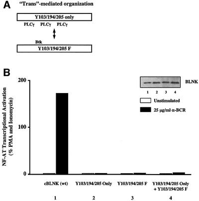
Finally, we analyzed the potential for cooperativity between distinct BLNK-binding effectors in regulating NF-AT transcriptional activation. We first examined whether PLCγ2 and non-PLCγ2 binding mutants of BLNK could complement each other in trans (Figure 8A). Consistent with the analysis in Figures 5 and 6, expression of either cBLNK molecules alone resulted in no BCR-inducible NF-AT transcriptional activation (Figure 8B). Moreover, co-expression of equal molar amounts of both mutant molecules in _blnk_–/– cells also failed to complement each other to restore NF-AT transcriptional activation. Hence, NF-AT transcriptional activation cannot be mediated through _‘trans’_-crosstalk between PLCγ2 and non-PLCγ2 binding BLNK molecules.
Fig. 8. ‘_Trans’_-mediated organization of signaling proteins with cBLNK does not reconstitute BCR activation. (A) Model for _‘trans’_-mediated organization of signaling proteins with cBLNK. In the trans model, the PLCγ2-binding mutant cBLNK (Y103/194/205 only) can cooperate with a non-PLCγ2-binding cBLNK mutant (Y103/194/205F) to regulate NF-AT transcriptional activation. (B) Lack of complementation by PLCγ and non-PLCγ binding cBLNK mutants in trans. _blnk_–/– DT40 cells were transiently transfected with 25 µg of wild-type cBLNK, Y103/Y194/Y205 only, Y103/Y194/Y205F, or both BLNK mutant cDNAs (12.5 µg each). The cells were analyzed for their ability to activate an NF-AT/AP-1 responsive element following incubation with media alone, in media containing anti-BCR mAb (M4, 25 µg/ml) or media containing PMA and ionomycin. Expression of BLNK was monitored by immunoblotting with an anti-BLNK antiserum (inset).
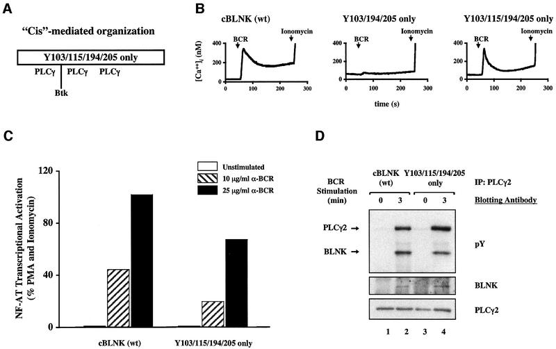
Conversely, we analyzed the ability of a cBLNK molecule containing both PLCγ2 and Btk binding sites in mediating BCR-activated signaling pathways in cis (Figure 9A). While expression of the PLCγ binding mutant, cBLNK(Y103/194/205 only), only minimally restored BCR induced [Ca2+]i mobilization (Figures 6A and 9B), the additional reconstitution of the Btk binding site, cBLNK(Y103/115/194/205 only), fully restored the initial and partially the latter phase of BCR-induced Ca2+ mobilization (Figure 9B). Additionally, while the PLCγ2 binding cBLNK (Y103/Y194/Y205 only) mutant was devoid of transcriptional activation of NF-AT, expression of cBLNK(Y103/115/194/205 only) restored ≥50% of the BCR-activated NF-AT transcriptional activity (Figure 9C) and tyrosine phosphorylation of PLCγ2 (Figure 9D). Together, these data suggest that phosphorylation of Ys on a single scaffold to bind PLCγ2 and other effectors (e.g. Btk) is required for normal BCR function.
Fig. 9. _‘Cis’_-mediated organization of signaling proteins with cBLNK. (A) Model for _‘cis’_-mediated organization of signaling proteins with cBLNK. In the ‘cis’ model, tyrosine phosphorylation of a single BLNK molecule provides docking sites for both PLCγ2 and Btk to regulate NF-AT transcriptional activation. (B) Restoration of [Ca2+]i by PLCγ and Btk binding cBLNK _in cis. blnk_–/– DT40 cells expressing wild-type cBLNK or Y103/115/194/205 only, were analyzed for their ability to induce [Ca2+]i mobilization following BCR crosslinking or ionomycin. (C) Restoration of NF-AT transcriptional activation by PLCγ and Btk binding cBLNK _in cis. blnk_–/– DT40 cells expressing wild-type cBLNK or Y103/115/194/205 only, were analyzed for their ability to activate an NF-AT/AP-1 responsive element following incubation with media alone, in media containing anti-BCR mAb (M4, 10 or 25 µg/ml, hatched or filled bar, respectively) or media containing PMA and ionomycin. (D) Normal tyrosine phosphorylation of PLCγ2. Transiently transfected PLCγ2 was immunoprecipitated from wild-type cBLNK or Y103/115/194/205 only expressing cells from resting and BCR- activated cells (M4, 4 µg/ml for 3 min at 37°C) and analyzed by immunoblotting with anti-pY mAb (top) or an anti-PLCγ2 antiserum (bottom).
Discussion
Assembly of macromolecular signaling complexes to coordinate the propagation of signaling pathways has gained great acceptance in a number of biological systems. Studies of yeast have demonstrated the critical importance of the Ste5p and Pbs2p scaffolding proteins in dictating the in vivo substrate specificities of the MAPK pathways and, in turn, specific biological responses of yeast mating and glycerol production reactions, respectively (reviewed in van Drogen and Peter, 2002). In lymphocytes, the specificity and sensitivity of signaling pathways is paramount to cellular fate. Lymphocytes must discern the fine differences between foreign and self-ligands that result in markedly distinct biological responses of cellular proliferation of pathogen-specific lymphocytes and apoptosis of self-reactive lymphocytes. One mechanism by which these ligands affect cellular fates is through different kinetics and dynamics of signaling pathways activated through the antigen receptors (Healy et al., 1997; Hippen et al., 2000; Kimura et al., 2000). Alterations in the generation of second messengers following receptor engagement, such as [Ca2+]i and small GTPases, are associated with the induction of lymphocyte unresponsiveness, a state known as anergy (Dolmetsch et al., 1997; Macian et al., 2002). Hence, the coordinated generation of second messengers in lymphocytes is required for normal cellular function.
In B cells, we and others have previously demonstrated the requirement of the BLNK adapter protein in B cell development and in BCR-signal transduction (Ishiai et al., 1999; Jumaa et al., 1999; Minegishi et al., 1999; Pappu et al., 1999; Xu et al., 2000; Yamazaki et al., 2002). Mice and humans deficient in BLNK demonstrate developmental blocks at the pro- to pre-B cell transition, and additionally, in mice, the immature to mature B cell transition and underscores the requirements for BLNK in both pre-BCR and IgM BCR signaling pathways, respectively. In the chicken DT40 B cell system, _blnk_–/– DT40 B cells exhibit absent [Ca2+]i and MAPK responses (Ishiai et al., 1999). In this study, we demonstrate the importance for the phosphorylation of multiple tyrosine residues within a single BLNK molecule to generate a single molecular scaffold. Fine mapping of BLNK phosphorylation sites permitted us to identify subsets of tyrosine residues that bind distinct effector molecules. Phosphorylation of Ys 103, 194 and 205 within cBLNK (and correspondingly Ys 84, 178 and 189 within hBLNK) facilitates PLCγ2 binding; Y115 within cBLNK (and correspondingly Y96 in hBLNK) facilitates Btk binding; Y91 within cBLNK (and correspondingly Y72 in hBLNK) facilitates Nck and Vav binding.
Mutation of the multiple PLCγ2 binding sites within BLNK reduces the association of PLCγ2 with BLNK and activation of PLCγ2-mediated signaling pathways. The correlative nature of the number of PLCγ2 sites within BLNK and the magnitude of PLCγ2 function supports an amplification role for the multiple PLCγ2 binding sites within BLNK. Furthermore, the inability of a mutant BLNK molecule that contains solely PLCγ2 binding sites [cBLNK(103/194/205 only)] to restore wild-type [Ca2+]i or NF-AT transcriptional activation hinted that other binding partners might be required for normal [Ca2+]i signaling. As Btk is also required for normal [Ca2+]i and in view of the reported association of BLNK with Btk (Hashimoto et al., 1999; Jumaa et al., 2001), we tested the hypothesis that the dual binding of Btk and PLCγ2 with BLNK may be required for normal [Ca2+]i. Indeed, while cBLNK(Y115F) bound PLCγ2 with similar stoichiometry as wild-type BLNK, cells expressing cBLNK(Y115F) still exhibited an attenuated [Ca2+]i response. The ability of BLNK, Btk and PLCγ2 to co-migrate as a large molecular weight complex (>600K Mr) in size fractionation studies as opposed to the ∼70K Mr BLNK in resting B cells also supports the notion that BLNK serves as a scaffold to nucleate a multi-component signaling (data not shown). Finally, the inability of two distinct PLCγ-binding site mutants to complement each other in NF-AT transcriptional activation (Figure 8B), and the partial restoration of NF-AT transcriptional activation by a BLNK molecule capable of binding both PLCγ2 and Btk (Figure 9C), is likewise consistent with the notion that BLNK provides a molecular platform on which specific signaling components are spatially organized in cis for appropriate activation (Figure 9A). The ability of cBLNK(Y103/115/194/205 only) to partially restore [Ca2+]i mobilization and NF-AT transcriptional activation (as compared with wild-type cBLNK) suggests that other signaling effector proteins through Ys 91 and 248 in cBLNK (and Y72 within hBLNK) are also required for normal levels of NF-AT activation. Candidates for Y91 include Vav and Nck, both of which can bind a phosphopeptide encompassing Y91 (Figure 4B) and have been demonstrated to play important roles in the [Ca2+]i and NF-AT transcriptional responses (Costello et al., 1999).
While others have reported a stable association of Btk with BLNK (Hashimoto et al., 1999), we have been unable to demonstrate a high stoichiometry of association of Btk with wild-type BLNK, although some degree of association has been intermittently observed (data not shown). This may reflect the transient nature of this interaction and/or the inability to co-immunoprecipitate complexes that are less accessible to detergent solubilization. As a result, we were unable to interpret the inability of cBLNK(Y115F) mutant to interact with Btk. A model has recently been proposed in which Btk tyrosine phosphorylation and activation occur in a BLNK-dependent fashion (Baba et al., 2001). However, we were unable to demonstrate any differences in Btk phosphorylation or kinase activity in BLNK-expressing or -deficient DT40 cells (data not shown). In addition, no differences in Btk phosphorylation and activation were observed in pro-B cell cultures derived from the bone marrows of blnk+/– and _blnk_–/– mice (our unpublished data). These data do not favor a model in which Btk activation is dependent upon BLNK, but rather a model in which Btk is activated in a BLNK-independent fashion. Additionally, the tyrosine phosphorylation of BLNK and association of PLCγ2 with BLNK occurs in a Btk-independent fashion (Fu et al., 1998; our unpublished data). While _btk_–/– DT40 B cells are unable to mobilize [Ca2+]i following BCR crosslinking (Ishiai et al., 1999), cells expressing cBLNK(Y115F) are still capable of mediating a moderate degree of BCR-induced [Ca2+]i. As the latter still express wild-type Btk, the binding of Btk to Tyr 115 of cBLNK may only partially contribute to PLCγ2 activation. Given the low affinities of most single domain mediated interactions, it is likely that the multi-modular nature of signaling proteins all contribute to the stable assembly and localization of signaling complexes. Hence, normal [Ca2+]i signaling requires not only both BLNK-dependent PLCγ2–BLNK interaction and BLNK-independent Btk activation, but also the assembly of Btk–BLNK–PLCγ2 macromolecular complexes.
The presence of three PLCγ2 binding sites within BLNK represents an atypical example of scaffolding. In growth factor receptors, a single predominant tyrosine (Y1021 in the PDGF receptor and Y992 in the EGF receptor) binds PLCγ1 (Rotin et al., 1992; Kashishian and Cooper, 1993; Larose et al., 1993; Valius et al., 1993). In T cells, phosphorylation of Y192 within the transmembrane ‘linker for activation of T cells’ (LAT) adapter protein binds PLCγ1 (Zhang et al., 2000; Paz et al., 2001). Our combinatorial analysis of the three PLCγ2 binding sites within BLNK supports an amplification role for PLCγ-mediated signaling. As [Ca2+]i represents a major regulator of biological function, differential BLNK phosphorylation may alter the dynamics and kinetics of second messenger generation. Finally, as tyrosine phosphorylation of BLNK regulates many signaling pathways, differential BLNK phosphorylation may also result in the activation of subsets of signaling functions that alter the cellular fate of a given B cell response. Additional studies are ongoing to determine if tyrosine phosphorylation of BLNK may represent such a key regulatory point in determining the outcome of B cell function.
Materials and methods
Cells, antibodies and plasmids
Parental DT40 B cells and their derivatives were cultured in RPMI-1640 supplemented with 10% FCS, 1% chicken serum, 50 µM 2-mercaptoethanol, 2 mM l-glutamine and antibiotics. Anti-BLNK antiserum was generated by immunizing rabbits with a bacterially expressed GST fusion protein containing human BLNK (aas 4–205). This antiserum reacts with both human and chicken BLNK (data not shown). Phosphospecific antibodies were generated at Biosource International (Hopkinton, MA) by immunizing rabbits with coupled peptides corresponding to each of the phosphorylated tyrosine residues of murine BLNK (Y72, aas 67–79; Y84, aas 79–90; Y96, aas 93–104; Y178, aas 172–184; and Y189, aas 184–196). Sera were negatively depleted by affinity chromatography using unphosphorylated peptide and the flow-through purified with phosphorylated peptide. These antibodies react with both human and murine BLNK (data not shown).
Additional antibodies used in this study include: anti-hBLNK mAb (2B11 or 2C9), anti-phosphotyrosine mAb (4G10, UBI and PY20; Transduction Labs), anti-chicken IgM mAb (M4; courtesy of Dr Max Cooper), anti-myc mAb (9E10), anti-β-actin (Sigma), anti-GST (Sigma), anti-pErk2 (Promega), anti-Erk2 (Santa Cruz Biotech), anti-pJNK (Promega), anti-PLCγ2 (Santa Cruz Biotech), anti-Btk (Pharmingen), anti-Vav (Santa Cruz Biotech), anti-Nck (Santa Cruz Biotech), anti-JNK (Pharmingen), anti-phospho p38 (New England Biolabs) and anti-p38 (Santa Cruz Biotech) antibodies.
GST fusion proteins were produced using the pGEX-KT vector (Hakes and Dixon, 1992). Mutants of BLNK were produced by a PCR-directed mutagenesis strategy. All junctions and PCR products were confirmed by standard dideoxy DNA sequencing.
Transfection of cells and biochemical analysis of cells
Stable transfectants were generated by electroporating 107 _blnk_–/– DT40 cells with 20 µg of pApuro containing wild-type BLNK or BLNK mutations as described previously (Kong et al., 1995). Analysis of NF-AT transcriptional activation was performed as described previously (Kong et al., 1995).
Cells were resuspended at 108 cells/ml in PBS for 15 min at 37°C. DT40 cells and their derivatives were crosslinked with an anti-BCR M4 mAb (4 µg/ml) for the indicated time periods described in the figures at 37°C. Cells were sedimented at 5000 g and lysed in an equal volume of 10 mM Tris pH 8.0, 150 mM NaCl, 1% NP-40 supplemented with protease and phosphatase inhibitors (lysis buffer) for 15 min at 4°C. Cellular debris was sedimented at 15 000 g for 10 min at 4°C and the supernatant harvested for studies.
For immunoprecipitations, cellular lysates were incubated with antiserum for 2 h and captured with protein A–Sepharose (Pharmacia) for 1 h at 4°C. Immune complexes were washed three times with lysis buffer (1 ml each) and prepared for analysis. Protocols for SDS–PAGE and immunoblotting have been published previously (Chan et al., 1995).
For the phosphorylated peptide precipitation assays, resting Daudi cell lysates were incubated with each of the peptides for 2 h and captured with streptavidin agarose (Sigma) for 20 min at 4°C. The associated complexes were washed three times with lysis buffer (1 ml each) and prepared for analysis.
Retroviral infections were performed utilizing the Phoenix amphotrophic packaging cell line using standard protocols (Swift et al., 1999). In brief, the Phoenix cells were transfected with a plasmid expressing human PLCγ2 and a GFP marker using calcium phosphate precipitation. The resulting supernatant was placed onto the target DT40 derivatives with 10 µg/ml protamine sulfate and spun at 2 000 r.p.m. for 1 h. The infected cells were subsequently sorted based on fluorescence.
Fluorimetry
For analysis of intracellular calcium levels, cells were loaded with Fura-2 (Molecular Probes) and calcium-sensitive fluorescence was monitored using a Hitachi F2000 Fluorescence spectrophotometer at wavelengths 340 nm and 540 nm. Cells were stimulated with soluble anti-BCR M4 mAb (4 µg/ml). Maximal fluorescence was determined following lysis of cells with Triton X-100 while minimum fluorescence was determined following chelation with EGTA.
Analysis of 32P-labeled BLNK peptides
Daudi B cells expressing a myc-epitope tagged hBLNK were labeled with [32P]orthophosphate for 4 h in phosphate-deficient media and analyzed under resting or BCR-activating conditions. BCR stimulation was performed by addition of anti-IgM F(ab′)2 for 2 min and terminated by the addition of cold lysis buffer. BLNK was immunoprecipitated from 32P-labeled cells with an anti-myc 9E10 mAb, resolved by SDS–PAGE, transferred to nitrocellulose and visualized by autoradiography. Tryptic peptide mapping was performed as described previously (Luo et al., 1991). Tryptic peptides were separated by TLC utilizing HTLE-7000 electrophoretic apparatus (CBS, Del Mar, CA) as described previously (Chan et al., 1995).
For sequencing analysis, labeled peptides were eluted in 0.1 ml pH 1.9 buffer and sonicated as described previously (Hellman et al., 1995). Eluted peptides were cleared by centrifugation at 15 000 g for 10 min at 4°C. Subsequent digestion with endo-gluC (Boehringer Mannheim) was accomplished by washing the peptide three times with H2O and resuspending it in 0.1 ml of 25 mM ammonium carbonate. Eluted peptides were then digested with 5 µg of endo-gluC overnight at room temperature. The sample was heated to 85°C for 30 min to inactivate the remaining endo-gluC, washed three times with H2O and resuspended in 0.02 ml of 30% acetonitrile for manual Edman sequencing. Manual Edman sequencing was performed using the Sequelon AA Reagent Kit (Millipore) according to manufacturer’s instructions.
Supplementary data
Supplementary data are available at The EMBO Journal Online.
Acknowledgments
Acknowledgements
The authors thank Alec Cheng and members of the Chan laboratory for critical review of the manuscript.
References
- Baba Y., Hashimoto,S., Matsushita,M., Watanabe,D., Kishimoto,T., Kurosaki,T. and Tsukada,S. (2001) BLNK mediates Syk-dependent Btk activation. Proc. Natl Acad. Sci. USA, 98, 2582–2586. [DOI] [PMC free article] [PubMed] [Google Scholar]
- Benschop R. and Cambier,J. (1999) B cell development: signal transduction by antigen receptors and their surrogates. Curr. Opin. Immunol., 11, 143–151. [DOI] [PubMed] [Google Scholar]
- Chan A.C., Dalton,M., Johnson,R., Kong,G.-h., Wang,T., Thoma,R. and Kurosaki,T. (1995) Activation of ZAP-70 kinase activity by phosphorylation of tyrosine 493 is required for lymphocyte antigen receptor function. EMBO J., 14, 2499–2508. [DOI] [PMC free article] [PubMed] [Google Scholar]
- Cheng A.M., Rowley,R.B., Pao,W., Hayday,A., Bolen,J.B. and Pawson,T. (1995) Syk tyrosine kinase required for mouse viability and B-cell development. Nature, 378, 303–306. [DOI] [PubMed] [Google Scholar]
- Costello P., Walters,A., Mee,P., Turner,M., Reynolds,L., Prisco,A., Sarner,N., Zamoyska,R. and Tybulewicz,V. (1999) The Rho-family GTP exchange factor Vav is a critical transducer of T cell receptor signals to the calcium, ERK, and NF-κ B pathways. Proc. Natl Acad. Sci. USA, 96, 3035–3040. [DOI] [PMC free article] [PubMed] [Google Scholar]
- Cox D., Chang,P., Kurosaki,T. and Greenberg,S. (1996) Syk tyrosine kinase is required for immunoreceptor tyrosine activation motif-dependent actin assembly. J. Biol. Chem., 271, 16597–16602. [DOI] [PubMed] [Google Scholar]
- DeFranco A.L. (1997) The complexity of signaling pathways activated by the BCR. Curr. Opin. Immunol., 9, 296–308. [DOI] [PubMed] [Google Scholar]
- Dolmetsch R.E., Lewis,R.S., Goodnow,C.C. and Healy,J.I. (1997) Differential activation of transcription factors induced by Ca2+ response amplitude and duration. Nature, 386, 855–858. [DOI] [PubMed] [Google Scholar]
- Fu C. and Chan,A.C. (1997) Identification of two tyrosine phosphoproteins, pp70 and pp68, that interact with PLCγ, Grb2, and Vav following B cell antigen receptor activation. J. Biol. Chem., 272, 27362–27368. [DOI] [PubMed] [Google Scholar]
- Fu C., Turck,C., Kurosaki,T. and Chan,A. (1998) BLNK: a central linker protein in B cell activation. Immunity, 9, 93–103. [DOI] [PubMed] [Google Scholar]
- Gangi-Peterson L., Peterson,S., Shapira,L., Golding,A., Caricchio,R., Cohen,D., Margulies,D. and Cohen,P. (1998) bca: an activation-related B-cell gene. Mol. Immunol., 35, 55–63. [DOI] [PubMed] [Google Scholar]
- Goitsuka R., Fujimura,Y., Mamada,H., Umeda,A., Morimura,T., Uetsuka,K., Doi,K., Tsuji,S. and Kitamura,D. (1998) BASH, a novel signaling molecule preferentially expressed in B cells of the Bursa of Fabricius. J. Immunol., 161, 5804–5808. [PubMed] [Google Scholar]
- Hakes D.J. and Dixon,J.E. (1992) New vectors for high level expression of recombinant proteins in bacteria. Anal. Biochem., 202, 293–298. [DOI] [PubMed] [Google Scholar]
- Hashimoto S. et al. (1999) Identification of the SH2 domain binding protein of Bruton’s tyrosine kinase as BLNK—functional significance of Btk-SH2 domain in B-cell antigen receptor-coupled calcium signaling. Blood, 94, 2357–2364. [PubMed] [Google Scholar]
- Healy J.I., Dolmetsch,R.E., Timmerman,L.A., Cyster,J.G., Thomas,M.L., Crabtree,G.R., Lewis,R.S. and Goodnow,C.C. (1997) Different nuclear signals are activated by the B cell receptor during positive versus negative signaling. Immunity, 6, 419–428. [DOI] [PubMed] [Google Scholar]
- Hellman U., Wernstedt,C., Gonez,J. and Heldin,C.H. (1995) Improve ment of an in-gel digestion procedure for the micropreparation of internal protein fragments for amino acid sequencing. Anal. Biochem., 224, 451–455. [DOI] [PubMed] [Google Scholar]
- Hendricks R.W., de Bruijn,M.F.T.R., Maas,A., Dingjan,G.M., Karis,A. and Grosveld,F. (1996) Inactivation of Btk by insertion of lacZ reveals defects in B cell development only past the pre-B cell stage. EMBO J., 15, 4862–4872. [PMC free article] [PubMed] [Google Scholar]
- Hibbs M.L., Tarlinton,D.M., Armes,J., Grail,D., Hodgson,G., Maglitto,R., Stacker,S.A. and Dunn,A.R. (1995) Multiple defects in the immune system of lyn-deficient mice, culminating in autoimmune disease. Cell, 83, 301–311. [DOI] [PubMed] [Google Scholar]
- Hippen K.L., Tze,L.E. and Behrens,T.W. (2000) CD5 maintains tolerance in anergic B cells. J. Exp. Med., 191, 883–889. [DOI] [PMC free article] [PubMed] [Google Scholar]
- Ishiai M. et al. (1999) BLNK required for coupling Syk to PLC-γ2 and Rac1-JNK in B cells. Immunity, 10, 117–125. [DOI] [PubMed] [Google Scholar]
- Jordan J.D., Landau,E.M. and Iyengar,R. (2000) Signaling networks: the origins of cellular multitasking. Cell, 103, 193–200. [DOI] [PMC free article] [PubMed] [Google Scholar]
- Jumaa H., Wollscheid,B., Mitterer,M., Weinands,J., Reth,M. and Nielsen,P.J. (1999) Abnormal development and function of B lymphocytes in mice deficient for the signaling adaptor protein SLP-65. Immunity, 11, 547–554. [DOI] [PubMed] [Google Scholar]
- Jumaa H., Mitterer,M., Reth,M. and Nielsen,P.J. (2001) The absence of SLP65 and Btk blocks B cell development at the pre-B cell receptor-positive stage. Eur. J. Immunol., 31, 2164–2169. [DOI] [PubMed] [Google Scholar]
- Kashishian A. and Cooper,J.A. (1993) Phosphorylation sites at the C-terminus of the platelet-derived growth factor receptor bind phospholipase C γ1. Mol. Biol. Cell, 4, 49–57. [DOI] [PMC free article] [PubMed] [Google Scholar]
- Kerner J.D., Appleby,M.W., Mohr,R.N., Chien,S., Rawlings,D.J., Maliszewski,C.R., Witte,O.N. and Perlmutter,R.M. (1995) Impaired expansion of mouse B cell progenitors lacking Btk. Immunity, 3, 301–312. [DOI] [PubMed] [Google Scholar]
- Khan W.N. et al. (1995) Defective B-cell development and function in Btk-deficient mice. Immunity, 3, 283–299. [DOI] [PubMed] [Google Scholar]
- Kimura M. et al. (2000) Impaired Ca/calcineurin pathway in in vivo anergized CD4 T cells. Int. Immunol., 12, 817–824. [DOI] [PubMed] [Google Scholar]
- Kong G.H., Bu,J.Y., Kurosaki,T., Shaw,A.S. and Chan,A.C. (1995) Reconstitution of syk function by the ZAP-70 protein tyrosine kinase. Immunity, 2, 485–492. [DOI] [PubMed] [Google Scholar]
- Kurosaki T. (1999) Genetic analysis of B cell antigen receptor signaling. Annu. Rev. Immunol., 17, 555–592. [DOI] [PubMed] [Google Scholar]
- Larose L., Gish,G., Shoelson,S. and Pawson,T. (1993) Identification of residues in the β platelet-derived growth factor receptor that confer specificity for binding to phospholipase C-γ1. Oncogene, 8, 2493–2499. [PubMed] [Google Scholar]
- Law C.-L., Chandra,K.A., Sidorenko,S.P. and Clark,E.A. (1996) Phospholipase C-γ1 interacts with conserved phosphotyrosyl residues in the linker region of Syk and is a substrate for Syk. Mol. Cell. Biol., 16, 1305–1315. [DOI] [PMC free article] [PubMed] [Google Scholar]
- Leo A., Wienands,J., Baier,G., Horejsi,V. and Schraven,B. (2002) Adapters in lymphocyte signaling. J. Clin. Invest., 109, 301–309. [DOI] [PMC free article] [PubMed] [Google Scholar]
- Luo K., Hurley,T.R. and Sefton,B.M. (1991) Cyanogen bromide cleavage and proteolytic peptide mapping of proteins immobilized to membranes. Methods. Enzymol., 201, 149–152. [DOI] [PubMed] [Google Scholar]
- Macian F., Garcia-Cozar,F., Im,S.H., Horton,H.F., Byrne,M.C. and Rao,A. (2002) Transcriptional mechanisms underlying lymphocyte tolerance. Cell, 109, 719–731. [DOI] [PubMed] [Google Scholar]
- Minegishi Y., Rohrer,J., Coustan-Smith,E., Lederman,H.M., Pappu,R., Campana,D., Chan,A.C. and Conley,M.E. (1999) An essential role for BLNK in human B cell development. Science, 286, 1954–1957. [DOI] [PubMed] [Google Scholar]
- Nishizumi H., Taniuchi,I., Yamanashi,Y., Kitamura,D., Ilic,D., Mori,S., Watanabe,T. and Yamamoto,T. (1995) Impaired proliferation of peripheral B cells and induction of autoimmune disease in lyn-deficient mice. Immunity, 3, 549–560. [DOI] [PubMed] [Google Scholar]
- Okada T., Maeda,A., Iwamatsu,A., Gotoh,K. and Kurosaki,T. (2000) BCAP: the tyrosine kinase substrate that connects B cell receptor to phosphoinositide 3-kinase activation. Immunity, 13, 817–827. [DOI] [PubMed] [Google Scholar]
- Pappu R., Cheng,A., Li,B., Gong,Q., Chiu,C., Griffin,N., White,M., Sleckman,B. and Chan,A. (1999) Requirement for B cell linker protein (BLNK) in B cell development. Science, 286, 1949–1954. [DOI] [PubMed] [Google Scholar]
- Paz P.E., Wang,S., Clarke,H., Lu,X., Stokoe,D. and Abo,A. (2001) Mapping the Zap-70 phosphorylation sites on LAT (linker for activation of T cells) required for recruitment and activation of signaling proteins in T cells. Biochem. J., 356, 461–471. [DOI] [PMC free article] [PubMed] [Google Scholar]
- Rotin D. et al. (1992) SH2 domains prevent tyrosine dephosphorylation of the EGF receptor: identification of Tyr992 as the high-affinity binding site for SH2 domains of phospholipase Cγ. EMBO J., 11, 559–567. [DOI] [PMC free article] [PubMed] [Google Scholar]
- Smith F.D. and Scott,J.D. (2002) Signaling complexes: junctions on the intracellular information super highway. Curr. Biol., 12, R32–R40. [DOI] [PubMed] [Google Scholar]
- Swift S., Lorens,J., Achacoso,P. and Nolan,G.P. (1999) Rapid production of retroviruses for efficient gene delivery to mammalian cells using 293T cell-based systems. In Coligan,J.E. (ed.), Current Protocols in Immunology. John Wiley and Sons, New York, NY, pp. 10.17.14–10.17.29. [DOI] [PubMed]
- Takata M., Sabe,H.,A,H., Inazu,T., Homma,Y., Nukada,T., Yamamura,H. and Kurosaki,T. (1994) Tyrosine kinases lyn and syk regulate B cell receptor-coupled Ca2+ mobilization through distinct pathways. EMBO J., 13, 1341–1349. [DOI] [PMC free article] [PubMed] [Google Scholar]
- Tsukada S., Saffran,D.C., Rawlings,D.J., Parolini,O., Allen,R.C., Klisak,I., Sparkes,R.S., Kubagawa,H. and Mohandas,T. (1993) Deficient expression of a B cell cytoplasmic tyrosine kinase in human X-linked agammaglobulinemia. Cell, 72, 279–290. [DOI] [PubMed] [Google Scholar]
- Turner M., Mee,P.J., Costello,P.S., Williams,O., Price,A.A., Duddy,L.P., Furlong,M.T., Geahlen,R.L. and Tybulewicz,V.L.J. (1995) Perinatal lethality and blocked B-cell development in mice lacking the tyrosine kinase Syk. Nature, 378, 298–302. [DOI] [PubMed] [Google Scholar]
- Valius M., Bazenet,C. and Kazlauskas,A. (1993) Tyrosines 1021 and 1009 are phosphorylation sites in the carboxy terminus of the platelet-derived growth factor receptor β subunit and are required for binding of phospholipase Cγ and a 64-kilodalton protein, respectively. Mol. Cell. Biol., 13, 133–143. [DOI] [PMC free article] [PubMed] [Google Scholar]
- van Drogen F. and Peter,M. (2002) MAP kinase cascades: scaffolding signal specificity. Curr. Biol., 12, R53–R55. [DOI] [PubMed] [Google Scholar]
- Wienands J., Schweikert,J., Wollscheid,B., Jumaa,H., Neilsen,P.J. and Reth,M. (1998) SLP-65: a new signaling component in B lymphocytes which requires expression of the antigen receptor for phosphorylation. J. Exp. Med., 188, 791–795. [DOI] [PMC free article] [PubMed] [Google Scholar]
- Xu S., Tan,J.E.L., Wong,P.Y., Manickam,S., Ponniah,S. and Lam,K.P. (2000) B cell development and activation defects in xid-like immunodeficiency in BLNK/SLP-65-deficient mice. Int. Immunol., 12, 397. [DOI] [PubMed] [Google Scholar]
- Yamazaki T., Takeda,K., Gotoh,K., Takeshima,H., Akira,S. and Kurosaki,T. (2002) Essential immunoregulatory role for BCAP in B cell development and function. J. Exp. Med., 195, 535–545. [DOI] [PMC free article] [PubMed] [Google Scholar]
- Zhang W., Trible,R.P., Zhu,M., Liu,S.K., McGlade,C.J. and Samelson,L.E. (2000) Association of Grb2, Gads, and phospholipase C-γ1 with phosphorylated LAT tyrosine residues. Effect of LAT tyrosine mutations on T cell antigen receptor-mediated signaling. J. Biol. Chem., 275, 23355–23361. [DOI] [PubMed] [Google Scholar]
