Tyrosine kinase signalling in breast cancer: Epidermal growth factor receptor and c-Src interactions in breast cancer (original) (raw)
Abstract
Both the non-receptor tyrosine kinase, c-Src, and members of the epidermal growth factor (EGF) receptor family are overexpressed in high percentages of human breast cancers. Because these molecules are plasma membrane-associated and involved in mitogenesis, it has been speculated that they function in concert with one another to promote breast cancer development and progression. Evidence to date supports a model wherein c-Src potentiates the survival, proliferation and tumorigenesis of EGF receptor family members, in part by associating with them. Phosphorylation of the EGF receptor by c-SRC is also critical for mitogenic signaling initiated by the EGF receptor itself, as well as by several G-protein coupled receptors (GPCRs), a cytokine receptor, and the estrogen receptor. Thus, c-Src appears to have pleiotropic effects on cancer cells by modulating the action of multiple growth-promoting receptors.
Keywords: c-Src, epidermal growth factor receptor, human epidermal growth factor receptor 2/neu, signal transducers and activators of transcription, tyrosine phosphorylation
Introduction
Recent evidence has implicated an involvement of tyrosine kinases in human breast cancer development. Two families in particular have been examined, namely, the human epidermal growth factor and the Src families of tyrosine kinases.
Human epidermal growth factor receptor (HER1) in human breast cancer
The human epidermal growth factor receptor (HER1) is the prototype of a family that consists of four known members (EGF receptor/HER1, neu/erbB2/HER2, erbB3/HER3, and erbB4/HER4). These receptor tyrosine kinases are characterized by an extracellular ligand-binding domain, an internal kinase domain, and a carboxyl-terminal domain that contains multiple tyrosine residues. Upon binding of EGF, HER1 dimerizes and becomes phosphorylated on these carboxyl-terminal tyrosyl residues, which in turn act as docking sites for multiple signaling proteins that contain SH2 domains. HER1 plays a variety of roles in normal development, and is found in ductal epithelial cells of normal breast tissue [1**].
The link between HER1 and human cancer initally came from studies by Velu et al [2], who demonstrated that cells that overexpress HER1 become transformed when they are grown in the continuous presence of EGF. HER1 is overexpressed in a variety of human cancers, including benign skin hyperplasia, glioblastoma and cancers of the breast, prostate, ovary, liver, bladder, esophagus, larynx, stomach, colon, and lung [3*]. Approximately 30% of human breast tumors overexpress HER1, and this over-expression is correlated with a loss of estrogen responsiveness and a poorer prognosis [4,5]. Much evidence suggests that HER1 is involved in later stages of human breast cancer and may play a role in the metastatic process [6].
Human epidermal growth factor receptor 2 (HER2) in breast cancer
Among the HER family members, HER2 is most closely related to HER1 [7] and has been found to be amplified in 10-35% of human breast carcinomas, an event that portends a poor disease prognosis [8**,9]. Overexpression of HER2 occurs more frequently in the early stages of breast cancer, and is therefore thought to be involved in tumor initiation and early stages of progression [10**]. The involvement of HER2 in human breast cancer is further supported by the success of recent immunotherapy trials, which targeted the receptor in conjunction with current chemotherapeutic protocols [11].
c-Src in breast cancer
c-Src, a nonreceptor tyrosine kinase that is localized to intracellular membranes of the cell, has also been found to be overexpressed or highly activated in a number of human neoplasms, including carcinomas of the breast, lung, colon, esophagus, skin, parotid, cervix, and gastric tissues, as well as in neuroblastomas and myeloproliferative disorders. In several studies that together examined over 125 human breast tumor specimens and cell lines [12,13,14*], more than 70% of the samples contained levels of c-Src tyrosine kinase activity that were two-fold to 50-fold greater than those found in normal breast epithelium or immortalized mammary epithelial cells. This elevated activity could be accounted for solely by an increase in c-Src protein levels, and did not reflect an increase in specific activity of the enzyme [14*]. Such striking increases in c-Src protein levels in a surprisingly high percentage of human breast neoplasias provide correlative evidence that c-Src is involved in some facet of breast cancer development.
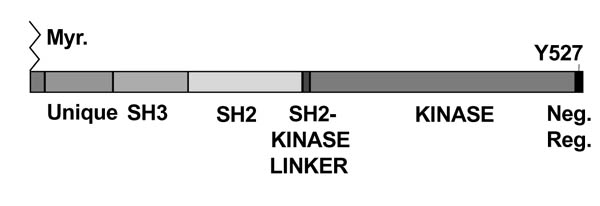
Unlike the EGF receptor, overexpression of c-Src alone is insufficient to transform murine fibroblasts in culture or to sustain tumor growth in intact animals [15,16,17**]. However, expression of dominant interfering forms of c-Src (Fig. 1) in cultured murine fibroblasts has shown that c-Src is required for EGF-induced mitogenesis [18*,19], and studies in transgenic mice have demonstrated that c-Src is necessary for induction of mammary tumors by the polyomavirus middle T oncogene [20]. These findings suggest that c-Src may function to promote growth of tumor cells by participating in or augmenting mitogenic signaling pathways that are initiated by extracellular growth factors or intracellular oncogenes. There appear to be two prominent roles of c-Src in this regard. One is to modulate receptor function by augmenting signals immediately downstream of the receptors and by regulating endocytosis [1**,21**,22**], and the other is to affect morphogenetic remodeling of the cell by phosphorylating proteins that associate with the actin cytoskeleton [23,24*].
Figure 1.

Structure of c-Src. C-Src is the prototype of a large family of cytoplasmic tyrosine kinases that associate with cellular membranes through lipid modifications at their amino-termini. As a linear molecule, the relationship between the various domains can be seen: an amino-terminal membrane-association domain that contains the site of myristylation; a unique domain that exhibits the widest sequence divergence among family members of any of the domains; an SH3 domain that binds polyproline motifs on target molecules; a SH2 domain that binds phosphotyrosine residues on target molecules; a SH2/kinase linker; the catalytic domain; and the negative regulatory domain that contains the predominant site of tyrosine phosphorylation on the inactive molecule (Y527 in chicken, Y530 in human). Mutations that abrogate myristylation, the SH2 domain, and the catalytic activity were shown to reduce EGF-stimulated DNA synthesis in C3H10T½ murine fibroblasts, providing evidence for the involvement of c-Src in mitogenic pathways [18*].
Synergism between c-Src and human epidermal growth factor receptor 1 in oncogenesis
The fact that c-Src and HER1 are co-overexpressed in many of the same tumor types suggests that these two kinases may participate in regulating the genesis and/or progression of human cancers. In a direct test to resolve this question, dual overexpression of both c-Src and HER1 in C3H10T ½ mouse fibroblasts was found to lead to synergistic increases in EGF-induced DNA synthesis, soft agar colony formation, and tumor formation in nude mice, when compared with cells that express only one of the pair [17**]. This enhanced oncogenesis correlated with the EGF-dependent physical association between c-Src and HER1, increased phosphorylation of the HER1 substrates, Shc and phospholipase Cγ, and the phosphorylation of two novel tyrosyl residues on the receptor, which have been identified by phosphotryptic mapping to be Tyr 845 and Tyr 1101 [17**,24*]. Stover et al [25] showed that activated Src can phosphorylate HER1 at Tyr 891 and Tyr 920 in vitro, and that these sites can mediate binding of the SH2 domains of phosphatidyl inositol-3 kinase (PI-3K) and Src itself. These same sites have been found to be phosphorylated in several colorectal carcinomas and MCF7 breast cancer cells, from which c-Src and PI-3K can be coimmunoprecitated with HER1. These findings support the idea that bidirectional interactions between c-Src and HER1 occur.
Interestingly, co-overexpression of both HER1 and c-Src also occurs in a subset of human breast cancer cell lines and breast tumor tissues [14*]. Like the mouse 10T½ fibroblasts, breast tumor cell lines that co-overexpress c-Src and HER1 display EGF-induced complex formation between c-Src and HER1, the appearance of Tyr 845 and Tyr 1101 phosphorylations on the c-Src-associated receptor, and increased phosphorylation of the HER1 effectors Shc and mitogen-activated protein kinase (MAPK), as well as increased tumor size in nude mice when compared with the majority of cell lines that do not overexpress these tyrosine kinases or express only one of the pair. Enhanced MAPK and MEK activity have also been found in human breast tumors that overexpress both c-Src and HER1 [26,27]. These results suggest that c-Src and HER1 act synergistically, and that this interaction is manifested by increased signaling through HER1, unregulated growth, and tumorigenesis. Because complex formation between HER1 and c-Src can be detected only under conditions of mutual overexpression, as is seen in many breast tumors and cell lines, disruption of this complex could provide the basis for novel therapeutic approaches.
Molecular mechanisms of c-Src/human epidermal growth factor receptor 1 synergism
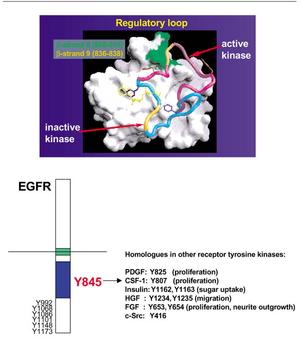
Although little is known about the biologic significance of Tyr 1101 phosphorylation, Tyr 845 is located in the activation loop of the catalytic domain and is particularly intriguing, because its homologs are found in a variety of receptor and nonreceptor tyrosine kinases (Fig. 2) [28]. In fact, substitution of Phe for the Tyr 845 homologs in other receptors renders these receptors catalytically impaired and defective in downstream signaling [29,30,31]. In contrast, Tyr 845 in the EGF receptor has never been identified as an autophosphorylation site. Failure to detect such a phosphorylation was interpreted to mean either that the site was not phosphorylated or that its phosphorylation was extremely short-lived.
Figure 2.

Tyr 845 is located in the catalytic domain of HER1 in a highly conserved subdomain that has functional homologs in other tyrosine kinases. Tyr 845 resides in the activation loop of the HER1 tyrosine kinase catalytic domain (upper panel), a region that shares a high degree of homology among all tyrosine kinases (lower panel). Upon ligand-binding to the EGF receptor (EGFR), the activation loop containing phosphorylated Tyr 845 is modeled (upper panel [63]) to flip into a configuration that promotes access to ATP and substrate, as is the case with phosphorylation of the analogous tyrosines within other tyrosine kinases. Mutation of sites in other tyrosine kinases that are analogous to Tyr 845 renders the molecules catalytically inactive and abrogates downstream biologic signaling. Similar effects of mutagenizing Tyr 845 have been observed [24*,32**]. A unique characteristic of Tyr 845 appears to be that its phosphorylation is mediated by c-Src, whereas homologs in other receptor tyrosine kinases are phosphorylated by the receptors themselves. The phosphorylation of Tyr 845 by c-Src is proposed to facilitate cross-talk between HER1 and signaling pathways activated by other cellular receptors, such as G-protein-coupled receptors and estrogen receptor.
Recently, it was shown [32**] that phosphorylation of Tyr 845 is dependent upon the catalytic activity of c-Src, suggesting that c-Src directly phosphorylates this site. Cells expressing kinase-inactive c-Src not only fail to support phosphorylation of Tyr 845, but also display a drastically decreased ability to grow in soft agar in the presence of EGF and to form tumors in nude mice, suggesting that phosphorylation of Tyr 845 may be critical for mitogenesis and transformation. Indeed, in 10T½ cells that express either increased or endogenous levels of c-Src, expression of a Tyr845Phe mutant form of HER1 results in a reduction in EGF-, serum-, and lysophosphatidic acid (LPA)-induced DNA synthesis [24*,32**]. Thus, c-Src-mediated phosphorylation of Tyr 845 appears to be necessary for the mitogenesis that emanates from HER1.
The finding that serum- and LPA-induced DNA synthesis are affected by the Tyr845Phe mutation suggests that other cell-surface receptors may mediate their effects in part through the EGF receptor. LPA, a major mitogen in serum, is known to activate a Gi-coupled receptor [33]. Several laboratories have recently reported that activation of certain GPCRs can trigger phosphorylation of the EGF receptor, as well as activation of its downstream effectors Shc and MAPK, and that this activation is dependent on c-Src kinase activity [34*,35,36]. In addition, stimulation of the growth hormone cytokine receptor has been found to induce EGF receptor phosphorylation via janus kinase 2 [37]. Recent work in our laboratory (Biscardi et al, unpublished data) has demonstrated that treatment of 10T½ cells with different GPCR ligands (thrombin, endothelin, and LPA) or with growth hormone induces increases in overall tyrosine phosphorylation of HER1, as well as in Tyr 845 phosphorylation. Interestingly, the kinase activity of c-Src is also required for phosphorylation of Tyr 845 via these alternate receptors, and mitogenesis is dependent on the phosphorylation of Tyr 845. 10T½ cells that express the Tyr845Phe variant of HER1 are impaired in their ability to synthesize DNA in response to these stimuli. However, the weakly mitogenic effects of isoproterenol, which signals through a Gsα-coupled pathway, are not affected by the Tyr845Phe mutation, indicating that this mutation does not act as a general inhibitor of mitogenesis.
Estrogen receptor, c-Src, and human epidermal growth factor receptor 1
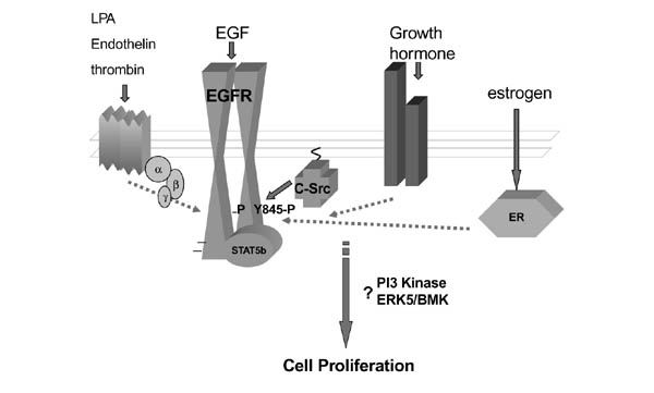
Accumulating evidence also points to an intricate network of cross-talk between the estrogen receptor and HER1. Early work by Ignar-Trowbridge et al [38,39] demonstrated that EGF can transcriptionally activate genes that contain estrogen response elements. More recently, Migliaccio et al [40,41] showed that estrogen is able to activate many of the effectors classically thought to be linked to the EGF receptor signaling pathway, including c-Src, Ras, and MAPK. These researchers have also shown that estrogen requires c-Src kinase activity in order to trigger its mitogenic effects [42**]. To further investigate this phenomenon, our laboratory has examined the effects of the Tyr845Phe mutation on estrogen-dependent DNA synthesis in the estrogen-responsive MCF7 breast cancer cell line. As was the case for the GPCR and cytokine receptor coupled agonists, estrogen-stimulated DNA synthesis was decreased to basal levels as a result of expression of the Y845F mutant (Biscardi et al, unpublished data). Taken together, these findings suggest the possibility that the EGF receptor plays an important, perhaps widespread, role in mediating the cell's response to an array of external signals and that the c-Src mediated phosphorylation of EGF receptor Tyr 845 appears to be a critical event in this process (Fig. 3).
Figure 3.

HER1 acts as a central mediator for multiple signaling pathways. A variety of extracellular ligands trigger the phosphorylation of HER1 on Tyr 845. These include the following: thrombin, endothelin, and LPA, which bind G-protein coupled receptors; growth hormone, which binds a cytokine receptor; and estrogen, which binds a steroid hormone receptor. Moreover, c-Src kinase activity is required for the ability of LPA, endothelin, growth hormone, and estrogen to induce phosphorylation of Tyr 845. We hypothesize that c-Src-mediated phosphorylation of Tyr 845 is a central signaling event and is required for mitogenesis to occur in response to a variety of external stimuli in addition to EGF. The signaling molecules that transmit mitogenic cues from phosphorylated Tyr 845 have yet to be delineated, but may include such effectors as STAT5b, PI-3K, or ERK5. ER, estrogen receptor.
Human epidermal growth factor receptor 2 and c-Src in breast cancer
Evidence supporting bidirectional interactions between c-Src and the EGF receptor raises the question of whether c-Src interacts in a similar manner with other HER family members. Some indications that HER2 and c-Src can physically and/or functionally interact have emerged over recent years, but little is currently known about the relationship between c-Src and either HER3 or HER4. In vitro studies have also demonstrated that HER2/neu can associate with the SH2 domain of c-Src in a tyrosine phosphorylation-dependent manner [43,44**], and in vivo coassociation between HER2/neu and c-Src has been detected in murine mammary tumors, human breast cancer cell lines, and human tumor tissues [44**] (Belsches-Jablonski AP et al. unpublished data). Furthermore, transgenic murine tumor tissues or human mammary epithelial cell lines expressing mutationally activated Neu exhibit a correlative increase in c-Src activity [44**,45]. These results suggest that c-Src may be downstream of HER2 signaling. It has also been demonstrated in vitro [25], however, that c-Src is able to phosphorylate HER2 at nonautophosphorylation sites. The identity of these sites, their existence in intact cells, and their functional significance have not yet been determined. Nevertheless, the currently available information suggests that HER2 and c-Src are able to interact physically and that bidirectional signaling may be a mechanism of interaction between these two tyrosine kinases, as it is for HER1 and c-Src.
The functional consequences of coassociation between HER2 and c-Src remain unclear. In fact, available evidence suggests that the HER2-c-Src interaction may affect different parameters of oncogenesis to different extents and perhaps by different mechanisms than the HER1-c-Src association. Results from recent studies of MCF10A cells that ectopically express mutationally activated rat p185_neu_ [45], and a panel of 13 human breast cancer cell lines and 13 human mammary tumor samples (Belsches-Jablonski AP et al. unpublished data) suggest that the HER2-c-Src complex may play an important role in heregulin-stimulated anchorage-independent growth and antiapoptotic or survival mechanisms, but have less of an effect on anchorage-dependent growth. In contrast, the HER1-c-Src complex has been found to have striking effects on anchorage-dependent growth and on anchorage-independent growth, but its role in survival signaling is unclear.
Whether c-Src mediates the phosphorylation of the Tyr 845 homolog in HER2 (Tyr 877) as it does in HER1 is not known. The comparable site in the activated, rat p185_neu_ protein (Tyr 882) is an autophosphorylation site, and mutation of this residue reduces the intrinsic kinase activity of the protein and its transforming potential [46*]. These findings suggest that the Tyr 845 homolog in p185_neu_ or HER2 functions in a manner more similar to the majority of tyrosine kinase receptors than it does to HER1, and raise the possibility that the mechanism by which c-Src interacts with HER2 may be distinct from that by which c-Src interacts with HER1.
Signals activated by c-Src/human epidermal growth factor receptor 1 interactions
Although mutation of Tyr 845 has profound effects on the cell's ability to respond mitogenically to EGF, many of the downstream targets of HER1 are unaffected. The Y845F mutant HER1 kinase activity appears to be unchanged, as does its ability to associate with c-Src. Moreover, the phosphorylation and/or activation of a number of HER1 effectors, including Shc, MAPK, signal transducer and activator of transcription (STAT)3, and phospholipase Cγ [32**] (Tice and Biscardi, unpublished data) are likewise unaffected. However, recent evidence, produced in a collaborative effort between our laboratory and that of Silva (unpublished data), suggests that STAT5b might be a physiologically relevant downstream effector of Tyr845.
The STATs are a family of transcription factors that are activated at the plasma membrane by tyrosine phosphorylation in response to signals from cytokine and growth factor receptors [47*,48]. Tyrosine phosphorylation results in STAT dimerization, nuclear translocation, and binding of STAT dimers to consensus elements upstream of regulated genes.
Increasing evidence indicates that STAT proteins are involved in the process of oncogenesis [49*,50]. Two laboratories have shown that STAT3 is required for v-Src transformation [51**,52**], whereas deGroot et al [53] demonstrated that active STAT5 is necessary for the soft agar growth of BCR-Abl transformed leukemia cells. Recent studies [54**] have also indicated a direct role for c-Src in the activation of STAT proteins. For example, c-Src was shown to mediate the EGF stimulation of STATs 1, 3, 5a, and 5b in NIH3T3 cells engineered to overexpress HER1, as well as in A431 cells, which endogenously express high levels of HER1. In contrast, another group [55] has recently described a role for c-Src in the tyrosine phosphorylation (but not the transcriptional activation) of STAT5a and STAT5b in a COS cell transfection model.
Our recent studies (Silva et al, unpublished data) indicate a role for the STAT proteins in signaling pathways that are activated in 10T½ and breast cancer cells co-overexpressing c-Src and EGF receptor. We have shown that c-Src tyrosine kinase activity is required for maximal transcriptional activation of STAT5b by EGF, and that phosphorylation of Tyr 845 is required for both the EGF-induced association between STAT5b and HER1 as well as tyrosine phosphorylation of STAT5b. These studies suggest a model whereby HER1 and c-Src overexpression and EGF stimulation lead to the phosphorylation of Tyr845 and the recruitment and activation of STAT5b.
A number of other signaling molecules have also been linked to EGF-induced mitogenesis in various cell systems, and should be considered as additional candidates for downstream effectors of Tyr845. These signaling molecules include PI-3K, big MAPK [BMK1 or extracellular-signal-regulated kinase (ERK)5], and the transcription factor Myc. After growth factor activation, PI-3K interacts, through its SH2 domain, with tyrosine phosphorylated growth factor receptors, resulting in an increase in PI-3K activity. Studies using specific antibodies to PI-3K [56*] demonstrated that its catalytic activity is required for EGF (and platelet-derived growth factor)-induced mitogenesis. Although PI-3K associates with the EGF receptor, the binding site has not been characterized and thus it is interesting to speculate that this function may be fulfilled by Tyr 845. ERK5, a member of the MAPK family, was first shown to be activated in response to oxidative stress, hyperosmolality, and serum. Recent studies [57**,58] have shown that this kinase is also activated in response to EGF and nerve growth factor. Furthermore, dominant-negative ERK5 blocks EGF-induced cell proliferation in a breast epithelial cell line by preventing cells from entering the S phase [57**]. Studies in mouse fibroblasts have shown that c-Src kinase is required for ERK5 activation in response to hydrogen peroxide. Although a role of c-Src in EGF activation of ERK5 has not yet been demonstrated, these studies provide the background for a potential role of ERK5 in the c-Src-mediated activation of HER1. One substrate of ERK5 is the early response gene c-myc [59]. C-myc encodes a nuclear phosphoprotein, which, in combination with Max, activates gene transcription. C-Myc expression correlates with the proliferative state [60], and has been shown to rescue platelet-derived growth factor signaling that is blocked by kinase-inactive c-Src [61]. Together these findings link c-Src and EGF with PI-3K, ERK5, and c-Myc, and are suggestive of a potential role for one or more of these molecules to function as downstream effectors of phosphorylated Tyr 845.
Conclusion
Substantial evidence is accumulating to indicate functional synergism between the nonreceptor tyrosine kinase c-Src and members of the EGF receptor family in promoting breast cancer progression. Members of both families are overexpressed in approximately 70% or more of human breast cancers, and the human EGF receptor (HER1) and HER2/neu portend a poor prognosis for the disease.
It has been demonstrated that c-Src, which is nontransforming when overexpressed alone, can potentiate the tumorigenic capacity of overexpressed HER1. Recently, one mechanism by which c-Src synergizes with the HER1 has been uncovered. This mechanism involves the EGF-dependent association of c-Src with HER1 and phosphorylation of the receptor by c-Src on residues Tyr 845 and Tyr 1101. The functional consequences of Tyr 1101 phosphorylation are unknown, but phosphorylation of Tyr 845 is required for EGF-induced DNA synthesis and activation of members of the STAT family of transcription factors, particularly STAT5b, but not activation of Shc or MAPK. Whether the STATs are the predominant mediators of Tyr 845-dependent mitogenesis or whether there are other mitogenic signaling pathways that emanate from phosphorylated Tyr 845 remains to be determined. Surprisingly, Tyr 845 phosphorylation has also been found to be an intermediate in mitogenic signaling from a variety of GPCR, as well as from certain cytokine receptors and the estrogen receptor. Thus, HER1, and specifically phosphorylation of Tyr 845 by c-Src, appears to play an important, perhaps widespread role in mediating cell responses to an array of external signals.
c-Src also complexes with another member of the EGF receptor family, namely HER-2 or erbB-2/neu. This association is independent of extracellular ligand and appears to contribute more to cell survival and anchorage-independent growth of breast cancer cells than to anchorage-dependent growth or migration. The mechanism of c-Src interaction with HER2 and how this interaction may transmit survival or anchorage-independent growth signals is not known, but it is speculated to be different than the interaction between c-Src and HER1.
Acknowledgments
Acknowledgements
The authors thank D Tice for his contributions to the figures. The work cited from our laboratories was supported by grants CA71449 and CA39438 from the DHHS (SJP), grant 4621 from the Council for Tobacco Research (SJP), and grant DAMB 17-99-1-9431 from the Army Breast Cancer Initiative, DOD (CMS). JSB is a recipient of a postdoctoral fellowship (DAMD17-97-7329) from the Army Breast Cancer Initiative, DOD.
References
- Biscardi JS, Tice DA, Parsons SJ. c-Src, receptor tyrosine kinases, and human cancer. Adv Cancer Res. 1999;76:61–119. doi: 10.1016/s0065-230x(08)60774-5. [DOI] [PubMed] [Google Scholar]
- Velu TJ, Beguinot L, Vass WC, et al. Epidermal growth factor-dependent transformation by a human EGF receptor proto-oncogene. Science. 1987;238:1408–1410. doi: 10.1126/science.3500513. [DOI] [PubMed] [Google Scholar]
- Khazaie K, Schirrmacher V, Lichtner RB. EGF receptor in neoplasia and metastasis. Cancer Metastasis Rev. 1993;12:255–274. doi: 10.1007/BF00665957. [DOI] [PubMed] [Google Scholar]
- Bolla M, Chedin M, Souvignet C, et al. Estimation of epidermal growth factor receptor in 177 breast cancers: correlation with prognostic factors. Breast Cancer Res Treat. 1990;16:97–102. doi: 10.1007/BF01809293. [DOI] [PubMed] [Google Scholar]
- Toi M, Osaki A, Yamada H, Toge T. Epidermal growth factor receptor expression as a prognostic indicator in breast cancer. . Eur J Cancer. 1991;27:977–980. doi: 10.1016/0277-5379(91)90262-c. [DOI] [PubMed] [Google Scholar]
- Lichtner RB, Kaufmann AM, Kittmann A, et al. Ligand mediated activation of ectopic EGF receptor promotes matrix protein adhesion and lung colonization of rat mammary adenocarcinoma cells. Oncogene. 1995;10:1823–1832. [PubMed] [Google Scholar]
- Coussens L, Yang-Feng TL, Liao YC, et al. Tyrosine kinase receptor with extensive homology to EGF receptor shares chromosomal location with neu oncogene. Science. 1985;230:1132–1139. doi: 10.1126/science.2999974. [DOI] [PubMed] [Google Scholar]
- Slamon DJ, Clark GM, Wong SG, et al. Human breast cancer:correlation of relapse and survival with amplification of the HER2/neu oncogene. Science. 1987;235:177–182. doi: 10.1126/science.3798106. [DOI] [PubMed] [Google Scholar]
- Slamon DJ, Godolphin W, Jones LA, et al. Studies of the HER2/neu proto-oncogene in human breast and ovarian cancer. Science. 1989;244:707–712. doi: 10.1126/science.2470152. [DOI] [PubMed] [Google Scholar]
- Hynes NE, Stern DF. The biology of erbB2/neu/HER2 and its role in cancer. Biochim Biophys Acta. 1994;1198:165–184. doi: 10.1016/0304-419x(94)90012-4. [DOI] [PubMed] [Google Scholar]
- Baselga J, Norton L, Albanell J, Kim YM, Mendelsohn J. Recombinant humanized anti-HER2 antibody (HERceptin) enhances the antitumor activity of paclitaxel and doxorubicin against HER2/neu overexpressing human breast cancer xenografts. Cancer Res. 1998;58:2825–2831. [PubMed] [Google Scholar]
- Ottenhoff-Kalff AE, Rijksen G, van Beurden EA, Hennipman A, Michels AA, Staal GE. Characterization of protein tyrosine kinases from human breast cancer: involvement of the c-Src oncogene product. Cancer Res. 1992;52:4773–4778. [PubMed] [Google Scholar]
- Verbeek BS, Vroom TM, Adriaansen-Slot SS, et al. c-Src protein expression is increased in human breast cancer. An immunohistochemical and biochemical analysis. J Pathol. 1996;180:383–388. doi: 10.1002/(SICI)1096-9896(199612)180:4<383::AID-PATH686>3.3.CO;2-E. [DOI] [PubMed] [Google Scholar]
- Biscardi JS, Belsches AP, Parsons SJ. Characterization of human epidermal growth factor receptor and c-Src in human breast tumor cells. Mol Carcinogen. 1998;21:261–272. doi: 10.1002/(sici)1098-2744(199804)21:4<261::aid-mc5>3.0.co;2-n. [DOI] [PubMed] [Google Scholar]
- Shalloway D, Coussens PM, Yaciuk P. Overexpression of the c-Src protein does not induce transformation of NIH3T3 cells. Proc Natl Acad Sci USA. 1984;81:7071–7075. doi: 10.1073/pnas.81.22.7071. [DOI] [PMC free article] [PubMed] [Google Scholar]
- Luttrell DK, Luttrell LM, Parsons SJ. Augmented mitogenic responsiveness to epidermal growth factor in murine fibroblasts that overexpress pp60c-src. Mol Cell Biol. 1988;8:497–501. doi: 10.1128/mcb.8.1.497. [DOI] [PMC free article] [PubMed] [Google Scholar]
- Maa MC, Leu TH, McCarley DJ, Schatzman RC, Parsons SJ. Potentiation of EGF receptor-mediated oncogenesis by c-Src: implications for the etiology of multiple human cancers. Proc Natl Acad Sci USA. 1995;92:6981–6985. doi: 10.1073/pnas.92.15.6981. [DOI] [PMC free article] [PubMed] [Google Scholar]
- Wilson LK, Luttrell DK, Parsons JT, Parsons SJ. pp60c-Src tyrosine kinase, myristylation, and modulatory domains are required for enhanced mitogenic responsiveness to epidermal growth factor seen in cells overexpressing c-src. Mol Cell Biol. 1989;9:1536–1544. doi: 10.1128/mcb.9.4.1536. [DOI] [PMC free article] [PubMed] [Google Scholar]
- Roche S, Koegl M, Barone MV, Roussel MF, Courtneidge SA. DNA synthesis induced by some but not all growth factors requires Src family protein tyrosine kinases. Mol Cell Biol. 1995;15:1102–1109. doi: 10.1128/mcb.15.2.1102. [DOI] [PMC free article] [PubMed] [Google Scholar]
- Guy CT, Muthuswamy SK, Cardiff RD, Soriano P, Muller WJ. Activation of the c-Src tyrosine kinase is required for the induction of mammary tumors in transgenic mice. Genes Dev. 1994;8:23–32. doi: 10.1101/gad.8.1.23. [DOI] [PubMed] [Google Scholar]
- Ware MF, Tice DA, Parsons SJ, Lauffenberger DA. Overexpression of cellular c-Src in fibroblasts enhances endocytic internalization of epidermal growth factor receptor. J Biol Chem. 1997;272:30185–30190. doi: 10.1074/jbc.272.48.30185. [DOI] [PubMed] [Google Scholar]
- Wilde A, Beattie EC, Lem L, et al. EGF receptor signaling stimulates src kinase phosphorylation of clathrin, influencing clathrin redistribution and EGF uptake. Cell. 1999;96:677–687. doi: 10.1016/s0092-8674(00)80578-4. [DOI] [PubMed] [Google Scholar]
- Belsches AP, Haskell MD, Parsons SJ. Role of c-Src tyrosine kinase in EGF-induced mitogenesis. Front Biosci. 1997;2:d501–d518. doi: 10.2741/a208. [DOI] [PubMed] [Google Scholar]
- Biscardi JS, Maa MC, Tice DA, et al. c-Src mediated phosphorylation of the epidermal growth factor receptor on Tyr845 and Tyr1101 is associated with modulation of receptor function. J Biol Chem. 1999;274:8335–8343. doi: 10.1074/jbc.274.12.8335. [DOI] [PubMed] [Google Scholar]
- Stover DR, Becker M, Liebetanz J, Lydon NB. Src phosphorylation of the epidermal growth factor receptor at novel sites mediates receptor interaction with Src and p85. J Biol Chem. 1995;270:15591–15597. doi: 10.1074/jbc.270.26.15591. [DOI] [PubMed] [Google Scholar]
- Salh B, Marotta A, Matthewson C, et al. Investigation of the Mek-MAP kinase-Rsk pathway in human breast cancer. Anticancer Res. 1999;19:731–740. [PubMed] [Google Scholar]
- Xing C, Imagawa W. Altered MAP kinase (ERK1,2) regulation in primary cultures of mammary tumor cells: elevated basal activity and sustained response to EGF. Carcinogenesis. 1999;20:1201–1208. doi: 10.1093/carcin/20.7.1201. [DOI] [PubMed] [Google Scholar]
- Hanks SJ, Quinn AM, Hunter T. The protein kinase family: conserved features and deduced phylogeny of the catalytic domains. Science. 1988;241:42–52. doi: 10.1126/science.3291115. [DOI] [PubMed] [Google Scholar]
- Ellis L, Clauser E, Morgan DO, et al. Replacement of insulin receptor tyrosine residues 1162 and 1163 compromises insulin-stimulated kinase activity and uptake of 2-deoxyglucose. Cell. 1986;45:721–732. doi: 10.1016/0092-8674(86)90786-5. [DOI] [PubMed] [Google Scholar]
- Fantl WJ, Escobedo JA, Williams LT. Mutations of the platelet-derived growth factor receptor that cause a loss of ligand-induced conformational change, subtle changes in kinase activity, and impaired ability to stimulate DNA synthesis. Mol Cell Biol. 1989;9:4473–4478. doi: 10.1128/mcb.9.10.4473. [DOI] [PMC free article] [PubMed] [Google Scholar]
- Van der Geer P, Hunter T. Tyrosine 706 and 807 phosphorylation site mutants in the murine colony-stimulating factor-1 receptor are unaffected in their ability to induce early response gene transcription. Mol Cell Biol. 1991;11:4698–4709. doi: 10.1128/mcb.11.9.4698. [DOI] [PMC free article] [PubMed] [Google Scholar]
- Tice DA, Biscardi JS, Nickles AL, Parsons SJ. Mechanism of biological synergy between cellular Src and epidermal growth factor receptor. Proc Natl Acad Sci USA. 1999;96:1415–1420. doi: 10.1073/pnas.96.4.1415. [DOI] [PMC free article] [PubMed] [Google Scholar]
- Moolenaar WH, Kranenburg O, Postma FR, Zondag GC. Lysophosphatidic acid: G-protein signalling and cellular responses. Curr Opin Cell Biol. 1997;9:168–173. doi: 10.1016/s0955-0674(97)80059-2. [DOI] [PubMed] [Google Scholar]
- Daub H, Wallasch C, Lankenau A, Herrlich A, Ullrich A. Signal characteristics of G protein-transactivated EGF receptor. EMBO J. 1997;16:7032–7044. doi: 10.1093/emboj/16.23.7032. [DOI] [PMC free article] [PubMed] [Google Scholar]
- Luttrell LM, Della Rocca GJ, van Biesen T, Luttrell DK, Lefkowitz RJ. Role of c-Src tyrosine kinase in G-protein coupled receptor and G beta gamma subunit-mediated activation of mitogen-activated protein kinases. J Biol Chem. 1997;272:4637–4644. doi: 10.1074/jbc.272.7.4637. [DOI] [PubMed] [Google Scholar]
- Cunnick JM, Dorsey JF, Standley T, et al. Role of tyrosine kinase activity of epidermal growth factor receptor in the lysophosphatidic acid-stimulated mitogen-activated protein kinase pathway. J Biol Chem. 1998;273:14468–14475. doi: 10.1074/jbc.273.23.14468. [DOI] [PubMed] [Google Scholar]
- Yamauchi T, Ueki K, Tobe K, et al. Tyrosine phosphorylation of the EGF receptor by the kinase JAK2 is induced by growth hormone. Nature. 1997;390:91–96. doi: 10.1038/36369. [DOI] [PubMed] [Google Scholar]
- Ignar-Trowbridge DM, Nelson KG, Bidwell MC, et al. Coupling of dual signaling pathways: EGF action involves the estrogen receptor. Proc Natl Acad Sci USA. 1992;89:4658–4662. doi: 10.1073/pnas.89.10.4658. [DOI] [PMC free article] [PubMed] [Google Scholar]
- Ignar-Trowbridge DM, Teng CT, Ross KA, et al. Peptide growth factors elicit estrogen receptor-dependent trasnscriptional activation of an estrogen-responsive element. Mol Endocrinol. 1993;7:992–998. doi: 10.1210/mend.7.8.8232319. [DOI] [PubMed] [Google Scholar]
- Migliaccio A, Pagano M, Auricchio F. Immediate and transient stimulation of protein tyrosine phosphorylation by estradiol in MCF7 cells. Oncogene. 1993;8:2183–2191. [PubMed] [Google Scholar]
- Migliaccio A, Di Domenico M, Castoria G, et al. Tyrosine kinase/p21ras/MAP-kinase pathway activation by estradiol-receptor. EMBO J. 1996;15:1292–1300. [PMC free article] [PubMed] [Google Scholar]
- Castoria G, Barone MV, Di Domenico M, et al. Non-transcriptional action of oestradiol and progestin triggers DNA synthesis. EMBO J. 1999;18:2500–2510. doi: 10.1093/emboj/18.9.2500. [DOI] [PMC free article] [PubMed] [Google Scholar]
- Luttrell DK, Lee A, Lansing TJ, et al. Involvement of pp60c-Src with two major signaling pathways in human breast cancer. Proc Natl Acad Sci USA. 1994;91:83–87. doi: 10.1073/pnas.91.1.83. [DOI] [PMC free article] [PubMed] [Google Scholar]
- Muthuswamy SK, Siegel PM, Dankort DL, Webster MA, Muller WJ. Mammary tumors expressing the neu proto-oncogene possess elevated c-Src tyrosine kinase activity. Mol Cell Biol. 1994;14:735–743. doi: 10.1128/mcb.14.1.735. [DOI] [PMC free article] [PubMed] [Google Scholar]
- Sheffield LG. C-Src activation by ErbB2 leads to attachment-independent growth of human breast epithelial cells. Biochem Biophys Res Commun. 1998;250:27–31. doi: 10.1006/bbrc.1998.9214. [DOI] [PubMed] [Google Scholar]
- Zhang HT, O'Rourke DM, Zhao H, et al. Absence of autophosphorylation site on Y882 in the p185neu oncogene product correlates with a reduction of transforming potential. . Oncogene. 1998;16:2835–2842. doi: 10.1038/sj/onc/1201820. [DOI] [PubMed] [Google Scholar]
- Leaman DW, Leung S, Li X, Stark GR. Regulation of STAT-dependent pathways by growth factors and cytokines. FASEB J. 1996;10:1578–1588. [PubMed] [Google Scholar]
- Wells JA, deVos A. Hematopoietic receptor complexes. Annu Rev Biochem. 1996;65:609–634. doi: 10.1146/annurev.bi.65.070196.003141. [DOI] [PubMed] [Google Scholar]
- Watson CJ, Miller WR. Elevated levels of members of the STAT family of transcription factors in breast carcinoma nuclear extracts. Br J Cancer. 1995;71:840–844. doi: 10.1038/bjc.1995.162. [DOI] [PMC free article] [PubMed] [Google Scholar]
- Sartor CI, Dziubinski ML, Yu CL, Jove R, Ethier SP. Role of epidermal growth factor receptor and STAT-3 activation in autonomous proliferation of SUM-102PT human breast cancer cells. Cancer Res. 1997;57:978–987. [PubMed] [Google Scholar]
- Bromberg JF, Horvath CM, Besser D, Lathem WW, Darnell JE. STAT3 activation is required for cellular transformation by v-src. Mol Cell Biol. 1998;18:2553–2558. doi: 10.1128/mcb.18.5.2553. [DOI] [PMC free article] [PubMed] [Google Scholar]
- Turkson J, Bowman T, Garcia R, et al. STAT3 activation by src induces specific gene regulation and is required for cell transformation. Mol Cell Biol. 1998;18:2545–2552. doi: 10.1128/mcb.18.5.2545. [DOI] [PMC free article] [PubMed] [Google Scholar]
- deGroot RP, Raaijmakers JAM, Lammers J-WJ, Jove R, Koenderman L. STAT5 activation by BCR-Abl contributes to transformation of K562 leukemia cells. Blood. 1999;94:1108–1112. [PubMed] [Google Scholar]
- Olayioye MA, Beuvink I, Horsch K, Daly JM, Hynes NE. ErbB receptor-induced activation of Stat transcription factors is mediated by Src tyrosine kinases. J Biol Chem. 1999;274:17209–17218. doi: 10.1074/jbc.274.24.17209. [DOI] [PubMed] [Google Scholar]
- Kazansky AV, Kabotyanski EB, Wyszomierski SL, Mancini MA, Rosen JM. Differential effects of prolactin and src/abl kinases on the nuclear translocation of STAT5b and STAT5a. J Biol Chem. 1999;274:22484–22492. doi: 10.1074/jbc.274.32.22484. [DOI] [PubMed] [Google Scholar]
- Roche S, Koegl M, Courtneidge SA. The phosphatidylinositol 3-kinase alpha is required for DNA synthesis induced by some, but not all, growth factors. Proc Natl Acad Sci USA. 1994;91:9185–9189. doi: 10.1073/pnas.91.19.9185. [DOI] [PMC free article] [PubMed] [Google Scholar]
- Kato Y, Tapping RI, Huang S, et al. Bmk1/Erk5 is required for cell proliferation induced by epidermal growth factor. Nature. 1998;395:713–716. doi: 10.1038/27234. [DOI] [PubMed] [Google Scholar]
- Kamakura S, Moriguchi T, Nishida E. Activation of the protein kinase ERK5/BMK1 by receptor tyrosine kinases. J Biol Chem. 1999;274:26563–26571. doi: 10.1074/jbc.274.37.26563. [DOI] [PubMed] [Google Scholar]
- English JM, Pearson G, Baer R, Cobb MH. Identification of substrates and regulators of the mitogen-activated protein kinase ERK5 using chimeric protein kinases. J Biol Chem. 1998;273:3854–3860. doi: 10.1074/jbc.273.7.3854. [DOI] [PubMed] [Google Scholar]
- Kelly K, Siebenlist U. The regulation and expression of c-myc in normal and malignant cells. Ann Rev Immunol. 1986;4:317–338. doi: 10.1146/annurev.iy.04.040186.001533. [DOI] [PubMed] [Google Scholar]
- Barone MV, Courtneidge SA. Myc but not Fos rescue of PDGF signaling block caused by kinase-inactive Src. Nature. 1995;378:509–512. doi: 10.1038/378509a0. [DOI] [PubMed] [Google Scholar]
- Groenen LC, Walker F, Burgess AW, Treutlein HR. A model for the activation of the epidermal growth factor receptor kinase: Involvement of an asymmetric dimer? Biochemistry. 1997;36:3826–3836. doi: 10.1021/bi9614141. [DOI] [PubMed] [Google Scholar]