Analysis of the Life Cycle of the Soil Saprophyte Bacillus cereus in Liquid Soil Extract and in Soil (original) (raw)
Abstract
Bacillus is commonly isolated from soils, with organisms of Bacillus cereus sensu lato being prevalent. Knowledge of the ecology of B. cereus and other Bacillus species in soil is far from complete. While the older literature favors a model of growth on soil-associated organic matter, the current paradigm is that B. cereus sensu lato germinates and grows in association with animals or plants, resulting in either symbiotic or pathogenic interactions. An in terra approach to study soil-associated bacteria is described, using filter-sterilized soil-extracted soluble organic matter (SESOM) and artificial soil microcosms (ASM) saturated with SESOM. B. cereus ATCC 14579 displayed a life cycle, with the ability to germinate, grow, and subsequently sporulate in both the liquid SESOM extract and in ASM inserted into wells in agar medium. Cells grew in liquid SESOM without separating, forming multicellular structures that coalesced to form clumps and encasing the ensuing spores in an extracellular matrix. Bacillus was able to translocate from the point of inoculation through soil microcosms as shown by the emergence of outgrowths on the surrounding agar surface. Microscopic inspection revealed bundles of parallel chains inside the soil. The motility inhibitor l-ethionine failed to suppress outgrowth, ruling out translocation by a flagellar-mediated mechanism such as swimming or swarming. Bacillus subtilis subsp. subtilis Marburg and four Bacillus isolates taken at random from soils also displayed a life cycle in SESOM and ASM and were all able to translocate through ASM, even in presence of l-ethionine. These data indicate that B. cereus is a saprophytic bacterium that is able to grow in soil and furthermore that it is adapted to translocate by employing a multicellular mode of growth.
Members of the aerobic spore-forming genus Bacillus are commonly isolated from many types of soil at a range of depths and altitudes, and under various climatic conditions (9, 24, 32, 36). Bacillus cereus group members are the most commonly observed of the soil-isolated Bacillus strains (36). B. cereus sensu lato comprises the species B. cereus, B. anthracis, B. thuringiensis, B. mycoides, B. pseudomycoides, and B. weihenstephanensis (11, 16, 26). While these species are genetically very closely related, their precise phylogenetic and taxonomic relationships are still debated (11). B. cereus is widely reported as a soil bacterium and also occurs in the rhizosphere of some plants (10, 32). Some strains of B. cereus produce antibiotics able to suppress fungal diseases of the rhizosphere (23). B. cereus is also a food poisoning bacterium that can occasionally be an opportunistic human pathogen (29). B. thuringiensis is an insect pathogen that produces insecticidal proteins (3), and B. anthracis is a pathogen of mammals (15). While all may reportedly be isolated from soils, our knowledge of the ecology of these Bacillus species in soil is far from complete.
Winogradsky detected saprophytic Bacillus species such as B. megaterium and B. cereus in soil, stating that the bacteria's spores became active when an excess of easily decomposable organic matter became available or when the moisture content of the soil was high (38). Waksman viewed Bacillus species as “saprophytic organisms whose natural habitat is the soil” (13). Bacillus was reported to occur in soil as spores, germinating and becoming active only when readily decomposable organic matter was available (37). Some later reports implied growth of B. thuringiensis in soil by suggesting germination in soil at a pH above 6.0 and temperatures above 15.5°C (27). Van Ness (35) draws correlations between anthrax outbreaks and soil pH, arguing that B. anthracis is able to grow in specific soil types. B. megaterium is able to grow in soil, as supported by proof for in situ transcription of the exponentially expressed protease gene nprM (14). Yet the recent literature implies that B. cereus sensu lato does not grow in soil (6, 16). The current paradigm is that B. cereus sensu lato germinates and grows either in an animal host or in the rhizosphere, resulting in either symbiotic or pathogenic interactions. So B. anthracis is able to grow in various mammalian species, B. thuringiensis proliferates in the guts of various insects before killing its host, and B. cereus is both a gut commensal of various insects and an inhabitant of the rhizosphere of certain plants (10, 16, 21). Defecation or death of the host releases cells and spores into the soil, where the vegetative cells may sporulate and survive until their uptake by another host (5, 6, 16).
We have initiated a program to investigate the behavior of the soil bacterium B. cereus when it occurs in soil. The aim of the work reported here was to determine whether B. cereus is able to grow and perform a full life cycle in liquid soil extract and in artificial soil microcosms. The results indicate that B. cereus ATCC 14579 and related soil isolates demonstrate a complete saprophytic life cycle in conditions mimicking the soil environment. B. cereus germinated, grew, and sporulated in both liquid soil extract and artificial soil microscosms. Furthermore, when B. cereus grew in soil, it switched from a single-cell to a multicellularity phenotype, translocating through the soil.
(Part of this research was presented at the 105th General Meeting of the American Society for Microbiology, Atlanta, Ga. [20]).
MATERIALS AND METHODS
Strains.
The strains used in this study were B. cereus ATCC 14579, obtained from the American Type Culture Collection, B. subtilis subsp. subtilis Marburg (NCIB 3610) and B. subtilis 168 obtained from the Bacillus genetic stock center, and four Bacillus isolates from soil samples collected randomly from experimental fields of South Dakota State University. These isolates were obtained by pasteurizing serial dilutions of soil samples for 10 min at 80°C, plating on LB agar, incubating for 24 h at 30°C, and picking four colonies at random. The phylogenetic positions of these isolates were determined by sequencing 1,368 bases of their respective 16S rRNA genes.
Culture media.
Five liquid culturing media were used in this study. LB broth (Fisher Scientific) was set to pH 6.5 after autoclaving. Liquid soil extract, termed soil-extracted solubilized organic matter (SESOM), was prepared by suspending 100 g of air-dried topsoil (commercial soil from Heartland Gardens, Apple Valley, Minn.) in 500 ml of 3-(_N_-morpholino) propanesulfonic acid (MOPS) buffer (10 mM; pH 7 at 40°C) and shaking at 200 rpm for 1 h. In order to avoid chemical modification of soil organic matter by the high temperature required for autoclaving, the extract was filtered sequentially through filter paper (Whatman), hydrophilic polyvinylidene difluoride membranes with 5-μm and 0.45-μm pore sizes (Durapore, Millipore), and 0.22-μm-pore-size polyethersulfone membranes (Nalgene Labware) in order to remove particulate matter. After filtration, the pH was adjusted to 6.5, and SESOM was supplemented where required. Sterilization was performed by filtration (0.22-μm-pore-size filter), and the sterility of each batch was controlled by depositing 5 μl of medium on LB agar and incubating at 30°C for 24 h. For SESOM+, SESOM was supplemented with glucose (100 mg/liter), casein hydrolysate (1 mg/liter; Sigma), and yeast extract (1 mg/liter; Fisher Scientific). MOPS buffer (10 mM), supplemented as described above and designated MOPS+, was used as a negative control. M9 minimal medium (28) was supplemented with 1g/liter humic acid (Aldrich catalog no. H1, 675-2) to evaluate humic acid as a sole carbon and energy source. For SESOM agar, 80 ml SESOM+ was mixed with 20 ml of tempered 7.5% agar.
Culturing conditions.
Suspended populations were cultured at 30°C with shaking at 150 rpm in a 250-ml Erlenmeyer flask containing 100 ml of culture medium. Vegetative inocula were prepared by overnight culturing in LB broth, followed by washing of cells in phosphate buffer (100 mM; pH 7.0). The initial bacterial inoculum was calibrated to yield 105 CFU/ml, corresponding to an initial optical density of 0.005 (λ of 546 nm). Spore inocula were prepared by culturing in SESOM+ for 48 h, when no vegetative cells could be detected.
Assays for swimming, swarming, and translocation.
Swimming and swarming were assessed by stabbing cultures into LB and SESOM swim and swarm agars, respectively. Agar plates were incubated at 30°C for 24 and 48 h, and then the diameters of colonies were measured. Swim agars were prepared by supplementing LB with 0.35% agar and mixing 80 ml of SESOM+ with 20 ml of tempered 1.75% agar. Swarm agars were prepared by supplementing LB with 1.0% agar and mixing 80 ml of SESOM+ with 20 ml of tempered 5.0% agar. Swim and swarm agars were supplemented where necessary with 10 mM CaCl2 or 1 mM l-ethionine (Sigma), an inhibitor of methyl group turnover on methyl-accepting chemotaxis proteins and thereby of flagellum-mediated motility, shown previously to repress swimming in B. sphaericus (1).
Artificial soil microcosms (ASM), rather than the generally used thrice-autoclaved soil, were used to study growth under soil conditions. The composition of ASM was similar to that described by Ellis (7) but with modifications, primarily including replacing humic acid with SESOM. ASM consisted of four parts acid-washed, autoclaved sand and one part montmorillonite, made homoionic for calcium before autoclaving, and saturated with either SESOM+ or LB. Ca-montmorillonite homoionic for calcium was prepared as described by Stotzky and Burns (34). Briefly, bentonite (Sigma) was washed three times with CaCl2 (100 mM; pH 7), which was followed by repeated wash steps in distilled H2O (dH2O) until no chloride was released, as revealed by the use of silver nitrate as an indicator. Wells (8 mm in diameter) were punched into LB or SESOM agar and filled aseptically with approximately 0.35 ml of ASM. The ASM were point inoculated by injecting 1 μl of water-washed vegetative cells or spore suspension (106 CFU/ml) below the surface at the center of the soil microcosm. In order to investigate whether cells translocated by convective forces, bromophenol blue and Coomassie R250 were spotted at the centers of ASM. To ascertain the role of flagella in translocation through soil, ASM and the surrounding agar were supplemented with 1 mM l-ethionine.
Quantification of biomass.
Vegetative cell and spore densities were determined as CFU per ml by using the droplet technique (19). Serial dilutions were prepared in sterile phosphate buffer (100 mM; pH 7.0) and 20-μl volumes were spotted in duplicate onto each of two LB agar plates. Plates were incubated at 30°C for 10 h, when colonies were counted. Spore densities were determined by pasteurizing serial dilutions at 80°C for 10 min prior to plating. Cells and spores in ASM were enumerated by suspending the entire microcosm (0.35 ml) in 0.65 ml of phosphate buffer, vortexing for 60 s, and preparing serial dilutions. Growth in suspended culture was characterized by periodically determining the optical density at 546 nm.
Microscopic investigation.
Bacteria were visualized by the Live/Dead BacLight stain (L7012; Molecular Probes). The SYTO 9 component conveys green fluorescence to intact cells, while the propidium iodide is taken up by cells with impaired membranes, making them fluoresce in the red spectrum. For liquid experiments, bacteria from 1 ml of bacterial suspension were harvested by centrifugation (10,000 × g; 1 min). The pellet was resuspended in 20 μl of sterile dH2O and 2 μl of BacLight stain before being subjected to fluorescence microscopy with an AX70 fluorescence microscope (Olympus). For ASM experiments, samples taken from the top, middle, and lower layers of ASM were suspended in 20 μl of sterile dH2O and 2 μl of Live/Dead BacLight stain and viewed by confocal scanning laser microscopy (CSLM) using an Olympus Fluoview FV300 Laser Scanning Confocal Microscope System interfaced with an inverted microscope (Olympus IX70) and blue argon (488 nm) and green helium neon (543 nm) excitation lasers.
Statistical analysis of data.
All experiments were performed in triplicate and on two or more separate occasions. Growth curve data and CFU counts were expressed as the mean ± standard error of the mean. Culturable count data were subjected to a Student's t test using Microsoft Office Excel 2003.
Nucleotide sequence accession numbers.
Sequences of the four Bacillus isolates taken from soil have been deposited in the GenBank database (http://www.ncbi.nlm.nih.gov/) under the accession numbers DQ005707 (Bacillus isolate 9), DQ005708 (Bacillus isolate 12), DQ005709 (Bacillus isolate 17), and DQ005710 (Bacillus isolate 20).
RESULTS
Support of growth of B. cereus by liquid soil extract.
A washed inoculum of vegetatively growing B. cereus increased 20-fold in SESOM before sporulating (Table 1). Spores were able to germinate in SESOM as supported by the 35-fold biomass increase (Table 1). Collectively these data demonstrated that B. cereus was able to grow in an extract of topsoil and that SESOM is a useful tool for the laboratory study of bacterial growth on soil-associated nutrients. In order to enhance biomass yields for further study, SESOM was supplemented to yield SESOM+, leading to a 10-fold increase in biomass (Table 1). B. cereus was not able to grow on this supplement alone in MOPS buffer (MOPS+), nor could germination of spores be demonstrated after 24 h of incubation (Table 1), indicating that components in SESOM were primarily responsible for supporting growth. M9 minimal medium with 1 g/liter humic acid as a sole carbon and energy source did not support the growth of vegetative cells or outgrowth from a spore inoculum of B. cereus (data not shown). This indicated that B. cereus ATCC 14579, while able to grow in SESOM, was unable to do so on humic substances alone.
TABLE 1.
Characteristics of growth parameters, yields, and sporulation of B. cereus under different culture conditionsa
| Culture condition (inoculum and medium) | OD at 24 hb | Duration of lag phase (min) | Generation time (min)c | CFU/ml after 8 h (n = 12) | CFU/ml after 24 h (n = 12) | ||
|---|---|---|---|---|---|---|---|
| Total | % Spores | Total | % Spores | ||||
| VI into SESOM | 0.145 | 0 | 48.3 ± 1.3 | ND | ND | 2.3 ± 0.1 × 10⁶ | 144.4 ± 11.1d |
| SI into SESOM | 0.155 | ND | ND | ND | ND | 3.5 ± 0.6 × 10⁶ | 133.2 ± 14.3d |
| VI into LB | 2.105 | 80 | 26.2 ± 0.6 | 4.4 ± 0.2 × 10⁸ | 0.0 ± 0.0 | 5.0 ± 0.2 × 10⁸ | 0.0 ± 0.0 |
| VI into SESOM+ | 0.265 | 0 | 36.9 ± 0.8 | 2.1 ± 0.1 × 10⁷ | 0.0 ± 0.0 | 2.8 ± 0.3 × 10⁷ | 96.5 ± 2.4 |
| SI into LB | 2.311 | 160 | 26.1 ± 1.4 | 3.9 ± 0.3 × 10⁸ | 0.0 ± 0.0 | 5.6 ± 1.0 × 10⁸ | 0.0 ± 0.0 |
| SI into SESOM+ | 0.269 | 200 | 38.1 ± 1.8 | 1.1 ± 0.1 × 10⁷ | 0.0 ± 0.0 | 1.8 ± 0.2 × 10⁷ | 96.3 ± 2.6 |
| VI into MOPS+e | 0.006 | No growth | No growth | ND | ND | 3.6 ± 0.2 × 10⁵ | 0.0 ± 0.0 |
| SI into MOPS+ | 0.008 | No growth | No growth | ND | ND | 2.5 ± 0.2 × 10⁵ | 95.8 ± 4.2 |
Growth of B. cereus in liquid soil extract.
B. cereus grew exponentially in SESOM+, albeit at a lower growth rate than in LB (Table 1). Yet a washed vegetative inoculum did not demonstrate a lag phase in SESOM+ or in nonsupplemented SESOM (Table 1), as is routinely observed in LB. This indicated that B. cereus is well adapted to growth on SOM and does not require the induction of specific catabolic pathways. SESOM+ inoculated with spores appeared to support germination since, after a 200-min lag period, exponential growth was as observed for a vegetative inoculum. Germination appeared to require substantially more time than in LB, as reflected by a longer lag period (Table 1). Vegetative inocula yielded a higher cell count, and therefore spore number, at 24 h than did spore inocula. A t test performed on culturable counts showed that cell yields were statistically different in SESOM+ according the nature of the inoculum (data not shown) (df = 22; P = 0.01). In contrast, there was no significant difference between LB-grown cultures from the two inocula. No spores could be detected in LB-grown cultures, even after 72 h of incubation.
Switch to multicellularity.
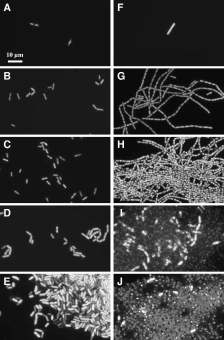
Cultures grown in SESOM and SESOM+ contained an abundance of clumps observable as flocs by visual inspection. In contrast, no such flocs could be observed in LB-grown cultures. Epifluorescence microscopy of Live/Dead BacLight-stained samples revealed that B. cereus switches from a single-cell to a multicellularity phenotype in SESOM+ (Fig. 1B and G), similar to the rhizoidal phenotype of B. mycoides. This was in stark contrast to the largely single-celled state in LB broth. At the onset of the stationary phase, these multicellular filaments coalesced to form large clusters (Fig. 1H). Clusters were not an artifact of centrifugation as clumps could be observed in SESOM by eye and by microscopy before centrifuging confirmed the presence of clusters, albeit at a low density. Sporulation was observed in all cluster-associated cells, and most spores remained associated with the clusters. Cells and their spores appeared to be encased in an extracellular matrix, as vigorous mixing did not lead to the dissociation of clumps.
FIG. 1.
Morphology of B. cereus growing in LB broth (A to E) or SESOM+ (F to J) at 30°C with shaking at 150 rpm for 4 h (A and F), 6 h (B and G), 10 h (C and H), 24 h (D and I), and 48 h (E and J). Culture samples were stained with Live/Dead BacLight stain and visualized by epifluorescence microscopy.
Multicellularity of various soil-isolated Bacillus species.
In order to investigate whether the observed multicellularity phenotype was a widely occurring phenomenon or unique to B. cereus ATCC 14579, we isolated several Bacillus organisms from local soils. Their 16S rRNA sequences (1,368 bp) placed isolates 9 and 20 in the B. cereus sensu lato assemblage and isolates 12 and 17 in the B. subtilis group. Isolate 9, closely related to B. mycoides, formed the characteristic rhizoidal counterclockwise outgrowths from points of inoculation on both LB and SESOM agar. All four isolates grew well in SESOM, forming large clumps (Fig. 2). Attempts at characterizing their growth by both optical density and culturable counting failed due to the large clumps that formed early during incubation. These clumps could not be dissociated to yield free cells by vigorous vortexing. Isolate 9 formed the largest clumps, with a high density of clumps observed by visual inspection although the culturable count at 24 h was only 8.4 × 10⁴ ± 1.2 × 10⁴ CFU/ml. B. subtilis subsp. subtilis Marburg also displayed clumping when cultured in SESOM (Fig. 2E), while its widely used derivative, B. subtilis 168, did not display this trait (results not shown).
FIG. 2.
Morphology of soil Bacillus isolates 9 (A), 20 (B), 12 (C), and 17 (D) and B. subtilis subsp. subtilis Marburg (E) cultured in SESOM+ for 24 h at 30°C with shaking at 150 rpm. Culture samples were stained with Live/Dead BacLight stain and visualized by epifluorescence microscopy.
Growth and sporulation in artificial soil microcosms.
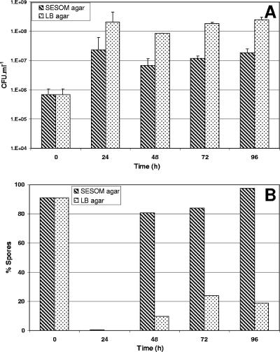
B. cereus was able to germinate in ASM surrounded by both SESOM and LB agar, as supported by both an increase of greater than 10-fold in the culturable count after 24 h (Fig. 3A) and the observation that not all cells occurred as spores (Fig. 3B). The minority of cells appeared to sporulate in the ASM surrounded by LB agar, even after 96 h. In contrast 97.6% of cells in microcosms surrounded by SESOM sporulated.
FIG. 3.
Culturable counts (A) and percentage of spores (B) of B. cereus in SESOM+-saturated artificial soil microcosms located in wells in SESOM+ or LB agar and incubated for 96 h at 30°C. Data are averages of three separate experiments, and error bars indicate standard errors of the means.
Translocation through soil microcosms.
B. cereus point inoculated as both spores and vegetative cells into the centers of SESOM+ and LB-saturated ASM inserted in both SESOM+ and LB agar grew out onto the surrounding agar (Fig. 4A and B). Outgrowth onto the agar surface was observed after 5 or more days, with a higher nutrient environment favoring more rapid outgrowth. So B. cereus appeared as visible outgrowth from both SESOM and LB-saturated ASM onto LB agar within 5 days and from LB-saturated ASM onto SESOM agar within 9 days. The 4-mm distance was therefore covered at overall translocation speeds of 33.3 and 18.5 μm/h, respectively. All four Bacillus soil isolates were able to translocate from their points of inoculation toward the surrounding agar surface, as indicated by outgrowth within 3 days (isolates 12, 17, and 20) and 4 days (isolate 9). B. subtilis subsp. subtilis Marburg traversed the distance of 4 mm in 5 days. Bromophenol blue diffused through the ASM matrix while the negatively charged Coomassie R250 did not, indicating that translocation of the negatively charged cells was unlikely due to convective forces.
FIG. 4.
Translocation of B. cereus and other soil Bacillus species through artificial soil microcosms by elongation and division of cells in chains. B. cereus point inoculated at the center of a soil microcosm (A) was able to translocate through the soil matrix containing a 1 mM concentration of the motility inhibitor l-ethionine, as demonstrated by outgrowths onto the surrounding agar surface (B). The bundles of chains observed in ASM at 72 h of incubation (C) were reminiscent of the multicellularity as induced in liquid SESOM after 10 h of incubation with shaking at 30°C (D). Samples were stained with Live/Dead BacLight stain and viewed by CLSM.
CSLM of BacLight-stained ASM revealed bundles of parallel chains inside the soil (Fig. 4C), reminiscent of the multicellular structures formed during growth in liquid SESOM (Fig. 4D). The bundles of chains spanned long distances, supporting the notion of translocation through the matrix by extension of multicellular filaments through growth and cell division. Surprisingly, B. cereus also translocated through ASM saturated with LB broth, and fluorescence microscopy revealed bundles of multicellular filaments (not shown).
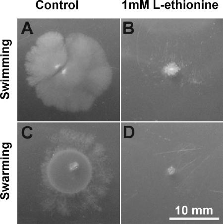
l-Ethionine at a 1 mM concentration inhibited both swimming (0.35% agar) and swarming (1.0% agar) with both SESOM (Fig. 5) and LB (results not shown) as nutrient sources. CaCl2 (10 mM) also inhibited both swimming and swarming in an LB background. B. cereus spores point inoculated into ASM or LB agar (both supplemented with 1 nM l-ethionine) grew out on the surrounding agar after the same period as ASM without the inhibitor. Bundles of chains like those observed in ASM without l-ethionine (Fig. 4C) could be viewed by CSLM. As the ASM were also saturated with Ca2+, motility was inhibited by two separate mechanisms, so that translocation through the artificial soil matrix was not likely to be a flagellum-mediated process. Rather, translocation appears to be a mechanism driven by elongation and division of cells in chains, leading to movement of the filaments between gaps in the soil matrix.
FIG. 5.
Swimming (A and B) and swarming (C and D) motility of B. cereus ATCC 14579 in SESOM swim and swarm agar without (A and C) and with (B and D) a 1 mM concentration of the motility inhibitor l-ethionine, viewed after incubation at 30°C for 72 h.
DISCUSSION
The role played by bacteria in soils and sediments is undisputed and yet poorly understood. Most predictions on their survival, growth, and activity are inferred from work performed under standard laboratory conditions. Soils and sediments, home to a large proportion of the prokaryotes on earth, are mixtures of sand, silt, and clay of various mineral compositions and particle sizes (33). Furthermore, they contain various concentrations of insoluble and soluble organic moieties or SOM (17). While these compounds may be quite different than the familiar glucose, tryptone, and yeast extract, they do contain components that support the growth of B. cereus (Table 1). Moreover B. cereus growing on SESOM switched to a multicellularity phenotype, in both liquid extract and ASM.
Growth of B. cereus in liquid soil extract and in soil.
When exposed to soil organic matter, B. cereus behaved as a saprophyte, apparently sensing available nutrients by germinating, growing to stationary phase, and subsequently sporulating. The data support the older views on the ecology of B. cereus (13, 35, 37), refuting the current paradigm where members of this group occur in soils only as spores. The absence of a lag phase in liquid culture indicated that early stationary phase vegetative populations did not require a period of adaptation to the energy and carbon source in SESOM and, therefore, that the species is well adapted to growth on soil organic carbon. This begs the question whether the insect and mammalian pathogens B. thuringiensis and B. anthracis are also able to grow in soils offering sufficient levels of amenable carbon and energy sources, multiplying outside their hosts. Ellis (7) recently reported growth of B. thuringiensis BT27a in an artificial soil with humic acid as the sole carbon and energy source. B. anthracis was found up to 5 m away from carcasses of bison that had died of anthrax (6). The authors ascribed this apparent spread to the activity of scavengers. This reported dissemination of B. anthracis could be due at least in part to translocation by growth. Based on long-term observations, previous investigators have proposed that B. anthracis does propagate in soils above pH 6.5, from where it may infect livestock (35).
A possible role for the multicellularity phenotype.
Bacteria have long been viewed as largely single-celled organisms, and multicellularity was recognized as a trait of selected groups such as the Actinomycetes and Myxobacteria (31). Coordinated multicellular behavior of bacteria has been reported with increasing frequency for a range of bacteria and is now an accepted bacterial trait (31). Bacterial biofilms have widely been defined as multicellular communities occurring at surfaces or interfaces (2, 25). The multicellular structures formed by B. cereus in liquid SESOM assembled in the absence of a distinct interface and cannot, therefore, be viewed as true biofilms. Rather, the multicellularity phenotype was reminiscent of the related B. mycoides growing in liquid medium (4). This filamentous or chain mode of growth is ascribed to incomplete cell separation after septation and appears as a constitutive trait in B. mycoides, leading to elaborate chiral colony patterns on agar (4). While constitutive in B. mycoides, the multicellularity phenotype is induced in B. cereus when it grows in liquid SESOM and in ASM. Multicellularity was also observed in ASM saturated with LB. This indicated that the switch to multicellularity was not due to SESOM per se, as it could also be elicited by either a chemical constituent associated with the Ca-montmorillonite or sand used or by association with the surfaces. LB broth supplemented with 10 and 100 mM CaCl2 yielded chains and some small clumps, indicating that calcium is one contributing factor in controlling the switch to multicellularity. B. cereus is able to grow as multicellular structures in the guts of some arthropods (21). Chains of bacteria growing in association with the wall of insect guts were initially described by Joseph Leidy (18) and termed Arthromitus, but they were later shown to be B. cereus organisms(8, 21). Wild strains of B. subtilis growing at the air-liquid interface form long chains of cells that associate to form biofilms, with apical cells sporulating (2). Collectively, these findings indicate a conserved ability among the Bacillus to switch to grow as chains, clumps, or biofilms under specific conditions. The chemical cues and genes involved in the switch to multicellularity by B. cereus are unknown at present.
Bacteria exhibit various modes of locomotion across interfaces, including swarming, twitching motility, swimming, gliding, darting, sliding (12), and macrofiber supercoiling (22). B. cereus is able to translocate by swimming through liquid and over agar surfaces due to a surface-induced differentiation to form hyperflagellated swarm cells (30). B. anthracis is also able to translocate across a surface by sliding, a surface translocation produced by the expansive forces in a growing culture in combination with surface properties which result in reduced friction between cell and surface (12). Both swimming and swarming of B. cereus were effectively inhibited by Ca2+ and by l-ethionine, which was shown previously to inhibit methyl group turnover on methyl-accepting chemotaxis proteins in B. sphaericus (1). Yet translocation was not inhibited in the presence of l-ethionine, excluding flagellar rotation as the effector of translocation through soil. This conclusion is strengthened by the additional suppression of motility and swarming exerted by the Ca2+ occurring in the ASM. Furthermore, the observed translocation of B. cereus and the other Bacillus soil isolates through the soil matrix appears distinct from movement on surfaces by macrofibers of B. subtilis. Macrofibers are formed by B. subtilis 168 mutants that do not separate after septation, forming filaments that self-assemble to form twisted fibers and that display a dragging movement across a solid surface (22). While macrofibers studied on solid surfaces are translocated by forces associated with the supercoiling of multiple filaments, the translocation of Bacillus through soil is more likely due to the forces inherent to cell elongation. This may entail a simplistic mode of bacterial locomotion, termed sliding by Henrichsen (12), that is driven by the elongation of cells that remain linked at their poles while constrained within the particulate soil environment.
The results presented here show that B. cereus can germinate, grow, and sporulate in extractable soil organic matter, thus displaying a saprophytic life cycle. Upon commencement of exponential growth in SESOM, cells switched to a multicellularity phenotype, growing to form filaments and then clumps. B. cereus and various soil-isolated Bacillus species are able to translocate from their point of inoculation in soil to a distal point. The switch to multicellular growth may play an important role in the ability of growing populations of Bacillus to translocate through soil by sliding. Collectively, these data support the older school of thought, i.e., that B. cereus is a saprophytic bacterium that is able to grow in soil and furthermore is adapted to translocate through soil.
.
Acknowledgments
We thank B. Bleakley and T. Schumacher for helpful discussions and S. Clay for critically reading the manuscript.
This work was supported by the South Dakota Agricultural Experiment Station, National Science Foundation/EPSCoR grant EPS-0091948, and by the State of South Dakota.
Footnotes
‡
Journal series publication 3509 from the South Dakota Agricultural Experiment Station.
REFERENCES
- 1.Andreev, J., P. A. Dibrov, D. Klein, and S. Braun. 1994. Chemotaxis, sporulation, and larvicide production in Bacillus sphaericus 2362. The influence of l-ethionine, and of aminophenylboronic acid. FEBS Lett. 349**:**416-419. [DOI] [PubMed] [Google Scholar]
- 2.Branda, S. S., J. E. González-Pastor, S. Ben-Yehuda, R. Losick, and R. Kolter. 2001. Fruiting body formation in Bacillus subtilis. Proc. Natl. Acad. Sci. USA 98**:**11621-11626. [DOI] [PMC free article] [PubMed] [Google Scholar]
- 3.Chattopadhyay, A., N. B. Bhatnagar, and R. Bhatnagar. 2004. Bacterial insecticidal toxins. Crit. Rev. Microbiol. 30**:**33-54. [DOI] [PubMed] [Google Scholar]
- 4.Di Franco, C., E. Beccari, T. Santini, G. Pisaneschi, and G. Tecce. 2002. Colony shape as a genetic trait in the pattern-forming Bacillus mycoides. BMC Microbiol. 2**:**33-47. [DOI] [PMC free article] [PubMed] [Google Scholar]
- 5.Dragon, D. C., and R. P. Rennie. 1995. The ecology of anthrax spores: tough but not invincible. Can. Vet. J. 36**:**295-301. [PMC free article] [PubMed] [Google Scholar]
- 6.Dragon, D. C., D. E. Bader, J. Mitchell, and N. Woollen. 2005. Natural dissemination of Bacillus anthracis spores in northern Canada. Appl. Environ. Microbiol. 71**:**1610-1615. [DOI] [PMC free article] [PubMed] [Google Scholar]
- 7.Ellis, R. J. 2004. Artificial soil microcosms: a tool for studying microbial autecology under controlled conditions. J. Microbiol. Methods 56**:**287-290. [DOI] [PubMed] [Google Scholar]
- 8.Feinberg, L., J. Jorgensen, A. Haselton, A. Pitt, R. Rudner, and L. Margulis. 1999. Arthromitus (Bacillus cereus) symbionts in the cockroach Blaberus giganteus: dietary influences on bacterial development and population density. Symbiosis 27**:**109-123. [PubMed] [Google Scholar]
- 9.Garbeva, P., J. A. Van Veen, and J. D. Van Elsas. 2003. Predominant Bacillus spp. in agricultural soil under different management regimes detected via PCR-DGGE. Microb. Ecol. 45**:**302-316. [DOI] [PubMed] [Google Scholar]
- 10.Halverson, L. J., M. K. Clayton, and J. Handelsman. 1993. Population biology of Bacillus cereus UW85 in the rhizosphere of field-grown soybeans. Soil Biol. Biochem. 25**:**485-493. [Google Scholar]
- 11.Helgason, E., N. J. Tourasse, R. Meisal, D. A. Caugant, and A. B. Kolstø. 2004. Multilocus sequence typing scheme for bacteria of the Bacillus cereus group. Appl. Environ. Microbiol. 70**:**191-201. [DOI] [PMC free article] [PubMed] [Google Scholar]
- 12.Henrichsen, J. 1972. Bacterial surface translocation: a survey and a classification. Bacteriol. Rev. 36**:**478-503. [DOI] [PMC free article] [PubMed] [Google Scholar]
- 13.Henrici, A. T. 1934. The biology of bacteria: an introduction to general microbiology. D.C. Heath and Company, Boston, Mass.
- 14.Hönerlage, W., D. Hahn, and J. Zeyer, J. 1995. Detection of mRNA of nprM in Bacillus megaterium ATCC 14581 grown in soil by whole-cell hybridization. Arch. Microbiol. 163**:**235-241. [Google Scholar]
- 15.Ivanova, N., A. Sorokin, I. Anderson, N. Galleron, B. Candelon, V. Kapatral, A. Bhattacharyya, G. Reznik, N. Mikhailova, A. Lapidus, L. Chu, M. Mazur, E. Goltsman, N. Larsen, M. D'Souza, T. Walunas, Y. Grechkin, G. Pusch, R. Haselkorn, M. Fonstein, S. D. Ehrlich, R. Overbeek, and N. Kyrpides. 2003. Genome sequence of Bacillus cereus and comparative analysis with Bacillus anthracis. Nature 423**:**87-91. [DOI] [PubMed] [Google Scholar]
- 16.Jensen, G. B., B. M. Hansen, J. Eilenberg, and J. Mahillon. 2003. The hidden lifestyles of Bacillus cereus and relatives. Environ. Microbiol. 5**:**631-640. [DOI] [PubMed] [Google Scholar]
- 17.Kalbitz, K., S. Solinger, J.-H. Park, B. Michalzik, and E. Matzner. 2000. Controls on the dynamics of dissolved organic matter in soils: a review. Soil Sci. 165**:**277-304. [Google Scholar]
- 18.Leidy, J. 1849. On the existence of entophyta in healthy animals as a natural condition. Proc. Natl. Acad. Sci. USA 4**:**225-233. [Google Scholar]
- 19.Lindsay, D., and A. von Holy. 1999. Different responses of planktonic and attached Bacillus subtilis and Pseudomonas fluorescens to sanitizer treatment. J. Food Prot. 62**:**68-379. [DOI] [PubMed] [Google Scholar]
- 20.Luo, Y., S. Vilain, and V. S. Brözel. 2005. Bacillus cereus grows in soil as mycoidal multicellular structures, abstr. N-223. Abstr. 105th Gen. Meet. Am. Soc. Microbiol., 2005. American Society for Microbiology, Washington, D.C.
- 21.Margulis, L., J. Z. Jorgensen, S. Dolan, R. Kolchinsky, F. A. Rainey, and S.-C. Lo. 1998. The Arthromitus stage of Bacillus cereus: intestinal symbionts of animals. Proc. Natl. Acad. Sci. USA 95**:**1236-1241. [DOI] [PMC free article] [PubMed] [Google Scholar]
- 22.Mendelson, N. H., L. Chen, and J. J. Thwaites. 2004. A new form of bacterial movement, dragging of multicellular aggregate structures over solid surfaces, is powered by macrofiber supercoiling. Res. Microbiol. 155**:**113-127. [DOI] [PubMed] [Google Scholar]
- 23.Milner, J. L., L. Silo-Suh, J. C. Lee, H. He., J. Clardy, and J. Handelsman. 1996. Production of kanosamine by Bacillus cereus UW85. Appl. Environ. Microbiol. 62**:**3061-3065. [DOI] [PMC free article] [PubMed] [Google Scholar]
- 24.Mishustin, E. N. (ed.) 1972. Microflora of soils in the northern and central USSR. Israel Program for Scientific Translations, Jerusalem, Israel.
- 25.O'Toole, G. A., H. B. Kaplan, and R. Kolter. 2000. Biofilm formation as microbial development. Annu. Rev. Microbiol. 54**:**49-79. [DOI] [PubMed] [Google Scholar]
- 26.Priest, F. G., M. Barker, L. W. Baillie, E. C. Holmes, and M. C. Maiden. 2004. Population structure and evolution of the Bacillus cereus group. J. Bacteriol. 186**:**7959-7970. [DOI] [PMC free article] [PubMed] [Google Scholar]
- 27.Saleh, S. M., R. F. Harris, and O. N. Allen. 1970. Fate of Bacillus thuringiensis in soil: effect of soil pH and organic amendment. Can. J. Microbiol. 16**:**677-680. [DOI] [PubMed] [Google Scholar]
- 28.Sambrook, J., and D. Russell. 2001. Molecular cloning: a laboratory manual, 3rd ed. Cold Spring Harbor Laboratory Press, Cold Spring Harbor, N.Y.
- 29.Schoeni, J. L., and A. C. Wong. 2005. Bacillus cereus food poisoning and its toxins. J. Food Prot. 68**:**636-648. [DOI] [PubMed] [Google Scholar]
- 30.Senesi, S., F. Celandroni, S. Salvetti, D. J. Beecher, A. C. L. Wong, and E. Ghelardi. 2002. Swarming motility in Bacillus cereus and characterization of a fliY mutant in swarm cell differentiation. Microbiology 148**:**1785-1794. [DOI] [PubMed] [Google Scholar]
- 31.Shapiro, J. A. 1998. Thinking about bacterial populations as multicellular organisms. Annu. Rev. Microbiol. 52**:**81-104. [DOI] [PubMed] [Google Scholar]
- 32.Stabb, E. V., L. Jacobson, and J. Handelsman. 1994. Zwittermicin A-producing strains of Bacillus cereus from diverse soils. Appl. Environ. Microbiol. 60**:**4404-4412. [DOI] [PMC free article] [PubMed] [Google Scholar]
- 33.Stotzky, G. 1986. Influence of soil mineral colloids on metabolic processes, growth, adhesion, and ecology of microbes and viruses, p. 305-428. In P. M. Huang and M. Schnitzner (ed.), Interactions of soil minerals with natural organics and microbes. Special publication 17. Soil Science Society of America, Madison, Wis.
- 34.Stotzky, G., and R. G. Burns. 1982. The soil environment: clay-humus-microbe interactions, p. 105-133. In R. G. Burns and J H. Slater (ed.), Experimental microbial ecology. Blackwell Scientific Publication, Ltd., Oxford, England.
- 35.Van Ness, G. B. 1971. Ecology of anthrax. Science 172**:**1303-1307. [DOI] [PubMed] [Google Scholar]
- 36.Von Stetten, F., R. Mayr, and S. Scherer. 1999. Climatic influence on mesophilic Bacillus cereus and psychrotolerant Bacillus weihenstephanensis populations in tropical, temperate and alpine soil. Environ. Microbiol. 1**:**503-515. [DOI] [PubMed] [Google Scholar]
- 37.Waksman, S. A. 1932. Principles of soil microbiology, 2nd ed. The Williams and Wilkins Co., Baltimore, Md.
- 38.Winogradsky, S. 1924. Microbiologie du sol. Sur l'étude de l'anaerobiose dans la terre. C. R. Acad. Sci. 1924**:**178-186. [Google Scholar]


