Modulation of the Activity of Secretory Phospholipase A2 by Antimicrobial Peptides (original) (raw)
Abstract
The antimicrobial peptides magainin 2, indolicidin, and temporins B and L were found to modulate the hydrolytic activity of secretory phospholipase A2 (sPLA2) from bee venom and in human lacrimal fluid. More specifically, hydrolysis of phosphatidylcholine (PC) liposomes by bee venom sPLA2 at 10 μM Ca2+ was attenuated by these peptides while augmented product formation was observed in the presence of 5 mM Ca2+. The activity of sPLA2 towards anionic liposomes was significantly enhanced by the antimicrobial peptides at low [Ca2+] and was further enhanced in the presence of 5 mM Ca2+. Similarly, with 5 mM Ca2+ the hydrolysis of anionic liposomes was enhanced significantly by human lacrimal fluid sPLA2, while that of PC liposomes was attenuated. These results indicate that concerted action of antimicrobial peptides and sPLA2 could improve the efficiency of the innate response to infections. Interestingly, inclusion of a cationic gemini surfactant in the vesicles showed an essentially similar pattern on sPLA2 activity, suggesting that the modulation of the enzyme activity by the antimicrobial peptides may involve also charge properties of the substrate surface.
During the past 2 decades, living organisms of all types have been found to produce a large repertoire of gene-encoded antimicrobial peptides that play an important role in innate immunity to microbial invasion. Antimicrobial peptides can be synthesized at low metabolic cost and easily stored in large amounts and are readily available shortly after an infection to rapidly neutralize a broad range of microbes. Magainins were discovered in the skin of the African clawed frog Xenopus laevis and show a broad spectrum of antimicrobial (40, 77, 78) and anticancer activities (4, 13) at nonhemolytic concentrations. In a manner similar to that of magainins, indolicidin is active against both gram-negative and -positive bacteria (60) and fungi (1), as well as protozoa (2). Indolicidin is cytotoxic also to rat and human T lymphocytes (57), lyses red blood cells (1), and has activity against human immunodeficiency virus type 1 (55). Indolicidin is found in cytoplasmic granules of bovine neutrophils (60) and has a unique amino acid composition with five Trp and three Pro residues in its 13-amino acid sequence. With a size similar to that of indolicidin, temporins are linear 10- to 13-residue-long peptides isolated from the skin of the european red frog, Rana temporaria (62). Temporin B has been found to be active against gram-positive bacteria and is not hemolytic (62). In contrast, temporin L is active against both gram-positive and-negative bacteria, is hemolytic, and is toxic to cancer cells (54).
Antimicrobial peptides are considered to kill bacteria by permeabilizing and/or disrupting their membranes (39, 71, 83). The molecular bases for the activity and selectivity of these peptides have been intensively studied using model membranes (39, 70). Being cationic, antimicrobial peptides interact preferentially with acidic lipids that are particularly abundant in bacteria (16, 17, 51), thus providing a basis for differences in cell specificity. The interaction of antimicrobial peptides with bilayers alters the organization of the membrane and makes them permeable to ions, for instance (32, 69), causing membrane depolarization (42). Various models accounting for the peptide-induced membrane permeation process have been proposed (7, 61). It has been reported that not only the nature of the peptide but also the characteristics of the cell membrane as well as the metabolic state of the target cells determine the mechanism of action of antimicrobial peptides (37). However, the action of cationic antimicrobial peptides is not limited to direct killing of microorganisms. Accordingly, they exhibit an impressive variety of additional activities, having an impact particularly on the quality and effectiveness of innate responses and inflammation (25). Stimulation of host defense mechanisms by antimicrobial peptides has been demonstrated (3, 74), and receptor-mediated signaling by some peptides have also been reported (26, 75). Some of the antimicrobial peptides could act synergistically with other host antimicrobial molecules to kill microbes (11), and positive cooperativity has been reported between peptides and lysozyme, as well as between different antimicrobial peptides (25, 43). It is also becoming clear that there exists a certain degree of coupling between the innate and adaptive immune systems and that antimicrobial peptides influence both the quality and effectiveness of immune and inflammatory responses.
Phospholipases A2 (PLA2s) are ubiquitous enzymes, which catalyze the hydrolysis of the sn-2 ester bond of phospholipids to release free fatty acids and lysophospholipids (for a review, see reference 15). PLA2s play a key role in various biological processes, including homeostasis of cellular membranes, lipid digestion, host defense, signal transduction, and production of lipid mediators such as eicosanoids and lysophospholipid derivatives, which exhibit diverse and potent biological actions (15, 44). Interestingly, it has been shown that secretory PLA2 (sPLA2) has antibacterial activity and represents an important host defense molecule (9, 50, 53). Mammalian group IIA sPLA2s have been found at high levels in inflammatory conditions and have the ability to kill gram-positive bacteria (9, 24). High concentrations of these enzymes showed some bactericidal effect also against gram-negative bacteria. Likewise, they are able to act as acute-phase proteins, in concert with other antibacterial proteins (9). Belonging to group IIA sPLA2s, the lacrimal fluid sPLA2 has been identified as the principal mediator of antistaphylococcal activity (53). Similarly to group II PLA2, mammalian group V PLA2 has been found to be bactericidal against gram-positive bacteria (24), suggesting potential as therapeutic agents against bacterial infections. The mechanisms of regulation of PLA2 activity have been subjected to intense research, and the control of phospholipid derivatives produced by its action has long been considered in the treatment of related diseases (18, 76). The discovery of specific inhibitory peptides has given some insight into the regulation of the activity of intracellular PLA2s (68).
Mellitin, a 26-amino-acid peptide present in bee venom, has been previously found to enhance the activity of sPLA2s (11). Yet, mellitin is also profoundly hemolytic (7, 11), which would eventually limit its possible application as an antimicrobial agent. In order to study if the activation of sPLA2 is a more general property of antimicrobial peptides and if the hemolytic activity is independent from this effect, we compared the influence of magainin 2, temporins B and L, and indolicidin on the activity of sPLA2s of bee venom and human lacrimal fluid. The two former peptides are nonhemolytic in contrast to indolicidin and temporin L (1, 54, 62, 77, 78). Our results reveal significant impact of these peptides on the hydrolytic activity of sPLA2.
MATERIALS AND METHODS
Materials.
Bee venom PLA2 was purchased from Sigma Chemical Co. (St. Louis, Mo.). 1-Palmitoyl-2-oleoyl-_sn_-glycero-3-phosphoglycerol (POPG) was from Avanti Polar Lipids (Alabaster, Ala.), and the fluorescent phospholipid analogs 1-palmitoyl-2-[6-(pyren-1-yl)]hexanoyl-sn_-glycero-3-phosphocholine (PPHPC) and 1-palmitoyl-2-[6-(pyren-1-yl)]hexanoyl-sn_-glycero-3-phosphomonomethylester (PPHPM) from K&V Bioware (Espoo, Finland). Concentrations of lipids were determined gravimetrically with a high-precision electrobalance (Cahn, Cerritos, Calif.), and those of the fluorescent lipids were determined spectrophotometrically by using 42,000 cm−1 at 342 nm as the molar extinction coefficient for pyrene. The gemini surfactant (2_S,3_R)-2,3-dimethoxy-1,4-bis(_N_-hexadecyl-N,_N_-dimethylammonnium) butane dibromide (SR-1) was synthesized as described previously (12), and its purity was verified by nuclear magnetic resonance. This compound was kindly provided by S. Borocci and G. Mancini (University of Rome, Rome, Italy). The purity of other lipids was checked by thin-layer chromatography on silicic acid-coated plates (Merck, Darmstadt, Germany) developed with chloroform/methanol/water (65:25:4 [vol/vol/vol]). Examination of the plates after iodine staining and, when appropriate, upon UV illumination revealed no impurities. Magainin 2 was from Sigma, indolicidin from Bachem (Bubendorf, Switzerland), temporin L from Synpep (Dublin, Calif.), and temporin B from Tana Laboratories (Houston, Tex.). The purities of the above synthetic peptides (>99, >95, >94, and >90% for magainin 2, indolicidin, and temporins L and B, respectively) were assessed by high-performance liquid chromatography, and their sequences were verified by both automated Edman degradation and mass spectrometry. Peptide concentrations were determined gravimetrically and by quantitative ion-exchange column chromatography and ninhydrin derivatization.
Collection of lacrimal fluid.
Tears were collected from healthy donors briefly exposed to the vapors of freshly minced onions. The collected fluid was stored at −20°C until used.
Assay for PLA2.
PLA2 activity was determined by a kinetic assay described previously (45, 46, 65-67). Appropriate amounts of the lipid stock solutions were mixed in chloroform to obtain the desired compositions and then dried under a stream of nitrogen followed by high vacuum for a minimum of 2 h. The lipid residues were subsequently dissolved in ethanol to yield a lipid concentration of 50 μM, and small unilamellar liposomes were formed by rapidly injecting this lipid ethanolic solution into the buffer (5). The lipid concentration was 1.25 μM for both PPHPC and PPHPM/POPG (5:5 [molar ratio]) liposomes in a total volume of 2 ml. All fluorescence measurements were performed in magnetically stirred quartz cuvettes (with 1-cm path length) at 37°C. After 15 min of equilibration the reactions were initiated by adding 50 ng of bee venom sPLA2 or 0.5 μl of human lacrimal fluid. The progress of phospholipid hydrolysis was followed by measuring the pyrene monomer intensity at 400 nm as a function of time. Fluorescence intensities were measured with a Perkin-Elmer LS 50B spectrometer with an excitation wavelength of 344 nm and both emission and excitation band-passes set at 4 nm. The assay was calibrated by adding known picomolar aliquots of (pyren-1-yl)hexanoate into the reaction mixture in the absence of enzyme while detecting pyrene monomer emission intensity. The activity of the enzyme was calculated from the initial velocity of the reaction kinetic curves and converted to the amount of product released per time unit. The assays were carried out both with and without added 5 mM Ca2+. Under the former conditions approximately 10 μM residual Ca2+ was present, as determined by titration with EDTA.
RESULTS
Effects of antimicrobial peptides on the activity of bee venom sPLA_2._
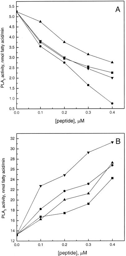
Increasing the concentrations of the antimicrobial peptides magainin 2, indolicidin, and temporins B and L progressively attenuated the hydrolysis of zwitterionic PPHPC liposomes by this enzyme at a low [Ca2+] (Fig. 1A). Indolicidin was most effective, and the hydrolytic activity of the enzyme decreased by approximately 86% at 0.4 μM peptide. Inhibition of the activity of sPLA2 by the antimicrobial peptides decreased in the sequence indolicidin > temporin L ≈ magainin 2 > temporin B. Interestingly, in the presence of 5 mM Ca2+, the above peptides had the opposite effect and the hydrolysis of PPHPC liposomes was enhanced (Fig. 1B), the activation being augmented with increasing concentrations of the peptides. The efficiency of equimolar concentrations of these antimicrobial peptides to activate bee venom sPLA2s increased in the sequence magainin < temporin B < indolicidin < temporin L.
FIG. 1.
Effects of magainin 2 (▪), indolicidin (•), and temporins L (▴) and B (▾) on the hydrolysis of PPHPC liposomes by bee venom sPLA2 without added Ca2+ (A) and in the presence of 5 mM Ca2+ (B). Final lipid concentration is 1.25 μM in 2 ml of 50 mM Tris-HCl, 150 mM NaCl, pH 7.4. The reactions were started by adding 50 ng of PLA2 into the cuvette, while temperature was maintained at 37°C with a circulating water bath. Each data point represents the mean of triplicate measurements. The standard deviation is less than 1 nmol of fatty acid/min and for the sake of clarity is not shown.
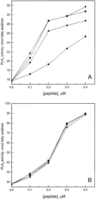
The outer leaflet of mammalian plasma membranes is exclusively composed of zwitterionic phospholipids, whereas bacterial membranes contain large amounts of negatively charged phospholipids (16, 17, 51). Accordingly, we studied the effects of these peptides on the hydrolysis of anionic liposomes by bee venom sPLA2. In behavior similar to that of other sPLA2s (66), this enzyme has been shown to hydrolyze the anionic liposomes more rapidly than neutral vesicles, with Ca2+ activating the enzyme (Fig. 1 and 2; see references 8 and 72). At 10 μM Ca2+, all four peptides activated the enzyme, this effect being augmented in the sequence indolicidin < temporin B < magainin 2 ≈ temporin L (Fig. 2A). These antimicrobial peptides activated bee venom sPLA2 also in the presence of 5 mM Ca2+ with essentially no differences between the four peptides (Fig. 2B). Compared to that of zwitterionic vesicles, the activation by the antimicrobial peptides of sPLA2 action on anionic liposomes with 5 mM Ca2+ was more pronounced (Fig. 1B and 2B), being maximally ≈ 400 and 136%, respectively.
FIG. 2.
Effects of magainin 2 (▪), indolicidin (•), and temporins L (▴) and B (▾) on the hydrolysis of PPHPM/POPG (50:50, molar ratio) liposomes by bee venom sPLA2 without added Ca2+ (A) and in the presence of 5 mM Ca2+ (B). Otherwise, conditions were as described in the legend for Fig. 1.
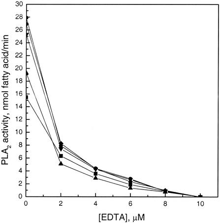
In order to exclude the possibility that trace amounts of Ca2+ may be present in the peptides used, which could enhance PLA2 activity, we measured enzyme activity in the presence of increasing concentrations of EDTA. Both with and without peptides, EDTA abolished sPLA2 activity at 10 μM (Fig. 3), thus suggesting the antimicrobial peptides to be essentially free of Ca2+.
FIG. 3.
Inhibition of EDTA on the hydrolysis of PPHPM/POPG (50:50 [molar ratio]) liposomes by bee venom sPLA2 without antimicrobial peptides (▪) and in the presence of 0.2 μM magainin 2 (•), indolicidin (▴), and temporins L (▾) and B (♦). Otherwise, conditions were as described in the legend for Fig. 1.
Effects of antimicrobial peptides on the activity of sPLA2 from human tears.
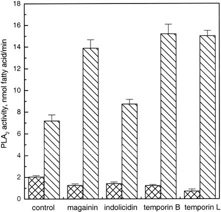
Human sPLA2 is present in many tissues and secretions, including rheumatoid synovial fluid (59), platelets (33), Paneth cells (49), neutrophils (56), and lacrimal glands (48). High concentrations of group II sPLA2 are present in human lacrimal fluid, an important host defense mechanism in eyes that kills gram-positive bacteria (53). This sPLA2 requires Ca2+ as an essential active site cofactor and, in keeping with the catalytic activity of this enzyme being important to its antimicrobial activity, also the latter is Ca2+ dependent (53). As reported previously (6) human lacrimal fluid sPLA2 has a marked preference for anionic phospholipids, hydrolyzing anionic vesicles more rapidly than neutral vesicles (Fig. 3). The hydrolysis of neutral liposomes by lacrimal fluid sPLA2 was inhibited by all four antimicrobial peptides, with temporin L being most effective (Fig. 4). Similar to the activity of bee venom sPLA2, the activity of lacrimal fluid enzyme towards anionic liposomes was significantly enhanced by the antimicrobial peptides (Fig. 4). The enzyme activity was significantly increased by magainin 2 and temporins B and L, and there were essentially no differences between these three peptides, while indolicidin was least effective.
FIG. 4.
Effects of 0.2 μM magainin 2, indolicidin, and temporins L and B on the hydrolysis of 1.25 μM PPHPC (crosshatched) and PPHPM/POPG (50:50, molar ratio, hatched) liposomes by human lacrimal fluid sPLA2 in the presence of 5 mM Ca2+. The reactions were started by adding 0.5 μl of human lacrimal fluid into the cuvette. *, P < 0.05. Each data point represents mean plus or minus standard error of the mean (n = 5).
Effects of cationic gemini surfactant on the activity of sPLA2.
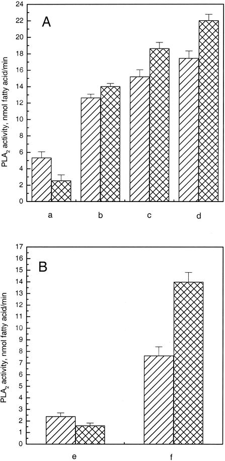
The above results readily showed that the positively charged antimicrobial peptides have pronounced effects on the activity of sPLA2. Interestingly, an essentially similar pattern was evident for the impact of other membrane-partitioning agents (45-47, 65) on sPLA2 activity. In order to elucidate if the positive charges of these cationic perturbants play a role in their effects on the activity of sPLA2, we included a small amount (5 mol%) of the cationic gemini surfactant SR-1 in the vesicles. Strikingly, this cationic surfactant showed qualitatively similar effects on sPLA2 activity (Fig. 5), suggesting that the positive charges of these perturbants play an important role in their modulation of sPLA2 activity.
FIG. 5.
Hydrolysis of 1.25 μM PPHPC and PPHPM/POPG (50:50, molar ratio) liposomes by bee venom (A) and human lacrimal fluid (B) sPLA2. The vesicle compositions were PPHPC with 10 μM Ca2+ (a), PPHPC with 5 mM Ca2+ (b and e), PPHPM/POPG with 10 μM Ca2+ (c), and PPHPM/POPG with 5 mM Ca2+ (d and f), in the presence (5 mol%, crosshatched) and absence (hatched) of the cationic gemini surfactant SR-1. *, P < 0.05. Each data point represents mean plus or minus standard error of the mean (n = 5).
DISCUSSION
sPLA2s constitute a large family of structurally related enzymes, which are found in numerous organisms, including mammalian tissues, venoms, and plants, and have been classified into 12 groups (I to XII) primarily based on sequence homology (14). Group III sPLA2 was first identified in bee venom (34) and is one of the most thoroughly studied of this group. This enzyme has been cloned (34), and its tertiary structure revealed an overall scaffold similar to that of group I and group II sPLA2 (58). Group IIA sPLA2s are secreted by a variety of cells involved in the inflammatory response. The presence of these enzymes in human macrophages, the Paneth cells of the intestine, and the lacrimal glands is significant because all these cells have an established antibacterial function (9). Human lacrimal fluid sPLA2 binds avidly to phosphatidylglycerol-rich membranes of bacteria, and quantities as low as 1 ng per ml are sufficient to kill gram-positive bacteria (53). The activity of this sPLA2 against gram-negative bacteria is considerably enhanced in the presence of agents that disrupt their lipopolysaccharide coat (73). It is apparent that, in order to hydrolyze the bacterial cell membrane, the enzyme must first penetrate the anionic peptidoglycan cell wall. This is related to the cell wall structures characterizing this bacterial species and is affected by factors such as growth state and cell wall-degrading enzymes and substrates. Accordingly, the sensitivity of gram-positive bacteria to sPLA2 varies greatly between species (19, 53). Agents disrupting the cell wall and/or facilitating the access of PLA2 to the membrane could be anticipated to promote the bactericidal effect of sPLA2.
Antimicrobial peptides show a broad range of activity against gram-positive and-negative bacteria, fungi, mycobacteria, and some enveloped viruses (79). With a net positive charge, antimicrobial peptides are characterized by their preference for anionic interfaces that confers relative specificity on the bacterial membranes as opposed to the zwitterionic membranes of host cells (35, 39, 41, 52). The same charge characteristics are inherent to human group IIA sPLA2s, which have a large number of cationic residues on their substrate binding surface (63). Our data demonstrate a pronounced influence of the antimicrobial peptides on the activities of sPLA2s, suggesting possible synergistic action of these two antibacterial components, irrespective of the hemolytic activity of some of the peptides. The effects of the four peptides on sPLA2 activity depend on lipid composition in a manner showing a bacterial membrane to be susceptible to enhanced hydrolysis, while the host cell membrane would remain intact. Our results thus highlight the potential of concerted action between sPLA2 and antimicrobial peptides as part of the innate response to infections, particularly when more antibiotic-resistant bacterial strains are emerging.
A variety of membrane perturbants have been shown to affect the adsorption and action of PLA2 (e.g., references 11, 29, and 45). In keeping with this, our present results demonstrate the profound impact of the four antimicrobial peptides studied on the sPLA2 reaction. In distinction from enzymes acting on monomeric soluble substrates, PLA2s have access to their substrate only within the membrane phase and are sensitive to the curvature and as well as physicochemical characteristics of the phospholipid membrane (10, 20, 21, 27, 36, 64). Also, “structural defects” in membranes have been proposed to affect PLA2 activity (31), with subtle changes in the structure of phospholipids having a profound influence on the affinity of the interface binding site of the enzyme to the substrate surface (30). Accordingly, defects in membranes caused by antimicrobial peptides could enhance the penetration of sPLA2 and the hydrolysis of vesicles would proceed at a much higher rate with augmented catalytic turnover of the bound enzyme. For kinetic analysis two-dimensional “scooting” of sPLA2 in the substrate surface has been suggested, with very slow exchange of PLA2 or phospholipid between the phospholipid vesicles (21, 22, 30). The antimicrobial peptides could modulate sPLA2s activity by substrate replenishment through peptide-mediated vesicle-vesicle contacts, similarly to mellitin (11). The effects of the four antimicrobial peptides on the structure of anionic membranes in particular are pronounced (38, 80, 81). As suggested for magainin 2 and indolicidin (80), the peptide would first bind to the outer leaflet of the bilayer and subsequently cosegregate in a cooperative manner with the bound acidic phospholipids into microdomains. Reorientation of peptides and formation of “channel”- or “pore”-like structures could occur, and the bilayer structure could become locally destabilized. Reorientation of temporin B (81) and two states of temporin L in bilayers (82) was also suggested. These peptides are likely to induce complex dynamic structures in the membranes with a variety of substrate configurations. Different effects of these peptides on membrane properties may thus influence sPLA2 activity to different degrees (Fig. 1 to 3).
Due to the neutral charge and high curvature of PPHPC small unilamellar liposomes, the four antimicrobial peptides studied can be expected to bind weakly to the zwitterionic membrane surface without inserting deeply into the hydrophobic core of the bilayers. At low [Ca2+], the coverage of the interface by the peptides would decrease the area available for the binding of sPLA2, resulting in inhibition due to a smaller fraction of the enzyme being bound to the interface. Repulsion between Ca2+ and membrane-bound, positively charged antimicrobial peptides could also affect the binding of sPLA2 to the membrane. However, in the presence of 5 mM Ca2+, the activity of bee venom sPLA2 towards PPHPC was enhanced by all four antimicrobial peptides. Ca2+ is essential for the activity of PLA2s (8), and activation of bee venom PLA2 by this ion could exceed the inhibition caused by the antimicrobial peptides with zwitterionic phosphatidylcholine (PC) as a substrate. Interestingly, hydrolysis of PC membrane by bee venom sPLA2 in the presence of 5 mM Ca2+ was augmented by the antimicrobial peptides, while under the same conditions the activity of lacrimal fluid sPLA2 was attenuated. Compared to that of the group III sPLA2s, the interface binding surface of group II sPLA2s contains a significantly larger number of basic amino acids and has a highly cationic character (23, 50). Accordingly, the effects of cationic antimicrobial peptides on the binding of these two enzymes to the substrate could differ, resulting in different effects on their impact on the catalytic activities of bee venom and lacrimal fluid sPLA2.
Interestingly, while magainin 2, indolicidin, and temporins B and L differ in structural parameters, they had qualitatively identical effects on the hydrolysis of zwitterionic and anionic vesicles by sPLA2 measured with and without added Ca2+. Intriguingly, comparison of zwitterionic and anionic vesicles as substrates for sPLA2 has revealed essentially similar pattern for the effects of adriamycin (45), phorbol esters (46), and polyamines (65) on the activity of PLA2. Modulation of sPLA2 activity via similar changes induced in the substrate by these peptides is thus suggested, implying a lack of a direct and specific effect on the enzyme. The enzyme action is thus likely to be affected via the physical properties of the substrate. Importantly, the cationic gemini surfactant SR-1 added to the vesicles also showed a similar pattern on sPLA2 activity (Fig. 5), indicating that modulation of sPLA2 reaction by the above cationic perturbants could involve alterations in the electric properties of the substrate surface. Subsequently, changes in the extent of the association of the enzyme to substrate could be involved, in keeping with the suggestion that the initial binding of the enzyme to the interface is limiting and that conditions favoring its surface association enhance the overall rate of hydrolysis (28). Possible variation in the bilayer curvature and presence of multilamellar vesicles would be unlikely to influence the comparison of the measured effects due to progressively increasing concentrations of the added antimicrobial peptides on the enzyme activity.
Due to the inherent complexity of the physicochemical characteristics of the dynamic phospholipid/water interface harboring the site of the catalytic action of PLA2, it is difficult to distinguish at this stage between the various mechanisms outlined above. Furthermore, there is no reason to assume the above possibilities to be mutually exclusive. More thorough studies are thus warranted to establish the molecular level mechanism(s) involved. Yet, taken the importance of the development of novel means to fight emerging antibiotic-resistant strains, such efforts are clearly needed.
Acknowledgments
This study was supported by the Technology Development Fund (TEKES) and the Finnish Academy. Memphys is supported by the Danish National Research Council.
We thank S. Borocci and G. Mancini (University of Rome) for kindly providing the gemini surfactant SR-1. The skillful technical assistance of Kaija Niva is appreciated.
REFERENCES
- 1.Ahmad, I. W., R. Perkins, D. M. Lupan, M. E. Selsted, and A. S. Janoff. 1995. Liposomal entrapment of the neutrophil-derived peptide indolicidin endows it with in vivo antifungal activity. Biochim. Biophys. Acta 1237**:**109-114. [DOI] [PubMed] [Google Scholar]
- 2.Aley, S. B., M. Zimmerman, M. Hetsko, M. E. Selsted, and F. D. Gillin. 1994. Killing of Giardia lamblia by cryptdins and cationic neutrophil peptides. Infect. Immun. 62**:**5397-5403. [DOI] [PMC free article] [PubMed] [Google Scholar]
- 3.Ammar, B., A. Perianin, A. Mor, G. Sarfati, M. Tissot, and P. Nicolas. 1998. Dermaseptin, a peptide antibiotic, stimulates microbicidal activities of polymorphonuclear leukocytes. Biochem. Biophys. Res. Commun. 247**:**870-875. [DOI] [PubMed] [Google Scholar]
- 4.Baker, M. A., W. L. Maloy, M. Zasloff, and L. S. Jacob. 1993. Anticancer efficacy of Magainin2 and analogue peptides. Cancer Res. 53**:**3052-3057. [PubMed] [Google Scholar]
- 5.Batzri, S., and E. D. Korn. 1973. Single bilayer liposomes prepared without sonication. Biochim. Biophys. Acta 298**:**1015-1019. [DOI] [PubMed] [Google Scholar]
- 6.Bayburt, T., B. Z. Yu, H. K. Lin, J. Browning, M. K. Jain, and M. H. Gelb. 1993. Human nonpancreatic secreted phospholipase A2-interfacial parameters, substrate specificities, and competitive inhibitors. Biochemistry 32**:**573-582. [DOI] [PubMed] [Google Scholar]
- 7.Bechinger, B. 1999. The structure, dynamics and orientation of antimicrobial peptides in membranes by multidimensional solid-state NMR spectroscopy. Biochim. Biophys. Acta 1462**:**157-183. [DOI] [PubMed] [Google Scholar]
- 8.Berg, O. G., M. H. Gelb, M. D. Tsai, and M. K. Jain. 2001. Interfacial enzymology: the secreted phospholipase A2-paradigm. Chem. Rev. 101**:**2613-2653. [DOI] [PubMed] [Google Scholar]
- 9.Buckland, A. G., and D. C. Wilton. 2000. The antibacterial properties of secreted phospholipases A2. Biochim. Biophys. Acta 1488**:**71-82. [DOI] [PubMed] [Google Scholar]
- 10.Burack, W. R., A. R. Dibble, M. M. Allietta, and R. L. Biltonen. 1997. Changes in vesicle morphology induced by lateral phase separation modulate phospholipase A2 activity. Biochemistry 36**:**10551-10557. [DOI] [PubMed] [Google Scholar]
- 11.Cajal, Y., and M. K. Jain. 1997. Synergism between mellitin and phospholipase A2 from bee venom: apparent activation by intervesicle exchange of phospholipids. Biochemistry 36**:**3882-3893. [DOI] [PubMed] [Google Scholar]
- 12.Cerichelli, G., L. Luchetti, and G. Mancini. 1996. Surfactant control of the ortho/para ratio in the bromination of anilines. 3. Tetrahedron 52**:**2465-2470. [Google Scholar]
- 13.Cruciani, R. A., J. L. Barker, M. Zasloff, H.-C. Chen, and O. Colamonici. 1991. Antibiotic magainins exert cytolytic activity against transformed cell lines through channel formation. Proc. Natl. Acad. Sci. USA 88**:**3792-3796. [DOI] [PMC free article] [PubMed] [Google Scholar]
- 14.Dennis, E. A. 1997. The growing phospholipase A2 superfamily of signal transduction enzymes. Trends Biochem. Sci. 22**:**1-2. [DOI] [PubMed] [Google Scholar]
- 15.Dennis, E. A., S. G. Rhee, M. M. Billah, and Y. A. Hannun. 1991. Role of phospholipase in generating lipid second messengers in signal transduction. FASEB J. 5**:**2068-2077. [DOI] [PubMed] [Google Scholar]
- 16.Devaux, P. F. 1991. Static and dynamic asymmetry in cell membranes. Biochemistry 30**:**1163-1173. [DOI] [PubMed] [Google Scholar]
- 17.Dolis, D., C. Moreau, C. Zachowski, and P. F. Devaux. 1997. Aminophospholipid translocase and proteins involved in transmembrane phospholipid traffic. Biophys. Chem. 68**:**221-231. [DOI] [PubMed] [Google Scholar]
- 18.Dunn, R. D., and K. W. Broady. 2001. Snake inhibitors of phospholipase A2 enzymes. Biochim. Biophys. Acta 1533**:**29-37. [DOI] [PubMed] [Google Scholar]
- 19.Foreman-Wykert, A. K., Y. Weinrauch, P. Elsbach, and J. Weiss. 1999. Cell-wall determinants of the bactericidal action of group IIA phospholipase A2 against Gram-positive bacteria. J. Clin. Investig. 103**:**715-721. [DOI] [PMC free article] [PubMed] [Google Scholar]
- 20.Gadd, M. E., and R. L. Biltonen. 2000. Characterization of the interaction of phospholipase A(2) with phosphatidylcholine-phosphatidylglycerol mixed lipids. Biochemistry 39**:**9623-9631. [DOI] [PubMed] [Google Scholar]
- 21.Gelb, M. H., M. K. Jain, A. M. Hanel, and O. G. Berg. 1995. Interfacial enzymology of glycerolipid hydrolases: lessons from secreted phospholipases A2. Annu. Rev. Biochem. 64**:**653-688. [DOI] [PubMed] [Google Scholar]
- 22.Gelb, M. H., J. H. Min, and M. K. Jain. 2000. Do membrane-bound enzymes access their substrates from the membrane or aqueous phase: interfacial versus noninterfacial enzymes. Biochim. Biophys. Acta 1488**:**20-27. [DOI] [PubMed] [Google Scholar]
- 23.Ghomashchi, F., Y. Lin, M. S. Hixon, B.-Z. Yu, R. Annand, M. K. Jain, and M. H. Gelb. 1998. Interfacial recognition by bee venom phospholipase A2: insights into nonelectrostatic molecular determinants by charge reversal mutagenesis. Biochemistry 37**:**6697-6710. [DOI] [PubMed] [Google Scholar]
- 24.Grönroos, J. O., V. J. O. Laine, M. J. W. Janssen, M. R. Egmond, and T. J. Nevalainen. 2001. Bactericidal properties of group IIA and group V phospholipases A2. J. Immunol. 166**:**4029-4034. [DOI] [PubMed] [Google Scholar]
- 25.Hancock, R. E. W., and G. Diamond. 2000. The role of cationic antimicrobial peptide in innate host defences. Trends Microbiol. 8**:**402-410. [DOI] [PubMed] [Google Scholar]
- 26.Hoffmann, J. A., F. C. Kafatos, C. A. Janeway, and R. A. Ezekowitz. 1999. Phylogenetic perspectives in innate immunity. Science 284**:**1313-1318. [DOI] [PubMed] [Google Scholar]
- 27.Honger, T., K. Jorgensen, R. L. Biltonen, and O. G. Mouritsen. 1996. Systematic relationship between phospholipase A2 activity and dynamic lipid bilayer microheterogeneity. Biochemistry 35**:**9003-9006. [DOI] [PubMed] [Google Scholar]
- 28.Jain, M. K., and O. G. Berg. 1989. The kinetics of interfacial catalysis by phospholipase A2 and regulation of interfacial activation: hopping versus scooting. Biochim. Biophys. Acta 1002**:**127-156. [DOI] [PubMed] [Google Scholar]
- 29.Jain, M. K., J. Rogers, O. Berg, and M. H. Gelb. 1991. Interfacial catalysis by phospholipase A2: activation by substrate replenishment. Biochemistry 30**:**7340-7348. [DOI] [PubMed] [Google Scholar]
- 30.Jain, M. K., M. H. Gelb, J. Rogers, and O. G. Berg. 1995. Kinetic basis for interfacial catalysis by phospholipase A2. Methods Enzymol. 249**:**567-614. [DOI] [PubMed] [Google Scholar]
- 31.Koumanov, K. S., A. B. Momchilova, P. J. Quinn, and C. Wolf. 2002. Ceramides increase the activity of the secretory phospholipase A2 and alter its fatty acid specificity. Biochem. J. 363**:**45-51. [DOI] [PMC free article] [PubMed] [Google Scholar]
- 32.Kourie, J. I., and A. A. Shorthouse. 2000. Properties of cytotoxic peptide-formed ion channels. Am. J. Physiol. Cell Physiol. 278**:**C1063-C1087. [DOI] [PubMed] [Google Scholar]
- 33.Kramer, R. M., C. Hession, B. Johansen, G. Hayes, P. McGray, E. P. Chow, R. Tizard, and R. B. Pepinsky. 1989. Structure and properties of a human non-pancreatic phospholipase A2. J. Biol. Chem. 264**:**5768-5775. [PubMed] [Google Scholar]
- 34.Kuchler, K., M. Gmachl, M. J. Sippl, and G. Kreil. 1989. Analysis of the cDNA for phospholipase A2 from honeybee venom glands. The deduced amino acid sequence reveals homology to the corresponding vertebrate enzymes. Eur. J. Biochem. 184**:**249-254. [DOI] [PubMed] [Google Scholar]
- 35.Ladokhin, A. S., M. E. Selsted, and S. H. White. 1997. Bilayer interactions of indolicidin, a small antimicrobial peptide rich in tryptophan, proline, and basic amino acids. Biophys. J. 72**:**794-805. [DOI] [PMC free article] [PubMed] [Google Scholar]
- 36.Lehtonen, J. Y. A., and P. K. J. Kinnunen. 1995. Phospholipase A2 as a mechanosensor. Biophys. J. 68**:**1888-1894. [DOI] [PMC free article] [PubMed] [Google Scholar]
- 37.Liang, J. F., and S. C. Kim. 1999. Not only the nature of the peptide but also the characteristics of cell membrane determine the antimicrobial mechanism of a peptide. J. Pept. Res. 53**:**518-522. [DOI] [PubMed] [Google Scholar]
- 38.Lohner, K., and E. Prenner. 1999. Differential scanning calorimetry and X-ray diffraction studies of the specificity of the interaction of antimicrobial peptides with membrane-mimetic systems. Biochim. Biophys. Acta 1462**:**141-156. [DOI] [PubMed] [Google Scholar]
- 39.Maget-Dana, R., D. Lelievre, and A. Brack. 1999. Surface active properties of amphiphilic sequential isopeptides: comparison between α-helical and β-sheet conformations. Biopolymers 49**:**415-423. [DOI] [PubMed] [Google Scholar]
- 40.Matsuzaki, K. 1999. Why and how are peptide-lipid interactions utilized for self-defense? Magainins and tachyplesins as archetypes. Biochim. Biophys. Acta 1462**:**1-10. [DOI] [PubMed] [Google Scholar]
- 41.Matsuzaki, K., K. Sugishita, N. Fujii, and K. Miyajima. 1995. Molecular basis for membrane selectivity of an antimicrobial peptide, magainin 2. Biochemistry 34**:**3423-3429. [DOI] [PubMed] [Google Scholar]
- 42.Matsuzaki, K., K. Sugishita, M. Harada, N. Fujii, and M. Miyajima. 1997. Interactions of an antimicrobial peptide, magainin 2, with outer and inner membranes of Gram-negative bacteria. Biochim. Biophys. Acta 1327**:**119-130. [DOI] [PubMed] [Google Scholar]
- 43.McCafferty, D. G., P. Cudic, M. K. Yu, D. C. Behenna, and R. Kruger. 1999. Synergy and duality in peptide antibiotic mechanisms. Curr. Opin. Chem. Biol. 3**:**672-680. [DOI] [PubMed] [Google Scholar]
- 44.Mukherjee, A. B., L. Miele, and N. Pattabiraman. 1994. Phospholipase A2 enzymes: regulation and physiological role. Biochem. Pharmacol. 48**:**1-10. [DOI] [PubMed] [Google Scholar]
- 45.Mustonen, P., and P. K. J. Kinnunen. 1991. Activation of phospholipase A2 by adriamycin in vitro. J. Biol. Chem. 266**:**6302-6307. [PubMed] [Google Scholar]
- 46.Mustonen, P., and P. K. J. Kinnunen. 1992. Substrate level modulation of the activity of phospholipase A2 in vitro by 12-O-tetradecanoylphorbol-13-acetate. Biochem. Biophys. Res. Commun. 185**:**185-190. [DOI] [PubMed] [Google Scholar]
- 47.Mustonen, P., J. Y. Lehtonen, and P. K. J. Kinnunen. 2057. 1998. Binding of quinacrine to acidic phospholipids and pancreatic phospholipase A2. Effects on the catalytic activity of the enzyme. Biochemistry 37**:**12051-12057. [DOI] [PubMed] [Google Scholar]
- 48.Nevalainen, T. J., H. J. Aho, and H. Peuravuori. 1994. Secretion of group 2 phospholipase A2 by lacrimal glands. Investig. Ophthalmol. Vis. Sci. 35**:**417-421. [PubMed] [Google Scholar]
- 49.Nevalainen, T. J., J. M., Grönroos, and M. Kallajoki. 1995. Expression of group II phospholipase A2 in the human gastrointestinal tract. Lab. Investig. 72**:**201-208. [PubMed] [Google Scholar]
- 50.Nevalainen, T. J., M. M. Haapamäki, and J. M. Grönroos. 2000. Roles of secretory phospholipases A2 in inflammatory diseases and trauma. Biochim. Biophys. Acta 1488**:**83-90. [DOI] [PubMed] [Google Scholar]
- 51.Op den Kamp, J. A. F. 1979. Lipid asymmetry in membranes. Annu. Rev. Biochem. 48**:**47-71. [DOI] [PubMed] [Google Scholar]
- 52.Oren, Z., J. Hong, and Y. Shai. 1999. A comparative study on the structure and function of a cytolytic α-helical peptide and its antimicrobial β-sheet diastereomer. Eur. J. Biochem. 259**:**360-369. [DOI] [PubMed] [Google Scholar]
- 53.Qu, X.-D., and R. I. Lehrer. 1998. Secretory phospholipase A2 is the principal bactericide for staphylococci and other gram-positive bacteria in human tears. Infect. Immun. 66**:**2791-2797. [DOI] [PMC free article] [PubMed] [Google Scholar]
- 54.Rinaldi, A. C., M. I. Mangoni, A. Rufo, C. Luzi, M. Simmaco, D. Barra, H. Zhao, P. K. J. Kinnunen, A. Bozzi, and A. Di Giulio. 2002. Temporin L: antimicrobial, cytotoxic activities and effects on membrane permeabilization in lipid vesicles. Biochem. J. 368**:**91-100. [DOI] [PMC free article] [PubMed] [Google Scholar]
- 55.Robinson, W. E. Jr., B. McDougall, D. Tran, and M. E. Selsted. 1998. Anti-HIV-1 activity of indolicidin, an antimicrobial peptide from neutrophils. J. Leukoc. Biol. 63**:**94-100. [DOI] [PubMed] [Google Scholar]
- 56.Rosenthal, M. D., M. N. Gordon, E. S. Buescher, J. H. Slusser, L. K. Harris, and R. C. Franson. 1995. Human neutrophils store type II 14-kDa phospholipase A2 in granules and secrete active enzyme in response to soluble stimuli. Biochem. Biophys. Res. Commun. 208**:**650-656. [DOI] [PubMed] [Google Scholar]
- 57.Schluesener, H. J., S. Radermacher, A. Melms, and S. Jung. 1993. Leukocytic antimicrobial peptides kill autoimmune T cells. J. Neuroimmunol. 47**:**199-202. [DOI] [PubMed] [Google Scholar]
- 58.Scott, D. L., Z. Otwinowski, M. H. Gelb, and P. B. Sigler. 1990. Crystal structure of bee-venom phospholipase A2 in a complex with a transition-state analogue. Science 250**:**1563-1566. [DOI] [PubMed] [Google Scholar]
- 59.Seilhamer, J. J., W. Pruzanski, P. Vadas, S. Plant, J. A. Miller, J. Kloss, and L. K. Johnson. 1989. Cloning and recombinant expression of phospholipase A2 present in rheumatoid arthritic synovial fluid. J. Biol. Chem. 264**:**5335-5338. [PubMed] [Google Scholar]
- 60.Selsted, M. E., M. J. Novotny, W. L. Morris, Y. O. Tang, W. Smith, and J. S. Cullor. 1992. Indolicidin, a novel bactericidal tridecapeptide amide from neutrophils. J. Biol. Chem. 267**:**4292-4295. [PubMed] [Google Scholar]
- 61.Shai, Y. 1999. Mechanism of the binding, insertion and destabilization of phospholipid bilayer membranes by α-helical antimicrobial and cell non-selective membrane-lytic peptides. Biochim. Biophys. Acta 1462**:**55-70. [DOI] [PubMed] [Google Scholar]
- 62.Simmaco, M., G. Mignogna, S. Canofeni, R. Miele, M. L. Mangoni, and D. Barra. 1996. Temporins, antimicrobial peptides from the European red frog Rana temporaria. Eur. J. Biochem. 242**:**788-792. [DOI] [PubMed] [Google Scholar]
- 63.Snitko, T., R. S. Koduri, S. K. Han, R. Othman, S. F. Baker, B. J. Molini, D. C. Wilton, M. H. Gelb, and W. H. Cho. 1997. Mapping the interfacial binding surface of human secretory group IIa phospholipase A(2). Biochemistry 36**:**14325-14333. [DOI] [PubMed] [Google Scholar]
- 64.Thuren, T., P. Vainio, J. A. Virtanen, P. Somerharju, K. Blomqvist, and P. K. J. Kinnunen. 1984. Evidence for the control of the action of phospholipases A by the physical state of the substrate. Biochemistry 23**:**5129-5134. [Google Scholar]
- 65.Thuren, T., J. A. Virtanen, and P. K. J. Kinnunen. 1986. Polyamine-phospholipid interaction probed by the accessibility of the phospholipid sn-2 ester bond to the action of phospholipase A2. J. Membr. Biol. 92**:**1-7. [DOI] [PubMed] [Google Scholar]
- 66.Thuren, T., J. A. Virtanen, R. Verger, and P. K. J. Kinnunen. 1987. Hydrolysis of 1-palmitoyl-2-[6-(pyren-1-yl)]hexanoyl-sn-glycero-3-phospholipids by phospholipase A2: effect of the polar head-group. Biochem. Biophys. Acta 917**:**411-417. [DOI] [PubMed] [Google Scholar]
- 67.Thuren, T., J. A. Virtanen, P. J. Somerharju, and P. K. J. Kinnunen. 1988. Phospholipase A2 assay using an intramolecularly quenched pyrene-labeled phospholipid analog as a substrate. Anal. Biochem. 170**:**248-255. [DOI] [PubMed] [Google Scholar]
- 68.Tseng, A., A. S. Inglis, and K. F. Scott. 1996. Native peptide inhibition. J. Biol. Chem. 271**:**23992-23998. [DOI] [PubMed] [Google Scholar]
- 69.Vaara, M. 1992. Agents that increase the permeability of the outer membrane. Microbiol. Rev. 56**:**395-411. [DOI] [PMC free article] [PubMed] [Google Scholar]
- 70.Van't Hof, W., E. C. I. Veerman, E. J. Helmerhorst, and A. V. N. Amerongen. 2001. Antimicrobial peptides: properties and applicability. Biol. Chem. 382**:**597-619. [DOI] [PubMed] [Google Scholar]
- 71.Wade, D., A. Boman, B. Wåhlin, C. M. Drain, D. Andreu, and H. G. Boman. 1990. All-D amino acid-containing channel-forming antibiotic peptides. Proc. Natl. Acad. Sci. USA 87**:**4761-4765. [DOI] [PMC free article] [PubMed] [Google Scholar]
- 72.Waite, M. 1987. Mechanism of phospholipase A2 action, p. 191-240. In D. J. Hanahan (ed.), The phospholipases. Plenum Press, New York, N.Y.
- 73.Weiss, J., G. Wright, A. C. Bekkers, C. J. van den Bergh, and H. M. Verheij. 1991. Conversion of pig pancreas phospholipase A2 by protein engineering into enzyme active against Escherichia coli treated with the bactericidal/permeability-increasing protein. J. Biol. Chem. 266**:**4162-4167. [PubMed] [Google Scholar]
- 74.Welling, M. M., P. S. Hiemstra, M. T. van den Barselaar, A. Paulusma-Annema, P. H. Nibbering, and E. K. Pauwels. 1998. Antibacterial activity of human neutrophil defensins in experimental infections in mice is accompanied by increased leukocyte accumulation. J. Clin. Investig. 15**:**1583-1590. [DOI] [PMC free article] [PubMed] [Google Scholar]
- 75.Yang, D., O. Chertov, S. N. Bykovskaia, Q. Chen, M. J. Buffo, and J. Shogan. 1999. β-Defensins: linking innate and adaptive immunity through dendritic and T cell CCR6. Science 286**:**525-528. [DOI] [PubMed] [Google Scholar]
- 76.Yedgar, S., D. Lichtenberg, and E. Schnitzer. 2000. Inhibition of phospholipase A2 as a therapeutic target. Biochim. Biophys. Acta 1488**:**182-187. [DOI] [PubMed] [Google Scholar]
- 77.Zasloff, M. 1987. Magainins, a class of antimicrobial peptides from Xenopus skin: isolation, characterization of two active forms, and partial cDNA sequence of a precursor. Proc. Natl. Acad. Sci. USA 84**:**5449-5453. [DOI] [PMC free article] [PubMed] [Google Scholar]
- 78.Zasloff, M., B. Martin, and H.-C. Chen. 1988. Antimicrobial activity of synthetic magainin peptides and several analogues. Proc. Natl. Acad. Sci. USA 85**:**910-913. [DOI] [PMC free article] [PubMed] [Google Scholar]
- 79.Zasloff, M. 2002. Antimicrobial peptides of muticellular organisms. Nature 415**:**389-395. [DOI] [PubMed] [Google Scholar]
- 80.Zhao, H., J. P. Mattila, J. M. Holopainen, and P. K. J. Kinnunen. 2001. Comparison of the membrane association of two antimicrobial peptides, magainin 2 and indolicidin. Biophys. J. 81**:**2979-2991. [DOI] [PMC free article] [PubMed] [Google Scholar]
- 81.Zhao, H., C. R. Rinaldi, A. Di Giulio, M. Simmaco, and P. K. J. Kinnunen. 2002. Interactions of the antimicrobial peptides temporins with model membranes. Comparison of temporins B and L. Biochemistry 41**:**4425-4436. [DOI] [PubMed] [Google Scholar]
- 82.Zhao, H., and P. K. J. Kinnunen. 2002. Binding of the antimicrobial peptide temporin L to liposomes assessed by Trp fluorescence. J. Biol. Chem. 277**:**25170-25177. [DOI] [PubMed] [Google Scholar]
- 83.Zhao, H., A. C. Rinaldi, A. Rufo, A. Bozzi, P. K. J. Kinnunen, and A. Di Giulio. 2002. Structural and charge requirements for antimicrobial peptide insertion into biological and model membranes. In G. Menestrina, M. Dalla Serra, and P. Lazarovici (ed.), Pore-forming peptides and protein toxins. Harwood Academic Publishers, Reading, United Kingdom.
