The BCR-ABL oncoprotein potentially interacts with the xeroderma pigmentosum group B protein (original) (raw)
Abstract
The previously uncharacterized CDC24 homology domain of BCR, which is missing in the P185 BCR-ABL oncogene of Philadelphia chromosome (Ph1)-positive acute lymphocytic leukemia but is retained in P210 BCR-ABL of chronic myelogeneous leukemia, was found to bind to the xeroderma pigmentosum group B protein (XPB). The binding appeared to be required for XPB to be tyrosine-phosphorylated by BCR-ABL. The interaction not only reduced both the ATPase and the helicase activities of XPB purified in the baculovirus system but also impaired XPB-mediated cross-complementation of the repair deficiency in rodent UV-sensitive mutants of group 3. The persistent dysfunction of XPB may in part underlie genomic instability in blastic crisis.
The BCR-ABL oncogene is responsible for pathogenesis of Philadelphia chromosome (Ph1)-positive human leukemias and is generated by a specific reciprocal translocation t(9; 22)(q34; q11) (1). Depending on the locations of the breakpoint on the BCR genome, there are two alternative forms of BCR-ABL: P210 BCR-ABL, found in chronic myelogenous leukemia (CML), and P185 BCR-ABL, found in acute lymphocytic leukemia (ALL). One of the unique clinical features in CML is that the chronic phase, which usually lasts for several years, ultimately is followed by an acute leukemic phase called blastic crisis (2). Increased myelopoiesis with cells of every maturation stage in the chronic phase is caused by massive expansion of myeloid progenitor cells that still retain a cytokine dependence for growth. However, immature cells predominate in blastic crisis. This transition is accompanied by second genetic alterations in >80% of the patients. Sometimes, mutations of specific genes have been observed, which include p53 (3), Rb (4), p16 (5), Ras (6), and AML-1 (7). Thus, blastic crisis may be characterized by an intrinsic genomic instability. However, there has been no consistent explanation for molecular mechanisms that underlie this transformation process.
Although P185 BCR-ABL contains only the BCR first exon-encoded sequence that has been postulated to be necessary and sufficient for activation in BCR-ABL, P210 BCR-ABL retains additional BCR sequences derived from exons 3 to 10. This region has a homology to the catalytic domains of GDP-GTP exchangers like CDC24, DBL, and VAV, but no substrate has been found so far (1). The only functional difference between P185 and P210 BCR-ABLs is that P185 is more potent than P210 in terms of enzymology, in vitro transforming potential for susceptible fibroblasts (8) as well as for hematopoietic cells, and leukemogenicity in transgenic mice (9).
To elucidate functions of the CDC24 homology domain of BCR, which may specifically link P210 BCR-ABL to its unique clinical features, we used the yeast two-hybrid system to identify cellular proteins binding to this region. Surprisingly, the only gene we could isolate has turned out to be XPB/ERCC-3 (10), a subunit of the basal transcription factor TFIIH (11). Somatic mutations of XPB/ERCC-3 induce a human repair disorder, xeroderma pigmentosum group B (XPB), with which patients have a high predisposition to skin cancer in sun-exposed parts of the body. Based on the first discovery of function of the CDC24 domain of BCR, we propose that blastic crisis in CML might be related to dysregulation of XPB in TFIIH by P210 BCR-ABL.
MATERIALS AND METHODS
Yeast Two-Hybrid Screening and Molecular Construction.
The BCR cDNA fragment (12) _Bgl_II(1704) to _Bgl_II(2839) encoding the CDC24 homology domain (amino acids 413–789) was subcloned in-frame into pGBT9 (CLONTECH) containing the GAL4 DNA-binding domain. A human placenta cDNA library based on pACT2 with GAL4 activation domain was screened with the yeast strain CG1945 according to manufacturer’s directions (CLONTECH). Double selection by HIS3 and lacZ followed by retransformation of yeasts with recovered DNAs eventually gave two independent clones, C8 (XPB 203–782) and C32 (XPB 1–782).
The XPB fragment N-ter was constructed by inserting _Xba_I linkers with stop codons at _Eco_RV (1011) (10), Hx was constructed by adding _Bam_HI linkers at _Eco_RV (1011) and _Xba_I linkers at _Nco_I (1980) after klenow treatment, and C-ter was constructed by adding _Bam_HI linkers at _Nco_I (1980) after klenow treatment. All inserts were cloned into the _Bam_HI/_Xho_I sites of pGEX-3X in which _Sma_I was converted to _Xho_I. _Escherichia coli_-expressed glutathione _S_-transferase (GST) fusion proteins were purified as described (13) and were incubated with protein lysates from P210-infected Sf9 cells. The in vitro kinase assay for P210 was performed as described (12).
Protein Analysis of Cultured Cells.
Retroviral gene transfer, transfection, protein extraction, preparation of glutathione Sepharose-bound proteins from mammalian cells, and Western blot analysis were performed as described (13). Anti-XPB and anti-p62 antibodies were purchased from Santa Cruz Biotechnology, and antiphosphotyrosine antibody PY20 was from ICN. Anti-ABL and anti-BCR antibodies were described previously (12, 14). Isolated XPB clones C8 and C32 were fused in-frame to the GST gene in pGEM4-GST:Asp718 to _Xba_I:_Eco_RI(-) described previously (13). A baculovirus that expresses GST-C32 was established as described (12). Sf9 cells were infected with either GST-C32 alone or in combination with P210 BCR-ABL virus reported previously (14). Coinfection was performed with a higher multiplicity of infection for P210 BCR-ABL (10: 1) than for GST-C32 (3: 1). The GST-C32 proteins were purified as described (13) from ≈5 × 10⁷ infected Sf9 cells.
ATPase and Helicase Assays.
ATPase assays in triplicate were performed with 2 μg of purified GST-C32 from baculovirus-infected Sf9 cells basically as described by Roy et al. (15) in the presence of M13 mp18 single-stranded DNAs, and released free phosphates were quantitated by colorimetric analysis described by Lanzetta et al. (16). With standard curves prepared by KH2PO4, OD660 values were converted to picomoles of free Pi released per nanogram of protein per hour, and means ± SD were calculated.
Direction-specific helicase assays were performed by modification of the method described by Schaeffer et al. (17) with purified GST-C32 preparations (usually 50–200 ng) before and after heat inactivation (boiling for 3 min). M13 mp18 annealed to minus strand oligonucleotide 44-mer (position 6211–6254) was digested by _Eco_RI (6230) and was labeled with [α-32P] dATP (3,000 Ci/mmol, Amersham) by Klenow. This procedure gives radioactive 24-mer released only by 3′-5′ helicase.
UV Treatment.
The UV-sensitive mutant 27–1 and its parental Chinese hamster ovary (CHO) 9 cells were transfected with the GST-C32 retroviral constructs alone or in combination with the P210 or P185 BCR-ABL construct in a hygromycin vector pME18SHygB, and cells were selected by hygromycin B at 400 μg/ml and/or by G418 at 1.6 mg/ml. The UV survival experiments based on procedures described by Ma et al. (18) were performed three times with two independent clones for each, and means of one representative clone were plotted. Asynchronously growing cells in monolayer at densities ≈1.0 × 10³ in a 6-cm dish were irradiated with germicidal UV (Blak-Ray Lamp, Ultraviolet Products, San Gabriel, CA) after rinsing with PBS, and cultures were continued before counting cells. The UV fluence rate was determined with radiometer UVX-25 from Ultraviolet Products.
RESULTS AND DISCUSSION
By screening 3.6 × 10⁵ clones in a human placenta cDNA library with the CDC24 homology domain of BCR (BCR 413–789) as bait, we eventually obtained two independent clones (C8 and C32). Nucleotide sequence analyses revealed that C32 was the full length form of XPB (xeroderma pigmentosum group B gene) (amino acids 1–782; nucleotides 75–2,750), a helicase of the basal transcription factor TFIIH (10, 11). C8 was identified as a truncated form of the same protein (amino acids 203–782; nucleotides 700–2,741). The binding ability between the CDC24 domain of BCR and C32 (9.95 ± 0.18 unit) was 74% of that observed in the well known interaction between p53 and the SV40 large T antigen (13.53 ± 0.61 unit) as measured in triplicate by liquid assay of β-galactosidase activities.
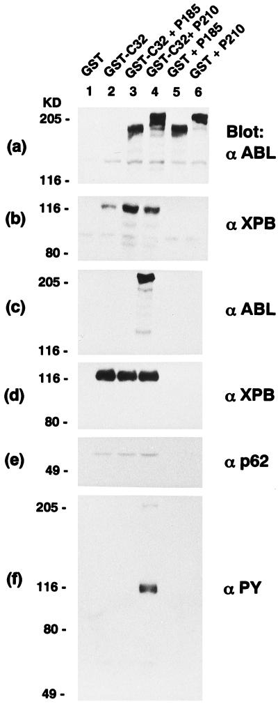
To test whether biologically significant interactions between P210 BCR-ABL and XPB take place in cells, we overexpressed BCR-ABL and GST-tagged XPB clones (C8 and C32) in 27–1 cells (Fig. 1). The 27–1 cell line is a UV-sensitive mutant of the CHO-9 cells, and its DNA repair deficiency has been reported to be corrected substantially by the introduction of XPB (10, 11, 18). Glutathione Sepharose-bound preparations of the lysates contained GST-C32, p62 (11), and P210 BCR-ABL but not P185 BCR-ABL (Fig. 1 c, d, and e). Tyrosine phosphorylation of GST-C32 also was observed in 27–1 cells when coexpressed with P210 BCR-ABL but not with P185 BCR-ABL (Fig. 1f).
Figure 1.
Expression of GST-C32 and BCR-ABL in 27–1 cells. GST-C32 was overexpressed alone or in combination with P210 or P185 BCR-ABL in 27–1 cells. Total cell lysates (a and b) or glutathione Sepharose-bound preparations (c_–_f) from cells expressing GST (vector alone) (lane 1), GST-C32 (lane 2), GST-C32/P185 BCR-ABL (lane 3), GST-C32/P210 BCR-ABL (lane 4), GST/P185 BCR-ABL (lane 5), and GST/P210 BCR-ABL (lane 6) were subjected to anti-ABL (a and c), anti-XPB (b and d), anti-p62 (e), and anti-phosphotyrosine (PY) (f) Western blotting.
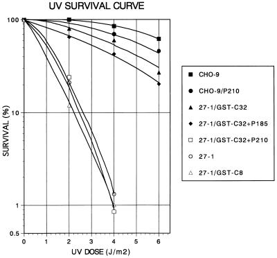
BCR-ABL has been shown to be a potent inhibitor of apoptosis (19). However, expression of P210 had a small but appreciable positive effect on the UV sensitivity of the parental CHO-9 cells (Fig. 2). Although GST-C32 containing a full length XPB could significantly restore UV resistance to 27–1 cells, GST-C8 with a similar expression level (data not shown) to that of GST-C32 could not (Fig. 2). C8 lacks the amino-terminal 202 amino acids that contain the putative nuclear localization signal as well as the DNA-binding motif (10). These data not only show that the isolated XPB (C32) is functionally intact but also are consistent with the previous finding that a mutation in the DNA-binding motif in XPB abrogated its repair activity (18). When P210 BCR-ABL was coexpressed with GST-C32, the repair activity was drastically inhibited (Fig. 2). We could detect the expression of P210 BCR-ABL 7 days after the exposure to UV at 2 J/m2 (data not shown). This blocking effect was not prominent in P185 BCR-ABL with a similar expression level to P210 (Figs. 1a and 2), indicating that this inhibition is mediated by the CDC24 homology domain of BCR.
Figure 2.
XPB cannot correct the nucleotide excision repair defect of 27–1 cells that express P210 BCR-ABL. Shown is a UV survival curve of 27–1 and parental CHO-9 cells with the expression of various constructs as indicated in the box (see Fig. 1).
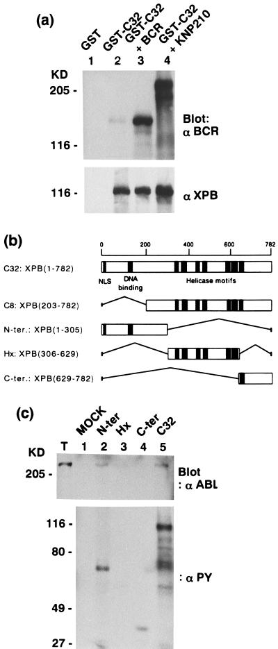
To examine whether tyrosine phosphorylation is involved in the interaction between XPB and P210 BCR-ABL, the intact BCR or a kinase negative mutant of the p210 BCR-ABL (KN P210) was coexpressed in 27–1 cells with GST-C32 as described in Fig. 1. In either case, the double transfectants showed no difference in the biological behavior from the 27–1 cells that expressed GST-C32 alone, and the GST-C32 proteins purified by the glutathione column were not tyrosine-phosphorylated (data not shown) but were bound to the overexpressed BCR or KN P210 BCR-ABL proteins (Fig. 3a). Binding of the endogenous BCR protein to the overexpressed GST-C32 also was observed (Fig. 3a, lane 2). This suggests that the interaction between the CDC24 homology domain of BCR and XPB may take place in a physiological setting.
Figure 3.
(a) Tyrosine phosphorylation is not required for the XPB/P210 BCR-ABL interaction. Glutathione Sepharose-bound preparations from 27–1 cells overexpressing GST (lane 1), GST-C32 (lane 2), GST-C32 with BCR (lane 3), and GST-C32 with a kinase negative mutant of P210 BCR-ABL (KN P210) (lane 4) were subjected to anti-BCR (Upper) and anti-XPB (Lower) Western blotting. (b) Schematic representation of the isolated XPB clones (C8 and C32) and three XPB fragments (N-ter, Hx, and C-ter) constructed from C32. The putative nuclear localization signal (NLS), the potential DNA-binding domain, and conserved helicase motifs are indicated. The amino acid numbers are shown on the top. (c) XPB N-ter and C-ter fragments were tyrosine-phosphorylated by P210 BCR-ABL. _E. coli_-expressed and glutathione Sepharose-bound GST (lane 1, MOCK), GST-N-ter (lane 2), GST-Hx (lane 3), GST-C-ter (lane 4) as shown in b, and GST-C32 (lane 5) were incubated with total cell lysates from Sf9 cells infected with P210 BCR-ABL baculovirus (T) to allow binding. After washing, each sample was subjected to an in vitro kinase assay followed by anti-ABL (Upper) and anti-PY (Lower) Western blotting.
To define the interacting region in XPB, the XPB sequence was divided to three fragments shown in Fig. 3b. Each fragment was expressed in E. coli in the form of GST fusion protein. An in vitro kinase assay was performed with those GST fusion proteins after binding to baculovirus-expressed P210 BCR-ABL. Although the bound P210 BCR-ABL protein was under detection when incubated with C-ter, both the N-ter and C-ter fragments were tyrosine-phosphorylated by P210 BCR-ABL (Fig. 3c). Considering that binding ability of CDC24-homology domain of BCR with C8 was 11.9% of that with C32 as judged by the β-galactosidase assay in yeast two hybrid system, we assume that the major BCR-binding region of XPB is in the N-ter region and that sites of tyrosine phosphorylation are in both N-ter and C-ter regions.
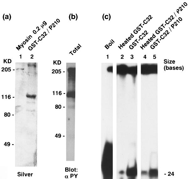
To examine biochemical effects of P210 BCR-ABL on XPB, we then expressed those proteins in baculovirus. Enzymatically active (data not shown) P210 BCR-ABL was efficiently copurified with GST-C32 from coinfected Sf9 cells (Fig. 4a). Tyrosine phosphorylation of GST-C32 was observed at a level comparable to that of heavily autophosphorylated P210 BCR-ABL (Fig. 4b). Purified baculovirus GST-C32 showed an ATPase activity of ≈0.81 ± 0.045 pmol/ng/hr in the presence of single-stranded DNAs, which is one magnitude less than that of purified TFIIH from HeLa cells and is in a similar range to that of _E. coli_-expressed recombinant XPB (15). The ATPase activity of tyrosine-phosphorylated GST-C32 purified from P210 BCR-ABL-coexpressing Sf9 cells was found to be reduced by 36% (0.52 ± 0.045 pmol/ng/hr). This inhibitory effect of P210 BCR-ABL on XPB also was found in the direction-specific helicase activity (≈25% reduction as judged by the radioactivity of the catalyzed substrates) (Fig. 4c). p53 also has been reported to function as an inhibitor for Rad3 ATPase/helicase when added in molar excess in a purified two components system (20). Given the facts that GST-C32 complexed to BCR purified from baculovirus-infected Sf9 cells has almost no change in ATPase activity (data not shown) and that both BCR and KN P210 BCR-ABL can bind to XPB but cannot exert similar biological effects to P210 in the 27–1 cell system, tyrosine phosphorylation of XPB may play a major role in the biochemical inhibition.
Figure 4.
P210-bound and tyrosine-phosphorylated GST-C32 (GST-tagged full length XPB) expressed in Sf9 cells has reduced ATPase and helicase activities. (a) Purified myosin (0.2 μg) (lane 1) and GST-C32 bound to P210 BCR-ABL (lane 2) expressed in and purified from Sf9 cells were stained by silver nitrate. (b) Purified GST-C32 complexed to P210 shown in a was subjected to anti-PY Western blotting. Note a high level of tyrosine phosphorylation in GST-C32 comparable to that of autophosphorylated P210 BCR-ABL. (c) Direction-specific helicase substrates (see Materials and Methods) were boiled for 2 min (positive control, lane 1), were treated with purified GST-C32 after (lane 2) or before (lane 3) heat inactivation or with purified GST-C32 complexed to P210 BCR-ABL after (lane 4) or before (lane 5) heat inactivation, and were run on a 10% polyacrylamide gel.
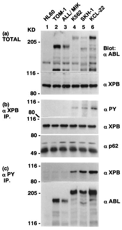
We then examined the interaction between BCR-ABL and XPB in cell lines derived from Ph1-positive human leukemic cells (Fig. 5). The XPB proteins were tyrosine-phosphorylated in all of the three CML cell lines (K562, SKH-1, and KCL-22) but not in either of the two ALL cell lines (TOM-1 and ALL/MIK) (Fig. 5b). Anti-ABL Western blotting of anti-XPB immunoprecipitates was unsuccessful to show the specific binding of P210 BCR-ABL to XPB. However, anti-PY immunoprecipitates from CML cells contained both P210 BCR-ABL and XPB, and those from Ph1-positive ALL cells had background levels of XPB (Fig. 5c). These data suggest that P210 BCR-ABL and XPB could interact, but the complex formation is difficult to show directly, possibly because of the ability of the antibody currently available or because of low stoichiometry of interaction in leukemic cells. The subcellular localization of BCR, BCR-ABL, and virally activated P160 v-ABL has been reported to be in the cytoplasm and XPB in the nucleus (18, 21). However, a fraction of the BCR proteins has been shown to be associated with heterochromatin (22). P120 v-ABL was shown to be largely in the nucleus in hematopoietic cells possibly by using a nuclear localization signal in the ABL sequence, which is retained also in BCR-ABL (23). When nuclear and cytoplasmic lysates were examined separately in KCL-22, 1.7% of XPB was calculated to be in the cytoplasm whereas 2.5% of P210 BCR-ABL was in the nucleus. This information suggests that P210 BCR-ABL and XPB could interact, but with a low stoichiometry.
Figure 5.
XPB is tyrosine-phosphorylated and possibly forms a complex with P210 BCR-ABL in CML cells. Total cell lysates (a), anti-XPB immunoprecipitates (b), and anti-phosphotyrosine immunoprecipitates (c) from human leukemic cell lines HL60 (derived from Ph1-negative acute promyelocytic leukemia) (lane 1), two cell lines from Ph1-positive ALL [TOM-1 (lane 2) and ALL/MIK (lane 3)] (28), three cell lines from Ph1-positive CML in blastic crisis [K562 (lane 4), SKH-1 (lane 5), and KCL-22 (lane 6)] (7) were subjected to anti-ABL (a, Upper and c, Lower), anti-XPB (a, Upper, b, Middle, and c, Upper), anti-PY (b, Top), and anti-p62 (b, Bottom) Western blot analyses.
Thus, our findings show that P210 BCR-ABL potentially inactivates XPB. Both purified TFIIH from XPB patients with DNA repair defects and recombinant XPB with a mutation from the same patient have been reported to contain reduced helicase activity (24). Given the inefficient in vivo binding between P210 BCR-ABL and XPB, we presume that the repair defect in the P210 BCR-ABL-expressing leukemic cells may not occur as efficiently as in cells in XPB patients. However, we emphasize that the small but persistent interaction between the two proteins might alter critical genes in the transformation process. Pane et al. have reported that benign neutrophilic CML, whose blastic transformation occurs much later, if any, than the classical CML, is specifically associated with P230 BCR-ABL that retains the XPB-interacting region of BCR (25). However, there have been increasing number of cases of typical CML associated with P230 BCR-ABL and of arguments against the claim (26, 27). Considering the antiapoptotic ability of BCR-ABL, the “no repair and no apoptosis result in cancer” theory might be applied to the molecular basis of blastic crisis in CML.
Acknowledgments
We thank Dr. A. Yasui for distribution of CHO-9 and 27–1 cells, Dr. H. Hirai and M. Okabe for Ph1-positive leukemic cell lines, Dr. Owen N. Witte for kinase negative ABL, Dr. K. Maruyama for pME18SHygB, and Dr. Y. Nakatsu and Dr. F. Hanaoka for discussions.
ABBREVIATIONS
CML
chronic myelogenous leukemia
ALL
acute lymphocytic leukemia
XBP
xeroderma pigmentosum group B protein
GST
glutathione _S_-transferase
CHO
Chinese hamster ovary
Footnotes
This paper was submitted directly (Track II) to the Proceedings Office.
References
- 1.Maru Y, Witte O N. In: Appplication of Basic Science to Hematopoiesis and Treatment of Disease. Thomas E D, editor. New York: Raven; 1993. pp. 123–143. [Google Scholar]
- 2.Champlin R E, Golde D W. Blood. 1985;65:1039–1047. [PubMed] [Google Scholar]
- 3.Feinstein E, Cimino G, Gale R P, Alimena G, Berthier R, Kishi K, Goldman J, Zaccaria A, Berrebi A, Canaani E. Proc Natl Acad Sci USA. 1991;88:6293–6297. doi: 10.1073/pnas.88.14.6293. [DOI] [PMC free article] [PubMed] [Google Scholar]
- 4.Towatari M, Adachi K, Kato H, Saito H. Blood. 1991;78:2178–2181. [PubMed] [Google Scholar]
- 5.Sill H, Goldman J M, Cross N C P. Blood. 1995;85:2013–2016. [PubMed] [Google Scholar]
- 6.Le Maistre A, Lee M S, Talpaz M, Kantarjian H M, Freireich E J, Deisseroth A B, Trujillo J M, Stass S A. Blood. 1989;73:889–891. [PubMed] [Google Scholar]
- 7.Mitani K, Ogawa S, Tanaka T, Miyoshi H, Kurokawa M, Mano H, Yazaki Y, Ohki M, Hirai H. EMBO J. 1994;13:504–510. doi: 10.1002/j.1460-2075.1994.tb06288.x. [DOI] [PMC free article] [PubMed] [Google Scholar]
- 8.Lugo T G, Pendergast A M, Muller A J, Witte O N. Science. 1990;247:1079–1082. doi: 10.1126/science.2408149. [DOI] [PubMed] [Google Scholar]
- 9.Voncken J W, Kaartinen V, Pattengale P K, Germeraad W T V, Groffen J, Heisterkamp N. Blood. 1995;86:4603–4611. [PubMed] [Google Scholar]
- 10.Weeda G, van Ham R C A, Vermeulen W, Bootsma D, van der Eb A J, Hoeijmakers J H J. Cell. 1990;62:777–791. doi: 10.1016/0092-8674(90)90122-u. [DOI] [PubMed] [Google Scholar]
- 11.Hoeijmakers J H J, Egly J M, Vermeulen W. Curr Opin Genet Dev. 1996;6:26–33. doi: 10.1016/s0959-437x(96)90006-4. [DOI] [PubMed] [Google Scholar]
- 12.Maru Y, Witte O N. Cell. 1991;67:459–468. doi: 10.1016/0092-8674(91)90521-y. [DOI] [PubMed] [Google Scholar]
- 13.Maru Y, Afar D E, Witte O N, Shibuya M. J Biol Chem. 1996;271:15353–15357. doi: 10.1074/jbc.271.26.15353. [DOI] [PubMed] [Google Scholar]
- 14.Pendergast A M, Clark R, Kawasaki E S, McCormick F P, Witte O N. Oncogene. 1989;4:759–766. [PubMed] [Google Scholar]
- 15.Roy R, Schaeffer L, Humbert S, Vermeulen W, Weeda G, Egly J M. J Biol Chem. 1994;269:9826–9832. [PubMed] [Google Scholar]
- 16.Lanzetta P A, Alvarez L J, Reinach P S, Candia O A. Anal Biochem. 1979;100:95–97. doi: 10.1016/0003-2697(79)90115-5. [DOI] [PubMed] [Google Scholar]
- 17.Schaeffer L, Moncollin V, Roy R, Staub A, Mezzina M, Sarasin A, Weeda G, Hoeijmakers J H J, Egly J M. EMBO J. 1994;13:2388–2392. doi: 10.1002/j.1460-2075.1994.tb06522.x. [DOI] [PMC free article] [PubMed] [Google Scholar]
- 18.Ma L, Westbroek A, Jochemsen A G, Weeda G, Bosch A, Bootsma D, Hoeijmakers J H J, van der Eb A J. Mol Cell Biol. 1994;14:4126–4134. doi: 10.1128/mcb.14.6.4126. [DOI] [PMC free article] [PubMed] [Google Scholar]
- 19.Amarante-Mendes G P, Kim C N, Liu L, Huang Y, Perkins C L, Green D R, Bhalla K. Blood. 1998;91:1700–1705. [PubMed] [Google Scholar]
- 20.Wang X W, Yeh H, Schaeffer L, Roy R, Moncollin V, Egly J M, Wang Z, Friedberg E C, Evans M K, Taffe B G, et al. Nat Genet. 1995;10:188–195. doi: 10.1038/ng0695-188. [DOI] [PubMed] [Google Scholar]
- 21.Wetzler M, Talpaz M, van Etten R A, Hirsh-Ginsberg C, Beran M, Kurzrock R. J Clin Invest. 1993;92:1925–1939. doi: 10.1172/JCI116786. [DOI] [PMC free article] [PubMed] [Google Scholar]
- 22.Wetzler M, Talpaz M, Yee G, Stass S A, Van Etten R A, Andreeff M, Goodacre A M, Kleine H D, Mahadevia R K, Kurzrock R. Proc Natl Acad Sci USA. 1995;92:3488–3492. doi: 10.1073/pnas.92.8.3488. [DOI] [PMC free article] [PubMed] [Google Scholar]
- 23.Birchenall-Roberts M C, Ruscetti F W, Kasper J J, Bertolette D C, III, Yoo Y D, Bang O, Roberts M S, Turley J M, Ferris D K, Kim S. Mol Cell Biol. 1995;15:6088–6099. doi: 10.1128/mcb.15.11.6088. [DOI] [PMC free article] [PubMed] [Google Scholar]
- 24.Hwang J R, Moncollin V, Vermeulen W, Seroz T, van Vuuren H, Hoeijmakers J H J, Egly J M. J Biol Chem. 1996;271:15898–15904. doi: 10.1074/jbc.271.27.15898. [DOI] [PubMed] [Google Scholar]
- 25.Pane F, Frigeri F, Sindona M, Luciano L, Ferrara F, Cimino R, Meloni G, Saglio G, Salvatore F, Rotoli B. Blood. 1996;88:2410–2414. [PubMed] [Google Scholar]
- 26.Briz M, Vilches C, Cabrera R, Fores R, Fernandez M N. Blood. 1997;90:5024–5025. [PubMed] [Google Scholar]
- 27.Wada H, Mizutani S, Nishimura J, Usuki Y, Kohsaki M, Komai M, Kaneko H, Sakamoto S, Delia D, Kanamaru A, et al. Cancer Res. 1995;55:3192–3196. [PubMed] [Google Scholar]
- 28.Okabe M, Matsushima S, Morioka M, Kobayashi M, Abe S, Sakurada K, Kakinuma M, Miyazaki T. Blood. 1987;69:990–998. [PubMed] [Google Scholar]
