Rescue of cardiomyocyte dysfunction by phospholamban ablation does not prevent ventricular failure in genetic hypertrophy (original) (raw)
Abstract
Cardiac hypertrophy, either compensated or decompensated, is associated with cardiomyocyte contractile dysfunction from depressed sarcoplasmic reticulum (SR) Ca2+ cycling. Normalization of Ca2+ cycling by ablation or inhibition of the SR inhibitor phospholamban (PLN) has prevented cardiac failure in experimental dilated cardiomyopathy and is a promising therapeutic approach for human heart failure. However, the potential benefits of restoring SR function on primary cardiac hypertrophy, a common antecedent of human heart failure, are unknown. We therefore tested the efficacy of PLN ablation to correct hypertrophy and contractile dysfunction in two well-characterized and highly relevant genetic mouse models of hypertrophy and cardiac failure, Gαq overexpression and human familial hypertrophic cardiomyopathy mutant myosin binding protein C (MyBP-CMUT) expression. In both models, PLN ablation normalized the characteristically prolonged cardiomyocyte Ca2+ transients and enhanced unloaded fractional shortening with no change in SR Ca2+ pump content. However, there was no parallel improvement in in vivo cardiac function or hypertrophy in either model. Likewise, the activation of JNK and calcineurin associated with Gαq overexpression was not affected. Thus, PLN ablation normalized contractility in isolated myocytes, but failed to rescue the cardiomyopathic phenotype elicited by activation of the Gαq pathway or MyBP-C mutations.
Introduction
In the face of unremitting hemodynamic stress, “adaptive” cardiac hypertrophy inevitably progresses to ventricular dilation and clinical heart failure, which affects an estimated 5 million Americans and has a mortality rate of approximately 50% in 4 years (1). A nearly universal characteristic of hypertrophied and failing myocardium is depressed sarcoplasmic reticulum (SR) Ca2+ cycling, caused by decreased expression of the cardiac SR Ca2+ ATPase (SERCA2a), by a relative overabundance of the SERCA2a inhibitory protein phospholamban (PLN), or by both (2, 3). Recent experimental successes have generated enthusiasm for treating heart failure by restoring SR Ca2+ cycling, either through adenoviral-mediated myocardial-targeted expression of SERCA2a itself (4) or through antisense suppression (5) or genetic ablation of PLN (6), to relieve SERCA2a from endogenous inhibition. The common result of each approach is enhanced SR Ca2+ cycling, which has improved energetics, survival, and cardiac function at the cellular, organ, and intact animal levels. Furthermore, there is no compromise in exercise performance or longevity in the case of PLN ablation (7, 8).
To date, PLN ablation or inhibition has improved SR Ca2+ cycling and/or contractile function in transgenic mice overexpressing slow skeletal troponin I (9), a nonphosphorylatable troponin I mutant (10), calsequestrin (CSQ) (11), SERCA1a (12), and a mutant myosin heavy chain (13). Most strikingly, PLN ablation prevented the functional, structural, and histological abnormalities in cardiomyopathic mice lacking muscle Lim protein (MLP) (14), and PLN inhibition attenuated heart failure progression in cardiomyopathic hamsters (15), suggesting that abnormal SR Ca2+ cycling may represent a root cause of heart failure. Thus, targeting PLN has been strikingly successful in preventing phenotypic progression in various experimental primary cardiomyopathies (14, 15). However, the efficacy of PLN inhibition in primary myocardial hypertrophy that progresses to dilated heart failure (i.e., decompensated hypertrophy), which is a common cause of heart failure in human hypertensive and valvular heart disease, remains unknown.
To further test whether normalizing depressed cardiomyocyte Ca2+ handling by PLN inhibition is likely to be therapeutically efficacious in decompensated hypertrophy, we examined the consequences of PLN ablation on two highly relevant mouse models. In the Gαq overexpressor (16), hypertrophy and failure are induced by Gαq, the proximal angiotensin, endothelin, and the α-adrenergic receptor signal transducer, which has been shown to transduce pressure-overload hypertrophy (17). In the mutant myosin binding protein C (MyBP-CMUT) mouse (18), a human mutation of this sarcomeric protein is expressed, as occurs in approximately 15% of patients with familial hypertrophic cardiomyopathy. Herein, we show that PLN ablation restores impaired Ca2+ cycling and contractility of isolated cardiomyocytes from both the Gαq and MyBP-CMUT mice but that there are no measurable benefits of these cellular enhancements on global cardiac function or on development of myocardial hypertrophy.
Methods
Experimental animals.
PLN homozygous knockout mice (PLNKO) (12) were mated with Gαq-40 overexpressors (16) to generate PLN heterozygous knockout/Gαq transgenic mice (F1). These F1 mice were crossed with PLN heterozygotes to create PLNKO/Gαq transgenic mice (PLNKO/Gαq) along with Gαq overexpressors and wild types. Mice bearing the truncated MyBP-C allele (MyBP-CMUT) were previously generated using homologous recombination (18). Double heterozygous PLN knockout/MyBP-CMUT mice were inbred to obtain homozygous MyBP-CMUT mice with PLN ablation (PLNKO/MyBP-CMUT) along with MyBP-CMUT and wild-type mice. PCR analysis of tail genomic DNA was employed to genotype all mice (6, 18). Animals used in this study were male littermates at 10–16 weeks of age and were handled and maintained according to protocols by the ethics committee of the University of Cincinnati. The investigation conforms with the Guide for the Care and Use of Laboratory Animals published by the NIH (19).
Immunoblotting.
Ventricular homogenates were analyzed by standard Western blotting to compare PLN, SERCA2a, CSQ, calcineurin (CaN), phosphorylated JNK, connexin 43 (Cx43), Wnt-1, β-catenin, and N-cadherin levels. Binding of the primary antibody was detected by peroxidase-conjugated secondary antibodies and enhanced chemiluminescence (Amersham, Piscataway, New Jersey, USA).
Cardiomyocyte mechanics and Ca2+ transients.
Isolated left ventricular myocytes from Langendorff-perfused hearts were perfused in 1.8 mM Ca2+-Tyrode at room temperature and field stimulated at 0.5 Hz. Mechanics and Ca2+ transients were measured and calibrated as described previously (12, 20). A minimum of 5 cells per heart were studied, and each animal was analyzed as a single n. Data were analyzed by Felix computer software (Photon Technology International, Lawrenceville, New Jersey, USA).
In vivo measurements of cardiac function.
Echocardiographic parameters were measured using standard techniques as reported previously (21). Wall stress was derived from the LaPlace law: (pressure × radius)/(2 × wall thickness) (22). Closed-chest invasive hemodynamic studies were performed under ketamine and thiobutabarbital anesthesia. β-adrenergic responsiveness was assessed after infusion of dobutamine as described previously (16).
Histology.
Mouse hearts were fixed with 4% paraformaldehyde followed by dehydration and paraffin embedding. Serial 4-μm sections were stained with hematoxylin and eosin or Masson’s trichrome.
RNA analysis.
Quantitative assessment of cardiac hypertrophy gene expression was performed by RNA dot blotting using gene-specific antisense oligonucleotides (16).
Statistical analysis.
Data are presented as means ± SEM, unless otherwise stated; n indicates the number of mice. Statistical analysis was performed by one-way or two-way analysis of variance, followed by the Student-Newman-Keuls test for multiple comparisons. Values of P < 0.05 were considered statistically significant.
Results
SR Ca2+-transport proteins in Gαq and MyBP-CMUT mice.
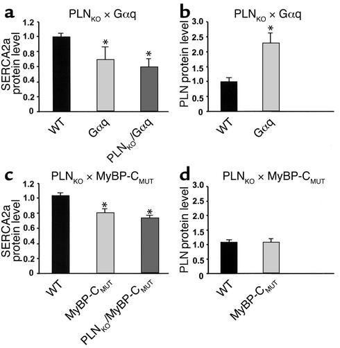
Defective SR Ca2+ cycling is indicated in the Gαq and MyBP-CMUT mice (16, 23). However, the mechanism for SR dysfunction has not been established as it relates to altered expression of SR Ca2+-handling proteins. Therefore, ventricular cardiac homogenates from Gαq-overexpressing and MyBP-CMUT hearts were assayed for SR Ca2+-transport protein levels by quantitative immunoblotting. Since initial studies indicated that levels of CSQ, the major SR Ca2+-storage protein, were not altered in these hearts, CSQ was used as an internal standard for assessment of SERCA2a, the principle Ca2+-handling protein in SR, and PLN, the endogenous inhibitor of SERCA2a. SERCA2a protein levels were significantly decreased in both Gαq and MyBP-CMUT hearts (Gαq: 0.70-fold ± 0.17-fold of wild type, n = 5, P < 0.05; MyBP-CMUT: 0.81-fold ± 0.05-fold of wild type, n = 6, P < 0.05) (Figure 1, a and c). Further impairing SERCA2a function in Gαq-overexpressing hearts, levels of PLN were markedly increased to 2.3-fold ± 0.33-fold of wild type (n = 5, P < 0.05) (Figure 1b), whereas PLN protein level was unaltered in MyBP-CMUT hearts (Figure 1d). Thus, impaired Ca2+ cycling in Gαq and MyBP-CMUT hearts is most likely the result of diminished SERCA2a activity.
Figure 1.
Quantitative immunoblotting of SERCA2a and PLN in mouse ventricular homogenate. (a and b) PLNKO × Gαq cross. (a) Relative SERCA2a protein level. (b) Relative PLN protein level. (c and d) PLNKO × MyBP-CMUT cross. (c) Relative SERCA2a protein level. (d) Relative PLN protein level. *P < 0.05 versus wild type; n = 5–6 hearts per group.
Normalization of cardiomyocyte Ca2+ handling and contraction by PLN ablation.
Downregulated SERCA2a protein in Gαq- and MyBP-CMUT–induced hypertrophy, and the consequent increase in proportional expression of the SERCA2a inhibitor PLN in both models, provided a compelling rationale for downregulating PLN to restore SR activity and thereby improve the depressed myocardial contractile function due to defective SR Ca2+ cycling. Furthermore, we considered that cardiac hypertrophy in these models might be attenuated if, as postulated for dilated cardiomyopathy (14, 15), abnormal Ca2+ signaling was an important factor. To test this notion, Gαq or MyBP-CMUT mice were mated with PLN-deficient mice to generate animals with PLN ablation in the context of cardiac-specific Gαq overexpression or MyBP-CMUT expression. Importantly, PLN ablation did not affect SERCA2a protein levels in either model (Figure 1, a and c).
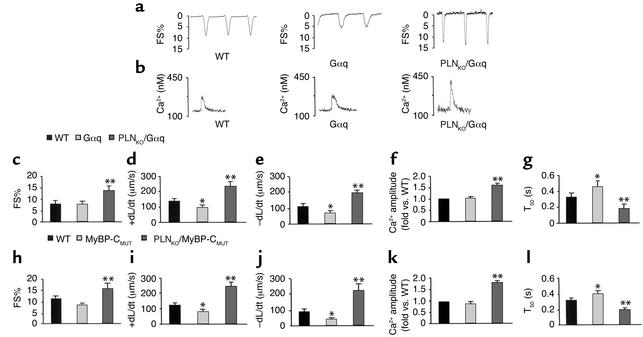
The effects of PLN ablation on Gαq or MyBP-CMUT cardiomyocyte contraction were assessed in isolated ventricular myocytes and compared with the effects in littermate controls. Consistent with previous reports (23), Gαq cardiac myocytes exhibited significantly depressed maximal rates of shortening and relengthening, but unloaded fractional shortening did not differ from that of nontransgenic controls (Figure 2, a and c–e). Likewise, cardiac myocytes from MyBP-CMUT hearts exhibited depressed maximal rates of shortening and relengthening, which in this case were associated with a modest decrease in the extent of unloaded shortening as compared with wild type (Figure 2, h–j).
Figure 2.
Myocyte (5–8 cells per heart, 6–8 hearts per group) contractility and Ca2+ kinetics in hearts from PLNKO × Gαq and PLNKO × MyBP-CMUT crosses. Representative recordings of cell shortening (a) and the intracellular Ca2+ concentration transient (b) in myocytes of the PLNKO × Gαq cross are shown. (c–g) PLNKO × Gαq myocytes. (c) FS%. (d) Rate of contraction (+dL/dt). (e) Rate of relaxation (–dL/dt). (f) Ca2+ transient amplitude (fold vs. WT). (g) Time to 50% decay of the Ca2+ signal (T50). (h–l) PLNKO × MyBP-CMUT myocytes. (h) FS%. (i) +dL/dt. (j) –dL/dt. (k) Ca2+ transient amplitude. (l) T50. *P < 0.05 versus wild type; **P < 0.05 versus Gαq or MyBP-CMUT myocytes.
These cardiomyocyte contractile abnormalities were associated with alterations in SR Ca2+ cycling that were consistent with the observed alterations in SR Ca2+-handling proteins. In neither model was the peak Ca2+ transient amplitude significantly different from that of wild-type controls (Figure 2, f and k). In both models, however, the time to 50% decay of the Ca2+ signal (T50) was prolonged as compared with nontransgenic controls, reflecting decreased uptake of Ca2+ by the SR (Figure 2, g and l). When PLN was eliminated by gene ablation, Ca2+ cycling was greatly enhanced in both models, to levels even exceeding those of wild-type controls (Figure 2, f, g, k, and l). A strikingly shortened duration of the Ca2+ transient (to approximately 60% of control) reflected enhanced SR Ca2+ cycling and resulted in substantial increases in peak Ca2+ amplitude, most likely reflecting increased SR Ca2+ stores.
Enhanced Ca2+ cycling in cardiac myocytes by ablation of PLN resulted in a parallel improvement of their contractile function. In general, when compared with the baseline Gαq and MyBP-CMUT models, PLN ablation doubled various aspects of mechanical function to levels that were significantly better than wild type (Figure 2, c–e, h–j). Specifically, PLNKO/Gαq myocytes showed approximately 70% improvements in +dL/dt, –dL/dt, and fractional shortening. Likewise, PLNKO/MyBP-CMUT myocytes resulted in full restoration of all depressed contractile parameters to levels higher than those observed in the wild-type cells. Indeed, each of the measured parameters of contractility in PLNKO/Gαq and PLNKO/MyBP-CMUT myocytes resembled those in PLNKO myocytes (data not shown), indicating that PLN ablation restored the defective SR Ca2+ cycling, which caused the decreased myocyte contractility in these two models of hypertrophy.
PLN ablation fails to improve in vivo global cardiac function.
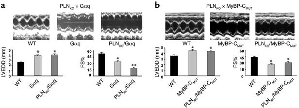
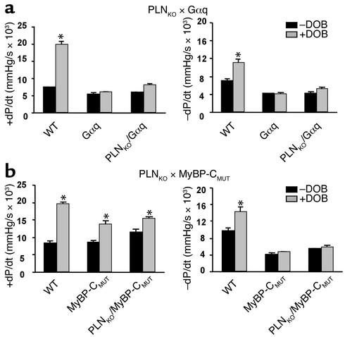
Overall cardiac performance is the result of a complex interplay between myocardial contractility, chamber geometry, loading conditions, extracellular resistance and elastic force produced by interstitial tissues, and neurohumoral factors. Thus, it was important to determine whether the successful strategy of PLN ablation to improve cardiomyocyte contractile function was translated into enhanced in vivo global cardiac function in Gαq-overexpressing and MyBP-CMUT mice. Surprisingly, echocardiography showed no improvement in left ventricular ejection performance upon PLN ablation in Gαq-overexpressing mice, as revealed by persistently increased left ventricular end diastolic dimension (LVEDD) and impaired percent fractional shortening (FS%). At 5 weeks of age, LVEDD was 3.87 ± 0.45 mm and 3.60 ± 0.22 mm, whereas FS% was 23.9 ± 2.9 and 28.7 ± 3.4 in PLNKO/Gαq and Gαq mice, respectively. Similar values were obtained in adult mice at 12–16 weeks of age (Figure 3a). In PLNKO/MyBP-CMUT mice, LVEDD was 3.7 ± 0.1 mm and FS% was 41.5 ± 2.8, as compared with 4.0 ± 0.1 mm and 31.1 ± 2.8 in MyBP-CMUT mice, at 5 weeks of age. These parameters in PLNKO/MyBP-CMUT mice further deteriorated and were similar to those in MyBP-CMUT mice by 12–16 weeks of age (Figure 3b). Likewise, PLN ablation did not significantly improve basal or β-adrenergic left ventricular contractility (+dP/dt) or relaxation (–dP/dt) in the Gαq mice (Figure 4a), although there were no alterations in β-adrenergic receptor density in these models (16, 24). Similar findings were obtained in MyBP-CMUT hearts (Figure 4b).
Figure 3.
M-mode echocardiographic tracings and in vivo global cardiac function of mice from PLNKO × Gαq (a) and PLNKO × MyBP-CMUT (b) crosses, determined by echocardiography. *P < 0.05 versus wild type; **P < 0.05 versus Gαq or MyBP-CMUT hearts; n = 6–8 per group.
Figure 4.
Hemodynamic characteristics and left ventricular response to β-adrenergic stimulation in hearts from PLNKO × Gαq (a) and PLNKO × MyBP-CMUT (b) crosses. +dP/dt indicates the rate of left ventricular contraction; –dP/dt, the rate of left ventricular relaxation; –DOB, the baseline without dobutamine; and +DOB, infusion of dobutamine at 32 ng/g of body weight per minute. *P < 0.05 versus baseline; n = 6–8 per group.
When considering the lack of apparent benefit to cardiac function of a maneuver that unambiguously enhanced cardiomyocyte contractility, it is important to note that there was no change in ventricular morphology — that is, chamber dimension and wall thickness — with PLN ablation (data not shown). These two morphometric measures, along with left ventricular pressure (which was not altered by PLN ablation) (data not shown), are the determinants of ventricular wall stress or afterload. Indeed, the left ventricular wall stress, which as expected was around twofold higher than that of wild type in the Gαq model, increased further with PLN ablation (wild type, 39.11 ± 1.96 dynes/mm2; Gαq, 77.60 ± 1.73 dynes/mm2; PLNKO/Gαq, 133.18 ± 6.31 dynes/mm2; n = 6–8; P < 0.01 vs. wild type). Similar alterations were observed in the MyBP-CMUT mice (wild type, 47.41 ± 5.3 dynes/mm2; MyBP-CMUT, 81.45 ± 14.3 dynes/mm2; PLNKO/MyBP-CMUT, 75.33 ± 12.4 dynes/mm2; n = 6; P < 0.01 vs. wild type). Thus, the restored cardiomyocyte Ca2+ cycling and contractility upon PLN ablation did not reflect similar improvement in global cardiac function, possibly because of the absence of reverse ventricular remodeling and the consequent high wall stress in PLNKO/Gαq and PLNKO/MyBP-CMUT hearts. Alternatively, Ca2+ measurements in vitro may not adequately reflect the in vivo Ca2+ signaling, which may not be influenced by PLN ablation, contributing to the observed cardiomyopathic phenotype.
Rescue of cardiomyocyte contractile function is not sufficient to reverse cardiac remodeling.
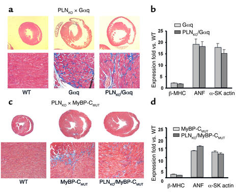
The surprising absence of any measurable functional benefit of enhancing cardiomyocyte contractility on in vivo cardiac function was most likely due to persistent ventricular enlargement in the PLNKO/MyBP-CMUT and PLNKO/Gαq mice. Therefore, a detailed analysis was undertaken of the effects of PLN ablation on the seminal characteristic of these two models, myocardial hypertrophy. Consistent with the echocardiographic findings (data not shown), the ratio of heart weight to body weight was not changed by PLN ablation in either the Gαq-overexpressing model (wild type, 4.02 ± 0.3 mg/g; Gαq, 5.8 ± 0.1 mg/g; PLNKO/Gαq, 5.72 ± 0.2 mg/g; n = 6; P < 0.01 vs. wild type) or the MyBP-CMUT model (wild type, 4.54 ± 0.4 mg/g; MyBP-CMUT, 6.66 ± 0.2 mg/g; PLNKO/MyBP-CMUT, 6.33 ± 0.33 mg/g; n = 6; P < 0.01 vs. wild type). Histological analyses confirmed that ablation of PLN did not prevent the characteristic morphological features of Gαq-overexpressing or MyBP-CMUT hearts (16, 18), including myocyte hypertrophy, myofibrillar disarray, and fibrosis (Figure 5, a and c).
Figure 5.
Histological studies and reactivation of fetal gene profile in the hearts from PLNKO × Gαq (a and b) and PLNKO × MyBP-CMUT (c and d) crosses. Cross-sectioning of hearts (×4) and trichrome staining (×200) were performed to assess chamber geometry and fibrosis. Transcriptional levels were normalized by GAPDH and expressed as fold over wild type; n = 4–6 per group.
A hallmark characteristic shared by myocardial hypertrophy and heart failure is reactivation of a fetal gene program, which is a highly sensitive indicator of cardiomyocyte hypertrophy. Therefore, to reveal improvements conferred by PLN ablation that were not evident by functional or pathological studies, expression of certain members of this genetic program — that is, β-myosin heavy chain (β-MHC), atrial natriuretic factor (ANF), and α-skeletal actin (α-SK actin) mRNA — were assayed by dot blot analysis (Figure 5, b and d). As previously reported, ventricles of Gαq-overexpressing and MyBP-CMUT mice exhibited markedly increased ANF, α-SK actin, and β-MHC mRNA levels. Interestingly, ablation of PLN did not alter expression of these genes (Figure 5, b and d). Taken together with the above studies, these findings demonstrate that ablation of PLN neither prevents nor reverses Gαq- and MyBP-CMUT–mediated myocardial hypertrophy.
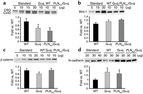
Previous studies have reported that among the three overall branches of the MAPK cascade (ERK, p38, and JNK), only JNK was activated in Gαq hearts (25). Increased JNK activation, using the phospho-specific antibody, was also observed in both the Gαq (1.2-fold) and PLNKO/Gαq (1.3-fold) hearts, as compared with wild-type (1.0-fold) hearts. In agreement with a recent report (26), JNK activation was associated with a significant reduction in the levels of the gap-junction protein Cx43 in both Gαq-overexpressing and PLNKO/Gαq hearts (Figure 6a). There were no significant differences in the levels of Wnt-1 and β-catenin, two principle regulators of the expression of Cx43 (27), in wild-type, Gαq-overexpressing, and PLNKO/Gαq hearts (Figure 6, b and c). In the same hearts, the protein levels of N-cadherin, the Cx43-associated protein, were persistently increased in both Gαq and PLNKO/Gαq hearts (Figure 6d). Interestingly, cardiac overexpression of N-cadherin has been observed to lead to dilated cardiomyopathy (28). Furthermore, since Gαq-triggered cardiac hypertrophy has been reported to be at least partially dependent on the CaN signaling pathway (29) and there is cross-talk between the CaN and JNK pathways (30), the protein levels of CaN were examined. A sustained increase in both Gαq (1.7-fold) and PLNKO/Gαq (1.7-fold) hearts as compared to wild type (1.0-fold) was observed. Taken together, these data indicate that JNK and CaN, putative mediators of Gαq-triggered cardiac hypertrophy, are not influenced by improved SR Ca2+ handling.
Figure 6.
Quantitative immunoblotting of gap-junction proteins and their regulators in mouse ventricular homogenates. Shown are Cx43 (a), Wnt-1 (b), β-catenin (c), and N-cadherin (d) in wild-type, Gαq, and PLNKO/Gαq hearts. *P < 0.05 versus wild type; n = 5 hearts per group.
Discussion
The most important finding of these studies is that normalization, even “supernormalization,” of cardiomyocyte Ca2+ cycling and contractility by PLN ablation is not sufficient to prevent cardiomyopathy in two highly relevant models of genetic myocardial hypertrophy. This finding contrasts with the apparent salutary effects of PLN inhibition on experimental dilated cardiomyopathies, in which correction of cardiomyocyte SR Ca2+ handling has had strikingly beneficial effects not only on cardiac function but on the histopathological features of these cardiomyopathies (14, 15).
Defects in SR Ca2+ uptake and release, which result in decreased contractility, are common features of hypertrophied and failing animal and human myocardia, although the mechanisms underlying the etiology of these defects have not been unambiguously defined. Recent evidence indicates that depressed cardiomyocyte Ca2+ cycling may contribute to decompensation of cardiac hypertrophy and progression of heart failure (2, 3). The present studies reveal that cardiomyocyte Ca2+ kinetics are depressed in the hypertrophies that develop as a consequence of myocardial-specific expression of Gαq and a MyBP-C mutation found in human familial hypertrophic cardiomyopathy. The prolonged decay of Ca2+ transients observed in field-stimulated ventricular cardiomyocytes from these models was associated with significant decreases in the level of SERCA2a protein expression and hence an increase in the relative ratio of PLN to SERCA2a. Since PLN is the primary determinant of SR function — that is, the so-called “physiological brake” on SR Ca2+ cycling — we hypothesized that improving SERCA2a activity through PLN ablation would enhance cardiac function and possibly attenuate the hypertrophic cardiomyopathy phenotype of Gαq-overexpressing and MyBP-CMUT hearts.
Our experimental approach paralleled that previously used by Minamisawa et al. to prevent dilated cardiomyopathy in MLP knockout mice (14) — that is, ablating PLN in the context of the underlying genetic stimulus for cardiac disease. As expected, PLN ablation strikingly improved SR Ca2+ cycling and cardiomyocyte dysfunction in both models. The direct effect, as demonstrated by the decrease in duration of the Ca2+ transient, was that diastolic SR reuptake of cytosolic Ca2+ was accelerated, leading to improved myocyte diastolic/relaxation function. An indirect effect of increased SR reuptake was enhanced SR Ca2+ content, reflected by the increased amplitude of the Ca2+ transient, which improved myocyte systolic/contractile function. Indeed, unloaded shortening of Gαq-overexpressing or MyBP-CMUT myocytes increased to levels significantly greater than “normal” in the context of PLN ablation, and the rates of contraction and relaxation were similarly improved. Notably, the levels of SERCA2a protein remained depressed, indicating the hypercontractile cardiomyocyte phenotype was largely, if not entirely, attributable to the relief of inhibition of SERCA2a by PLN, although possible contributions of other important Ca2+-handling proteins such as the ryanodine receptor, junctin, triadin, and the L-type Ca2+ channel were not assessed (31). A final theoretical benefit of the high-amplitude Ca2+ transient resulting from PLN ablation is activation of CaMKII (32), which has the potential to phosphorylate SERCA2a and stimulate its Ca2+-transport activity.
Whatever the precise biochemical mechanisms transducing the benefits of PLN ablation and enhanced SR function, the effects on cardiomyocyte systolic and diastolic mechanical function were unarguable. However, both invasive and noninvasive assessments of left ventricular function revealed that the enhanced cardiomyocyte function afforded by PLN ablation failed to improve a variety of measures of cardiac function. Unlike isolated, unloaded cardiac myocytes, in which intrinsic changes in Ca2+ cycling and contractile function are directly translated into enhanced mechanical performance, ventricular ejection is affected by additional factors such as hemodynamic load, chamber geometry, and cell-cell or cell-matrix connectivity. Indeed, systolic wall stress, which is dependent on ventricular chamber dimension, pressure, and wall thickness, is a critical determinant of left ventricular function in vivo (22), and neither ventricular morphometry nor wall stress was improved by PLN ablation in Gαq or MyBP-CMUT mice. Furthermore, the levels of phosphorylated JNK, associated with decreases in Cx43, as well as the levels of CaN, which were increased in Gαq-overexpressing hearts, remained elevated upon PLN ablation.
The absence of a beneficial effect of restoring cardiomyocyte Ca2+ signaling and mechanical function by PLN ablation on the overall cardiac phenotype of Gαq or MyBP-CMUT mice is in sharp contrast to previously published studies of a similar design using the MLPKO dilated cardiomyopathy model (14). In this instance, PLN ablation prevented not only cardiac dysfunction and ventricular dilation, but also the profound histopathological abnormalities that characterize this model (14). Since the MLPKO dilated cardiomyopathy is associated with defects in the cytoarchitecture of the myocyte, including SR organization (33), SR dysfunction may be a triggering event in this model. In preventing the resulting Ca2+ cycling and cellular abnormalities induced by MLP ablation, PLN ablation can therefore also prevent development of the full-blown phenotype. Likewise, PLN ablation has also prevented the functional and cellular alterations induced by CSQ overexpression (11), in which SR dysfunction is an obvious cause for remodeling. In this instance, augmentation of SR Ca2+ transport restored myocyte Ca2+ homeostasis, relieved wall stress, and prevented the hypertrophic phenotype. On the other hand, PLN ablation was not expected to benefit the dilated cardiomyopathy caused by tropomodulin overexpression, since myocyte basal Ca2+ cycling is already increased in that model (34, 35). Finally, PLN ablation has improved cardiac function in a transgenic mouse model of hypertrophic cardiomyopathy caused by a mutant form of myosin heavy chain, without compromising longevity or exercise tolerance (13).
The inability of PLN to rescue cardiac remodeling in the Gαq-overexpressing hearts suggests that the depressed SR function may represent one of the multiple modulators of the hypertrophic response in this model. Gαq represents the common signaling effector for endothelin I, angiotensin II, and α-adrenergic receptors, and Gαq activity is necessary for development of pressure-overload hypertrophy (17). Overexpression of Gαq recapitulates many aspects of both experimental cardiac hypertrophy and failure and the analogous human syndromes (16, 23). Previous genetic manipulations have targeted proximal players of the Gαq signaling pathway to improve cardiac function and attenuate hypertrophy (25, 36–39). Furthermore, a recent study has revealed that targeting molecules in the cell survival and death pathway may be especially critical to delay or prevent the progression of hypertrophy to heart failure in the Gαq-overexpressing mice (40). Taken together, the multiple factors modulated in the above studies demonstrate the pleiotrophic effects of Gαq overexpression and emphasize the different roles that multiple individual perturbations along the proximal Gαq hypertrophy signaling pathway can have in the development of myocardial hypertrophy and cardiac dysfunction.
For similar reasons, the inability of PLN to rescue the fundamental structural, functional, and histopathological characteristics of the MyBP-CMUT mouse may suggest that impaired SR Ca2+ cycling is one of the multiple phenotypic effectors in this model. MyBP-C is a major protein component of the striated muscle sarcomere (41). Besides its role in myofibrillogenesis, MyBP-C has been suggested to maintain overall sarcomere stability and to modify force production (41). Mutations leading to truncation of the C-terminal part of MyBP-C, as modeled in the present study, may exert two additive effects contributing to contractile dysfunction. The truncated protein may be ineffective in transmitting force, since it has lost its ability to bind to titin and myosin heavy chain (41–43), and may result in a high concentration of the unphosphorylated N-terminal fragment, which has been shown to produce a decrease in the generation of force (44). Therefore, ablation of PLN would probably compensate for a defect in force generation but may fail to transmit this force at the intact organ level, due to the mutation or the change in left ventricular geometry by remodeling.
Nevertheless, caution should prevail in interpreting the results of this and other studies on rescue of mouse cardiomyopathic phenotypes by genetic ablation of PLN. PLN deficiency is associated with a number of cellular adaptations to accommodate the enhanced Ca2+ cycling and energetic demand of the hyperdynamic cardiac function, which may contribute to or even mask the effects of PLN loss. Such compensatory mechanisms include the downregulation of the ryanodine receptor (45) and the increased inactivation kinetics of the L-type Ca2+ channel (46), which may contribute to the altered Ca2+ homeostasis in the cross models. Interestingly, the Ca2+ sensitivity or myosin ATPase activity of the contractile proteins is not altered (47). Furthermore, PLN ablation is associated with blunted cardiac β-adrenergic contractile responses (6), which may be restored upon decreases in the perfusate Ca2+ (48), consistent with the lack of alterations in β-adrenergic receptor density or coupling in this model (24).
In conclusion, the present study used PLN ablation to correct SR Ca2+ signaling and contractility in hypertrophied cardiac myocytes but failed to improve whole-heart contractile function. Even supernormalization of cardiomyocyte contractility did not prevent Gαq- or MyBP-CMUT–induced myocardial hypertrophy and cardiac dilation. In the context of prior studies showing complete phenotypic reversal of dilated cardiomyopathy by PLN ablation or inhibition (14, 15), and therefore demonstrating the critical role of SR Ca2+ cycling in cardiac remodeling, the current findings indicate that additional strategies for therapeutic intervention need to be identified that will prevent or modify the progression of hypertrophy to heart failure.
Acknowledgments
This work was supported by NIH grants HL-26057, HL-64018, HL-52318, and P40RR12358 to E. G. Kranias; HL-58010, HL-52318, and HL-59888 to G. W. Dorn; and 5T32HL-07382 to A. N. Carr; and by a grant from the Howard Hughes Medical Institute to C. E. Seidman and J. G. Seidman.
Footnotes
See the related Commentary beginning on page 801.
Qiujing Song and Albrecht G. Schmidt contributed equally to this work.
Conflict of interest: The authors have declared that no conflict of interest exists.
Nonstandard abbreviations used: sarcoplasmic reticulum (SR); phospholamban (PLN); mutant myosin binding protein C (MyBP-CMUT); cardiac SR Ca2+ ATPase (SERCA2a); calsequestrin (CSQ); muscle Lim protein (MLP); PLN homozygous knockout mice (PLNKO); calcineurin (CaN); connexin 43 (Cx43); left ventricular end diastolic dimension (LVEDD); percent fractional shortening (FS%); β myosin heavy chain (β-MHC); atrial natriuretic factor (ANF); and α-skeletal actin (α-SK actin) mRNA.
References
- 1.Ho KK, Pinsky JL, Kannel WB, Levy D. The epidemiology of heart failure: the Framingham Study. J. Am. Coll. Cardiol. 1993;22:6–13. doi: 10.1016/0735-1097(93)90455-a. [DOI] [PubMed] [Google Scholar]
- 2.Hasenfuss G, et al. Calcium handling proteins in the failing human heart. Basic. Res. Cardiol. 1997;92(Suppl. 1):87–93. doi: 10.1007/BF00794072. [DOI] [PubMed] [Google Scholar]
- 3.Houser SR, Piacentino V, 3rd, Weisser J. Abnormalities of calcium cycling in the hypertrophied and failing heart. J. Mol. Cell. Cardiol. 2000;32:1595–1607. doi: 10.1006/jmcc.2000.1206. [DOI] [PubMed] [Google Scholar]
- 4.del Monte F, et al. Improvement in survival and cardiac metabolism after gene transfer of sarcoplasmic reticulum Ca2+-ATPase in a rat model of heart failure. Circulation. 2001;104:1424–1429. doi: 10.1161/hc3601.095574. [DOI] [PMC free article] [PubMed] [Google Scholar]
- 5.del Monte F, Harding SE, Dec GW, Gwathmey JK, Hajjar RJ. Targeting phospholamban by gene transfer in human heart failure. Circulation. 2002;26:904–907. doi: 10.1161/hc0802.105564. [DOI] [PMC free article] [PubMed] [Google Scholar]
- 6.Luo W, et al. Targeted ablation of the phospholamban gene is associated with markedly enhanced myocardial contractility and loss of β-agonist stimulation. Circ. Res. 1994;75:401–409. doi: 10.1161/01.res.75.3.401. [DOI] [PubMed] [Google Scholar]
- 7.Desai KH, Schauble E, Luo W, Kranias E, Bernstein D. Phospholamban deficiency does not compromise exercise capacity. Am. J. Physiol. 1999;276:H1172–H1177. doi: 10.1152/ajpheart.1999.276.4.H1172. [DOI] [PubMed] [Google Scholar]
- 8.Slack JP, et al. The enhanced contractility of the phospholamban-deficient mouse heart persists with aging. J. Mol. Cell. Cardiol. 2001;33:1031–1040. doi: 10.1006/jmcc.2001.1370. [DOI] [PubMed] [Google Scholar]
- 9.Wolska BM, et al. Expression of slow skeletal troponin I in hearts of phospholamban knockout mice alters the relaxant effect of beta-adrenergic stimulation. Circ. Res. 2002;90:882–888. doi: 10.1161/01.res.0000016962.36404.04. [DOI] [PubMed] [Google Scholar]
- 10.Pi Y, Kemnitz KR, Zhang D, Kranias EG, Walker JW. Phosphorylation of troponin I controls cardiac twitch dynamics: evidence from phosphorylation site mutants expressed on a troponin I-null background in mice. Circ. Res. 2002;90:649–656. doi: 10.1161/01.res.0000014080.82861.5f. [DOI] [PubMed] [Google Scholar]
- 11.Sato Y, et al. Rescue of contractile parameters and myocyte hypertrophy in calsequestrin overexpressing myocardium by phospholamban ablation. J. Biol. Chem. 2001;276:9392–9399. doi: 10.1074/jbc.M006889200. [DOI] [PubMed] [Google Scholar]
- 12.Zhao W, et al. Combined phospholamban ablation and SERCA1a overexpression result in a new hyperdynamic cardiac state. Cardiovasc. Res. 2003;57:71–81. doi: 10.1016/s0008-6363(02)00609-0. [DOI] [PubMed] [Google Scholar]
- 13.Freeman K, et al. Alterations in cardiac adrenergic signaling and calcium cycling differentially affect the progression of cardiomyopathy. J. Clin. Invest. 2001;107:967–974. doi: 10.1172/JCI12083. [DOI] [PMC free article] [PubMed] [Google Scholar]
- 14.Minamisawa S, et al. Chronic phospholamban-sarcoplasmic reticulum calcium ATPase interaction is the critical calcium cycling defect in dilated cardiomyopathy. Cell. 1999;99:313–322. doi: 10.1016/s0092-8674(00)81662-1. [DOI] [PubMed] [Google Scholar]
- 15.Hoshijima M, et al. Chronic suppression of heart-failure progression by a pesudophosphorylated mutant of phospholamban via in vivo cardiac rAAV gene delivery. Nat. Med. 2002;8:864–871. doi: 10.1038/nm739. [DOI] [PubMed] [Google Scholar]
- 16.D’Angelo DD, et al. Transgenic Gαq overexpression induces cardiac contractile failure in mice. Proc. Natl. Acad. Sci. U. S. A. 1997;94:8121–8126. doi: 10.1073/pnas.94.15.8121. [DOI] [PMC free article] [PubMed] [Google Scholar]
- 17.Akhter SA, et al. Targeting the receptor-Gαq interference to inhibit in vivo pressure overload myocardial hypertrophy. Science. 1998;280:574–577. doi: 10.1126/science.280.5363.574. [DOI] [PubMed] [Google Scholar]
- 18.McConnell BK, et al. Dilated cardiomyopathy in homozygous myosin-binding protein-C mutant mice. J. Clin. Invest. 1999;104:1235–1244. doi: 10.1172/JCI7377. [DOI] [PMC free article] [PubMed] [Google Scholar]
- 19.National Research Council. 1996. Guide for the care and use of laboratory animals. National Academy Press. Washington, D.C., USA. 125 pp.
- 20.Grynkiewicz G, Poenie M, Tsien RY. A new generation of Ca2+ indicators with greatly improved fluorescence properties. J. Biol. Chem. 1985;260:3440–3450. [PubMed] [Google Scholar]
- 21.Mochly-Rosen D, et al. Cardiotrophic effects of protein kinase C epsilon: analysis by in vivo modulation of PKC epsilon translocation. Circ. Res. 2000;86:1173–1179. doi: 10.1161/01.res.86.11.1173. [DOI] [PubMed] [Google Scholar]
- 22.Opie, L.H. 1991. The heart: physiology and metabolism. Raven Press. New York, New York, USA. 322–332.
- 23.Sakata Y, Hoit BC, Liggett SB, Walsh RA, Dorn GW., 2nd Decompensation of pressure-overload hypertrophy in Gαq-overexpressing mice. Circulation. 1998;97:1488–1495. doi: 10.1161/01.cir.97.15.1488. [DOI] [PubMed] [Google Scholar]
- 24.Kiss E, et al. β-adrenergic regulation of cAMP and protein phosphorylation in phospholamban-knockout mouse hearts. Am. J. Physiol. 1997;272:H785–H790. doi: 10.1152/ajpheart.1997.272.2.H785. [DOI] [PubMed] [Google Scholar]
- 25.Minamino T, et al. MEKK1 is essential for cardiac hypertrophy and dysfunction induced by Gαq. Proc. Natl. Acad. Sci. U. S. A. 2002;99:3866–3871. doi: 10.1073/pnas.062453699. [DOI] [PMC free article] [PubMed] [Google Scholar]
- 26.Petrich BG, et al. c-Jun N-terminal kinase activation mediates downregulation of connexin43 in cardiomyocytes. Circ. Res. 2002;91:640–647. doi: 10.1161/01.res.0000035854.11082.01. [DOI] [PubMed] [Google Scholar]
- 27.Ai Z, Fischer A, Spray DC, Brown AM, Fishman GI. Wnt-1 regulation of connexin43 in cardiac myocytes. J. Clin. Invest. 2000;105:161–171. doi: 10.1172/JCI7798. [DOI] [PMC free article] [PubMed] [Google Scholar]
- 28.Ferreira-Cornwell MC, et al. Remodeling the intercalated disc leads to cardiomyopathy in mice misexpressing cadherins in the heart. J. Cell. Sci. 2002;115:1623–1634. doi: 10.1242/jcs.115.8.1623. [DOI] [PubMed] [Google Scholar]
- 29.Mende U, et al. Transient cardiac expression of constitutively active Gαq leads to hypertrophy and dilated cardiomyopathy by calcineurin-dependent and independent pathways. Proc. Natl. Acad. Sci. U. S. A. 1998;95:13893–13898. doi: 10.1073/pnas.95.23.13893. [DOI] [PMC free article] [PubMed] [Google Scholar]
- 30.De Windt LJ, Lim HW, Haq S, Force T, Molkentin JD. Calcineurin promotes protein kinase C and c-Jun NH2-terminal kinase activation in the heart. Cross-talk between cardiac hypertrophic signaling pathways. J. Biol. Chem. 2000;275:13571–13579. doi: 10.1074/jbc.275.18.13571. [DOI] [PubMed] [Google Scholar]
- 31.Marks AR. Cardiac intracellular calcium release channels: role in heart failure. Circ. Res. 2000;87:8–11. doi: 10.1161/01.res.87.1.8. [DOI] [PubMed] [Google Scholar]
- 32.De Koninck P, Schulman H. Sensitivity of CaM kinase II to the frequency of Ca2+ oscillations. Science. 1998;279:227–230. doi: 10.1126/science.279.5348.227. [DOI] [PubMed] [Google Scholar]
- 33.Arber S, et al. MLP-deficient mice exhibit a disruption of cardiac cytoarchitectural organization, dilated cardiomyopathy, and heart failure. Cell. 1997;88:393–403. doi: 10.1016/s0092-8674(00)81878-4. [DOI] [PubMed] [Google Scholar]
- 34.Delling U, Sussman MA, Molkentin JD. Re-evaluating sarcoplasmic reticulum function in heart failure. Nat. Med. 2000;6:942–943. doi: 10.1038/79592. [DOI] [PubMed] [Google Scholar]
- 35.Sussman MA, et al. Pathogenesis of dilated cardiomyopathy: molecular, structural, and population analyses in tropomodulin-overexpressing transgenic mice. Am. J. Pathol. 1999;155:2101–2113. doi: 10.1016/S0002-9440(10)65528-9. [DOI] [PMC free article] [PubMed] [Google Scholar]
- 36.Rogers JH, et al. RGS4 reduces contractile dysfunction and hypertrophic gene expression in Gαq overexpressing mice. J. Mol. Cell. Cardiol. 2001;33:209–218. doi: 10.1006/jmcc.2000.1307. [DOI] [PubMed] [Google Scholar]
- 37.Wu G, Toyokawa T, Hahn H, Dorn GW., 2nd Epsilon protein kinase C in pathological myocardial hypertrophy. Analysis by combined transgenic expression of translocation modifiers and Gαq. J. Biol. Chem. 2000;275:29927–29930. doi: 10.1074/jbc.C000380200. [DOI] [PubMed] [Google Scholar]
- 38.Dorn GW, 2nd, Tepe NM, Lorenz JN, Koch WJ, Liggett SB. Low- and high-level transgenic expression of β2-adrenergic receptors differentially affect cardiac hypertrophy and function in Gαq overexpressing mice. Proc. Natl. Acad. Sci. U. S. A. 1999;96:6400–6405. doi: 10.1073/pnas.96.11.6400. [DOI] [PMC free article] [PubMed] [Google Scholar]
- 39.Tepe NM, Liggett SB. Transgenic replacement of type V adenylyl cyclase identifies a critical mechanism of β-adrenergic receptor dysfunction in the Gαq overexpressing mouse. FEBS Lett. 1999;458:236–240. doi: 10.1016/s0014-5793(99)01147-3. [DOI] [PubMed] [Google Scholar]
- 40.Yussman MG, et al. Mitochondrial death protein Nix is induced in cardiac hypertrophy and triggers apoptotic cardiomyopathy. Nat. Med. 2002;8:725–730. doi: 10.1038/nm719. [DOI] [PubMed] [Google Scholar]
- 41.Yang Q, et al. In vivo modeling of myosin binding protein C familial hypertrophic cardiomyopathy. Circ. Res. 1999;85:841–847. doi: 10.1161/01.res.85.9.841. [DOI] [PubMed] [Google Scholar]
- 42.Winegrad S. Cardiac myosin binding protein C. Circ. Res. 1999;84:1117–1126. doi: 10.1161/01.res.84.10.1117. [DOI] [PubMed] [Google Scholar]
- 43.McClellan G, Kulikovskaya I, Winegrad S. Changes in cardiac contractility related to calcium-mediated changes in phosphorylation of myosin-binding protein C. Biophys. J. 2001;81:1083–1092. doi: 10.1016/S0006-3495(01)75765-7. [DOI] [PMC free article] [PubMed] [Google Scholar]
- 44.Kunst G, et al. Myosin binding protein C, a phosphorylation-dependent force regulator in muscle that controls the attachment of myosin heads by its interaction with myosin S2. Circ. Res. 2000;86:51–58. doi: 10.1161/01.res.86.1.51. [DOI] [PubMed] [Google Scholar]
- 45.Chu G, et al. Compensatory mechanisms associated with the hyperdynamic function of phospholamban-deficient mouse hearts. Circ. Res. 1996;79:1064–1076. doi: 10.1161/01.res.79.6.1064. [DOI] [PubMed] [Google Scholar]
- 46.Masaki H, Sato Y, Luo W, Kranias EG, Yatani A. Phospholamban deficiency alters inactivation kinetics of L-type Ca2+ channel currents in mouse ventricular myocytes. Am. J. Physiol. 1997;272:H606–H612. doi: 10.1152/ajpheart.1997.272.2.H606. [DOI] [PubMed] [Google Scholar]
- 47.Schwinger RH, et al. The enhanced contractility in phospholamban deficient mouse hearts is not associated with alterations in Ca2+-sensitivity or myosin ATPase-activity of the contractile proteins. Basic Res. Cardiol. 2000;95:12–18. doi: 10.1007/s003950050003. [DOI] [PubMed] [Google Scholar]
- 48.Serikov VB, Petrashevskaya NN, Canning AM, Schwartz A. Reduction of [Ca2+](i) restores uncoupled beta-adrenergic signaling in isolated perfused transgenic mouse hearts. Circ. Res. 2001;88:9–11. doi: 10.1161/01.res.88.1.9. [DOI] [PubMed] [Google Scholar]
