Reverse Genetics for Crimean-Congo Hemorrhagic Fever Virus (original) (raw)
Abstract
The widespread geographical distribution of Crimean-Congo hemorrhagic fever (CCHF) virus (more than 30 countries) and its ability to produce severe human disease with high mortality rates (up to 60%) make CCHF a major public health concern worldwide. We describe here the successful establishment of a reverse genetics technology for CCHF virus, a member of the genus Nairovirus, family Bunyaviridae. The RNA polymerase I (pol I) system was used to generate artificial viral RNA genome segments (minigenomes), which contained different reporter genes in antisense (virus RNA) or sense (virus-complementary RNA) orientation flanked by the noncoding regions of the CCHF virus S segment. Reporter gene expression was observed in different eukaryotic cell lines following transfection and subsequent superinfection with CCHF virus, confirming encapsidation, transcription, and replication of the pol I-derived minigenomes. The successful transfer of reporter gene activity to fresh cells demonstrated the generation of recombinant CCHF viruses, thereby confirming the packaging of the pol I-derived minigenomes into progeny viruses. The system offers a unique opportunity to study the biology of nairoviruses and to develop therapeutic and prophylactic measures against CCHF infections. In addition, we demonstrated for the first time that the human pol I system can be used to develop reverse genetics approaches for viruses in the family Bunyaviridae. This is important since it might facilitate the manipulation of bunyaviruses with cell and host tropisms restricted to primates.
Crimean-Congo hemorrhagic fever (CCHF) virus is the causative agent of a serious human hemorrhagic fever with mortality rates ranging from 15 to 60% (16, 31, 39, 42, 44). CCHF is thought to be an old disease, with reports from southeastern Russia as early as the 12th century (15). The first well-documented outbreaks of CCHF were reported in 1944 to 1945 from the western Crimea region of the former Soviet Union (3, 23). The isolated agent, Crimean hemorrhagic fever virus (4), was indistinguishable from Congo virus, which was isolated 1956 from a febrile child in Stanleyville (now Kisangani, Democratic Republic of the Congo), leading to the current designation, CCHF virus (2). Today there are reports of the disease from more than 30 countries in Africa, Asia, southeast Europe, and the Middle East (4, 20, 31).
CCHF virus is a member of the genus Nairovirus within the family Bunyaviridae (41). This family comprises more than 300 virus species grouped into five distinct genera: Orthobunyavirus, Hantavirus, Phlebovirus, Nairovirus, and Tospovirus (41). Bunyaviruses are enveloped particles with a tripartite, single-stranded RNA genome of negative polarity; the particles contain highly conserved complementary nucleotide stretches at the segment ends (5, 7, 10, 34, 38). An intrastrand base pair interaction between these terminal nucleotides leads to noncovalently closed, circular RNA molecules providing the functional promoter region for interaction with the viral polymerase (10, 26). Together with hantaviruses, nairoviruses seem to have the simplest genome expression strategy among Bunyaviridae. There is no convincing evidence for an ambisense strategy (as in, e.g., genera Phlebovirus and Tospovirus) or the expression of a nonstructural protein by an overlapping open reading frame (ORF) (as in, e.g., genus Orthobunyavirus). The three genome segments encode four structural proteins: the RNA-dependent RNA polymerase (L protein) is encoded by the large (L) segment, the glycoproteins (G1 and G2) are encoded by the medium (M) segment, and the nucleocapsid protein (N) is encoded by the small (S) segment (16, 35).
The Nairovirus genus includes 34 described viruses and is divided into seven different serogroups (41). Only three viruses are known to cause disease: CCHF virus, Dugbe virus, and Nairobi sheep disease virus. All members of the genus Nairovirus seem to be transmitted mainly by hard ticks (family Ixodidae); CCHF virus is transmitted most efficiently by members of the genus Hyalomma (e.g., Hyalomma marginatum), followed by Rhipicephalus and Dermacentor spp. (20, 24). The natural cycle of CCHF virus includes transovarial and transstadial transmission among ticks and a tick-vertebrate host cycle involving wild and domestic animals (20, 45). In addition to tick bites, transmission to humans can occur through direct contact with blood or tissues from viremic animals or humans (37, 38). Nosocomial cases are common (8) and are a major public health concern (27).
The widespread geographical distribution of CCHF virus, its ability to produce severe human disease with high mortality rates, and fears about its intentional use in a bioterrorism attack (http://www.bt.cdc.gov/Agent/Agentlist.asp) make CCHF virus an extremely important human pathogen and a worldwide public health concern. Case management and treatment would benefit from knowledge of the biology and pathogenesis of the virus, which would be promoted by the generation of CCHF virus particles from an infectious clone. To make a first step into this direction, this study had as its objective the development of a reverse genetics system for CCHF virus, which would be helpful for studying the transcription and replication of the CCHF virus and, thus, for developing new antiviral drugs.
MATERIALS AND METHODS
Cells and virus.
BHK-21 (baby hamster kidney), 293T (human embryonic kidney), HeLa (human cervical carcinoma [21]), HuH-7 (human hepatoma), VeroE6 (monkey kidney), and SW13 (human adenocarcinoma) cells (American Type Culture Collection) were grown on plastic dishes in Glasgow (BHK-21), Eagle's minimal essential medium (293T, HeLa, VeroE6, and HuH-7), or Leibovitz L15 medium (SW13) supplemented with 5 to 10% fetal calf serum, 2 mM l-glutamine, 100 IU of penicillin/ml, and 100 μg of streptomycin/ml (Invitrogen/Life Technologies).
CCHF virus, strain IbAr10200, isolated in 1970 from ticks (Hyalomma excavatum) in Nigeria (Sokoto), was used for all experiments. The CCHF virus stocks were prepared on SW13 cells (18) by infection of T162 cell culture flasks with a 1:100 virus dilution. Supernatant was collected 7 days postinfection (p.i.) and centrifuged at 4°C (3,000 × g, 10 min), and aliquots were stored in liquid nitrogen. Virus titer was determined either by plaque assay (18) or 50% tissue culture infectious dose determination (22).
RNA isolation.
Total RNA was isolated with TRIzol reagent (Invitrogen/Life Technologies) by following the instructions provided by the supplier.
Construction of plasmids.
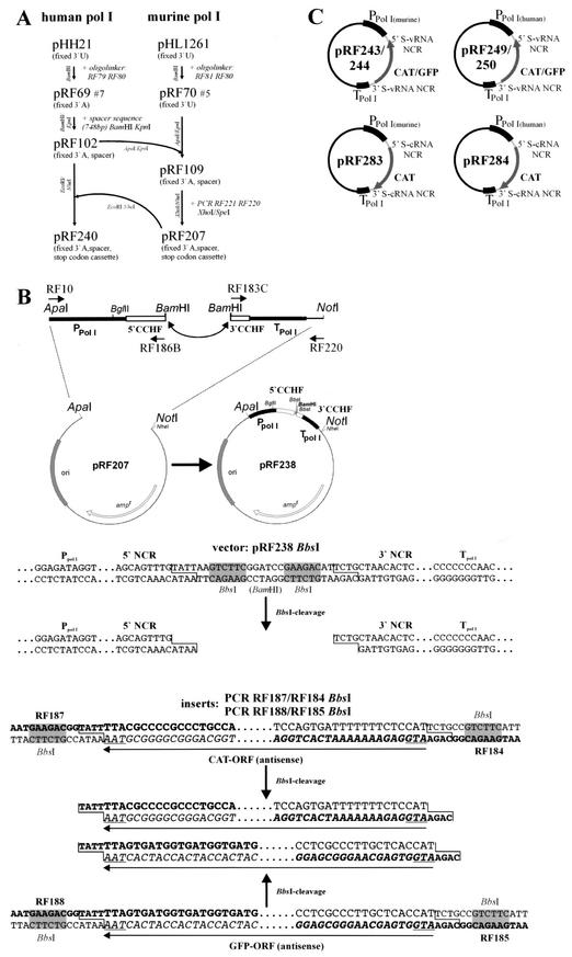
Different artificial CCHF virus minigenomes were designed as schematically outlined in Fig. 1C. Briefly, the terminal noncoding regions (NCRs) of the CCHF virus S segment (168 nucleotides [nt] from the 5′ end and 55 nt from the 3′ end of virion RNA) were combined with chloramphenicol acetyltransferase (CAT) or green fluorescent protein (GFP) reporter genes for expression under the control of the cellular RNA polymerase I (pol I) promoter. To be able to use different eukaryotic cell lines, we generated all constructs for the utilization of either the human (h) or murine (m) pol I systems (19, 29, 46). The basic cloning constructs for the human and murine pol I systems were plasmids pRF240 and pRF207 (Fig. 1A), respectively. Both plasmids were based on the original pol I vector plasmids pHH21 (19, 29), pHL1261 (11), and pRF42 (13) but were constructed to contain a pol I terminator region followed by stop codons for all six possible ORFs (sense and antisense) to prevent false-positive signals after transfection (13) (Fig. 1A). Additional features of the plasmids were a foreign spacer sequence located between two _Bsm_BI sites for more-efficient and controllable restriction enzyme digestion and a fixed adenosine nucleotide at the 3′ end after pol I transcription (Fig. 1B). This facilitates cloning and precise pol I transcription with a terminal adenine (e.g., nairovirus and hantavirus genome segments), rather than a uracil residue (e.g., influenza virus and phleboviruses) (30, 46).
FIG.1.
Design of plasmids. (A) Adaptation and improvement of pol I cloning vectors. The original pol I vectors pHH21 (19, 29) and pHL1261 (11) were modified in several steps. The terminal 3′ nucleotide of the pol I transcript was changed (U→A), which facilitates cloning and precise pol I transcription with a terminal adenine (e.g., nairovirus and hantavirus genome segments), rather than a uracil residue (e.g., influenza virus and phleboviruses) (30, 46). A stop codon cassette to prevent false-positive background signals (13) and a spacer region between the two _Bsm_BI cloning sites for a more efficient and controllable cleavage were also inserted. (B) Construction of chimeric CCHF virus reporter plasmids. Two PCR fragments, one containing the murine (m) pol I promoter and the CCHF virus S vRNA 5′ NCR (primers RF10 and RF186B [Fig. 2]) and the other containing the pol I terminator and the CCHF virus S vRNA 3′ NCR (primers RF183C and RF220 [Fig. 2]), were ligated with the large _Apa_I-_Not_I fragment obtained from plasmid pRF207 containing the components for bacterial amplification and selection. This resulted in plasmid pRF238 (pol I [m] CCHF virus S NCR), containing both CCHF virus S segment NCRs separated by a _Bbs_I-_Bam_HI-_Bbs_I cassette. To construct CCHF virus minigenome plasmids, PCR-amplified reporter gene cassettes were inserted between the _Bbs_I sites (shaded boxes) in the pol I-driven expression plasmid pRF238. The special feature (cleavage outside of the recognition site) of the restriction enzyme _Bbs_I was used for exact insertion of the reporter gene sequence between the two NCRs of the CCHF virus S segment. Oligonucleotide sequences are in bold. Start and stop codons are underlined. Restriction enzyme cleavage sites are shown with a line between the sequences. Restriction enzyme recognition sites are shaded. Reporter gene ORFs are in italics. (C) Schematic diagram of the different CCHF virus S CAT and GFP minigenomes used for reverse genetics studies. PpolI(human) and PpolI(murine), human and murine pol I promoters, respectively; TpolI, murine pol I terminator sequence. vRNA minigenome constructs pRF243 and pRF249 contain CAT reporter genes in antisense orientation, whereas in pRF244 and pRF250 the CCHF virus N ORF is replaced by a GFP gene. cRNA transcription cassettes in sense orientation were inserted in constructs pRF283 and pRF284. The CAT and GFP genes are flanked by the NCR of the CCHF virus S segment, 168 nt representing the 5′ vRNA (or 3′ cRNA NCR) part and 55 nt representing the 3′ vRNA (or 5′ cRNA NCR) part.
vRNA constructs.
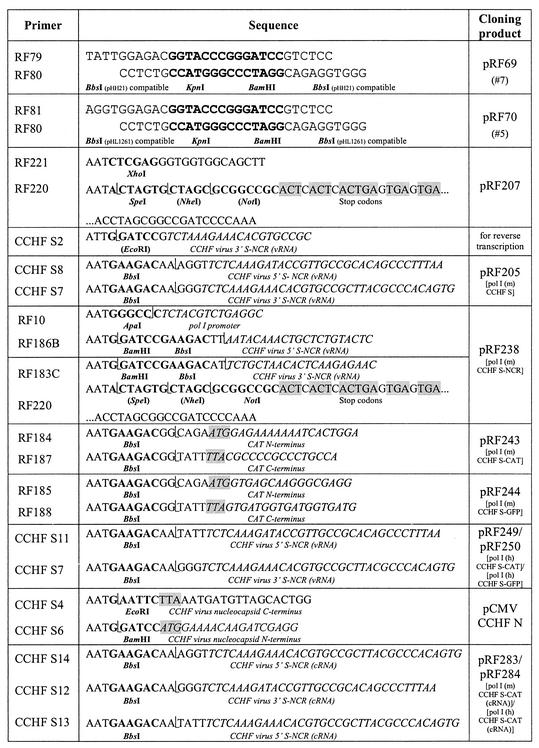
To generate the different CCHF virus RNA minigenomes, the entire S segment was initially reverse transcribed with primer CCHF S2 (Fig. 2) by using total RNA extracted from CCHF virus-infected VeroE6 cells and subsequently amplified by PCR with primers CCHF S7 and S8 (Fig. 2). The resulting PCR fragment of 1,701 bp was cleaved with _Bbs_I and inserted into plasmid pRF109, containing the pol I component (_Bsm_BI fragment; 2,922 bp; Fig. 1A). This resulted in plasmid pRF205 (pol I [m] CCHF virus S [virus RNA {vRNA}]), which can be used for transcription of the full-length CCHF virus S segment by the cellular pol I. To facilitate the exact replacement of the nucleoprotein (N) ORF by different reporter genes (e.g., the CAT and GFP genes), pRF238 was constructed to contain the pol I components and the 5′ and 3′ NCRs of the CCHF virus S segment separated by a _Bbs_I-_Bam_HI-_Bbs_I cassette (Fig. 1B). The construct pRF205 (pol I [m] CCHF virus S [vRNA]) was used as a PCR template to amplify the two CCHF virus S segment NCRs (RF10/RF186B, _Apa_I/_Bam_HI; RF183C/RF220, _Bam_HI/_Not_I) for insertion into the _Apa_I/_Not_I fragment of vector pRF207, resulting in pRF238 (Fig. 1B). In the next step, PCR products containing the CAT gene (RF184 and RF187, _Bbs_I) or the GFP gene (RF185/RF188, _Bbs_I) were inserted into the construct pRF238 (_Bbs_I fragment; 3,186 bp) (Fig. 1B), resulting in pRF243 (pol I [m] CCHF virus S CAT [vRNA]) and pRF244 (pol I [m] CCHF virus S GFP [vRNA]) (Fig. 1C). To utilize the human pol I system, the entire pol I cassettes of pRF243 and pRF244 were amplified (primers CCHF S7 and S11; Fig. 2) and inserted into _Bsm_BI-cleaved pRF240 (Fig. 1A). This resulted in pRF249 (pol I [h] CCHF virus S CAT [vRNA]) and pRF250 (pol I [h] CCHF virus S GFP [vRNA]) (Fig. 1C).
FIG. 2.
Oligonucleotide primers used to construct plasmids. Boldface sequences, restriction enzyme recognition sites; italic sequences, template-complementary nucleotides; shaded sequences, start and stop codons. L-shaped lines mark cleavage sites.
cRNA constructs.
For the construction of virus-complementary RNA (cRNA)-transcribing pol I-driven plasmids, pRF243 or pRF249 were used to amplify the CAT gene flanked by the two NCRs of the CCHF virus S segment. _Bbs_I-restricted PCR fragments (CCHF S12/S14, template pRF243; CCHF S12/S13, template pRF249) were inserted into plasmid pRF207, containing the murine or human pol I components (_Bsm_BI fragment; 2,964 bp), or pRF240 (_Bsm_BI fragment; 2,871 bp), respectively. This resulted in pRF284 (pol I [m] CCHF virus S CAT [cRNA]) and pRF283 (pol I [h] CCHF virus S CAT [cRNA]) (Fig. 1C).
Expression plasmids.
For expression of the CCHF virus-specific N, the N ORF was amplified from plasmid pRF205 (pol I [m] CCHF virus S [vRNA]) with the primers CCHF S4 and S6 (Fig. 2). The 1,467-bp PCR fragment was cleaved with _Eco_RI and _Bam_HI and ligated into the _Eco_RI/_Bam_HI fragment of the vector pcDNA3(+) (Invitrogen/Life Technologies). This resulted in a cytomegalovirus (CMV) promoter-driven expression plasmid for the CCHF virus nucleoprotein (pCMV CCHF N).
Transfection and superinfection with CCHF virus.
Cell lines were seeded in tissue culture dishes (diameter, 6 cm) and transfected with the different minigenome plasmid DNAs by using Lipofectamine Plus reagent (Invitrogen/Life Technologies). Transfections were performed as described previously (10, 13). To determine the efficiency of transfection, the plasmid pHL2823, which contained an enhanced GFP gene under the control of the CMV promoter (R. Flick and G. Hobom, unpublished data), was similarly transfected. Transfected cells were superinfected 24 h posttransfection with CCHF virus at a multiplicity of infection (MOI) of 0.1 to 0.001 PFU/cell under biosafety level 4 (BSL-4) conditions. Briefly, 2 ml of virus dilutions in cell culture medium (without fetal calf serum [FCS] and antibiotics) was incubated for 1 h in 6-cm-diameter cell culture plates (37°C, 5% CO2). Subsequently, virus was removed and 5 ml of cell culture medium (with 5% FCS and antibiotics) was added. Superinfection experiments with CCHF virus were performed in the BSL-4 laboratories at the Canadian Science Centre for Human and Animal Health (Winnipeg, Canada).
Passaging of recombinant CCHF virus.
BHK-21 or 293T cells were transfected as described above and superinfected 24 h later with CCHF virus at an MOI of 0.001 PFU/cell. Cells were analyzed for CAT activity 48 h p.i., and the corresponding supernatants were used for virus passaging. Cell debris was removed by centrifugation at 3,000 × g for 10 min, and cells (approximately 106 HuH-7 or VeroE6 cells) were infected with 2 ml of undiluted supernatant (passage 1). After 1 h of incubation (37°C, 5% CO2), the inoculum was replaced by fresh medium (Dulbecco's modified Eagle medium, 5% FCS, antibiotics), and cells were incubated for 30 to 48 h. This procedure was repeated twice (passages 2 and 3).
CAT assays.
Cell extracts were prepared as described by Gorman et al. (17), and CAT activity was determined by using the commercially available Flash Cat kit (Stratagene) as described previously (10, 13). The reaction products were visualized by UV illumination, documented by photography, and evaluated with WinCam (Cybertech, Berlin, Germany) or Quantity One (Bio-Rad) software. Ratios of activities were calculated based on at least three independent sets of serial dilutions of cell lysates down to a level of 30 to 50% product formation for better quantification within a linear range. CAT activities after transfection of 8 μg of pRF243, for the analysis of vRNA minigenomes, and pRF283, for cRNA minigenome studies, followed by CCHF virus superinfection were arbitrarily set at 100% in all experiments.
Cell fixation and UV microscopy.
Cells transfected with GFP-containing minigenome constructs and superinfected with CCHF virus were fixed with 4% paraformaldehyde for at least 12 h at 4°C. The paraformaldehyde was changed prior to removal from the BSL-4 laboratory. GFP expression was visualized with an Axioplan 2 microscope (Zeiss) and documented with a 3CDC color video camera (DXC-970MD; Sony) and the Northern Eclipse 6 imaging software (Empix Imaging, Inc.).
RESULTS
Susceptibility of eukaryotic cells to infection with CCHF virus.
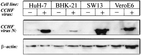
A helper virus-driven reverse genetics system is dependent on the capacity of a cell line to propagate the virus. Therefore, we initially investigated the susceptibilities of different eukaryotic cell lines (BHK-21, VeroE6, SW13, HuH-7, and 293T) to CCHF virus, strain IbAr10200. Cells were infected at an MOI of 0.001 PFU/cell, harvested 48 h p.i., and subjected to immunoblot analysis using a polyclonal antiserum against the nucleoprotein (A. Holmstroem and R. Flick, unpublished data). All tested cell lines promoted the growth of CCHF virus, as indicated by the expression of the viral nucleoprotein (N) (Fig. 3). SW13 cells displayed the highest N expression levels and release of progeny viruses, as determined by a plaque assay (18) and 50% tissue culture infectious dose analysis (21) (data not shown) and were, therefore, used for the preparation of virus stocks.
FIG. 3.
Susceptibilities of different eukaryotic cell lines for CCHF virus. Different eukaryotic cells (HuH-7, BHK-21, SW13, and VeroE6) were infected with CCHF virus at an MOI of 0.001 PFU/cell, harvested 48 h p.i., and subjected to immunoblot analysis using a polyclonal antiserum against the nucleoprotein (A. Holmstroem and R. Flick, unpublished data) (top). β-Actin was used as a loading control (bottom).
GFP expression from CCHF virus-specific minigenomes.
For the development of a CCHF virus reverse genetics system, we designed different artificial virus-specific minigenomes as schematically outlined in Fig. 1C. In a first approach different eukaryotic cell lines were tested for the rescue of reporter gene expression. Prior to transfection with the reporter gene constructs, the transfection efficiencies of the different cell lines were determined by using a CMV promoter-driven enhanced GFP expression plasmid (pHL2823; R. Flick and G. Hobom, unpublished data). The 293T cells showed the highest efficiency (70 to 80%), followed by BHK-21 cells (15 to 20%). HeLa and SW13 cells displayed a transfection rate of less than 5% (data not shown).
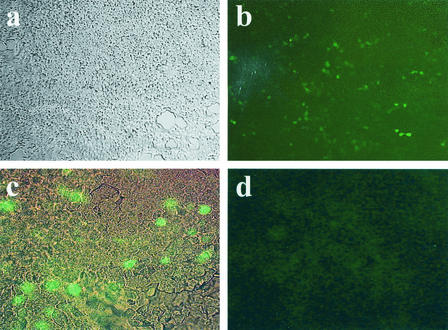
Subsequently, 293T, HeLa, and SW13 cells were transfected with the CCHF virus GFP minigenome plasmids driven by the human pol I system (pRF250; Fig. 1C), whereas the murine pol I system (pRF244; Fig. 1C) was tested in BHK-21 cells, which had previously been demonstrated as suitable cells for this particular promoter (13). The transfected plasmids were transcribed by the cellular pol I, resulting in vRNA-like minigenome molecules (46). For encapsidation, transcription, and replication of the pol I-generated artificial RNA segments, the vRNA-dependent RNA polymerase protein (L) and the nucleoprotein (N) were provided by superinfection with CCHF helper virus under BSL-4 conditions. GFP expression was detected in cells transfected with plasmids pRF244 or pRF250 24 h postsuperinfection (Fig. 4; data shown for pRF250 in 293T cells). The number of GFP-expressing cells varied from 10% for 293T cells (pRF250) to 5% for BHK-21 cells (pRF244) to less than 1% for HeLa and SW13 cells (pRF250) (data not shown). These results were consistent with the different transfection rates (see above) and clearly demonstrated that CCHF virus was able to recognize and transcribe the artificial RNA segments generated by the cellular pol I. GFP expression levels increased within the next 2 days (48 and 72 h postsuperinfection [data not shown]). This indicates that a continuous production of pol I-driven transcripts and their transcription driven by viral proteins lead to an accumulation of reporter gene product (GFP) in the cells (Fig. 4). The successful rescue of CCHF virus minigenomes by superinfection proves that the features important for recognition, encapsidation, and transcription of the artificial RNA segments by the viral RNA polymerase and nucleoprotein are located in the terminal NCRs of the CCHF virus S segment, as recently described in detail for the phlebovirus Uukuniemi (UUK) virus (10).
FIG. 4.
GFP vRNA minigenome rescue by CCHF virus superinfection. 293T cells were transfected with 4 μg of plasmid pRF250 (GFP gene-containing CCHF virus minigenome construct [pol I {h} CCHF virus S CAT vRNA) and superinfected with CCHF virus 24 h later. Cell monolayers were fixed and inactivated 48 h p.i. with 4% paraformaldehyde overnight prior to removal from the containment laboratory. (a) Light microscopy of transfected and superinfected cells. (b) Immunofluorescence of transfected and superinfected cells. (c) Same cells as in panel b but at higher magnification. (d) Immunofluorescence of transfected but nonsuperinfected cells.
CAT expression from CCHF virus-specific vRNA minigenomes.
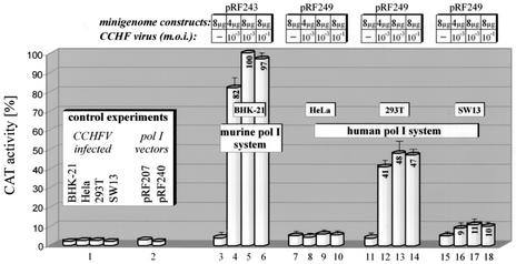
To confirm the results and develop a system for a more objective quantification, we used CCHF virus-specific minigenome constructs containing the CAT reporter gene. The same cell lines (see above) were transfected with plasmids carrying the CCHF virus S CAT minigenomes (pRF243 or pRF249; Fig. 1C) and were subsequently superinfected with CCHF virus 24 h later. High CAT activity, comparable to that for our recently published UUK virus reverse genetics system (13), was detected only in 293T and BHK-21 cell extracts prepared 48 h postsuperinfection (Fig. 5, lanes 4 to 6 and 12 to 14, respectively), whereas SW13 cells showed only very weak signals (Fig. 5, lanes 16 to 18), and in HeLa cells no reporter gene activity could be determined (Fig. 5, lanes 8 to 10). This may be due to the low transfection efficiency of the HeLa and SW13 cell lines as described above. As with the GFP reporter minigenome (Fig. 4), the CAT activity data confirmed that CCHF virus was able to recognize and transcribe an artificial minigenome RNA generated by the cellular pol I. CAT activity could not be increased following superinfection at increasing MOI (0.0001 and 0.1 PFU/cell) (Fig. 5, lanes 5 versus 6 and 13 versus 14), which might be due to an increased cytopathic effect upon superinfection at a higher MOI. However, an increase in CAT activity was observed with larger amounts of pol I cassette DNA (8 μg), especially for BHK-21 cells (Fig. 5, lane 4 versus 5). A similar increase was not detected when DNA was transfected without superinfection (Fig. 5, lane 3).
FIG. 5.
CAT vRNA minigenome rescue by CCHF virus superinfection. Different eukaryotic cell lines were transfected with different amounts of CAT-containing CCHF virus minigenome constructs (pRF243 [pol I {m} CCHF virus S CAT {vRNA}] and pRF249 [pol I {h} CCHF virus S CAT {vRNA}]) and subsequently superinfected with CCHF virus (MOIs of 0.1 and 0.001 PFU/cell). Cells were harvested 72 h posttransfection (48 h p.i.) and analyzed for CAT activity. Lane 1, reporter gene background activity in BHK-21, HeLa, 293T, and SW13 cells upon CCHF virus infection; lane 2, reporter gene background activity after transfection of pol I vector plasmids (pRF207 and pRF240); lanes 3, 7, 11, and 15, reporter gene background activity after transfection of different CAT vRNA minigenomes (nonsuperinfected); lanes 4 to 6, CAT activity in BHK-21 cells transfected with plasmid pRF243 (Fig. 1C) and superinfected with CCHF virus; lanes 8 to 10, HeLa cells transfected with plasmid pRF249 (Fig. 1C) and superinfected with CCHF virus; lanes 12 to 14, 293T cells transfected with plasmid pRF249 and superinfected with CCHF virus; lanes 16 to 18, SW13 cells transfected with plasmid pRF249 and superinfected with CCHF virus.
Optimization of reporter gene expression.
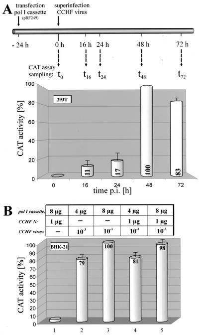
In the next step, we tried to optimize the system by determining the time course of reporter gene expression. Following transfection with the human pol I-driven CCHF virus S CAT minigenome construct pRF249 (Fig. 1C) and subsequent superinfection with CCHF virus, 293T cells were harvested at different time points p.i. and subsequently analyzed for CAT activity (Fig. 6A). CAT signals were clearly detectable at 16 h postsuperinfection and significantly increased over time, with the strongest signals at 48 h p.i. Longer incubation periods did not increase CAT activity due to virus-induced cytopathic effect in infected cultures. Based on these results cells were harvested and analyzed for CAT activity in all following experiments at 48 h p.i.
FIG. 6.
Optimization of the reverse genetics system. (A) Time course of reporter gene expression. 293T cells were transfected with the pol I expression cassette plasmid pRF249 (pol I [h] CCHF virus S CAT [vRNA]; Fig. 1C) 24 h prior to superinfection with CCHF virus (see scheme for the experimental procedure). Cells were harvested at different time points postsuperinfection and analyzed for CAT activity. The diagram summarizes the results from two independent experiments. (B) Effect of additional nucleoprotein on reporter gene expression. BHK-21 cells were transfected with 4 (lanes 2 and 4) or 8 μg (lanes 1, 3, and 5) of plasmid pRF243 (pol I [m] CCHF virus S CAT [vRNA]; Fig. 1C) and cotransfected with 1 μg of plasmid pCMV CCHF N (lanes 1, 4, and 5) or 1 μg of plasmid pcDNA3(+) (lanes 2 and 3) to maintain similar transfection conditions. Subsequently, cells were superinfected with CCHF virus 24 h posttransfection, harvested 72 h posttransfection (48 h p.i.), and analyzed for CAT activity. A transfected (8 μg of RF243) but noninfected culture was used as a control (lane 1).
To further improve the reporter gene expression from artificial RNA segments, a CMV-driven plasmid for the expression of the CCHF virus nucleoprotein was constructed (pCMV CCHF N). Following cotransfection with pCMV CCHF N and pRF243 (pol I [m] CCHF virus S CAT [vRNA]; Fig. 1C), BHK-21 cells were superinfected with CCHF virus, harvested 48 h postsuperinfection, and analyzed for CAT activity. As a control experiment for determining the effect of the CCHF virus N expression plasmid on minigenome reporter gene expression and to maintain similar transfection conditions, pRF243-transfected cells were cotransfected with 1 μg of the pcDNA3(+) plasmid. As already demonstrated (Fig. 5, lane 4 versus 5), CAT activity could be increased by transfecting larger amounts of the pol I-driven minigenome plasmid (Fig. 6B, lane 2 versus 3 and 4 versus 5). Coexpression of the viral nucleoprotein did not result in any change in CAT activity (Fig. 6B, lane 2 versus 4 and 3 versus 5), indicating that the viral N protein is not a limiting factor for this reverse genetics system. In addition, an inhibiting effect of the overexpressed CCHF virus N could not be detected. This is interesting, since it was speculated that, due to their large sizes, the nucleoproteins of nairoviruses (∼50 kDa) and hantaviruses (∼48 kDa) might have additional functions as do the NSs proteins of other bunyaviruses (∼26 to 28 kDa). However, for some bunyaviruses (e.g., Bunyamwera and Rift Valley fever viruses) an inhibitory effect on minigenome rescue appears to be associated with the NSs proteins (43; unpublished data).
Replication and transcription of CCHF virus-specific cRNA minigenomes.
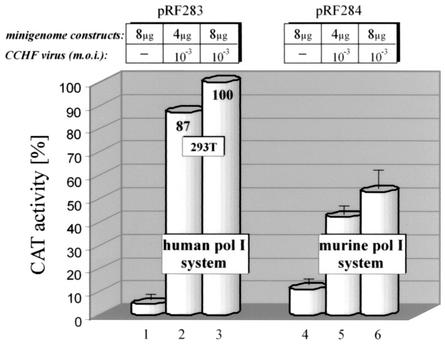
vRNA minigenomes are a perfect tool to analyze the recognition of the promoter site(s) by the viral polymerase, the use of the encapsidation signal(s) by the viral nucleoproteins, and transcription into a functional mRNA. CAT activity can theoretically be obtained either by direct transcription of the pol I-generated minigenome RNAs (primary transcription) or by transcription of the vRNA minigenomes after replication (vRNA→cRNA→vRNA) and, therefore, amplification of the pol I transcripts (secondary transcription). To address this question and study the replication of the pol I-generated CCHF virus minigenomes, constructs were generated with cRNA transcription cassettes inserted in sense orientation between the pol I promoter and terminator sequences. This resulted in pRF284, a CCHF virus minigenome driven by the murine pol I promoter, and pRF283 for pol I transcription in primate cells (Fig. 1C). These CCHF virus cRNA minigenomes have to undergo replication (cRNA→vRNA) prior to serving as templates for the generation of functional viral mRNA molecules (vRNA→mRNA). Therefore, they are useful tools for analyzing minigenome replication in the described reverse genetics system. Following transfection of pRF283 into 293T cells or pRF284 into BHK-21 cells and subsequent superinfection with CCHF helper virus, CAT activity could be detected in both cell extracts (Fig. 7, lanes 2 and 3 and 5 and 6, respectively). This demonstrates that the viral L protein mediates transcription and replication of pol I-driven minigenomes, confirming that the terminal 168 nt (3′ cRNA NCR) and 55 nt (5′ cRNA NCR) contain all necessary sequence information for encapsidation, transcription, and replication.
FIG. 7.
CCHF virus S CAT cRNA minigenome rescue. 293T and BHK-21 cell lines were transfected with different amounts of the human (pRF283) and murine (pRF284) pol I-driven, CAT-containing CCHF virus cRNA minigenome plasmids (Fig. 1C) as indicated. Twenty-four hours later, cells were superinfected with CCHF virus (MOI as indicated), harvested 72 h posttransfection (48 h p.i.), and analyzed for CAT activity. Lanes 1 and 4, transfected but noninfected controls; lanes 2 and 3, 293T cells transfected with pRF283 and superinfected with CCHF virus; lanes 5 and 6, BHK-21 cells transfected with pRF284 and superinfected with CCHF virus.
Generation of recombinant CCHF virus.
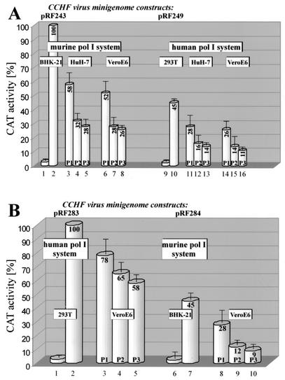
Following the successful rescue (encapsidation, replication, and transcription) of CCHF helper virus-driven minigenomes (vRNA and cRNA) with different reporter genes (CAT and GFP genes), we analyzed the capacity for packaging of pol I transcripts and production of recombinant CCHF virus particles. For this we transfected BHK-21 cells with pRF243 for vRNA minigenome analysis or with pRF284 for cRNA-transcribed minigenomes. In parallel, we transfected 293T cells with pRF249 or pRF283 for vRNA or cRNA analyses, respectively. All cells were superinfected with CCHF virus 24 h posttransfection, and supernatants were collected 30 to 48 h later (i.e., 54 to 72 h posttransfection). The supernatants were used to infect new HuH-7 and VeroE6 cultures, a step that was repeated two more times. Cell lysates from each passage were analyzed for CAT activity. As shown in Fig. 8, CAT activity could be detected in cells transfected with the minigenome plasmids and superinfected with CCHF virus (Fig. 8A, lanes 2 and 10, and B, lanes 2 and 7). In addition CAT activity could be passaged at least three times, regardless of whether vRNA or cRNA minigenomes were transfected, (Fig. 8A, lanes 3 to 5, 6 to 8, 11 to 13, and 14 to 16, and B, lanes 3 to 5 and 8 and 9), but the activity decreased with each passage. If superinfection was omitted, no CAT activity could be detected in minigenome construct-transfected cells and upon passaging (Fig. 8A, lanes 1 and 9, and B, lanes 1 and 6; passaging data not shown). The data clearly demonstrated that CAT activity was serially transferred to new cells. This indicated that the chimeric reporter RNA was packaged into progeny virus particles, which were subsequently released; this sequence of events is similar to what occurs in a normal infectious cycle. This is a very important step in the generation of recombinant CCHF viruses in a helper virus-free system (infectious clone). It confirms that the pol I-generated minigenomes are transcribed, replicated, and packaged into progeny viruses and forms the basis for the development of an infectious clone system. Surprisingly, CAT activity remained at fairly high levels (58% with cRNA minigenomes in 293T cells, 26% with vRNA minigenomes in BHK-21 cells) even after three passages. This is in contrast to data obtained with the UUK virus reverse genetics system, where only a weak (6%) CAT activity could be detected in the first passage (13). This indicates a strong rescue of CCHF virus-specific minigenomes by CCHF helper virus superinfection.
FIG. 8.
Packaging of pol I-driven minigenome segments into CCHF particles. Murine (pRF243 and pRF284) and human (pRF249 and pRF283) pol I-driven CCHF virus vRNA and cRNA minigenomes (Fig. 1C) were transfected into BHK-21 or 293T cells 24 h prior to superinfection with CCHF virus (MOI, 0.001 PFU/cell). Cell lysates were prepared 48 h later and assayed for CAT activity, while the cell culture media were collected and 2-ml undiluted samples were transferred to fresh HuH-7 or VeroE6 cells (P1). This was repeated twice (P2 and P3), and CAT activity was assayed after each passage. (A) vRNA minigenomes. Two different cell lines (HuH-7 and VeroE6) were used for passaging recombinant CCHF viruses derived from either the murine (lanes 3 to 8) or human (lanes 11 to 16) pol I system. Lane 1, pRF243-transfected but nonsuperinfected BHK-21 cells; lane 2, pRF243-transfected and superinfected BHK-21 cells; lanes 3 to 5, P1 to P3 in HuH-7 cells; lanes 6 to 8, P1 to P3 in VeroE6 cells; lane 9, pRF249-transfected but nonsuperinfected 293T cells; lane 10, pRF249-transfected and superinfected 293T cells; lanes 11 to 13, P1 to P3 in HuH-7 cells; lanes 14 to 16, P1 to P3 in VeroE6 cells. (B) cRNA minigenomes. VeroE6 cells were used for passaging recombinant CCHF viruses derived from either the human (lanes 2 to 5) or murine (lane 7 to 10) pol I system. Lane 1, pRF283-transfected but nonsuperinfected 293T cells; lane 2, pRF283-transfected and superinfected 293T cells; lanes 3 to 5, P1 to P3 in VeroE6 cells; lane 6, pRF284-transfected but nonsuperinfected BHK-21 cells; lane 7, pRF284-transfected and superinfected BHK-21 cells; lanes 8 to 10, P1 to P3 in VeroE6 cells. The results of two independent experiments are summarized.
DISCUSSION
The recent development of a reverse genetics system for bunyaviruses (13) based on the pol I transcription system (30, 46) paved the way to manipulate these viruses without using the traditional but cumbersome T7 polymerase expression system (14). The pol I system, pioneered by Hobom and colleagues (30, 46), uses the DNA-dependent pol I, an enzyme that is already part of all eukaryotic cells and that thus does not have to be provided in trans. Furthermore, the initiation and termination of pol I-derived transcripts are clearly defined (11, 46), and correct transcript ends can be produced without the need for inserted hepatitis virus δ ribozyme sequences (32). Most importantly, pol I-driven transcripts also lack 5′ and 3′ modifications (e.g., cap structure and poly[A] tail) (11, 46) and are thus similar to the genomic ends of negative-strand RNA viruses.
The approach described here represents the first successful reverse genetics system for nairoviruses. Rescue of reporter gene expressing minigenomes for other members of the Bunyaviridae family were reported only for Bunyamwera virus, genus Bunyavirus (6), Rift Valley fever (25) and UUK (13) viruses, both genus Phlebovirus, and very recently Hantaan virus, genus Hantavirus (9). We demonstrated that CCHF virus vRNA and even cRNA minigenomes could be generated by pol I and rescued by a helper virus-dependent reverse genetics system (Fig. 4, 5, and 7). In particular, the use of cRNA transcripts in a recombinant vaccinia virus T7-driven system is limited, because of the strong false-positive signals that can be found after transfection of cRNA minigenomes into vaccinia virus-infected eukaryotic cells. This is due to a vaccinia virus-encoded capping mechanism, which enables translation of the transfected cRNA molecules (6). Furthermore, this is the only study that has reported helper virus-driven rescue of bunyavirus cRNA minigenomes based on the pol I system (Fig. 7). The functionality of similar minigenomes had been demonstrated before, but only with an expression plasmid-driven bunyavirus reverse genetics system (13). Attempts to rescue cRNA minigenomes by using a UUK helper virus-driven reverse genetics system failed (13). In addition, we describe successful transfer of reporter gene activity via passaging of recombinant viruses using bunyavirus vRNA and even cRNA minigenomes for the primary transfection step, demonstrating efficient replication and packaging of pol I-driven CCHF virus minigenomes (Fig. 8).
Surprisingly, reporter gene activity could be transferred at relatively high levels by passaging recombinant bunyaviruses several times to fresh cells (Fig. 8). There is no apparent need for the virus to keep a CAT gene-containing artificial RNA genome segment, particularly since no pressure was used to select recombinant virus over wild-type helper virus. It remains to be analyzed if recombinant CCHF viruses contain the minigenome as an additional fourth genome segment or if the minigenome replaces a wild-type genomic segment. In the latter case a double infection would be necessary to complement the missing gene function in order to obtain reporter gene activity. This seems rather unlikely given the low MOI used in these studies.
The pol I system was originally developed for reverse genetics on influenza A viruses (12, 28, 30, 46), which use the nucleus during their replication cycle. Data presented here confirm our recently published results for UUK virus (13) indicating that this system can also be used for the development of reverse genetics approaches for negative-strand RNA viruses with a cytoplasmic replication cycle. Our data also showed that human pol I-derived artificial RNAs can be used for reverse genetics studies of viruses of the Bunyaviridae family. This is of particular importance, since it will allow the establishment of a reverse genetics systems for viruses with cell and host tropisms restricted to primates. In addition, pol I-driven transcripts in cRNA orientation could facilitate the future rescue of recombinant virus entirely from cDNA (infectious clone), since the formation of RNA double strands with virus-specific mRNA molecules could be avoided (36). The missing L segment sequence and L protein clone of CCHF virus are currently the limiting factors to performing this study.
The system described here will facilitate studies on the biology of nairoviruses in general and, following the development of an animal model, the pathogenesis of CCHF in particular. Thus, it will greatly improve the chances of developing urgently needed prophylactic measures and more feasible therapeutic interventions. The established helper virus system can already be used to modify the viral genome segments and study the impact on the viral replication cycle, as demonstrated before for other bunyaviruses (1, 6, 10, 13, 25, 33, 40). Insertions of genetic markers into the viral genome will later facilitate the identification of recombinant viruses and document reassortment events. Nucleotide exchanges in the noncoding regions will provide us with information regarding the _cis_-acting elements within these terminal nucleotides and, therefore, will help in determining important regulatory elements of virus replication and in performing antiviral-drug screening. It will also help in the study of the function of viral proteins in the replication cycle of the virus by allowing introduction of mutations into the different proteins. Furthermore, this reverse genetics system provides the opportunity to identify attenuation markers within the CCHF virus genome segments and, thus, helps in the development of attenuated recombinant viruses that could be evaluated as vaccine candidates.
Acknowledgments
We thank Anna Wallin, Daryl Dick, and Michael Garbutt for technical assistance. We thank Connie Schmaljohn (U.S. Army Medical Research Institute for Infectious Diseases, Frederick, Md.) and Markus Czub (Special Pathogens Program, National Microbiology Laboratory, Health Canada) for helpful discussions and suggestions. The CCHF virus was kindly provided by the Special Pathogens Branch, Centers for Disease Control and Prevention, Atlanta, Ga. (T. G. Ksiazek). We thank Jonathan Smith (Alphavax, Durham, N.C.) for initial help in setting up the CCHF virus system.
Kirsten Flick held a Short Term Fellowship from the Human Frontier Science Program, Strasbourg, France. She currently holds a postdoctoral fellowship from the Canadian Natural Sciences and Engineering Research Council. This work was financially supported by a grant from the National Institutes of Health (Rapid Response Grant Program for Bioterrorism-Related Research no. 1R21 AI 053560-01) and was funded by the Department of Emergency and Disaster Planning, National Board of Health and Welfare, Sweden.
The work was equally performed at the Canadian Science Centre for Human and Animal Health (Winnipeg, Canada) and the Swedish Institute for Infectious Disease Control (Solna, Sweden).
REFERENCES
- 1.Bridgen, A., and R. M. Elliott. 1996. Rescue of a segmented negative-strand RNA virus entirely from cloned complementary DNAs. Proc. Natl. Acad. Sci. USA 93**:**15400-15404. [DOI] [PMC free article] [PubMed] [Google Scholar]
- 2.Casals, J. 1969. Antigenic similarities between the virus causing Crimean hemorrhagic fever and Congo virus. Proc. Soc. Exp. Biol. Med. 131**:**233-236. [DOI] [PubMed] [Google Scholar]
- 3.Chumakov, M. P. 1945. A new tick-borne virus disease—Crimean hemorrhagic fever, p. 13. In A. A. Sokolov, M. P. Chumakov, and A. A. Kolachev (ed.), Crimean hemorrhagic fever (acute infectious capillary toxicosis). Izd. Otd. Primorskoi Armii, Simferopol, Russia.
- 4.Chumakov, M. P. 1974. Contribution to 30 years of investigation of Crimean haemorrhagic fever. Med. Virol. 22**:**5-18. [Google Scholar]
- 5.Clerex-van Haaster, C. M., J. P. M. Clerex, H. Ushijima, H. Akashi, F. Fuller, and D. H. Bishop. 1982. The 3′ terminal RNA sequences of bunyaviruses and nairoviruses (Bunyaviridae): evidence of end sequence generic differences within the virus family. J. Gen. Virol. 61**:**289-292. [DOI] [PubMed] [Google Scholar]
- 6.Dunn, E. F., D. C. Pritlove, H. Jin, and R. M. Elliott. 1995. Transcription of a recombinant bunyavirus RNA template by transiently expressed bunyavirus proteins. Virology 211**:**133-143. [DOI] [PubMed] [Google Scholar]
- 7.Elliott, R. M., C. S. Schmaljohn, and M. S. Collett. 1991. Bunyaviridae genome structure and gene expression. Curr. Top. Microbiol. Immunol. 169**:**91-141. [DOI] [PubMed] [Google Scholar]
- 8.Fisher-Hoch, S. P., J. B. McCormick, R. Swanepoel, A. Van Middlekoop, S. Harvey, and H. G. Kustner. 1992. Risk of human infections with Crimean-Congo hemorrhagic fever virus in a South African rural community. Am. J. Trop. Med. Hyg. 47**:**337-345. [DOI] [PubMed] [Google Scholar]
- 9.Flick, K., J. Hooper, C. S. Schmaljohn, R. F. Pettersson, H. Feldmann, and R. Flick. 2002. Rescue of Hantaan virus minigenomes. Virology 306**:**219-224 [DOI] [PubMed]
- 10.Flick, R., F. Elgh, G. Hobom, and R. F. Pettersson. 2002. Mutational analysis of the Uukuniemi virus (Bunyaviridae) promoter reveals two regions of functional importance. J. Virol. 76**:**10849-10860. [DOI] [PMC free article] [PubMed] [Google Scholar]
- 11.Flick, R., and G. Hobom. 1999. Transient bicistronic vRNA segments for indirect selection of recombinant influenza viruses. Virology 262**:**93-103. [DOI] [PubMed] [Google Scholar]
- 12.Flick, R., G. Neumann, E. Hofmann, E. Neumeier, and G. Hobom. 1996. Promoter elements in the influenza vRNA terminal structure. RNA 2**:**1046-1057. [PMC free article] [PubMed] [Google Scholar]
- 13.Flick, R., and R. F. Pettersson. 2001. Reverse genetics system for Uukuniemi virus (Bunyaviridae): RNA polymerase I-catalyzed expression of chimeric viral RNAs. J. Virol. 75**:**1643-1655. [DOI] [PMC free article] [PubMed] [Google Scholar]
- 14.Fuerst, T. R., E. G. Niles, F. W. Studier, and B. Moss. 1986. Eukaryotic transient-expression system based on recombinant vaccinia virus that synthesizes bacteriophage T7 RNA polymerase. Proc. Natl. Acad. Sci. USA 83**:**8122-8126. [DOI] [PMC free article] [PubMed] [Google Scholar]
- 15.Gear, J. H. S. 1988. Crimean-Congo hemorrhagic fever, p. 121-129. In J. H. S. Gear (ed.): CRC handbook of viral and rickettsial hemorrhagic fevers. CRC Press, Inc., Boca Raton, Fla.
- 16.Gonzales-Scarano, F., and N. Nathanson. 1996. Bunyaviridae, p. 1473-1504. In B. N. Fields, D. M. Knipe, and P. M. Howley (ed.), Fields virology, 3rd ed. Lippincott-Raven Publishers, Philadelphia, Pa.
- 17.Gorman, C. M., L. F. Moffat, and B. H. Howard. 1982. Recombinant genomes which express chloramphenicol acetyltransferase in mammalian cells. Mol. Cell. Biol. 2**:**1044-1051. [DOI] [PMC free article] [PubMed] [Google Scholar]
- 18.Hodgson, L. 1992. The sequence, genomic organization, and expression of the small genomic segment of Crimean-Congo hemorrhagic fever virus for the potential use as a diagnostic antigen. M.S. thesis. Hood College, Frederick, Md.
- 19.Hofmann, E. 1997. Ph.D. thesis. Justus Liebig University, Giessen, Germany.
- 20.Hoogstral, H. 1979. The epidemiology of tickborne Crimean-Congo hemorrhagic fever in Asia, Europe and Africa. J. Med. Entomol. 15**:**307-417. [DOI] [PubMed] [Google Scholar]
- 21.Jones, H. W., Jr., V. A. McKusick, P. S. Harper, and K. D. Wuu. 1971. George Otto Gey (1899-1970). The HeLa cell and a reappraisal of its origin. Obstet. Gynecol. 38**:**945-949. [PubMed] [Google Scholar]
- 22.Karber, G. 1931. 50% end-point calculation. Arch. Exp. Pathol. Pharmakol. 162**:**480-483. [Google Scholar]
- 23.Leshchinskaya, E. V. 1965. Clinical picture of Crimean hemorrhagic fever. Tr. Inst. Polio. Virusn. Entsefalitov Akad. Med. Nauk SSSR **7:**226-236. (In Russian; in English at NAMRU3-1856).
- 24.Logan, T. M., K. J. Linthicum, C. L. Bailey, D. M. Watts, and J. R. Moulton. 1989. Experimental transmission of Crimean-Congo hemorrhagic fever virus by Hyalomna truncatum Koch. Am. J. Trop. Med. Hyg. 40**:**207-212. [DOI] [PubMed] [Google Scholar]
- 25.Lopez, N., R. Muller, C. Prehaud, and M. Bouloy. 1995. The L protein of Rift Valley fever virus can rescue viral ribonucleoproteins and transcribe synthetic genome-like RNA molecules. J. Virol. 69**:**3972-3979. [DOI] [PMC free article] [PubMed] [Google Scholar]
- 26.Marriott, A. C., and P. A. Nuttall. 1996. Molecular biology of nairoviruses, p. 91-104. In R. M. Elliott (ed.), The Bunyaviridae. Plenum Press, New York, N.Y.
- 27.Mayers, D. L. 1999. Exotic virus infections of military significance: hemorrhagic fever viruses and pox virus infections. Dermatol. Clin. 17**:**29-40. [DOI] [PubMed] [Google Scholar]
- 28.Neumann, G., and G. Hobom. 1995. Mutational analysis of influenza virus promoter elements in vivo. J. Gen. Virol. 76**:**1709-1717. [DOI] [PubMed] [Google Scholar]
- 29.Neumann, G., T. Watanabe, H. Ito, S. Watanabe, H. Goto, P. Gao, M. Hughes, D. R. Perez, R. Donis, E. Hoffmann, G. Hobom, and Y. Kawaoka. 1999. Generation of influenza A viruses entirely from cloned cDNAs. Proc. Natl. Acad. Sci. USA 96**:**9345-9350. [DOI] [PMC free article] [PubMed] [Google Scholar]
- 30.Neumann, G., A. Zobel, and G. Hobom. 1994. RNA polymerase I-mediated expression of influenza viral RNA molecules. Virology 202**:**477-479. [DOI] [PubMed] [Google Scholar]
- 31.Oldfield, E. C., M. R. Wallace, K. C. Hyams, A. A. Yousif, D. D. Lewis, and A. L. Bourgeois. 1991. Endemic infectious diseases of the Middle East. Rev. Infect. Dis. 13(Suppl. 3)**:**S199-S217. [DOI] [PubMed] [Google Scholar]
- 32.Perrotta, A. T., and M. D. Been. 1991. A pseudoknot-like structure required for efficient self-cleavage of hepatitis delta virus RNA. Nature 350**:**434-436. [DOI] [PubMed] [Google Scholar]
- 33.Prehaud, C., N. Lopez, M. J. Blok, V. Obry, and M. Bouloy. 1997. Analysis of the 3′ terminal sequence recognized by the Rift Valley fever virus transcription complex in its ambisense S segment. Virology 227**:**189-197. [DOI] [PubMed] [Google Scholar]
- 34.Schmaljohn, C. S. 1996. Bunyaviridae: the viruses and their replication, p.1447-1471. In B. N. Fields, D. M. Knipe, and P. M. Howley (ed.), Fields virology, 3rd ed. Lippincott-Raven Publishers, Philadelphia, Pa.
- 35.Schmaljohn, C. S., and J. W. Le Duc. 1998. Bunyaviridae, p. 601-628. In L. H. Collier (ed.), Topley and Wilson's microbiology and microbial infections, 9th ed. Edward Arnold, London, United Kingdom.
- 36.Schnell, M. J., T. Mebatsion, and K.-K. Conzelmann. 1994. Infectious rabies viruses from cloned cDNA. EMBO J. 13**:**4195-4203. [DOI] [PMC free article] [PubMed] [Google Scholar]
- 37.Schwarz, T. F., H. Nitschko, G. Jager, H. Nsanze, M. Longson, R. N. Pugh, and A. K. Abraham. 1995. Crimean-Congo haemorrhagic fever in Oman. Lancet 346**:**1230. [DOI] [PubMed] [Google Scholar]
- 38.Swanepoel, R. 1995. Nairovirus infections, p. 285-294. In J. S. Porterfield (ed.), Kass handbook of Infectious diseases. Exotic viral infections. Chapman and Hall, London, United Kingdom.
- 39.Swanepoel, R., D. E. Gill, A. J. Shepherd, P. A. Leman, J. H. Mynhardt, and S. Harvey. 1989. The clinical pathology of Crimean-Congo hemorrhagic fever. Rev. Infect. Dis. 11(Suppl. 4)**:**S794-S800. [DOI] [PubMed] [Google Scholar]
- 40.Tchatalbachev, S., R. Flick, and G. Hobom. 2001. The packaging signal of influenza viral RNA molecules. RNA 7**:**979-989. [DOI] [PMC free article] [PubMed] [Google Scholar]
- 41.van Regenmortel, M. H. V., C. M. Fauquet, D. H. L. Bishop, E. B. Carstens, M. K. Estes, S. M. Lemon, J. Maniloff, M. A. Mayo, D. J. McGeoch, C. R. Pringle, and R. B. Wickner (ed.). 2000. Family Bunyaviridae, p. 599-621. In Virus taxonomy: classification and nomenclature. Seventh report of the International Committee on Taxonomy of Viruses. Academic Press, New York, N.Y.
- 42.Watts, D. M., T. G. Ksiazek, K. J. Linthicum, and H. Hoogstraal. 1988. Crimean-Congo hemorrhagic fever, p. 177-222. In T. P. Monath (ed.), The arboviruses: epidemiology and ecology, vol. II. CRC Press, Inc., Boca Raton, Fla.
- 43.Weber, F., E. F. Dunn, A. Bridgen, and R. M. Elliott. 2001. The Bunyamwera virus nonstructural protein NSs inhibits viral RNA synthesis in a minireplicon system. Virology 281**:**67-74. [DOI] [PubMed] [Google Scholar]
- 44.Yu-Chen, Y., K. Ling-Xiong, L. Ling, Z. Yu-Qin, L. Feng, C. Bao-Jian, and G. Shou-Yi. 1985. Characteristics of Crimean-Congo hemorrhagic fever virus (Xinjiang strain) in China. Am. J. Trop. Med. Hyg. 34**:**1179-1182. [PubMed] [Google Scholar]
- 45.Zeller, H. G., J.-P. Cornet, and J.-L. Camicas. 1994. Experimental transmission of Crimean-Congo hemorrhagic fever virus by west African wild ground-feeding birds to Hyalomma marginatum rufipes ticks. Am. J. Trop. Med. Hyg. 50**:**676-681. [DOI] [PubMed] [Google Scholar]
- 46.Zobel, A., G. Neumann, and G. Hobom. 1993. RNA polymerase I catalysed transcription of insert viral cDNA. Nucleic Acids Res. 21**:**3607-3614. [DOI] [PMC free article] [PubMed] [Google Scholar]
