Recovery of Virulent and RNase-Negative Attenuated Type 2 Bovine Viral Diarrhea Viruses from Infectious cDNA Clones (original) (raw)
Abstract
Cloned cDNA derived from the genome of the virulent type 2 bovine viral diarrhea virus (BVDV) strain NY'93/C was sequenced and served for establishment of the infectious cDNA clone pKANE40A. Virus recovered from pKANE40A exhibited growth characteristics similar to those of wild-type BVDV NY'93/C and proved to be clinically indistinguishable from the wild-type virus in animal experiments. A virus mutant in which the RNase residing in the viral glycoprotein Erns was inactivated, revealed an attenuated phenotype. The plasmid pKANE40A represents the first infectious cDNA clone established for a type 2 BVDV and offers a variety of new approaches to analyze the mechanisms of BVDV-induced disease in cattle.
Bovine Viral Diarrhea Virus (BVDV) is a member of the genus Pestivirus within the family Flaviviridae (62). The genus also includes Classical Swine Fever Virus (CSFV) and Border Disease Virus (BDV) of sheep. The pestiviral genome consists of a single-stranded positive-sense RNA of about 12.3 kb that contains one open reading frame (ORF) coding for a polyprotein of about 4,000 amino acids (38). The ORF is at both ends flanked by untranslated regions (UTR) (2, 6, 29, 40). The 5′ UTR serves as an internal ribosomal entry site (34-36, 42). The viral polyprotein is co- and posttranslationally processed by both viral and host proteases into at least 11 mature proteins arranged in the polyprotein in the order NH2-Npro, C, Erns, E1, E2, p7, NS2-3, NS4A, NS4B, NS5A, and NS5B-COOH (7, 8, 12, 14, 44, 48, 50, 64).
The nucleocapsid protein C and the glycoproteins Erns, E1, and E2 are structural components of the virion (52). Both Erns and E2 are found on virions (61), induce neutralizing antibodies (11, 59, 60), and elicit protective immunity in their natural host (16, 19, 44, 56). A considerable amount of Erns is also secreted from infected culture cells (44); the mechanism by which this protein is bound to the virion has yet to be elucidated. This protein is particularly interesting, since it exhibits an intrinsic RNase activity (18, 47, 63). Abrogation of the RNase activity by point mutations resulted in fully viable virus mutants for CSFV (17, 25). Infection of pigs with the RNase-negative CSFV strain Alfort/Tübingen revealed that inactivation of the RNase is correlated with clinical attenuation (25). However, the pathogenetic mechanism behind the attenuation remains to be solved, and whether the attenuation would be restricted to the particular virus strain or to the swine as a host could not be ruled out.
BVDV induces a variety of symptoms upon infection of its natural host, including fever, leukopenia, respiratory problems, and diarrhea (1, 30, 51). The clinical picture is variable, and in many cases, the infection remains unnoticed. Therefore, difficulties were encountered in reproducing disease by experimental infection with BVDV, and in consequence, it was not possible to clearly define attenuating mutations in the BVDV system for quite a long time. Some years ago, a new type of BVDV infections causing severe thrombocytopenia and hemorrhages in calves and adult cattle was described in North America and, sporadically, Europe (13, 33, 37, 41). Virus isolates from these outbreaks were antigenically and genetically distinct from the known BVDV strains. This group of viruses, which also comprises less-pathogenic viruses, was classified as BVDV type 2 (BVDV-2). The classification was based on nucleotide sequence characteristics of different parts of the genome (3, 33, 41). Although only some of the type 2 strains induce fatal clinical symptoms, they are a major threat to the cattle industry. Vaccines against the classical BVDV-1 provide only partial protection from BVDV-2 infection, and vaccinated dams may produce calves that are persistently infected with BVDV-2 (5, 41). This problem is probably due to the great antigenic diversity between type 1 and type 2 strains, which is most pronounced in the glycoprotein E2, the major target for pestivirus neutralizing antibodies (53, 55, 60). As a consequence of the antigenic variation, most monoclonal antibodies specific for BVDV-1 fail to bind to type 2 viruses (41).
The highly virulent isolate New York '93 (32) (in our case field isolate VLS 399) provided the basis for establishing the first BVDV-2 infectious cDNA clone. Strain NY'93/C was reisolated from the blood of a calf that had been infected with BVDV New York '93 and developed fatal disease. RNA was prepared from cells infected with NY'93/C from the first tissue culture passage and directly used for subsequent work. Northern blot analysis showed that, contrary to BVDV-2 strain 890 (40), the genome of NY'93/C contains no large insertions or deletions (data not shown). A cDNA library was established in phage lambda ZAPII and screened as described before (27). The cDNA synthesis was primed with BVDV-specific oligonucleotides with sequences deduced from published genomic sequences (Ol-BVD13, Ol-BVD14, and Ol-BVD15) (27) and the following oligonucleotides in the 5′ to 3′ orientation: Ol-B22.1R (GTTGACATGGCATTTTTCGTG), Ol-B12.1R (CCTCTTATACGTTCTCACAACG), Ol-BVD33 (GCATCCATCATXCCRTGDAT), Ol-N7-3-7 (CAAATCTCTGATCAGTTGTTCCAC), Ol-B23-RII (TTGCACACGGCAGGTCC), and Ol-B-3′ (GTCCCCCGGGGGCTGTTAAGGGTTTTCCTAGTCCA). BVDV-specific clones were identified by plaque hybridization (probe: _Xho_I-_Aat_II insert of full-length cDNA clone pA/BVDV; GenBank accession no. U63479) (28). Nucleotide sequence analysis of positive clones revealed that the complete viral genome had been cloned, except for the extreme 5′-terminal part and a region covering the sequence coding for the E2 protein. Apparently, the latter sequence was unstable in the vector system employed and was therefore analyzed from cDNA amplified by reverse transcription-PCR (RT-PCR [Titan One Tube RT-PCR System; Boehringer Mannheim, Germany]), with 2 μg of total RNA from cells infected with field isolate VLS 399 and primers CM29 (GATGTAGACACATGCGACAAGAACC) and CM51 (GCTTCCACTCTTATGCCTTG). The amplified RT-PCR products were purified by preparative agarose gel electrophoresis and elution with the Nucleotrap kit as recommended by the manufacturer (Macherey-Nagel, Düren, Germany).
The 5′ UTR (positions 1 to 385) was determined by rapid amplification of cDNA ends (RACE) technology. Single-stranded (−) DNA from the 5′ end of the virus genome was generated with displayThermo-RT reverse transcriptase (Display Systems Biotech, Copenhagen, Denmark) by using 2 μg of total RNA from infected cells and 100 pmol of primer CM79 (CTCCATGTGCCATGTACAGCAGAG), following the manufacturer's instructions (reaction of 65°C for 10 min, 42°C for 40 min, and 65°C for 15 min). The DNA was purified by two sequential phenol-chloroform extractions and ethanol precipitations with 1/4 volume of 10 M ammonium acetate (46). A poly(dA) tail was added to the first cDNA strand with terminal transferase (TdT; Roche Molecular Biochemicals, Mannheim, Germany) with 50% of the “first-strand” product, 50 U of TdT, 6.25 μM dATP, and 1.5 mM CoCl2 in 50 μl of TdT buffer as recommended by the manufacturer. After incubation at 37°C for 30 min, the product was purified by phenol-chloroform extraction and ethanol precipitation. Subsequently, two rounds of PCR were conducted with Tfl polymerase (Promega, Mannheim, Germany) under the conditions recommended by the supplier and primers T25V primer (Display Systems Biotech, Copenhagen, Denmark) plus either CM79 or CM86 (CTCGTCCACATGGCATCTCGAGA; nested PCR primer). The sequence of the PCR products revealed that the 5′-terminal region of the genome was identical to the New York '93 5′ end published by Topliff and Kelling (54), except for position 21. In contrast to other known type 2 genomes (40, 54), strain NY'93/C has adenosine at this position instead of uridine. According to the structure predicted for this region, the nucleotide at position 21 would be located in the loop of the stem-loop structure Ia. Mutagenesis experiments showed that exchanges of nucleotides in this region had no or minor effects on virus translation or replication activity (66). However, the presence of adenosine instead of uridine at position 21 in the RNA of NY'93/C could result in elongation of the stem by interaction of residues U11/A21 and U12/G20 and thus should lead to a more stable secondary structure.
Sequences derived from the utmost 3′-terminal end of the BVDV genome were represented in the cDNA library, since a specific primer covering the genomic 3′ end was included during cDNA synthesis. To verify the 3′-terminal sequence, a DNA oligonucleotide was ligated to the viral RNA and served in a second step as a template for a primer in 3′-end-specific RT-PCRs. The DNA primer (10 μg of Ol-nls+; CCTAAAAAGAAGCGGAAAGTC) was phosphorylated at the 5′ end with T4 polynucleotide kinase (New England Biolabs, Schwalbach, Germany) according to standard procedures, passed through a Sephadex G-50 spin column, and further purified by phenol-chloroform extraction and ethanol precipitation (45). Ligation was carried out with 5 μg of total RNA prepared from infected culture cells and 150 pmol of the phosphorylated primer with 20 U of T4 RNA ligase (New England Biolabs) in 50 μl of ligase mix (50 mM Tris-HCl [pH 7.8], 10 mM MgCl2, 10 mM dithiothreitol, 1 mM ATP, 40% polyethylene glycol, 50 U of RNA Guard [Amersham, Freiburg, Germany]) for 16 h at 17°C. The product was purified by phenol-chloroform extraction and ethanol precipitation. The 3′ end was amplified by two rounds of PCR conducted with primer Ol-nls− (GACTTTCCGCTTCTTTTTAGG) and either CM46 (GCACTGGTGTCACTCTGTTG) or CM80 (GAGAAGGCTGAGGGTGATGCTGATG; nested PCR primer) and subsequently sequenced. Similar to other pestivirus RNAs, the NY'93/C genome ends with the sequence…GCCCC. The length of the oligo(C) stretch at the end, which represents a single-stranded region in the predicted secondary structure (65), shows some variation among different pestiviruses, but this feature has apparently no influence on the infectivity of the RNA (26, 28).
The NY'93/C genome is 12,332 nucleotides long (GenBank accession no. AF502399). The complete sequence was determined from at least two independent cDNA clones or cDNA fragments. Exonuclease III and nuclease S1 were used to establish deletion libraries of cDNA clones (15). Nucleotide sequencing of double-stranded DNA was carried out with the BigDye Terminator Cycle Sequencing kit (PE Applied Biosystems, Weiterstadt, Germany). Both DNA strands of the cDNA clones were sequenced. Sequence analysis and alignments were done with Genetics Computer Group software (10). In total, about 47,000 nucleotides were analyzed, which equals an overall coverage of about 3.8 for the entire genome. The viral RNA contains one long ORF encoding a polyprotein of 3,913 amino acids.
The strain NY'93/C is the second BVDV-2 for which the genome has been fully sequenced. As expected, the degree of sequence homology with regard to BVDV-1 isolates is rather low (about 70%). In contrast, the sequence determined shows 96% homology to that of BVDV strain 890 (accession no. U18059) (40), the other BVDV-2 that was fully sequenced. Such a high level of similarity indicates that the two viruses, which were isolated at different times and places (4, 32), have evolved from a common ancestor only recently. The most obvious differences were found in the genomic region coding for NS2. BVDV 890 contains a large duplication of about 230 nucleotides (positions 3866 to 4093; reference for nucleotide positions, BVDV-1 SD1 [9], a virus without duplications or deletions in its RNA) in this region of the genome. This duplication is absent from the NY'93/C genome. However, the NY'93/C RNA contains a small insertion of 48 residues located rather close to the 3′ end of the NS2-coding region between residues 4987 and 4988 (positions corresponding to BVDV SD1). The inserted sequence shows no significant homology to BVDV sequences, and a GenBank search did not reveal any obvious similarity to known sequences. A variety of insertions within the NS2-coding regions of BVDV genomes have been correlated with the cleavage of NS2-3 into NS2 and NS3 and the exhibition of a cytopathogenic phenotype of the viruses (for review, see references 21 and 24). However, BVDV NY'93/C is noncytopathogenic, and protein analyses revealed that NS2-3 is not processed into the smaller products (data not shown). The same is true for the type 2 strain 890, so that, for the time being, no function can be assigned to the presence of the additional sequences in the viral genomes.
Construction and analysis of an infectious cDNA clone for NY'93/C.
Although a number of infectious cDNA clones have been established for CSFV and BVDV-1 (20, 23, 26, 28, 31, 43, 57), this is the first report of an infectious clone from a BVDV-2 strain. The clone was designed for runoff transcription with T7 RNA polymerase, resulting in a genome-like RNA without any heterologous additions.
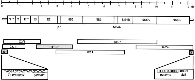
The full-length clone was constituted from five cDNA plasmids selected from the initial phage library and one RT-PCR product encompassing the region between positions 2265 and 4301. At the 5′ end, the sequence of the T7 promoter was added for in vitro transcription, and an _Srf_I site was introduced at the 3′ end for plasmid linearization (Fig. 1). The assembly of the different cDNA fragments in plasmid pACYC177 (New England Biolabs) was done according to standard procedures; details of the cloning strategy are available from the authors upon request. The full-length clone was named pKANE40A.
FIG. 1.
Construction of the infectious cDNA clone. The upper part sketches a BVDV genome with a scale in kilobases (kB) and the encoded polyprotein. The middle part shows the cDNA clones (white), the RT-PCR product (light gray), and the PCR products (dark gray) used for engineering the infectious cDNA clone. The lower part depicts the ends of the genomic cDNA sequences (underlined) and the sequences added at the 5′ and 3′ ends for in vitro transcription.
MDBK cells were transfected with RNA generated from the linearized pKANE40A template by in vitro transcription. A runoff transcript from plasmid pKANE28AII, which terminates 19 codons upstream of the NS5B coding region, served as a negative control. Three days posttransfection, immunofluorescence staining was conducted with a mix of monoclonal antibodies raised against BVDV E2 (58) as described before (28), and BVDV-specific signals were detected in cells transfected with RNA from pKANE40A, but not in the control. The virus recovered from pKANE40A was termed BVDV XIKE-A. The recovered virus was passaged twice, and the stock of the second passage was used for all further experiments. A pKANE40A specific nucleotide exchange from C to T at position 1630 was demonstrated by sequencing of an RT-PCR fragment and taken as proof of the identity of XIKE-A.
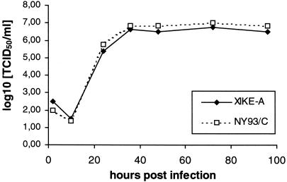
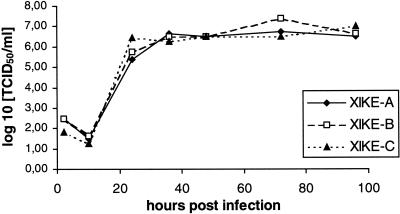
The specific infectivity of the RNA derived from pKANE40A (4.32 × 10² PFU/μg) was demonstrated to be very similar to that of BVDV NY'93/C (4 × 10² PFU/μg, determined with RNA from infected cells), and the growth characteristics of the two viruses were found to be equivalent (Fig. 2). Thus, the nucleotide exchanges identified in pKANE40A with regard to the consensus sequence (Table 1) had no significant influence, and XIKE-A was deemed suitable for further experiments.
FIG. 2.
Growth curves of the recombinant virus XIKE-A and the wild-type BVDV isolate NY'93/C. MDBK cells were infected with the viruses at an MOI of 0.1 and harvested by freezing and thawing at the indicated time points. Titers were determined after infection of MDBK cells by immunofluorescence staining 72 h p.i.
TABLE 1.
Sequence differences between the full-length cDNA clone pKANE40A and the BVDV NY'93/C consensus sequence
| Nucleotide position | Exchange | Amino acid substitution | Protein coded for |
|---|---|---|---|
| 1630 | C → T | Silent | Ems |
| 2410 | C → A | Silent | E1 |
| 4081 | G → A | Silent | NS2 |
| 4698 | T → C | Val → Ala | NS2 |
| 7648 | A → G | Silent | NS4B |
| 11671 | G → A | Silent | NS5B |
| 11868 | A → C | Asp → Ala | NS5B |
| 11898 | A → C | Asn → Thr | NS5B |
| 11941 | A → G | Silent | NS5B |
Animal experiment with XIKE-A and NY'93/C.
Some of the BVDV-2 isolates are known to be considerably more virulent than standard BVDV-1 (4, 13, 32, 39). The purpose of the animal experiment was to test the virulence of our reisolated virus, BVDV NY'93/C, and to compare its pathogenicity with that of the recombinant virus, XIKE-A, derived from the infectious cDNA clone. Two groups of three calves (8- to 9-month-old flecked cattle female calves, free of BVDV-specific antibody or antigen, with each group housed in a separate isolation unit) were infected intranasally with 105 50% tissue culture infective doses (TCID50) of either XIKE-A or NY'93/C per animal. Body temperatures and clinical signs were recorded daily; blood samples (stabilized with ca. 35 IU of heparin/ml) were taken on days 0, 2 to 16, and 21 postinfection (p.i.) for leukocyte counts and detection of viremia. Sera from all calves were collected for detection of neutralizing antibodies against NY'93/C on days 0, 7, 14, 21, 29, and 35 p.i. Nasal swabs for virus isolation were taken on days 0, 2 to 16, and 21 p.i.
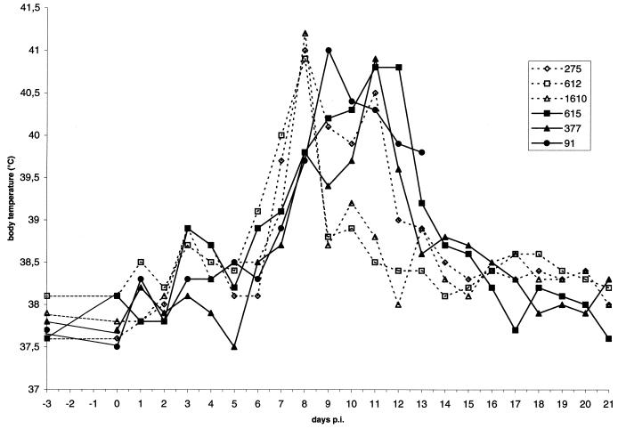
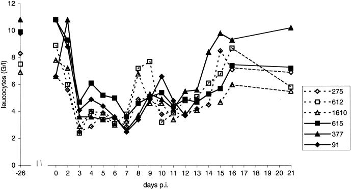
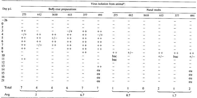
All animals in both groups developed fever (Fig. 3) and a broad spectrum of clinical signs, including respiratory symptoms and gastrointestinal disorders. Animal 091 was euthanized on day 13 p.i. for welfare reasons. All calves in both groups showed leukopenia starting on day 3 p.i. and persisting up to day 15 p.i. (Fig. 4). For detection of viremia, buffy coats were prepared by addition of 5 ml of ice-cold lysis buffer (150 mM NH4Cl, 10 mM KHCO3, 1 mM EDTA [pH 7.4]) to an aliquot of heparin-stabilized blood (containing ca. 107 leukocytes) and incubation on ice for 10 min, followed by centrifugation. The pellet was washed once with lysis buffer and twice with ice-cold PBS-A (phosphate-buffered saline [PBS] without Ca2+ and Mg2+) before it was resuspended in 2 ml of PBS-A. MDBK cells seeded in 24-well plates were inoculated with 200 μl of the buffy coat preparations (106 cells) and incubated for 5 days. Virus was detected by immunofluorescence in buffy coat preparations from animals infected with NY'93/C for 5 days and with XIKE-A for 7 days (Table 2). The identity of the viruses reisolated from the animals was verified by nucleotide sequencing of RT-PCR products from RNA isolated from buffy coat preparations from all animals.
FIG. 3.
Body temperatures of animals infected with NY'93/C (animals 275, 612, and 1610 [broken lines]) or XIKE-A (animals 615, 377, and 091 [solid lines]).
FIG. 4.
Leukocyte (WBC) counts of animals infected with NY'93/C (animals 275, 612, and 1610 [broken lines]) or XIKE-A (animals 615, 377, and 091 [solid lines]).
TABLE 2.
Virus isolation from buffy coat preparations and nasal swabs of animals infected with the wild-type virus NY'93/C (animals 615, 377, and 091) or with the recombinant virus XIKE-A (animals 275, 612, and 1610)
To detect virus in nasal discharge, nasal swabs were taken at the time points mentioned above, diluted in 2 ml of transport buffer (PBS supplemented with 5% fetal calf serum [FCS], 100 IU of penicillin G per ml, 0.1 mg of streptomycin per ml, and 2.5 μg of amphotericin B per ml), and passed through a 0.2-μm-pore-diameter filter. MDBK cells were inoculated in 24-well plates with 100 μl of these preparations and analyzed by indirect immunofluorescence microscopy after 5 days. Nasal shedding was found for 1 or 2 days (Table 2).
The presence of virus-neutralizing antibodies was tested in serum samples that had been inactivated by incubation at 56°C for 30 min. The sera were diluted in steps of 1:2 on 96-well microtiter plates and inoculated with a suspension of strain NY'93/C (100 TCID50 per well) for 1 h at 37°C. A total of 1.75 × 10⁴ MDBK cells were added to each well, incubated for 5 days, and analyzed by indirect immunofluorescence. Neutralization titers were calculated by the method of Kaerber (22) and expressed as the endpoint dilution that neutralized ca. 100 TCID50. Neutralizing antibodies were found in the serum of all calves starting on day 14 p.i.
The results of this study demonstrated that the recombinant virus XIKE-A is highly similar to the wild-type virus NY'93/C with regard to both pathogenicity and induction of an immune response in the natural host. It is therefore plausible to assume that any deviation from this clinical picture that might be observed in a virus mutant generated on the basis of the infectious clone pKANE40A would indeed be caused by the desired mutation.
Construction and analysis of Erns mutants.
Previous experiments with CSFV had shown that the RNase activity of the glycoprotein Erns is destroyed by substitution or deletion of histidine 297 or 346 (the numbers represent the residue positions in CSFV strain Alfort/Tübingen) (25). Viruses recovered from the mutated full-length clones were clinically attenuated. In BVDV strain NY'93/C, the two histidine residues are located at positions 300 and 349, respectively. To test whether the effects of mutations at these positions would be similar to those of CSFV in a BVDV-2 genome, two infectious clones were engineered with either a deletion of codon H349 or a substitution of codon H300 by leucine. The mutants were generated with the QuikChange site-directed mutagenesis kit (Stratagene, Amsterdam, Netherlands) according to the manufacturer's instructions. The plasmid used for introducing mutations into the region coding for Erns was cDNA clone C5/9 (insert corresponding to positions 100 to 2400 of the genome). The oligonucleotides used to generate mutant H346Δ were CM126 (GAGTGGAATAAAGGTTGGTGTAAC) and CM127 (GTTACACCAACCTTTATTCCACTC), and those used to generate mutant H297L were CM128 (AACAGGAGTCTATTAGGAATTTGGCCA) and CM129 (TGGCCAAATTCCTAATAGACTCCTGTT). The presence of the desired mutations and the absence of second-site mutations were verified by nucleotide sequencing. Fragments with the desired mutations cut with _Eag_I and PshAI were inserted into pKANE40A restricted with the same enzymes. The resulting recombinant virus mutants were named BVDV XIKE-B (H349Δ) and BVDV XIKE-C (H300L).
Both mutants were stable for at least five passages in MDBK cells. With regard to the growth characteristics, the two mutant viruses exhibited no significant differences compared with virus XIKE-A (Fig. 5), indicating that also in the BVDV system, the RNase function of Erns is dispensable for tissue culture propagation. Hulst et al. reported for the noncytopathogenic CSFV C strain that inactivation of the RNase activity of Erns resulted in a cytopathogenic virus (17). In contrast to these results, but similar to the data obtained for CSFV Alfort (25), the cells infected with the mutant viruses XIKE-B or XIKE-C did not show any signs of a cytopathic effect. Thus, it is very likely that the effect observed by Hulst and coworkers represents a special feature restricted to their individual virus or infectious clone, and not a general characteristic of RNase-negative pestiviruses.
FIG. 5.
Growth curves of the recombinant virus XIKE-A and the Erns mutants XIKE-B (H349Δ) and XIKE-C (H300L) (see Fig. 2).
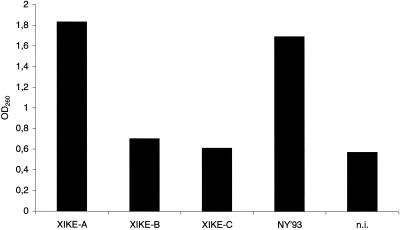
The RNase activity of XIKE-A, XIKE-B, and XIKE-C was determined in crude cell extracts as established before for CSFV (25). Cells were infected with the same multiplicity of infection (MOI) of either virus and lysed at 2 days p.i. Cell extracts were purified by centrifugation, and aliquots of the preparations were tested for their ability to convert poly(U) to acid-soluble low-molecular-weight RNA measured by photometric analysis as described before (25). High RNase activity was found in the NY'93/C and XIKE-A samples, whereas the two mutants XIKE-B and XIKE-C were in the same range as the negative control (Fig. 6). Thus, the enzymatic activity of Erns is obviously not restored in the recovered viruses and therefore seems to have no advantage for virus propagation in tissue culture. These results are in agreement with the data observed for CSFV before.
FIG. 6.
Determination of RNase activity of the recombinant viruses XIKE-A (wild-type sequence), XIKE-B (H349Δ), and XIKE-C (H300L) in comparison with the wild-type strain NY'93/C from crude cell extracts of MDBK cells infected with the respective viruses. MDBK cells that were not infected (n.i.) served as a negative control. The enzymatic degradation of poly(U) was determined by measuring the optical density at 260 nm (OD260) as a marker of the release of small RNA fragments into the supernatant (25, 47).
Animal experiment with XIKE-B.
In a second animal experiment, the clinical and immunological characteristics of the RNase-negative mutant XIKE-B were analyzed in comparison with XIKE-A. The H349Δ mutant was given precedence over the H300L mutant to minimize the danger of a genomic reversion to wild type.
Two groups of three calves (7- to 10-week-old Holstein and Holstein-cross calves) each were inoculated intranasally with a dose of 5 × 10⁵ TCID50 of either XIKE-A (group 1) or XIKE-B virus (group 2). The groups were housed in separate isolation units. Rectal temperatures and clinical symptoms were monitored daily; nasal swabs and blood samples were taken on days −8, 0, 2 to 14, 17, and 21. Serum samples were collected on days 0, 8, 12 or 14, 21, 28, and 38.
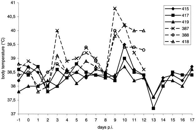
Nine to 10 days p.i., the calves of group 1 developed fever for up to 4 days (Fig. 7), which was accompanied by diarrhea and respiratory symptoms. Calf 388 showed convulsions. The group was euthanized for welfare reasons on day 12 p.i. because they were in a state of marked depression and anorexia. None of the calves from group 2 had body temperatures above 39.5°C (Fig. 7). No diarrhea was detected, and only mild respiratory symptoms were observed for up to 6 days, which might, however, be of secondary origin due to bacterial infection. Antibiotic treatment of animal 417 on day 10 p.i. resulted in a considerable improvement in the symptoms. Respiratory symptoms were not detected in further animal experiments with XIKE-B (data not shown).
FIG. 7.
Body temperatures of animals infected with XIKE-A (animals 387, 388, and 418 [broken lines]) or XIKE-B (animals 415, 417, and 419 [solid lines]).
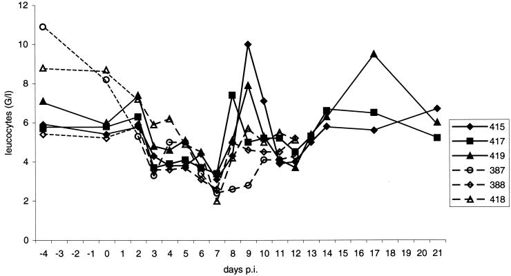
Leukopenia was detected in all calves; however, in contrast to the calves infected with XIKE-A, the animals from group 2 showed an early recovery of leukocyte numbers starting at day 7 p.i. that finally stabilized after day 13 p.i. (Fig. 8). Neutralizing antibodies were first detected on day 12 p.i. in the serum of the calves infected with XIKE-A and on day 14 p.i. in the serum of calves infected with the Erns mutant (Table 3).
FIG. 8.
Leukocyte (WBC) counts of animals infected with XIKE-A (animals 387, 388, and 418 [broken lines]) or XIKE-B (animals 415, 417, and 419 [solid lines]).
TABLE 3.
Neutralizing antibody titers determined in serum samples of all calves after experimental infection with XIKE-A (wild-type sequence) or XIKE-B (H346Δ)
| Day p.i. | Neutralizing antibody titer in serum samplea | |||||
|---|---|---|---|---|---|---|
| 387 | 388 | 418 | 415 | 417 | 419 | |
| 0 | <2 | <2 | <2 | <2 | <2 | <2 |
| 8 | <2 | <2 | <2 | <2 | <2 | <2 |
| 12 or 14 | 20 | 8 | 128 | 51 | 203 | 64 |
| 21 | eu | eu | eu | 512 | 1,024 | 406 |
| 28 | eu | eu | eu | 2,048 | 1,024 | 4,096 |
| 38 | eu | eu | eu | 8,182 | 4,096 | 4,096 |
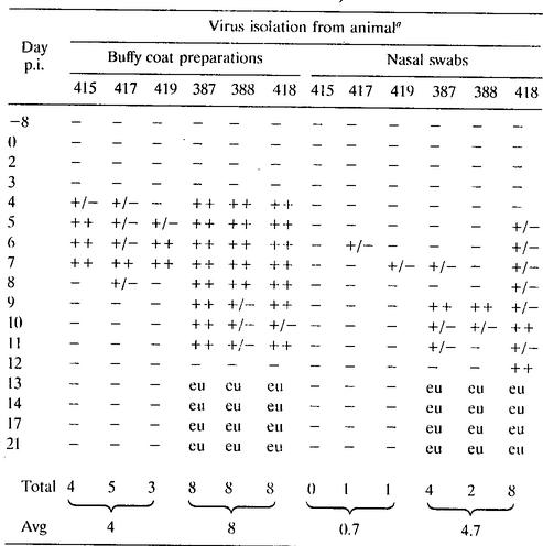
Virus was found in buffy coat preparations of all calves starting for all but one animal on day 4 p.i.; however, viremia was briefer for the Erns mutant (average, 4 days) than for XIKE-A (average, 8 days). Nasal shedding of virus was observed for up to 8 days (average, 4.7 days) with XIKE-A animals, but for a maximum of 1 day (average, 0.7 day) with XIKE-B animals (Table 4). Again, nucleotide sequencing of RT-PCR products encompassing the entire Erns coding region was used to confirm the presence of XIKE-A or B in buffy coat preparations from animals of group 1 or 2, respectively. Interestingly, an additional point mutation was found in RT-PCR products from two animals from group 2 (no. 415 and 419): nucleotide position 1246 of the RNA genome was changed from guanosine to uridine, resulting in the amino acid substitution Q287H within the Erns sequence. It is difficult to guess the effect of this mutation—especially since no three-dimensional structure of Erns is known. However, Erns belongs to the group of T2 RNases, and based on a three-dimensional model for this class of enzymes, Q287 is located at considerable distance from the active center of the enzyme. It therefore can be expected that the Q287H mutation has no influence on the enzymatic activity of Erns, and indeed an RNase test conducted with the reisolated viruses revealed that the mutation did not restore the RNase activity (data not shown). Taking together the results of the animal experiments, two important points have to be mentioned. First, BVDV-2 NY'93/C represents a highly pathogenic virus that consistently induces severe disease in infected animals. This was also shown in further animal experiments, not only with calves, but also with adult animals (data not shown). Thus, NY'93/C can be used as a standard challenge virus for experiments in which a stringent BVDV-2 challenge is needed. Alternatively, BVDV XIKE-A could be used for this purpose, since it also proved to be highly pathogenic in our experiments. Having established an infectious clone from which a pathogenic BVDV can be recovered, new approaches to the analysis of BVDV-induced disease, BVDV pathogenicity markers, and identification of mutations leading to virus attenuation can be pursued. Such analyses were simply not possible before, since the available infectious cDNA clones for BVDV were all derived from BVDV-1, which usually induces only variable mild symptoms or no symptoms at all. Thus, pKANE40A could be a starting point for the identification of attenuating mutations and the development of live marker vaccines against BVDV. As a first step, virus mutants were generated with changes abrogating the RNase activity residing in the glycoprotein Erns. Equivalent mutations have recently been tested for CSFV (25; M. von Freyburg, C. Meyer, and G. Meyers, unpublished results.). Even though some differences were observed with regard to individual clinical parameters, a clear correlation between RNase inactivation and virus attenuation was observed for both CSFV and BVDV. This finding raises questions about the attenuating principle of the RNase-negative mutants. Specific reduction of B-cell numbers, which represents a well-known characteristic of CSFV infection in pigs (49), was not observed in an experiment with CSFV mutant H346Δ (25). However, prevention of B-cell reduction most likely represents a characteristic of the type of mutant tested in this experiment and not a general feature of RNase-negative mutants, since it was not observed for another CSFV mutant (von Freyburg et al., unpublished). Moreover, induction of a specific drop in B-cell numbers has not been described for BVDV and could also not be identified in our experiments with either wild-type or mutant viruses (data not shown). Thus, the function of the RNase, and therefore also the molecular basis of attenuation of RNase-negative viruses, remains obscure for the moment. However, a role of the RNase in suppression of the host's immune response represents a fascinating hypothesis that fits well with the data obtained in our experiments. Further analyses are necessary to provide more data on the nature of the attenuation principle and to establish more defined hypotheses on the function of the RNase. The now established BVDV-2 infectious clone allows such studies to be conducted with the bovine system and thus offers the opportunity of comparative analyses for two different pestiviruses that both express the curious structural glycoprotein Erns, which has RNase activity.
TABLE 4.
Virus isolation from buffy coat preparations and nasal swabs of animals infected with the recombinant virus XIKE-A (animals 387, 388, and 418) or the Ems mutant XIKE-B (animals 415, 417, and 419)
Acknowledgments
We thank Maren Ziegler, Petra Wulle, and Silke Esslinger for excellent technical assistance and E. J. Dubovi for the kind gift of the BVDV isolate VLS 399.
This study was supported by a grant from Boehringer Ingelheim Vetmedica GmbH.
REFERENCES
- 1.Baker, J. C. 1987. Bovine viral diarrhea virus: a review. J. Am. Vet. Med. Assoc. 190**:**1449-1458. [PubMed] [Google Scholar]
- 2.Becher, P., A. D. Shannon, N. Tautz, and H.-J. Thiel. 1994. Molecular characterization of border disease virus, a pestivirus from sheep. Virology 198**:**542-551. [DOI] [PubMed] [Google Scholar]
- 3.Becher, P., M. Orlich, A. Kosmidou, M. König, M. Baroth, and H.-J. Thiel. 1999. Genetic diversity of pestivirus: identification of novel groups and implications for classification. Virology 262**:**64-71. [DOI] [PubMed] [Google Scholar]
- 4.Bolin, S. R., and J. F. Ridpath. 1992. Differences in virulence between two noncytopathic bovine viral diarrhea viruses in calves. Am. J. Vet. Res. 53**:**2157-2163. [PubMed] [Google Scholar]
- 5.Bolin, S. R., E. T. Littledike, and J. F. Ridpath. 1991. Serologic detection and practical consequences of antigenic diversity among bovine viral diarrhea viruses in a vaccinated herd. Am. J. Vet. Res. 52**:**1033-1037. [PubMed] [Google Scholar]
- 6.Collett, M. S., R. Larson, C. Gold, D. Strick, D. K. Anderson, and A. F. Purchio. 1988. Molecular cloning and nucleotide sequence of the pestivirus bovine viral diarrhea virus. Virology 165**:**191-199. [DOI] [PubMed] [Google Scholar]
- 7.Collett, M. S., R. Larson, S. K. Belzer, and E. Retzel. 1988. Proteins encoded by bovine viral diarrhea virus: the genomic organization of a pestivirus. Virology 165**:**200-208. [DOI] [PubMed] [Google Scholar]
- 8.Collett, M. S., M. A. Wiskerchen, E. Welniak, and S. K. Belzer. 1991. Bovine viral diarrhea virus genomic organization. Arch. Virol. Suppl. 3**:**19-27. [DOI] [PubMed] [Google Scholar]
- 9.Deng, R., and K. Brock. 1992. Molecular cloning and nucleotide sequence of a pestivirus genome, noncytopathogenic bovine viral diarrhea virus strain SD-1. Virology 191**:**867-879. [DOI] [PubMed] [Google Scholar]
- 10.Devereux, J., P. Haeberli, and O. Smithies. 1984. A comprehensive set of sequence analysis programs for the VAX. Nucleic Acids Res. 12**:**387-395. [DOI] [PMC free article] [PubMed] [Google Scholar]
- 11.Donis, R. O., W. Corapi, and E. J. Dubovi. 1988. Neutralizing monoclonal antibodies to bovine viral diarrhea virus bind to the 56K to 58K glycoprotein. J. Gen. Virol. 69**:**77-86. [DOI] [PubMed] [Google Scholar]
- 12.Elbers, K., N. Tautz, P. Becher, D. Stoll, T. Rümenapf, and H.-J. Thiel. 1996. Processing in the pestivirus E2-NS2 region: identification of proteins p7 and E2p7. J. Virol. 70**:**4131-4135. [DOI] [PMC free article] [PubMed] [Google Scholar]
- 13.Hamers, C., B. Couvreur, P. Dehan, C. Letellier, P. Lewalle, P. P. Pastoret, and P. Kerkhofs. 2000. Differences in experimental virulence of bovine viral diarrhoea viral strains isolated from haemorrhagic syndromes. Vet. J. 160**:**250-258. [DOI] [PubMed] [Google Scholar]
- 14.Harada, T., N. Tautz, and H.-J. Thiel. 2000. E2-p7 region of the bovine viral diarrhea virus polyprotein: processing and functional studies. J. Virol. 74**:**9498-9506. [DOI] [PMC free article] [PubMed] [Google Scholar]
- 15.Henikoff, S. 1987. Unidirectional digestion with exonuclease III in DNA sequence analysis. Methods Enzymol. 155**:**156-165. [DOI] [PubMed] [Google Scholar]
- 16.Hulst, M. M., D. F. Westra, G. Wensvoort, and R. J. M. Moormann. 1993. Glycoprotein E1 of hog cholera virus expressed in insect cells protects swine from hog cholera. J. Virol. 67**:**5435-5442. [DOI] [PMC free article] [PubMed] [Google Scholar]
- 17.Hulst, M. M., F. E. Panoto, A. Hoekmann, H. G. P. van Gennip., and R. J. M. Moormann. 1998. Inactivation of the RNase activity of glycoprotein Erns of classical swine fever virus results in a cytopathogenic virus. J. Virol. 72**:**151-157. [DOI] [PMC free article] [PubMed] [Google Scholar]
- 18.Hulst, M. M., G. Himes, E. Newbigin, and R. J. M. Moormann. 1994. Glycoprotein E2 of classical swine fever virus: expression in insect cells and identification as a ribonuclease. Virology 200**:**558-565. [DOI] [PubMed] [Google Scholar]
- 19.König, M., T. Lengsfeld, T. Pauly, R. Stark, and H.-J. Thiel. 1995. Classical swine fever virus: independent induction of protective immunity by two structural glycoproteins. J. Virol. 69**:**6479-6486. [DOI] [PMC free article] [PubMed] [Google Scholar]
- 20.Kümmerer, B. M., and G. Meyers. 2000. Correlation between point mutations in NS2 and the viability and cytopathogenicity of bovine viral diarrhea virus strain Oregon analyzed with an infectious cDNA clone. J. Virol. 74**:**390-400. [DOI] [PMC free article] [PubMed] [Google Scholar]
- 21.Kümmerer, B. M., N. Tautz, P. Becher, H.-J. Thiel, and G. Meyers. 2000. The genetic basis for cytopathogenicity of pestiviruses. Vet. Microbiol. 77**:**117-128. [DOI] [PubMed] [Google Scholar]
- 22.Mayr, A., P. A. Bachmann, B. Bibrac, and G. Wittmann. 1974. Virologische Arbeitsmethoden Band I. Gustav Fischer Verlag, Stuttgart, Germany.
- 23.Mendez, E., N. Ruggli, M. S. Collett, and C. M. Rice. 1998. Infectious bovine viral diarrhea virus (strain NADL) RNA from stable cDNA clones: a cellular insert determines NS3 production and viral cytopathogenicity. J. Virol. 72**:**4737-4745. [DOI] [PMC free article] [PubMed] [Google Scholar]
- 24.Meyers, G., and H.-J. Thiel. 1996. Molecular characterization of pestiviruses. Adv. Virus Res. 47**:**53-117. [DOI] [PubMed] [Google Scholar]
- 25.Meyers, G., A. Saalmüller, and M. Büttner. 1999. Mutations abrogating the RNase activity in glycoprotein Erns of the pestivirus classical swine fever virus lead to virus attenuation. J. Virol. 73**:**10224-10235. [DOI] [PMC free article] [PubMed] [Google Scholar]
- 26.Meyers, G., H.-J. Thiel, and T. Rümenapf. 1996. Classical swine fever virus: recovery of infectious viruses from cDNA constructs and generation of recombinant cytopathogenic defective interfering particles. J. Virol. 70**:**1588-1595. [DOI] [PMC free article] [PubMed] [Google Scholar]
- 27.Meyers, G., N. Tautz, E. J. Dubovi, and H.-J. Thiel. 1991. Viral cytopathogenicity correlated with integration of ubiquitin-coding sequences. Virology 180**:**602-616. [DOI] [PMC free article] [PubMed] [Google Scholar]
- 28.Meyers, G., N. Tautz, P. Becher, H.-J. Thiel, and B. M. Kümmerer. 1996. Recovery of cytopathogenic and noncytopathogenic bovine viral diarrhea viruses from cDNA constructs. J. Virol. 70**:**8606-8613. (Erratum, **71:**173, 1997.) [DOI] [PMC free article] [PubMed] [Google Scholar]
- 29.Meyers, G., T. Rümenapf, and H.-J. Thiel. 1989. Molecular cloning and nucleotide sequence of the genome of hog cholera virus. Virology 171**:**555-567. [DOI] [PubMed] [Google Scholar]
- 30.Moennig, V., and J. Plagemann. 1992. The pestiviruses. Adv. Virus Res. 41**:**53-91. [DOI] [PubMed] [Google Scholar]
- 31.Moormann, R. J. M., H. G. P. van Gennip, G. K. W. Miedema, M. M. Hulst, and P. A. van Rijn. 1996. Infectious RNA transcribed from an engineered full-length cDNA template of the genome of a pestivirus. J. Virol. 70**:**763-770. [DOI] [PMC free article] [PubMed] [Google Scholar]
- 32.Odeon, A. C., C. L. Kelling, D. J. Marshall, E. S. Estela, E. J. Dubovi, and R. O. Donis. 1999. Experimental infection of calves with bovine viral diarrhea virus genotype II (NY-93). J. Vet. Diagn. Investig. 11**:**221-228. [DOI] [PubMed] [Google Scholar]
- 33.Pellerin, C., J. van den Hurk, J. Lecomte, and P. Tijssen. 1994. Identification of a new group of bovine viral diarrhea virus (BVDV) strains associated with severe outbreaks and high mortalities. Virology 203**:**260-268. [DOI] [PubMed] [Google Scholar]
- 34.Pestova, T. V., and C. U. Hellen. 1999. Internal initiation of translation in bovine viral diarrhea virus. Virology 258**:**249-256. [DOI] [PubMed] [Google Scholar]
- 35.Pestova, T. V., I. N. Shatsky, S. P. Fletcher, R. J. Jackson, and C. U. Hellen. 1998. A prokaryotic-like mode of cytoplasmic eukaryotic ribosome binding to the initiation codon during internal translation initiation of hepatitis C and classical swine fever virus RNAs. Genes Dev. 12**:**67-83. [DOI] [PMC free article] [PubMed] [Google Scholar]
- 36.Poole, T. L., C. Wang, R. A. Popp, L. N. Potgieter, A. Siddiqui, and M. S. Collett. 1995. Pestivirus translation initiation occurs by internal ribosome entry. Virology 206**:**750-754. [DOI] [PubMed] [Google Scholar]
- 37.Rebhun, W. C., T. W. French, J. A. Perdrizet, E. J. Dubovi, S. G. Dill, and L. F. Karcher. 1989. Thrombocytopenia associated with acute bovine virus diarrhea infection in cattle. J. Vet. Intern. Med. 3**:**42-46. [DOI] [PubMed] [Google Scholar]
- 38.Rice, C. M. 1996. Flaviviridae: the viruses and their replication, p. 931-959. In B. N. Fields, D. M. Knipe, and P. M. Howley (ed.), Fields virology, 3rd ed. Lippincott-Raven, Philadelphia, Pa.
- 39.Ridpath, J. F., J. D. Neill, M. Frey, and J. G. Landgraf. 2000. Phylogenetic, antigenic and clinical characterization of type 2 BVDV from North America. Vet. Microbiol. 77**:**145-155. [DOI] [PubMed] [Google Scholar]
- 40.Ridpath, J. F., and S. R. Bolin. 1995. The genomic sequence of a virulent bovine viral diarrhea virus (BVDV) from the type 2 genotype: detection of a large genomic insertion in a noncytopathic BVDV. Virology 212**:**39-46. [DOI] [PubMed] [Google Scholar]
- 41.Ridpath, J. F., S. R. Bolin, and E. J. Dubovi. 1994. Segregation of bovine viral diarrhea virus into genotypes. Virology 205**:**66-74. [DOI] [PubMed] [Google Scholar]
- 42.Rijnbrand, R., T. van der Straaten, P. A. van Rijn, W. J. M. Spaan, and P. J. Bredenbeek. 1997. Internal entry of ribosomes is directed by the 5′ noncoding region of classical swine fever virus and is dependent on the presence of an RNA pseudoknot upstream of the initiation codon. J. Virol. 71**:**451-457. [DOI] [PMC free article] [PubMed] [Google Scholar]
- 43.Ruggli, N., J.-D. Tratschin, C. Mittelholzer, and M. A. Hofmann. 1996. Nucleotide sequence of classical swine fever virus strain Alfort/187 and transcription of infectious RNA from stably cloned full-length cDNA. J. Virol. 70**:**3478-3487. [DOI] [PMC free article] [PubMed] [Google Scholar]
- 44.Rümenapf, T., G. Unger, J. H. Strauss, and H.-J. Thiel. 1993. Processing of the envelope glycoproteins of pestiviruses. J. Virol. 67**:**3288-3294. [DOI] [PMC free article] [PubMed] [Google Scholar]
- 45.Sambrook, S., E. F. Fritsch, and T. Maniatis. 1989. Molecular cloning: a laboratory manual, 2nd ed. Cold Spring Harbor Laboratory Press, Cold Spring Harbor, N.Y.
- 46.Schaefer, B. C. 1995. Revolutions in rapid amplification of cDNA ends: new strategies for polymerase chain reaction cloning of full-length cDNA ends. Anal. Biochem. 227**:**255-273. [DOI] [PubMed] [Google Scholar]
- 47.Schneider, R., G. Unger, R. Stark, E. Schneider-Scherzer, and H.-J. Thiel. 1993. Identification of a structural glycoprotein of an RNA virus as a ribonuclease. Science 261**:**1169-1171. [DOI] [PubMed] [Google Scholar]
- 48.Stark, R., G. Meyers, T. Rümenapf, and H.-J. Thiel. 1993. Processing of pestivirus polyprotein: cleavage site between autoprotease and nucleocapsid protein of classical swine fever virus. J. Virol. 67**:**7088-7095. [DOI] [PMC free article] [PubMed] [Google Scholar]
- 49.Šuša, M., M. König, A. Saalmüller, M. J. Reddehase, and H. J. Thiel. 1992. Pathogenesis of classical swine fever: B-lymphocyte deficiency caused by hog cholera virus. J. Virol. 66**:**1171-1175. [DOI] [PMC free article] [PubMed] [Google Scholar]
- 50.Tautz, N., K. Elbers, D. Stoll, G. Meyers, and H.-J. Thiel. 1997. Serine protease of pestiviruses: determination of cleavage sites. J. Virol. 71**:**5415-5422. [DOI] [PMC free article] [PubMed] [Google Scholar]
- 51.Thiel, H.-J., G. W. Plagemann, and V. Moennig. 1996. The pestiviruses, p.1059-1073. In B. N. Fields, D. M. Knipe, and P. M. Howley (ed.), Fields virology, 3rd ed. Lippincott-Raven, Philadelphia, Pa.
- 52.Thiel, H.-J., R. Stark, E. Weiland, T. Rümenapf, and G. Meyers. 1991. Hog cholera virus: molecular composition of virions from a pestivirus. J. Virol. 65**:**4705-4712. [DOI] [PMC free article] [PubMed] [Google Scholar]
- 53.Tijssen, P., C. Pellerin, J. Lecomte, and J. van den Hurk. 1996. Immunodominant E2 (gp53) of highly virulent bovine viral diarrhea group II viruses indicates a close resemblance to a subgroup of border disease viruses. Virology 217**:**356-361.8599222 [Google Scholar]
- 54.Topliff, C. L., and C. L. Kelling. 1998. Virulence markers in the 5′ untranslated region of genotype 2 bovine viral diarrhea virus isolates. Virology 250**:**164-172. [DOI] [PubMed] [Google Scholar]
- 55.van Rijn, P. A., H. G. van Gennip, E. J. de Meijer, and R. J. Moormann. 1993. Epitope mapping of envelope glycoprotein E1 of hog cholera virus strain Brescia. J. Gen. Virol. 74**:**2053-2060. [DOI] [PubMed] [Google Scholar]
- 56.van Zijl, M., G. Wensvoort, E. de Kluyver, M. Hulst, H. van der Gulden, A. Gielkens, A. Berns, and R. Moormann. 1991. Live attenuated pseudorabies virus expressing envelope glycoprotein E1 of hog cholera virus protects swine against both pseudorabies and hog cholera. J. Virol. 65**:**2761-2765. [DOI] [PMC free article] [PubMed] [Google Scholar]
- 57.Vassilev, V. B., M. S. Collett, and R. O. Donis. 1997. Authentic and chimeric full-length genomic cDNA clones of bovine viral diarrhea virus that yield infectious transcripts. J. Virol. 71**:**471-478. [DOI] [PMC free article] [PubMed] [Google Scholar]
- 58.Weiland, E., H.-J. Thiel, G. Hess, and F. Weiland. 1989. Development of monoclonal neutralizing antibodies against bovine viral diarhea virus after pretreatment of mice with normal bovine cells and cyclophosphamide. J. Virol. Methods 24**:**237-244. [DOI] [PubMed] [Google Scholar]
- 59.Weiland, E., R. Ahl, R. Stark, F. Weiland, and H.-J. Thiel. 1992. A second envelope glycoprotein mediates neutralization of a pestivirus, hog cholera virus. J. Virol. 66**:**3677-3682. [DOI] [PMC free article] [PubMed] [Google Scholar]
- 60.Weiland, E., R. Stark, B. Haas, T. Rümenapf, G. Meyers, and H.-J. Thiel. 1990. Pestivirus glycoprotein which induces neutralizing antibodies forms part of a disulfide-linked heterodimer. J. Virol. 64**:**3563-3569. [DOI] [PMC free article] [PubMed] [Google Scholar]
- 61.Weiland, F., E. Weiland, G. Unger, A. Saalmüller, and H.-J. Thiel. 1999. Localization of pestiviral proteins Erns and E2 at the cell surface and on isolated particles. J. Gen. Virol. 80**:**1157-1165. [DOI] [PubMed] [Google Scholar]
- 62.Wengler, G., D. W. Bradley, M. S. Collett, F. X. Heinz, R. W. Schlesinger, and J. H. Strauss. 1995. Flaviviridae, p. 425-427. In F. A. Murphy, C. M. Fauquet, D. H. L. Bishop, S. A. Ghabrial, A. W. Jarvis, G. P. Martelli, M. A. Mayo, and M. D. Summers (ed.), Virus taxonomy. Sixth report of the International Committee on Taxonomy of Viruses. Springer-Verlag, Vienna, Austria.
- 63.Windisch, J. M., R. Schneider, R. Stark, E. Weiland, G. Meyers, and H.-J. Thiel. 1996. RNase of classical swine fever virus: biochemical characterization and inhibition by virus-neutralizing monoclonal antibodies. J. Virol. 70**:**352-358. [DOI] [PMC free article] [PubMed] [Google Scholar]
- 64.Xu, J., E. Mendez, P. R. Caron, C. Lin, M. A. Murcko, M. S. Collett, and C. M. Rice. 1997. Bovine viral diarrhea virus NS3 serine proteinase: polyprotein cleavage sites, cofactor requirements, and molecular model of an enzyme essential for pestivirus replication. J. Virol. 71**:**5312-5322. [DOI] [PMC free article] [PubMed] [Google Scholar]
- 65.Yu, H., C. W. Grassmann, and S.-E. Behrens. 1999. Sequence and structural elements at the 3′ terminus of bovine viral diarrhea virus genomic RNA: functional role during RNA replication. J. Virol. 73**:**3638-3648. [DOI] [PMC free article] [PubMed] [Google Scholar]
- 66.Yu, H., O. Isken, C. W. Grassmann, and S.-E. Behrens. 2000. A stem-loop motif formed by the immediate 5′ terminus of the bovine viral diarrhea virus genome modulates translation as well as replication of the viral RNA. J. Virol. 74**:**5825-5835. [DOI] [PMC free article] [PubMed] [Google Scholar]
