Amplification of the neu/erbB-2 oncogene in a mouse model of mammary tumorigenesis (original) (raw)
Abstract
The neu (c-erbB-2, Her-2) protooncogene is amplified and overexpressed in 20–30% of human breast cancers. Although transgenic mouse models have illustrated the role of Neu in the induction of mammary tumors, Neu expression in these models is driven by a strong viral promoter of questionable relevance to the human disease. To ascertain whether expression of activated Neu under the control of the endogenous promoter in the mammary gland could induce mammary tumors we have generated mice that conditionally express activated Neu under the transcriptional control of the intact endogenous Neu promoter. Expression of oncogenic neu in the mammary gland resulted in accelerated lobulo-alveolar development and formation of focal mammary tumors after a long latency period. However, expression of activated Neu under the normal transcriptional control of the endogenous promoter was not sufficient for the initiation of mammary carcinogenesis. Strikingly, all mammary tumors bear amplified copies (2–22 copies) of the activated neu allele relative to the wild-type allele and express highly elevated levels of neu transcript and protein. Thus, like human _erbB-2_-positive breast tumors, mammary tumorigenesis in this mouse model requires the amplification and commensurate elevated expression of the neu gene.
The neu (c-erbB2, Her-2) protooncogene is a member of the epidermal growth factor receptor (EGFR) family of receptor tyrosine kinases (1, 2). This family is comprised of several members including; EGFR (ErbB-1, HER) (3), Neu(ErbB-2, HER2) (4–8), ErbB-3 (HER3) (9) and ErbB-4 (HER4) (10). Enhanced expression of members of the EGFR family have been implicated in human breast cancer. In particular, amplification and consequent overexpression of neu has been observed in a significant proportion of human breast cancers (11–15). Consistent with these clinical observations, expression of the neu oncogene under the transcriptional control of the mouse mammary tumor virus (MMTV) long terminal repeat results in the rapid induction of multifocal mammary tumors (16–18). In contrast to the rapid tumor progression observed with these activated neu strains, mammary epithelial specific expression of the neu protooncogene in transgenic mice results in the induction of focal mammary tumors that occur only after a long latency period (19). Tumor progression in these strains further correlates with the occurrence of activating mutations in the juxta-transmembrane region of Neu (20). These alterations within Neu involve a set of cysteine residues and result in constitutive disulfide-mediated dimerization of the Neu receptor (20). Although comparable mutations have not yet been observed in Neu-induced human breast cancer, a splice variant of neu that closely resembles these sporadic neu transgene mutations has been demonstrated to be oncogenic because of its capacity to undergo disulfide-mediated dimerization (21, 22). Taken together, these observations suggest that oncogenic activation of neu may be a critical step in mammary tumor progression.
One limitation of these transgenic studies is that activated neu expression is driven by a strong viral promoter that is of questionable physiological relevance to human breast cancer. In addition, because the MMTV promoter activity is modulated by steroid hormones such as progesterone and estrogen, the use of these MMTV-based transgenic breast cancer models in elucidating the role of these sex hormones in tumor progression is problematic. One possible solution to these limitations is to generate transgenic mice that carry the activated neu oncogene under the transcriptional control of the endogenous neu promoter. However, given the importance of neu in embryonic development (23), inappropriate expression of activated neu may have deleterious consequences. Indeed expression of the neu oncogene in organs such as the kidney and lung has been documented to result in perinatal lethality (24).
To circumvent these limitations, we used a gene targeting strategy that replaced the first exon of endogenous neu with a Cre-inducible activated neu cDNA. To induce expression of this activated neu allele in the mammary gland, these targeted knock-in strains of mice subsequently were interbred with transgenic mice expressing Cre recombinase under the transcriptional control of the MMTV promoter enhancer. Mammary-specific expression of activated neu, mediated by Cre recombinase, initially resulted in accelerated mammary epithelial development. Subsequently, focal mammary tumors arose in these mice after a long latency period. Further, tumorigenesis in this model was associated with selective amplification of the activated neu allele (2–22 copies) that correlated with elevated levels of neu transcript and protein. These observations suggest that like human breast cancer, mammary tumorigenesis in this transgenic mouse model requires the concerted amplification and overexpression of the Neu protein.
Materials and Methods
Generation of MMTV-Cre Transgenics.
The cDNA encoding Cre recombinase was subcloned into pMMTV-SV40Pa (p206) (25). This construct was prepared for and injected as described (20). Transgenic progeny were identified by Southern analysis (20) using the _Bam_HI–_Cla_I (840–1209) fragment of the Cre cDNA (26) as a probe.
Gene Targeting.
ErbB-2 genomic sequences were isolated from a 129/SvJ genomic library. A 2.5-kb 5′ arm of homology (_Sph_I to _Nar_1) 5′ to exon one and a 8-kb 3′ arm of homology (_Kpn_I to _Sal_I) including exons 2 and 3 were designed. A loxP-flanked phosphoglycerate kinase(PGK)-neomycin-herpes simplex virus (HSV) poly(A) cassette was placed 5′ of the neuNT cDNA, which was flanked at the 3′ end by a simian virus 40 poly(A) sequence. This loxP-flanked Neo NeuNT sequence replaced exon 1 of the genomic allele. The construct was linearized by a _Not_I site 5′ to the 5′ arm of homology before R1 embryonic stem (ES) cell electroporation, and G418 selection. After screening 288 G418-resistant colonies by Southern analysis using a probe 5′ to the _Sph_I site, six positive cell lines were identified. Six- to 8-week-old BALB/c females were used to generate blastocysts that were harvested 3.5 days postcoitum. ES cells were injected into the blastocoel cavity, and the blastocysts then were reimplanted into pseudopregnant CD1 females. The chimeras were identified on the basis of coat color and were interbred with BALB/c mice. The genotype of the mice was determined by Southern analysis on tail DNA using a _Hin_dIII digest and the 600-bp external probe (_Sma_I fragment). All techniques were performed as described (27).
Whole-Mount and Immunohistochemical Analysis.
A whole-mount analysis was completed on the #4 mammary gland of mature virgin females as described (28, 29) because of the ease of examination and the presence of a lymph node, which can be used to quantitate the epithelial penetration of the fat pad. Immunohistochemistry was performed according to standard methods using a rabbit polyclonal antibody (800–874-8667) against Neu (Ciba Corning) as described (30).
RNA Analysis.
RNA was isolated and RNase protections were carried out as described (20). The antisense neu riboprobe was the _Sma_I–_Xba_I fragment of neu cDNA including the transmembrane domain (22). The antisense cre riboprobe was directed against 480 bp of the HSV pA sequence. The control pgk riboprobe was prepared as described (22).
Immunoprecipitation and Immunoblotting.
Protein lysates from tissues were prepared and quantified by using conventional methods (20). Neu was detected by a mouse mAb (AB-3, Oncogene Research Products). Immunoblotting for ErbB-3 (C-17) and Grb-2 (C-23) was performed by using rabbit polyclonal antibodies (Santa Cruz Biotechnology). Immunoprecipitations were completed in accordance with standard protocols using a mouse mAb for Neu (7.16.4) and were probed by using an antiphosphotyrosine antibody (PY20, Transduction Laboratories, Lexington, KY) (22).
Quantification of Amplification.
To determine the extent of amplification, a Southern analysis was conducted on samples that were restricted with _Hin_dIII and probed by using the external probe using methods described above. The intensity of the wild-type and recombinant alleles was measured and compared by PhosphorImager analysis (Molecular Dynamics).
Results
Generation and Characterization of Transgenic Mice that Conditionally Expressed Activated neu cDNA.
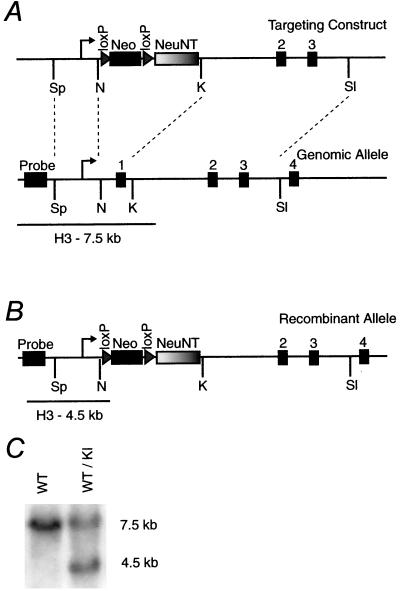
To place an oncogenic neu allele under the transcriptional control of the endogenous promoter in the mammary gland, we have used a strategy where the first coding exon of the endogenous neu gene was replaced by an oncogenic neu cDNA containing an activating point mutation in the transmembrane domain. We previously have demonstrated that replacement of the first coding exon of neu with the wild-type neu cDNA can completely rescue the embryonic lethality associated with the germ-line inactivation of neu (W.R.H., R. Chan, M. A. Laing, and W.J.M., unpublished observations). To circumvent the potential deleterious consequences of expressing an activated neu oncogene, we designed a targeting vector in which a loxP-flanked neomycin cassette was placed upstream of the oncogenic neu cDNA, preventing activated neu expression in the absence of Cre recombinase (Fig. 1A). We previously have shown that this loxP neo cassette can block neu expression from the Moloney murine leukemia viral enhancer (E.R.A., W.R.H., and W.J.M., unpublished observations). Electroporation of this targeting vector resulted in the generation of several independent embryonic stem cell lines carrying the expected targeted allele (Fig. 1B). After germ-line transmission of this recombinant allele (Fig. 1C), interbreeding of mice carrying this activated neu allele generated only wild-type and heterozygous pups, indicating that the nonexcised recombinant neu allele was indeed acting as a null allele, resulting in embryonic lethality.
Figure 1.
Targeting of the conditionally activated neu allele. (A) A schematic representation of the targeting construct and the genomic allele. The 2.5-kb 5′ arm of homology (_Sph_I to _Nar_1) and the 8-kb 3′ arm of homology (_Kpn_I to _Sal_I) were used to direct the homologous recombination to the wild-type allele, illustrated by the dashed lines. Exon 1 of the endogenous allele was replaced by a loxP (triangle)-flanked PGK-neomycin-HSV poly(A) (Neo) cassette, followed by the neuNT cDNA and a simian virus 40 poly(A) (NeuNT). The loxP-flanked Neo NeuNT cDNA has replaced exon 1 contained within the _Nar_1/_Kpn_I fragment. The size of the genomic _Hin_dIII (H3)-restricted fragment when detected by a probe 5′ to the site of homologous recombination also is depicted. Sp, _Sph_I; N, _Nar_1; K, _Kpn_I; Sl, _Sal_I. (B) The recombinant allele containing the loxP-flanked Neo NeuNT cassette in place of exon 1 and the corresponding size of the _Hin_dIII restriction fragment detected by the external probe are shown. (C) A representative Southern blot of tail DNA from mice that are wild type (WT) and heterozygous for the knock-in (KI) allele.
Induction of Activated neu Expression in the Mammary Epithelium.
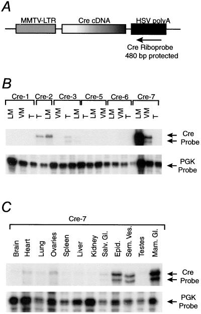
To induce expression of the activated neu allele in the mammary epithelium, transgenic mice expressing Cre recombinase in the mammary epithelium were generated by placing the Cre cDNA under the control of the MMTV promoter (Fig. 2A). Seven transgenic founders were identified, of which six transmitted the transgene to their progeny. An RNase protection assay on RNA isolated from testes, and mammary glands revealed three lines that expressed Cre in the mammary gland (Fig. 2B). Because leaky Cre expression in other organs could potentially result in _neu-_induced lethality, we performed a further RNase protection analyses on RNA harvested from major organs in these three lines. These analyses revealed that the Cre-2 and Cre-3 lines were unsuitable for our experiments because of expression in the brain and heart, respectively. Importantly, the RNase protection analysis, which surveyed major organs from the Cre-7 line, revealed that there was a high level of expression in the mammary gland with expression in other tissues primarily limited to the male accessory reproductive organs (Fig. 2C).
Figure 2.
Generation of MMTV-Cre transgenics. (A) A schematic of the transgenic construct is shown. The MMTV-long terminal repeat (MMTV-LTR) (gray) was used to drive expression of the cre cDNA (gradient fill). The cre cDNA was followed by the HSV polyadenylation [HSV poly(A)] sequence (black). The 480-bp antisense riboprobe is depicted by the arrow and is directed against the HSV poly(A). (B) The RNase protection on the lactating mammary gland (L), virgin mammary gland (V), and testes (T) for the six lines that passed the transgene shows that expression was occurring in the Cre-2, Cre-3, and Cre-7 lines. (C) An RNase protection screening major organs of the Cre-7 line. Note the expression primarily limited to the mammary gland and male accessory reproductive organs. The pgk riboprobe was included as an internal control for equal RNA loading.
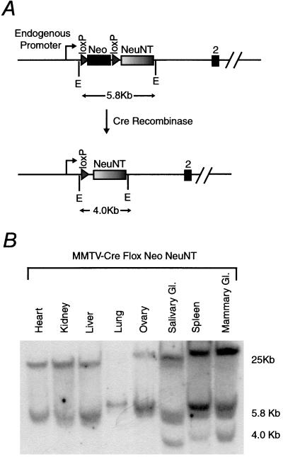
Mating the MMTV-Cre-7 mice with the mice harboring the inducible activated neu allele resulted in bigenic mice at the expected Mendelian ratio. Consistent with the RNase protection results, Southern blot analyses of DNA from all major organs revealed that excision of the loxP-flanked neomycin cassette was restricted primarily to the mammary gland, although spleen and salivary excision also were noted (Fig. 3). Quantitative PhosphorImager analyses of the Southern blots demonstrated that excision of the loxP-flanked neomycin cassette was 30–40% complete in the mammary gland. Although excision of the neomycin cassette was noted in the spleen and salivary glands, examination of these organs at the gross or histological level failed to reveal any abnormalities.
Figure 3.
Excision of the loxP- flanked neomycin cassette. (A) The recombinant allele is shown before and after Cre recombinase-mediated excision. Before excision the endogenous promoter will not regulate neuNT expression because of the poly(A) in the neomycin cassette. To detect removal of the neomycin cassette, the neuNT cDNA was used as a probe in an _Eco_RI (E) digest. The size of this fragment is depicted in both the excised and nonexcised forms. (B) A Southern blot shows that excision of the loxP-flanked Neo is limited to the spleen, salivary, and mammary glands by the presence of a 4-kb band in those samples. The wild-type allele also is detected by the neuNT cDNA probe, resulting in a faint band of 6.0 kb and a stronger band of 25 kb.
Whole-Mount and Immunohistochemical Analysis of Mammary Glands and Tumors.
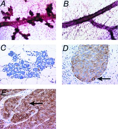
Using whole-mount analysis to examine the mammary epithelium, no significant deviation from the wild-type mammary structure was observed in either the Cre-7 or Flneo NeuNT mice (data not shown). However, when virgin bigenic mammary glands were examined, an abnormal mammary structure was observed (Fig. 4A). In contrast to the normal pattern of lobuloalveolar development (Fig. 4B), numerous lobular side buds were observed in the mammary glands of mice carrying the activated neu allele (Fig. 4A). High magnification histology of this abnormal structure revealed that the lobular side buds have formed acinar structures and are not solid dysplasias (Fig. 4C). Interestingly, these early lesions have no detectable staining for Neu. Although the initial mammary phenotype observed was limited to enhanced branching, older female mice developed focal comedo-adenocarcinomas (n = 9, 45% penetrant in mice over 1 year of age). Immunohistochemical examination of these tumors expressing activated Neu revealed both membrane and cytoplasmic immunoreactivity for Neu (Fig. 4D), in contrast to the MMTV-Neu induced tumors where the staining for Neu is distinctly membrane specific (Fig. 4E). Importantly, this MMTV-Neu transgenic control is expressing a wild-type neu allele, which does not force the constitutive dimerization associated with the activated allele. Despite the difference in localization of Neu, the mammary tumors from these strains were histologically identical. The apparent difference in localization likely reflects the rapid endocytosis of activated Neu, which is known to undergo constitutive dimerization (31).
Figure 4.
Digitized micrographic images of whole-mount (A and B) and immunohistochemical (C–E) analysis of the MMTV-Cre Flneo NeuNT mammary gland and tumors. (A) Whole mount of a virgin MMTV-Cre Flneo NeuNT mammary gland at 9 months. The extensive side branching terminating in lobuolalveolar units should be noted. (B) Whole mount of a virgin wild-type control mammary gland at a similar stage of development illustrating a normal duct with few side branches. (C) Immunohistochemical analysis of the same virgin gland shown in A. Note the absence of anti-Neu staining in the acinar structures, which are not dysplastic. Immunohistochemical analysis on the bigenic (D) and a control MMTV-Neu (E) induced tumor shows high levels of Neu expression in these tumors. The contrast in the cytoplasmic and membrane localization of the stain indicated by the arrows in D and E, respectively should be noted. (Magnifications: A and B, × 40; C_–_E, ×320.)
Induction of Mammary Tumors Involves Elevated Expression and Amplification of the Activated neu cDNA.
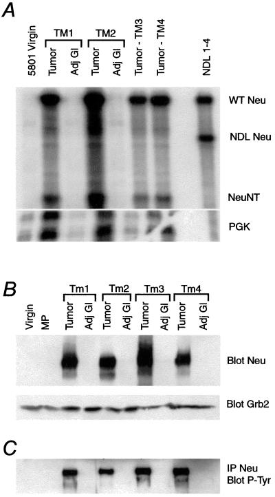
To ascertain whether tumor progression in these strains involved the up-regulation of activated neu allele, we determined the levels of neu transcript and protein in mammary tumors and adjacent normal mammary gland. To assess the levels of neu transcript in these tissues we performed RNase protection analyses on RNA from these tissues with a probe complementary to the transmembrane domain of Neu. In contrast to the normal mammary epithelium, elevated levels of protected fragment corresponding to the expression of the oncogenic neu allele were detected in mammary tumors (Fig. 5A, NeuNT band). Given that the NDL 1–4 control does not express wild-type neu, the presence of the protected fragment corresponding to the wild-type allele in these tumor samples likely reflects the nonstringent hybridization and digestion conditions used in these analyses. Further, because the activated rat neu cDNA differs from the endogenous mouse cDNA in its _Bam_HI restriction profile, we recently have shown by reverse transcriptase–PCR analyses that the majority of neu transcripts from the tumor RNA are derived from the activated rat allele (data not shown). Consistent with the RNase protection analyses, immunoprecipitations and immunoblots with Neu and phosphotyrosine-specific antibodies revealed elevated levels of both Neu and tyrosine-phosphorylated Neu protein in the tumor tissues compared with matched adjacent mammary glands. (Fig. 5 B and C).
Figure 5.
Overexpression of activated NeuNT in mammary tumors. (A) Using a probe that spans the transmembrane domain of Neu, an RNase protection reveals that MMTV-Cre Flneo NeuNT tumors (TM1–4) overexpress the activated form of neu. The mammary gland from a virgin MMTV-Cre Flneo NeuNT mouse (5801) was included, as was a positive control known to overexpress an activated form of neu (NDL 1–4). The full-length protected fragment is shown at the band labeled WT Neu. The mutant alleles are labeled NDL Neu and NeuNT, corresponding to their activating mutations. Thirty micrograms of RNA was used, except in the control NDL 1–4 tumor sample, where 20 μg was used. pgk was included as an internal control for equal loading of the samples. Adj. Gl., adjacent mammary gland. (B) Blotting for Neu reveals that only the tumors have elevated levels of Neu. Grb-2 was used to control for equal loading of the 120 μg of protein in each sample. MP, multiparious. (C) After immunoprecipitating for Neu, a blot was probed for phosphorylated tyrosine (P-Tyr), which demonstrated that catalytically active Neu was overexpressed in the tumors. Protein (1.3 mg) was used in this immunoprecipitation.
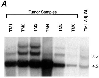
Given the elevated expression of neu in the mouse mammary tumor samples, they were examined for amplification of the recombinant and wild-type alleles. A Southern blot analyses of genomic DNA from tumor tissues revealed that in all tumors examined (n = 6), there was a selective amplification of the recombinant neu allele relative to the wild-type neu allele (Fig. 6). Additionally, a third band above the wild-type and recombinant bands appeared with the same number of copies as the wild-type allele in half of the surveyed tumors, likely a result of the amplification process. Quantitative PhosphorImager analyses revealed a 2- to 22-fold amplification of this activated neu allele relative to the wild-type allele (Table 1), suggesting that mammary tumorigenesis in this strain requires both the amplification and elevated expression of the neu oncogene and does not appear to correlate with pariety status.
Figure 6.
Amplification of neu in mammary tumors. Southern analysis showing the amplification of the recombinant allele (4.5 kb) in respect to the wild-type allele (7.5 kb) in the MMTV-Cre Flneo NeuNT tumors (TM1–6). The TM1 adjacent gland (TM1 Adj. Gl.) also is shown, illustrating that detectable amplification has not occurred in the normal mammary gland. The samples were restricted with _Hin_dIII and probed with the external probe using the same strategy described in Fig. 1. Interestingly, a new band has appeared above the wild-type band in TM2, TM3, and TM5, likely a result of the amplification process. This Southern analysis was subjected to a quantitative PhosphorImager analysis where the increase in neuNT copy number was determined relative to the wild-type allele. The results are shown in Table 1.
Table 1.
Amplification of NeuNT
| Sample | # of copies relative to wild type | Age at tumor detection, months | Pariety |
|---|---|---|---|
| TM1 | 8.7 | 8 | MP |
| TM2 | 6.0 | 16 | MP |
| TM3 | 6.1 | 15.3 | V |
| TM4 | 21.6 | 15 | V |
| TM5 | 4.8 | 13 | MP |
| TM6 | 1.7 | 14 | V |
| TM7 | NC | 17 | V |
| TM8 | NC | 17 | V |
| TM9 | NC | 17.3 | V |
| Average | 8.0 | 14.7 |
Discussion
We have demonstrated that mammary epithelial-specific expression of oncogenic neu under the transcriptional control of its physiological relevant promoter is capable of inducing abnormal mammary development and ultimately mammary tumors. To achieve mammary-specific induction of activated neu expression, we have taken advantage of a transgenic mice expressing Cre recombinase under the transcriptional control of the MMTV promoter enhancer. A previous report that suggested that MMTV-directed Cre expression resulted in widespread excision in many different tissues (32). In contrast to that report, our studies have revealed that induction of activated neu expression by the Cre recombinase was limited to relatively narrow set of tissues, including the mammary gland, spleen, and salivary gland (Fig. 3). The discordance between studies likely reflects differences in the tissue specificity of Cre transgene expression. Indeed, more recently, mammary-specific excision of the BRCA1 gene has achieved by crossing loxP-flanked BRCA1 strains with a separate MMTV/Cre transgenic strain (33). Future studies with these MMTV/Cre strains should prove useful in creating mammary-specific activation or deletion of genes critical for normal development.
In contrast to the rapid tumor progression observed in transgenic mice carrying MMTV-driven activated neu alleles, expression of activated neu under transcriptional control of its physiological promoter is not sufficient for induction of mammary tumors. Rather, these transgenic mice have accelerated lobuloalveolar development that correlates with the low levels of activated neu expression. However, female mice that express neu from the endogenous promoter eventually develop mammary tumors after a long latency period. Tumor progression in these strains was correlated with a dramatic elevation of both neu transcript and protein when compared with adjacent gland controls. One possible explanation for the elevated level of neu transcripts is that the selective amplification of the recombinant neu allele during tumor progression has occurred, analogous to the amplification and overexpression of erbB-2 observed in human breast cancer (11–15). Indeed, when the tumors were analyzed for amplification, they were found to have between two and 22 additional copies of the recombinant neu allele. The strong selection for amplification of the activated neu allele in this model may reflect the requirement of a critical threshold level of Neu for oncogenic conversion of the mammary epithelial cell. However, because the levels of activated neu do not precisely correspond to the extent of neu overexpression, there may be other molecular mechanisms involved in the observed elevated expression of the activated neu allele. In this regard, it has been reported that a certain percentage of _neu_-expressing human breast carcinomas do not exhibit evidence of neu amplification (13). Taken together, these observations suggest that amplification and the consequent elevated expression of activated neu is required for efficient transformation of the mammary epithelial cell.
In addition to closely mimicking the human disease, another important feature of this model is that activated neu expression ultimately is controlled by transcription factors that regulate the endogenous neu promoter sequence. Given the importance of number of transcription factors such as c-myc (34) and the estrogen receptor (35) in the progression of breast cancer, future studies with these mice should provide insight into the role that these critical transcription factors play in _neu_-induced breast cancer. In addition to breast cancer, a number of recent studies have suggested that neu may be involved in the induction of ovarian, lung, gastric, and prostate cancers (2, 36, 37). Given the Cre-inducible nature of this activated neu allele, future studies with tissue-specific expression of Cre should allow investigators to directly assess the role of activated neu in these tissue sites.
Acknowledgments
We thank Dinsdale Gooden for oligonucleotide synthesis and Brian Allore for automated DNA sequence analysis (MOBIX Central Facility, McMaster University). We are grateful to Margaret Hibbs and Ashley Dunn for generously providing ErbB-2 genomic clones. We are also grateful to Monica Graham and Judy Walls for technical support. R.D.C. acknowledges the generous support of Dr. Carol MacLeod at the University of California, Davis Cancer Center. We also thank John A. Hassell for critical reading of this manuscript. This work was supported by grants awarded to W.J.M by the Medical Research Society of Canada and the Canadian Breast Cancer Research Initiative. W.J.M. is a recipient of a Medical Research Council of Canada Scientist award, P.M.S. received a studentship from the Medical Research Council of Canada, and E.R.A. was supported by the Cancer Research Society Studentship and a scholarship from the United States Army Medical Research's Breast Cancer Research Program.
Abbreviations
MMTV
mouse mammary tumor virus
HSV
herpes simplex virus
PGK
phosphoglycerate kinase
Footnotes
This paper was submitted directly (Track II) to the PNAS office.
Article published online before print: Proc. Natl. Acad. Sci. USA, 10.1073/pnas.050408497.
Article and publication date are at www.pnas.org/cgi/doi/10.1073/pnas.050408497
References
- 1.Dougall W C, Qian X, Peterson N C, Miller M J, Samanta A, Greene M I. Oncogene. 1994;9:2109–2123. [PubMed] [Google Scholar]
- 2.Hynes N E, Stern D F. Biochim Biophys Acta. 1994;1198:165–184. doi: 10.1016/0304-419x(94)90012-4. [DOI] [PubMed] [Google Scholar]
- 3.Ullrich A, Coussens L, Hayflick J S, Dull T J, Gray A, Tam A W, Lee J, Yarden Y, Libermann T A, Schlessinger J. Nature (London) 1984;309:418–425. doi: 10.1038/309418a0. [DOI] [PubMed] [Google Scholar]
- 4.Bargmann C I, Hung M C, Weinberg R A. Nature (London) 1986;319:226–230. doi: 10.1038/319226a0. [DOI] [PubMed] [Google Scholar]
- 5.Coussens L, Yang-Feng T L, Liao Y C, Chen E, Gray A, McGrath J, Seeburg P H, Libermann T A, Schlessinger J, Francke U. Science. 1985;230:1132–1139. doi: 10.1126/science.2999974. [DOI] [PubMed] [Google Scholar]
- 6.Plowman G D, Whitney G S, Neubauer M G, Green J M, McDonald V L, Todaro G J, Shoyab M. Proc Natl Acad Sci USA. 1990;87:4905–4909. doi: 10.1073/pnas.87.13.4905. [DOI] [PMC free article] [PubMed] [Google Scholar]
- 7.Schechter A L, Stern D F, Vaidyanathan L, Decker S J, Drebin J A, Greene M I, Weinberg R A. Nature (London) 1984;312:513–516. doi: 10.1038/312513a0. [DOI] [PubMed] [Google Scholar]
- 8.Yamamoto T, Ikawa S, Akiyama T, Semba K, Nomura N, Miyajima N, Saito T, Toyoshima K. Nature (London) 1986;319:230–234. doi: 10.1038/319230a0. [DOI] [PubMed] [Google Scholar]
- 9.Kraus M H, Issing W, Miki T, Popescu N C, Aaronson S A. Proc Natl Acad Sci USA. 1989;86:9193–9197. doi: 10.1073/pnas.86.23.9193. [DOI] [PMC free article] [PubMed] [Google Scholar]
- 10.Plowman G D, Culouscou J M, Whitney G S, Green J M, Carlton G W, Foy L, Neubauer M G, Shoyab M. Proc Natl Acad Sci USA. 1993;90:1746–1750. doi: 10.1073/pnas.90.5.1746. [DOI] [PMC free article] [PubMed] [Google Scholar]
- 11.Slamon D J, Clark G M, Wong S G, Levin W J, Ullrich A, McGuire W L. Science. 1987;235:177–182. doi: 10.1126/science.3798106. [DOI] [PubMed] [Google Scholar]
- 12.Slamon D J, Godolphin W, Jones L A, Holt J A, Wong S G, Keith D E, Levin W J, Stuart S G, Udove J, Ullrich A. Science. 1989;244:707–712. doi: 10.1126/science.2470152. [DOI] [PubMed] [Google Scholar]
- 13.van de Vijver M J, Peterse J L, Mooi W J, Wisman P, Lomans J, Dalesio O, Nusse R. N Engl J Med. 1988;319:1239–1245. doi: 10.1056/NEJM198811103191902. [DOI] [PubMed] [Google Scholar]
- 14.Venter D J, Tuzi N L, Kumar S, Gullick W J. Lancet. 1987;2:69–72. doi: 10.1016/s0140-6736(87)92736-x. [DOI] [PubMed] [Google Scholar]
- 15.Zeillinger R, Kury F, Czerwenka K, Kubista E, Sliutz G, Knogler W, Huber J, Zielinski C, Reiner G, Jakesz R. Oncogene. 1989;4:109–114. [PubMed] [Google Scholar]
- 16.Bouchard L, Lamarre L, Tremblay P J, Jolicoeur P. Cell. 1989;57:931–936. doi: 10.1016/0092-8674(89)90331-0. [DOI] [PubMed] [Google Scholar]
- 17.Guy C T, Cardiff R D, Muller W J. J Biol Chem. 1996;271:7673–7678. doi: 10.1074/jbc.271.13.7673. [DOI] [PubMed] [Google Scholar]
- 18.Muller W J, Sinn E, Pattengale P K, Wallace R, Leder P. Cell. 1988;54:105–115. doi: 10.1016/0092-8674(88)90184-5. [DOI] [PubMed] [Google Scholar]
- 19.Guy C T, Webster M A, Schaller M, Parsons T J, Cardiff R D, Muller W J. Proc Natl Acad Sci USA. 1992;89:10578–10582. doi: 10.1073/pnas.89.22.10578. [DOI] [PMC free article] [PubMed] [Google Scholar]
- 20.Siegel P M, Dankort D L, Hardy W R, Muller W J. Mol Cell Biol. 1994;14:7068–7077. doi: 10.1128/mcb.14.11.7068. [DOI] [PMC free article] [PubMed] [Google Scholar]
- 21.Kwong K Y, Hung M C. Mol Carcinog. 1998;23:62–68. doi: 10.1002/(sici)1098-2744(199810)23:2<62::aid-mc2>3.0.co;2-o. [DOI] [PubMed] [Google Scholar]
- 22.Siegel P M, Ryan E D, Cardiff R D, Muller W J. EMBO J. 1999;18:2149–2164. doi: 10.1093/emboj/18.8.2149. [DOI] [PMC free article] [PubMed] [Google Scholar]
- 23.Lee K F, Simon H, Chen H, Bates B, Hung M C, Hauser C. Nature (London) 1995;378:394–398. doi: 10.1038/378394a0. [DOI] [PubMed] [Google Scholar]
- 24.Stocklin E, Botteri F, Groner B. J Cell Biol. 1993;122:199–208. doi: 10.1083/jcb.122.1.199. [DOI] [PMC free article] [PubMed] [Google Scholar]
- 25.Guy C T, Cardiff R D, Muller W J. Mol Cell Biol. 1992;12:954–961. doi: 10.1128/mcb.12.3.954. [DOI] [PMC free article] [PubMed] [Google Scholar]
- 26.Sternberg N, Sauer B, Hoess R, Abremski K. J Mol Biol. 1986;187:197–212. doi: 10.1016/0022-2836(86)90228-7. [DOI] [PubMed] [Google Scholar]
- 27.Joyner A L. Gene Targeting: A Practical Approach. New York: Oxford Univ. Press; 1993. [Google Scholar]
- 28.Vonderhaar B K, Greco A E. Endocrinology. 1979;104:409–418. doi: 10.1210/endo-104-2-409. [DOI] [PubMed] [Google Scholar]
- 29.Webster M A, Hutchinson J N, Rauh M J, Muthuswamy S K, Anton M, Tortorice C G, Cardiff R D, Graham F L, Hassell J A, Muller W J. Mol Cell Biol. 1998;18:2344–2359. doi: 10.1128/mcb.18.4.2344. [DOI] [PMC free article] [PubMed] [Google Scholar]
- 30.Deckard-Janatpour K, Muller W J, Chodosh L A, Gardner H P, Marquis S T, Coffey R J, Cardiff R D. Int J Oncol. 1997;11:235–241. doi: 10.3892/ijo.11.2.235. [DOI] [PubMed] [Google Scholar]
- 31.Bargmann C I, Weinberg R A. EMBO J. 1988;7:2043–2052. doi: 10.1002/j.1460-2075.1988.tb03044.x. [DOI] [PMC free article] [PubMed] [Google Scholar]
- 32.Wagner K U, Wall R J, St-Onge L, Gruss P, Wynshaw-Boris A, Garrett L, Li M, Furth P A, Hennighausen L. Nucleic Acids Res. 1997;25:4323–4330. doi: 10.1093/nar/25.21.4323. [DOI] [PMC free article] [PubMed] [Google Scholar]
- 33.Xu X, Wagner K U, Larson D, Weaver Z, Li C, Ried T, Hennighausen L, Wynshaw-Boris A, Deng C X. Nat Genet. 1999;22:37–43. doi: 10.1038/8743. [DOI] [PubMed] [Google Scholar]
- 34.Schoenenberger C A, Andres A C, Groner B, van de Vijver M J, LeMeur M, Gerlinger P. EMBO J. 1988;7:169–175. doi: 10.1002/j.1460-2075.1988.tb02797.x. [DOI] [PMC free article] [PubMed] [Google Scholar]
- 35.Osborne C K. Breast Cancer Res Treat. 1998;51:227–238. doi: 10.1023/a:1006132427948. [DOI] [PubMed] [Google Scholar]
- 36.Ishikawa T, Kobayashi M, Mai M, Suzuki T, Ooi A. Am J Pathol. 1997;151:761–768. [PMC free article] [PubMed] [Google Scholar]
- 37.Ross J S, Yang F, Kallakury B V, Sheehan C E, Ambros R A, Muraca P J. Am J Clin Pathol. 1999;111:311–316. doi: 10.1093/ajcp/111.3.311. [DOI] [PubMed] [Google Scholar]
