γ-Secretase is a membrane protein complex comprised of presenilin, nicastrin, aph-1, and pen-2 (original) (raw)
Abstract
γ-Secretase catalyzes the intramembrane proteolysis of Notch, β-amyloid precursor protein, and other substrates as part of a new signaling paradigm and as a key step in the pathogenesis of Alzheimer's disease. This unusual protease has eluded identification, though evidence suggests that the presenilin heterodimer comprises the catalytic site and that a highly glycosylated form of nicastrin associates with it. The formation of presenilin heterodimers from the holoprotein is tightly gated by unknown limiting cellular factors. Here we show that Aph-1 and Pen-2, two recently identified membrane proteins genetically linked to γ-secretase, associate directly with presenilin and nicastrin in the active protease complex. Coexpression of all four proteins leads to marked increases in presenilin heterodimers, full glycosylation of nicastrin, and enhanced γ-secretase activity. These findings suggest that the four membrane proteins comprise the limiting components of γ-secretase and coassemble to form the active enzyme in mammalian cells.
Regulated intramembrane proteolysis is an evolutionarily conserved biochemical mechanism that has been recognized only recently (1). Although hydrolysis of a peptide bond within the hydrophobic environment of a lipid bilayer seems counter-intuitive, several enzymes nevertheless appear to carry out this process. One such enzyme, termed γ-secretase, is a founding member of a new class of intramembrane-cleaving proteases (2). γ-Secretase was first recognized because of its role in the production of the amyloid-β protein (Aβ), a 40- to 42-residue peptide that is pathogenic in Alzheimer's disease (3). In addition, γ-secretase catalyzes the proteolytic release of the intracellular domains of Notch, β-amyloid precursor protein, and numerous other type I transmembrane receptors, indicating that it normally serves as a mediator of diverse signaling pathways.
Despite substantial progress in understanding its normal and abnormal biology, the complete identity of γ-secretase has remained elusive. However, the burden of evidence has suggested that presenilin (PS) is the active site of the protease (2). PS is cleaved into N-terminal and C-terminal fragments (NTF and CTF) that remain associated, and these heterodimers appear to be the biologically active form of the protein (4). Indeed, compounds designed as transition-state analogue inhibitors of γ-secretase bind specifically to PS heterodimers (5, 6). Moreover, mutation of two conserved intramembrane aspartates (7) or genetic deletion of PS (8) interferes with γ-secretase activity. These data, plus the recognition that PS contains an aspartic protease motif (9), strongly implicate PS as the active site of γ-secretase. However, PS does not act alone, as the levels of PS heterodimers are tightly regulated by other limiting factors (10), and overexpression of PS does not increase γ-secretase activity and produce more product.
Nicastrin (NCT), discovered via its association with PS (11), may be one of the hypothesized limiting factors. Genetic ablation of NCT in Caenorhabditis elegans and Drosophila results in a phenotype similar to the deletion of PS (11–15), and recent evidence has shown that NCT maturation depends on PS (16–19) and is directly associated with γ-secretase (19, 20). However, overexpression of both PS and NCT is not sufficient to generate more γ-secretase activity (19), suggesting that additional limiting factors exist. Genetic screens designed to modify a PS-deficient phenotype in C. elegans have recently yielded two novel genes, APH-1 (21) and PEN-2 (22). Genetic analyses demonstrated that these proteins are essential for PS endoproteolysis and γ-secretase function (22), but their biochemical role remained undefined. Here, we characterize the human forms of these two proteins and demonstrate that they are physical members of the γ-secretase complex. We further show that the combination of the four human proteins (PS1, NCT, Aph-1, and Pen-2) is sufficient to circumvent the tight regulation of PS heterodimer levels and thus augment γ-secretase activity in mammalian cells.
Methods
Immunoprecipitation and Western Blotting. CHAPSO [3-[(3-cholamidopropyl)dimethylammonio]-2-hydroxy-1-propanesulfonate] lysates were prepared as described (20). Coimmunoprecipitations (co-IPs) and III-31C precipitations were performed in 1% CHAPSO as described (20). For blue-native electrophoresis, samples were analyzed as described (23). Detection on Western blots used R302 (NCT, 1:4,000), guinea pig anti-NCT (1:2,000, Chemicon), Ab14 (PS1 NTF, 1:2,000), goat α-PS1 N-19 (PS1 NTF, 1:200, Santa Cruz Biotechnology), 13A11 (PS1 CTF, 5 μg/ml), M2 Flag (1:1,000, Sigma) or 3F10 (1:2,000, Roche).
Glycerol Gradients and in Vitro Activity Assay. Glycerol gradients contained 0.2% digitonin in Hepes buffer (50 mM Hepes/150 mM NaCl/5 mM MgCl2/5 mM CaCl2) and 18–28% glycerol in 1% increments. For co-IP from the gradient fractions, samples were adjusted to 0.5% digitonin and 6% glycerol. C100Flag activity assays were as described (20).
Stable Cell Lines. PS70 Chinese hamster ovary (CHO) cells, which stably express wild-type human PS1, have been described (24) and are the parental cell line for all stable transfectants prepared in this study. The A-5 clone additionally expresses Aph-1α2-HA, whereas the P-2 and P-22 clones express Flag-Pen2. The γ-23 and γ-30 cell lines express Flag-Pen2 in the A-5 background.
Results
Two homologues of APH-1 exist in the human genome, here termed Aph-1α and Aph-1β. Both genes encode proteins predicted to contain seven transmembrane domains (Fig. 1_a_). Additionally, Aph-1α occurs in two splice isoforms (Aph-1α1 and Aph-1α2) that diverge at the C terminus (22). To ascertain which isoform is most abundant, we performed Northern blotting on various human tissues. Both splice forms of Aph-1α were expressed at nearly equal abundance within each tissue surveyed (see Supporting Results and Fig. 5, which are published as supporting information on the PNAS web site, www.pnas.org). Moreover, their expression profile across tissues matched that of PS1. We detected little or no Aph-1β mRNA in these human tissues, suggesting that this transcript is not as widely expressed as Aph-1α. In accord, reverse transcription and PCR generated only Aph-1α1 and Aph-1α2 from human brain mRNA (but all three transcripts from human testis mRNA). We also performed Northern blots of multiple human tissues for Pen-2 and found a similar expression profile as those of PS1 and Aph-1α (Supporting Results).
Fig. 1.
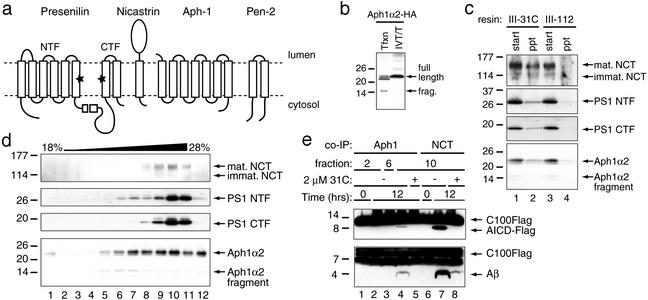
Aph1α2 is associated with the γ-secretase complex. (a) Schematic of the four candidate members of the γ-secretase complex. PS1 contains two intramembrane aspartates (stars) that constitute the probable active site. The membrane orientations of Aph-1 and Pen-2 are not yet verified. (b) Aph1α2-HA was transfected into COS7 cells (Tfxn) or generated by in vitro transcription/translation (IVT/T) in rabbit reticulocyte lysate. The highest band in the COS7 lysate comigrates with the IVT/T product. (c) Affinity precipitation of Aph1α2 by III-31C but not III-112 resin. Start lysate identical to that in b was subjected to batch mode pull-downs with each resin. Bound proteins (ppt) were blotted with R302 to NCT, Ab14 to PS1 NTF, 13A11 to PS1 CTF, and 3F10 to HA of Aph-1α2-HA. (d) One percent digitonin-solubilized membranes were fractionated on an 18–28% glycerol gradient. Fractions were blotted for NCT, PS1, and Aph-1α2 with antibodies as in c. The γ-secretase complex in COS7 reproducibly migrates into denser gradient fractions than do the complexes in HEK293, CHO, or HeLa membranes (see Fig. 4_c_, and data not shown). Although the reason for this consistent difference is unknown, we note that the γ-secretase complex also migrates differently in CHAPSO than in digitonin detergent gradients. (e) Fractions 2, 6, and 10 from the glycerol gradient were subjected to co-IP with 3F10 antibody (Aph1α2-HA) or R302 (NCT). Beads were exchanged into 0.25% CHAPSO/Hepes, adjusted to contain 0.0125% phosphatidylethanolamine and 0.1% phosphati-dylcholine, and incubated with C100Flag without or with 31C inhibitor. AICD-Flag was detected with M2 Flag antibody and Aβ with 6E10.
Aph-1 Is Physically Associated with the Active γ-Secretase Complex. To determine whether Aph-1α and Pen-2 are associated with γ-secretase, we evaluated each protein individually. We first focused on the transient expression in COS7 cells of Aph-1α2. We placed a hemagglutinin (HA) tag on the C terminus of Aph-1α2 for ease of detection. In whole lysates, we noted bands in the ≈23- and ≈14-kDa regions of the gel (Fig. 1_b_, lane 1). Although we did not detect a prominent band at the predicted ≈30-kDa size, the ≈23-kDa band likely represents the full-length protein because (i) an N-terminal cleavage fragment that would account for the 7-kDa difference would span more than one of the hydrophobic domains (and is longer than most signal peptides), (ii) the 23-kDa band comigrated with an in vitro transcribed and translated product (Fig. 1_b_, lane 2), and (iii) it was the only form present in stable cells (see Fig. 3_b_). Interestingly, full-length PS1 migrates 6–8 kDa faster than its predicted 53-kDa size in SDS gels. The fast migration of both proteins suggests that the multiple hydrophobic domains adopt a compact conformation during denaturing gel electrophoresis.
Fig. 3.
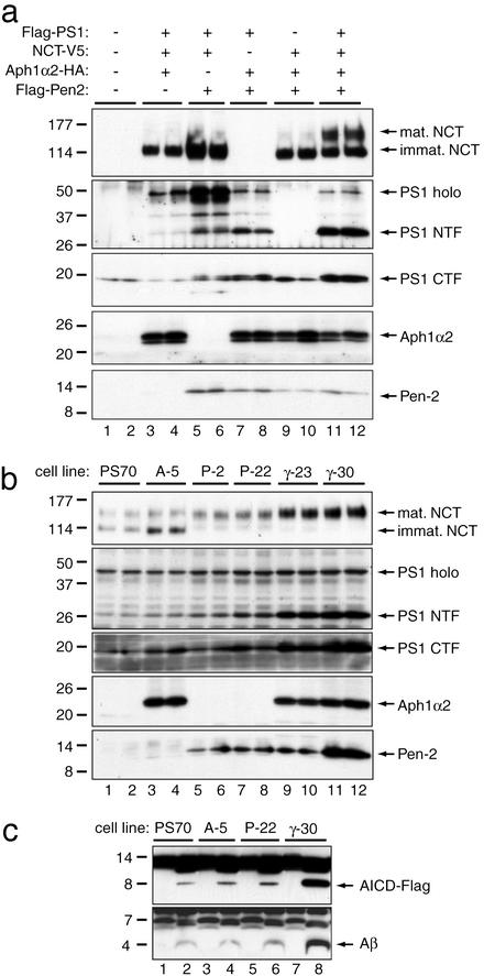
PS1, NCT, Aph-1α2, and Pen-2 are sufficient to generate more γ-secretase in mammalian cells. (a) CHO cells were transiently transfected with the indicated constructs. Equal amounts of total protein were blotted for NCT (V5), PS1 holoprotein and NTF (M2 Flag), PS1 CTF (13A11; which recognizes both endogenous and exogenous CTF), Aph-1α2 (3F10), and Pen-2 (M2 Flag). Note the increases in mature NCT and PS1 heterodimers when all four proteins are coexpressed (lanes 11 and 12). Exogenous human PS1 CTF migrates slightly higher than endogenous hamster CTF. (b) Stable CHO cell lines expressing various combinations of human PS1, Aph-1α2, and/or Pen-2. Note the nearly equal abundance of immature and mature forms of NCT in the parental PS70 cells (lanes 1 and 2); this obviates the need to overexpress NCT. There is a slight increase in the immature, ≈110-kDa NCT in the A-5 stable line, suggesting that excess Aph-1α2 may stabilize immature NCT. However, only when PS1, Aph-1α2, and Pen-2 are all overexpressed is there a marked increase in mature NCT and PS1 NTF and CTF. PS70, CHO cells stably expressing PS1; A-5, PS70 cells with Aph-1α2-HA; P-2 and P-22, PS70 cells with Flag-Pen2; γ-23 and γ-30, A-5 cells with Flag-Pen2. Antibodies: R302 (NCT), Ab14 (PS1 holoprotein and NTF), 13A11 (PS1 CTF), 3F10 (Aph-1α2), and M2 Flag (Pen-2). (c) Direct assessment of γ-secretase activity in membrane lysates of the indicated stable cell lines. Membranes from equal numbers of cells were solubilized in 1% CHAPSO, and a C100Flag activity assay was performed (20). Samples were probed for the AICD-Flag (Upper) and Aβ (Lower) products, as in Fig. 2_b_.
Next, we solubilized Aph1-transfected COS7 membrane preparations in 1% CHAPSO/Hepes buffer and performed precipitations with III-31C affinity resin, because this inhibitor matrix specifically binds active PS/γ-secretase complexes (20). As a control, we used an inactive compound of closely similar structure but lacking the inhibitory moiety. NCT, PS1 NTF, and PS1 CTF were specifically precipitated by the 31C resin (Fig. 1_c_, lanes 1 and 2), as expected (20). In parallel, Aph-1α2-HA also bound specifically to the affinity matrix. Importantly, none of these proteins was precipitated by the control III-112 resin (Fig. 1_c_, lanes 3 and 4), nor did they bind to III-31C resin in the presence of 1% Triton X-100, which disrupts γ-secretase complexes (20) (not shown). These data strongly suggest that Aph-1α2 is associated with PS1 and NCT in the active γ-secretase complex.
To establish whether the full-length Aph-1α2 molecule or its ≈14-kDa fragment was associated with γ-secretase activity, the COS7 lysates were subjected to glycerol velocity gradient fractionation. When we probed the resultant 12 gradient fractions for the presence of NCT, PS1 NTF, and PS1 CTF, we found that these three proteins codistributed in the gradient (Fig. 1_d_), confirming their physical association. However, only the full-length ≈23-kDa Aph-1α2 molecule comigrated with NCT and PS, whereas its ≈14-kDa fragment remained in less dense fractions. To confirm that the ≈23-kDa Aph-1α2 was physically associated with γ-secretase activity, we performed co-IP from three representative gradient fractions by using an HA antibody. After co-IP, the precipitated beads were incubated with purified, recombinant C100Flag substrate and probed for the time-dependent generation of the Aβ and AICD-Flag products (20) (Fig. 1_e_). In fraction 2, where there was essentially no ≈23-kDa Aph-1α2, no cleavage of C100Flag was detectable. In fraction 6, which contained roughly similar amounts of the ≈23- and ≈14-kDa forms of Aph1α2 but little or no PS1 NTF, PS1 CTF, or NCT, there was also no detectable γ-secretase activity. Only in fraction 10, which contained the ≈23-kDa form along with NCT, PS1 NTF, and PS1 CTF, was there measurable production of Aβ and AICD-Flag, and this proteolytic activity was completely inhibited by a well characterized γ-secretase inhibitor. As a positive control, co-IP with a NCT antibody also resulted in γ-secretase-specific cleavage of C100Flag (Fig. 1_e_).
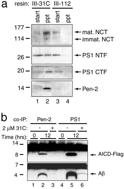
Pen-2 Is Physically Associated with the Active γ-Secretase Complex. We next performed a similar analysis with Pen-2 engineered with an N-terminal Flag tag. After transient transfection into COS7 cells, we prepared membrane lysates in 1% CHAPSO. We performed activity-dependent precipitations and found that Pen-2 bound specifically to the active III-31C resin but not the control III-112 resin (Fig. 2_a_). Not all of the transiently expressed Pen-2 protein was associated with the PS/NCT/γ-secretase complex, suggesting that like Aph-1α2, exogenously and transiently expressed Pen-2 enters into the endogenous complex with low efficiency. Indeed, this phenomenon is well described for PS: stable PS1 overexpression can largely replace the endogenous PS heterodimers (10), indicating competition for limiting cellular factors, whereas transient expression of PS1 results in small amounts of exogenous heterodimers (e.g., see Fig. 3_a_, lanes 3 and 4). This phenomenon has also been reported recently for the ≈150-kDa fully glycosylated (“mature”) form of NCT (19, 25), suggesting that transient overexpression of any component of γ-secretase results in inefficient incorporation into endogenous γ-secretase complexes.
Fig. 2.
Pen-2 is associated with the γ-secretase complex. (a) Affinity precipitation of Pen-2. Pen-2 is pulled down by III-31C resin but not the control III-112 resin. (b) M2 Flag resin was used to co-IP Pen-2 (lanes 1–3), which was then incubated as in Fig. 1_e_. A Flag-tagged PS1 construct was transfected and analyzed in parallel as a positive control (lanes 4–6).
To directly test whether precipitation of Pen-2 can pull down active γ-secretase, we coimmunoprecipitated CHAPSO lysates of the COS transfectants with M2 Flag antibody. We incubated the beads with C100Flag substrate and probed for the generation of γ-secretase products (Fig. 2_b_). After overnight incubation, both Aβ and AICD-Flag products were generated, and their production was blocked by a γ-secretase inhibitor. As a positive control, an N-terminally Flag-tagged PS1 construct was transiently transfected, coimmunoprecipitated and incubated with C100Flag in parallel, yielding the two products. Taken together, these data directly implicate both Aph-1α2 and Pen-2 as members of the active γ-secretase complex.
PS1, NCT, Aph-1, and Pen-2 Serve as Limiting Components of the γ-Secretase Complex. To determine whether these four proteins were themselves sufficient to generate additional γ-secretase complexes (i.e., to overcome the tight regulation of PS/γ-secretase levels), we transiently overexpressed each possible combination of three of the proteins in CHO cells and compared the effects to that of overexpressing all four proteins together. Because PS1 heterodimers (26) and the ≈150-kDa fully glycosylated NCT (16–19, 25) are specifically associated with γ-secretase activity while their immature forms (holoPS1 and ≈110-kDa NCT) are not, we used the levels of the mature forms as surrogates for the amount of active γ-secretase present in the cell. When Pen-2 was not present in the transfected mixture, we observed very little conversion of exogenous PS holoprotein into heterodimers and minimal maturation of exogenous NCT into its fully glycosylated form (Fig. 3_a_, lanes 3 and 4). When Aph-1α2 was not present, a similar result was obtained, although the slightly higher expression level of PS in this experiment resulted in a proportional increase in heterodimer formation (Fig. 3_a_, lanes 5 and 6).
When only exogenous NCT was not present in the mixture, we obtained a mild increase in the amount of PS1 NTF and CTF (Fig. 3_a_, lanes 7 and 8), without the high levels of PS1 holoprotein that occurred in the absence of Aph-1 (lanes 5 and 6). In this regard, it should be noted that CHO cells possess an excess of immature NCT that is not associated with γ-secretase (19), and this endogenous NCT is presumably available for association with our transiently expressed PS1, Aph-1α2, and Pen-2. When PS1 was omitted from the mixture, no significant conversion of the exogenous NCT into its mature form was observed (Fig. 3_a_, lanes 9 and 10), as expected (17). However, when all four proteins were coexpressed, there was a marked increase in both PS heterodimers and mature NCT (Fig. 3_a_, lanes 11 and 12), indicating that the transient overexpression of these four components together is sufficient to relieve the tight limit on PS heterodimer formation and generate excess γ-secretase complexes.
Stable Coexpression of the Four Components Reconstitutes γ-Secretase Activity. Because PS heterodimers are believed to constitute the active site of the protease (5, 6), the above results suggest that the stepwise addition of each of the four components results in increased levels of γ-secretase only when all four proteins are overexpressed. To confirm this conclusion, we raised stable cell lines. As CHO cells already possess excess endogenous NCT (19), we sought to stably express the other three putative components of γ-secretase in this line. Stable expression of PS1 results in the nearly complete replacement of endogenous hamster PS heterodimers with exogenous human PS1 heterodimers, as previously described for this PS70 cell line (24) (Fig. 3_b_, lanes 1 and 2). On stable coexpression of Aph-1α2-HA in the PS70 cells (yielding the A-5 line), little or no additional PS heterodimers or mature NCT were formed (Fig. 3_b_, compare lanes 1 and 2 with 3 and 4). When Pen-2 instead was stably coexpressed in the PS70 cell line (yielding the P-2 and P-22 lines), there was a modest increase in PS heterodimers and mature NCT (Fig. 3_b_, lanes 5–8). This result suggests that in CHO cells, endogenous Pen-2 levels are slightly limiting. When PS1, Aph-1α2 and Pen-2 were all stably overexpressed together (yielding the γ-23 and γ-30 lines), we observed a marked increase in the levels of PS heterodimers and a virtually complete conversion of all immature NCT into the mature form (Fig. 3_b_, lanes 9–12). This finding is consistent with the transient transfection results (Fig. 3_a_, lanes 11 and 12) and confirms that an excess of the four proteins increases the amounts of PS1 heterodimers and mature NCT simultaneously.
To directly test whether the increases in the mature forms of PS and NCT corresponded to increases in γ-secretase activity, we prepared membranes from each of the above stable cell lines and solubilized them in CHAPSO. We performed C100Flag in vitro activity assays on each membrane lysate, normalized for equal cell number (Fig. 3_c_). PS70 preparations were able to generate the appropriate γ-secretase products, and there was little increase in proteolytic activity in the A-5 line (Fig. 3_c_, lanes 1–4). A very slight increase in γ-secretase activity was observed in the P-22 cell line (Fig. 3_c_, lanes 5 and 6), which is consistent with the slight increase in PS heterodimers and mature NCT seen in this line (Fig. 3_b_, lanes 7 and 8). By comparison, the γ-30 cell lysates generated markedly more AICD-Flag and Aβ products (Fig. 3_c_, lanes 7 and 8). This observation indicates that the γ-30 cells, which express high levels of all four components, possess considerably more active γ-secretase complexes, in agreement with their large increases in PS heterodimers and mature NCT (Fig. 3_b_, lanes 11 and 12). By quantitative Western blotting, the levels of both PS1 NTF/CTFs and mature NCT were ≈3.5-fold higher in the γ-30 cells than in the parental PS70 cells; in accord, Aβ and AICD production were both ≈2.7-fold elevated.
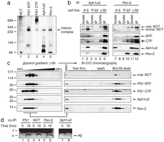
If these four proteins are indeed sufficient to constitute γ-secretase, then the size of the complex should approximate the sum of the individual molecular weights. We therefore used blue native (BN)-PAGE (23) of the γ-30 cell line to evaluate the size of the complex. We solubilized the cells in digitonin, because this detergent not only preserves the γ-secretase complex but is compatible with γ-secretase activity (20). On BN-PAGE gels, all four proteins comigrated in a band of ≈250 kDa (Fig. 4_a_), which is highly consistent with size estimates of the complex on glycerol velocity gradients made previously (27) and here (Figs. 1_d_ and 4_c_). Although there are various estimates for the molecular weight of the complex (16, 26), we favor ≈250 kDa, because it is the smallest sized complex in which we have observed γ-secretase activity (see Fig. 4 c and d).
Fig. 4.
NCT, PS 1, Aph1α2, and Pen-2 remain quantitatively associated after a partial purification of γ-secretase. (a) Digitonin lysate from γ-30 cells was run on a blue native-PAGE gel. Identically run strips of membrane were probed with antibodies as in Fig. 3_b_. A band containing all four proteins (mature complex) runs at ≈250 kDa. We observed lower migrating bands that contained some, but not all, of the proteins; these may represent intermediates in the assembly of γ-secretase. (b) Membranes from the indicated stable cell lines were solubilized in 1% CHAPSO and subjected to co-IPs. Immunoprecipitations were then probed with antibodies to NCT, PS NTF, PS CTF, Aph1α2, and Pen-2 (as in Fig. 3_b_). Asterisk, cross-reactive band (because of immunoprecipitation antibody light chains). (c) Digitonin-solubilized γ-30 cell membranes were fractionated on an 18–28% glycerol gradient. γ-Secretase-containing fractions were then pooled and injected over a 31C affinity column. The flow through, wash, and Brij-35 eluate fractions were probed for NCT, PS1 NTF and CTF, Aph-1α2, and Pen-2 as in Fig. 3_b_. (d) The elute fractions from the 31C column were pooled and subjected to co-IP with X81 (to PS1), R302 (to NCT), 3F10 (to Aph-1α2-HA), and M2 Flag (to Flag-Pen-2). The beads were washed and incubated in an in vitro C100 activity assay. Aβ was detected with 6E10. A longer exposure was needed to detect the Aβ generated from the Aph-1α2 co-IP (lanes 7 and 8), presumably because of a less efficient co-IP or steric interference of the 3F10 antibody.
Because all four proteins comigrated as a complex, we evaluated their interactions directly by using co-IP. We prepared lysates from the A-5, P-22 and γ-30 cell lines in 1% CHAPSO/Hepes. 3F10, which recognizes the HA tag of Aph-1α2, was able to coprecipitate NCT, PS NTF, and PS CTF in the A-5 cell line (Fig. 4_b_, lanes 1 and 2). As a negative control, the same antibody was used to precipitate lysates from the P-22 cell line, which does not stably express Aph-1α2. As expected, there was no NCT or PS heterodimer in the precipitate (Fig. 4_b_, lane 4), confirming the specificity of our co-IP. In the γ-30 lysate, 3F10 was able to co-IP NCT, PS heterodimers and Flag-Pen-2 (Fig. 4_b_, lanes 5 and 6), confirming that these four proteins interact in a stable complex. Conversely, when M2 Flag antibody (which recognizes the Flag tag of Pen-2) was used for co-IP of the P-22 lysate, NCT and the PS heterodimers were coprecipitated (Fig. 4_b_, lanes 9 and 10). No co-IP was observed with the M2 Flag antibody in the control A-5 lysate (Fig. 4_b_, lanes 7 and 8). Finally, M2 Flag coimmunoprecipitated all four proteins from the γ-30 lysate (Fig. 4_b_, lanes 11 and 12). Therefore, antibodies directed to either Aph-1α2 or Pen-2 effected specific pull-down of all four proteins.
Partial Purification Yields All Four Components in a Proteolytically Active Complex. Because the four proteins coimmunoprecipitated robustly in the γ-30 stable line (Fig. 4_b_), we performed a partial purification of γ-secretase from these cells to determine whether the proteins quantitatively maintain their association and can together confer proteolytic activity. We solubilized γ-30 cell membranes in 1% digitonin and fractionated the lysate over a glycerol gradient (Fig. 4_c_ Left). As in the COS7 transient transfectants (Fig. 1_d_), the four proteins were all present in the same gradient fractions. Those fractions were pooled and injected over a III-31C affinity column. The flow through, wash, and elute samples were collected and probed for the presence of the proteins (Fig. 4_c_ Right). All four membrane proteins specifically coeluted. Finally, to demonstrate that γ-secretase activity was preserved after these two purification steps, we coimmunoprecipitated the specific eluate from the III-31C column with antibodies to PS1, NCT, Aph-1α2, and Pen-2. We incubated these samples with substrate in the in vitro γ-secretase activity assay (28). In each case, the coprecipitated material was able to generate Aβ in a time-dependent manner (Fig. 4_d_). A longer exposure was required to detect the Aβ generated from the Aph-1α2 co-IP, probably because of a less efficient co-IP or steric interference of the 3F10 antibody, which is still present during the reaction. Nevertheless, in view of the interaction and retention of Aβ-generating proteolytic activity in these purification experiments, our results indicate that these four components stably associate into the active γ-secretase complex.
Discussion
Genetic complementation and RNAi analyses in invertebrates identified Aph-1 and Pen-2 as essential for the activity and accumulation of PS/γ-secretase complexes (21, 22). However, several possibilities remained for the actual roles of these two proteins, including as transient assembly factors, trafficking regulators, or actual members of the mature protease complex. Our biochemical approach clarifies these possibilities and provides evidence that the four proteins stably associate with each other into a proteolytically active complex. In this regard, recent progress in our multistep affinity purification of γ-secretase confirms that the four proteins indeed co-purify as a complex (P. Fraering, M.J.L., W.Y., B.L.O., D.J.S., and M.W., unpublished data). We also show that these four components cooperatively regulate each other's maturation and that they comprise the long-sought “limiting cofactors” of PS. In other words, coexpression of all four proteins unleashes γ-secretase and leads to more product formation. Moreover, our data suggest that the component lowest in abundance in a cell acts as the limiting factor that gates PS endoproteolysis, NCT maturation, and γ-secretase activity. Further work will determine which protein is limiting at the endogenous level in different cell types. Taken together with the genetic observations (21, 22), our new findings strongly support a model in which PS is the catalytic component of γ-secretase, a novel intramembrane aspartyl protease activated by autoproteolysis (7). NCT, Aph-1 and Pen-2 (29) apparently interact with PS to permit its autoproteolysis, with the active site of the mature protease located at the interface between the two PS subunits (see Fig. 1_a_).
Our data clearly show that overexpression of all four components substantially increases γ-secretase-mediated cleavage of C100 in vitro. We also attempted to quantify endogenous Aβ production among our various stably transfected cell lines (Fig. 3). However, the coexpression of several γ-components led to wide variation in the expression levels of the transfected β-amyloid precursor protein substrate, precluding accurate quantification of endogenous Aβ (W.T.K., M.J.L., and D.J.S., unpublished data).
Saturation mutagenesis in C. elegans identified no additional candidate genes encoding potential γ-secretase complex members (22). Moreover, the sum of the apparent molecular masses of PS (≈50 kDa), mature NCT (≈150 kDa), Aph-1 (≈23 kDa), and Pen-2 (≈10 kDa) is consistent with the observed size of the γ-secretase complex in digitonin glycerol gradients in HEK293 cells (200–250 kDa) (27) and in our blue native-PAGE analysis (Fig. 4_a_). Nevertheless, our data cannot rule out the requirement for another small member of the complex that is normally in excess in the cells we evaluated. Addressing this issue will require purification of the γ-secretase complex to homogeneity with retention of protease activity and complete analysis of all of the purified components. This is a difficult goal for a complex of several integral membrane proteins whose combined 18 transmembrane domains (at a minimum) must all fold together properly to allow catalytic activity. Such purification and in vitro reconstitution is the next challenge in this fascinating new area of intramembrane protease biochemistry. In the meantime, the identification of these four proteins as limiting biochemical components of the active γ-secretase complex should facilitate the development of high-throughput in vitro screens for inhibitors that are more effective, and perhaps even selective, for APP proteolysis. The new knowledge will also further the search for natural or induced mutations in Aph-1 and Pen-2 that could alter substrate cleavage specificity and perhaps even contribute to disease.
Supplementary Material
Supporting Information
Acknowledgments
We thank D. Curtis for helpful discussions, V. Khurana for technical assistance, J. Gao for genome database analysis, W. Esler for III-31C resin, S. Gandy for Ab14, D. Miller and P. Savam for R302, and P. Seubert and D. Schenk for 13A11. M.J.L. is the recipient of grants from the Alzheimer's Association and the National Institutes of Health. M.S.W. is supported by the National Institutes of Health and the Alzheimer's Association, and D.J.S. is supported by the National Institutes of Health and a Pioneer Award from the Alzheimer's Association.
This paper was submitted directly (Track II) to the PNAS office.
Abbreviations: Aβ, amyloid-β protein; PS, presenilin; NTF, N-terminal fragment; CTF, C-terminal fragment; co-IP, coimmunoprecipitation; NCT, nicastrin; CHO, Chinese hamster ovary; HA, hemagglutinin; CHAPSO, 3-[(3-cholamidopropyl)dimethylammonio]-2-hydroxy-1-propanesulfonate.
References
- 1.Brown, M. S., Ye, J., Rawson, R. B. & Goldstein, J. L. (2000) Cell 100**,** 391–398. [DOI] [PubMed] [Google Scholar]
- 2.Wolfe, M. S. & Selkoe, D. J. (2002) Science 296**,** 2156–2157. [DOI] [PubMed] [Google Scholar]
- 3.Selkoe, D. J. (2001) Physiol. Rev. 81**,** 741–766. [DOI] [PubMed] [Google Scholar]
- 4.Thinakaran, G., Borchelt, D. R., Lee, M. K., Slunt, H. H., Spitzer, L., Kim, G., Ratovitsky, T., Davenport, F., Nordstedt, C., Seeger, M., et al. (1996) Neuron 17**,** 181–190. [DOI] [PubMed] [Google Scholar]
- 5.Li, Y. M., Xu, M., Lai, M. T., Huang, Q., Castro, J. L., DiMuzio-Mower, J., Harrison, T., Lellis, C., Nadin, A., Neduvelil, J. G., et al. (2000) Nature 405**,** 689–694. [DOI] [PubMed] [Google Scholar]
- 6.Esler, W. P., Kimberly, W. T., Ostaszewski, B. L., Diehl, T. S., Moore, C. L., Tsai, J. Y., Rahmati, T., Xia, W., Selkoe, D. J. & Wolfe, M. S. (2000) Nat. Cell Biol. 2**,** 428–434. [DOI] [PubMed] [Google Scholar]
- 7.Wolfe, M. S., Xia, W., Ostaszewski, B. L., Diehl, T. S., Kimberly, W. T. & Selkoe, D. J. (1999) Nature 398**,** 513–517. [DOI] [PubMed] [Google Scholar]
- 8.De Strooper, B., Saftig, P., Craessaerts, K., Vanderstichele, H., Gundula, G., Annaert, W., Von Figura, K. & Van Leuven, F. (1998) Nature 391**,** 387–390. [DOI] [PubMed] [Google Scholar]
- 9.Steiner, H., Kostka, M., Romig, H., Basset, G., Pesold, B., Hardy, J., Capell, A., Meyn, L., Grim, M. L., Baumeister, R., et al. (2000) Nat. Cell Biol. 2**,** 848–851. [DOI] [PubMed] [Google Scholar]
- 10.Thinakaran, G., Harris, C. L., Ratovitski, T., Davenport, F., Slunt, H. H., Price, D. L., Borchelt, D. R. & Sisodia, S. S. (1997) J. Biol. Chem. 272**,** 28415–28422. [DOI] [PubMed] [Google Scholar]
- 11.Yu, G., Nishimura, M., Arawaka, S., Levitan, D., Zhang, L., Tandon, A., Song, Y. Q., Rogaeva, E., Chen, F., Kawarai, T., et al. (2000) Nature 407**,** 48–54. [DOI] [PubMed] [Google Scholar]
- 12.Levitan, D., Yu, G., St George Hyslop, P. & Goutte, C. (2001) Dev. Biol. 240**,** 654–661. [DOI] [PubMed] [Google Scholar]
- 13.Chung, H. M. & Struhl, G. (2001) Nat. Cell Biol. 3**,** 1129–1132. [DOI] [PubMed] [Google Scholar]
- 14.Lopez-Schier, H. & St. Johnston, D. (2002) Dev. Cell 2**,** 79–89. [DOI] [PubMed] [Google Scholar]
- 15.Hu, Y., Ye, Y. & Fortini, M. E. (2002) Dev. Cell 2**,** 69–78. [DOI] [PubMed] [Google Scholar]
- 16.Edbauer, D., Winkler, E., Haass, C. & Steiner, H. (2002) Proc. Natl. Acad. Sci. USA 99**,** 8666–8671. [DOI] [PMC free article] [PubMed] [Google Scholar]
- 17.Leem, J. Y., Vijayan, S., Han, P., Cai, D., Machura, M., Lopes, K. O., Veselits, M. L., Xu, H. & Thinakaran, G. (2002) J. Biol. Chem. 277**,** 19236–19240. [DOI] [PubMed] [Google Scholar]
- 18.Tomita, T., Katayama, R., Takikawa, R. & Iwatsubo, T. (2002) FEBS Lett. 520**,** 117–121. [DOI] [PubMed] [Google Scholar]
- 19.Kimberly, W. T., LaVoie, M. J., Ostaszewski, B. L., Ye, W., Wolfe, M. S. & Selkoe, D. J. (2002) J. Biol. Chem. 277**,** 35113–35117. [DOI] [PubMed] [Google Scholar]
- 20.Esler, W. P., Kimberly, W. T., Ostaszewski, B. L., Ye, W., Diehl, T. S., Selkoe, D. J. & Wolfe, M. S. (2002) Proc. Natl. Acad. Sci. USA 99**,** 2720–2725. [DOI] [PMC free article] [PubMed] [Google Scholar]
- 21.Goutte, C., Tsunozaki, M., Hale, V. A. & Priess, J. R. (2002) Proc. Natl. Acad. Sci. USA 99**,** 775–779. [DOI] [PMC free article] [PubMed] [Google Scholar]
- 22.Francis, R., McGrath, G., Zhang, J., Ruddy, D. A., Sym, M., Apfeld, J., Nicoll, M., Maxwell, M., Hai, B., Ellis, M. C., et al. (2002) Dev. Cell 3**,** 85–97. [DOI] [PubMed] [Google Scholar]
- 23.Schagger, H. & von Jagow, G. (1991) Anal. Biochem. 199**,** 223–231. [DOI] [PubMed] [Google Scholar]
- 24.Xia, W., Zhang, J., Kholodenko, D., Citron, M, Podlisny, M. B., Teplow, D. B., Haass, C., Seubert, P., Koo, E. H. & Selkoe, D. J. (1997) J. Biol. Chem. 272**,** 7977–7982. [DOI] [PubMed] [Google Scholar]
- 25.Yang, D. S., Tandon, A., Chen, F., Yu, G., Yu, H., Arawaka, S., Hasegawa, H., Duthie, M., Schmidt, S. D., Ramabhadran, T. V., et al. (2002) J. Biol. Chem. 277**,** 28135–28142. [DOI] [PubMed] [Google Scholar]
- 26.Li, Y.-M., Lai, M.-T., Xu, M., Huang, Q., DiMuzio-Mower, J., Sardana, M. K., Shi, X.-P., Yin, K.-C., Shafer, J. A. & Gardell, S. J. (2000) Proc. Natl. Acad. Sci. USA 97**,** 6138–6143. [DOI] [PMC free article] [PubMed] [Google Scholar]
- 27.Yu, G., Chen, F., Levesque, G., Nishimura, M., Zhang, D. M., Levesque, L., Rogaeva, E., Xu, D., Liang, Y., Duthie, M., et al. (1998) J. Biol. Chem. 273**,** 16470–16475. [DOI] [PubMed] [Google Scholar]
- 28.Kimberly, W. T., Esler, W. P., Ye, W., Ostaszewski, B. L., Gao, J., Diehl, T., Selkoe, D. J. & Wolfe, M. S. (2003) Biochemistry 42**,** 137–144. [DOI] [PubMed] [Google Scholar]
- 29.Steiner, H., Winkler, E., Edbauer, D., Prokop, S., Basset, G., Yamasaki, A., Kostka, M. & Haass, C. (2002) J. Biol. Chem. 277**,** 39062–39065. [DOI] [PubMed] [Google Scholar]
Associated Data
This section collects any data citations, data availability statements, or supplementary materials included in this article.
Supplementary Materials
Supporting Information


