Plasticity in the organization and sequences of human KIR/ILT gene families (original) (raw)
Abstract
The ≈1-Mb leukocyte receptor complex at 19q13.4 is a key polymorphic immunoregion containing all of the natural killer-receptor KIR and related ILT genes. When the organization of the leukocyte receptor complex was compared from two haplotypes, the gene content in the KIR region varied dramatically, with framework loci flanking regions of widely variable gene content. The ILT genes were more stable in number except for _ILT_6, which was present only in one haplotype. Analysis of Alu repeats and comparison of KIR gene sequences, which are over 90% identical, are consistent with a recent origin. KIR genesis was followed by extensive duplication/deletion as well as intergenic sequence exchange, reminiscent of MHC class I genes, which provide KIR ligands.
Natural Killer (NK) cells are regulated by interaction of surface receptors with MHC class I molecules (1, 2). In humans, these interactions are mediated by two structurally distinct receptor families, the C-type lectin-like (CD94/NKG2) heterodimers and Ig-like receptors, generically known as KIR for killer Ig receptor (3–5). The KIR loci are located on chromosome 19q13.4 near genes encoding related molecules such as the Ig-like transcripts (_ILT_s), also known as monocyte inhibitory receptors (_MIR_s) or leukocyte Ig-like receptors (LIR), and the leukocyte associated inhibitory receptors (LAIR) (6–12). The region has been called the leukocyte receptor complex or cluster (LRC) (10, 13).
Over 100 highly homologous KIR sequences have been deposited in databases (2). Although most _KIR_s show 90–95% identity, some cDNAs may represent different alleles (14, 15). Similarly, a vast number of transcripts belonging to the ILT family show high identity (8, 9). There are about 10 expressed ILT loci in addition to ILT9 and 10 genes (12) for which no functional transcripts have been found.
Different cells within an individual may each express a subset of the available KIR repertoire. Haploid genomes may encode different numbers of KIR genes (16). ILT polymorphism may not be so extensive except in certain loci, such as ILT5 (17).
Published data suggested that patterns of KIR expression relate to isotypic and allotypic variation in addition to differential regulation of gene expression. To clarify the nature of polymorphism and variation in gene number in the polygenic LRC, we undertook a complete analysis of two different haplotypes.
Methods
P1 Artificial Chromosome (PAC) Clones.
PACs were identified from the RPCI-1 library (18) with a KIR probe for exon 3 and an ILT2 cDNA, from the Human Genome Mapping Project Resource Center.
DNA Sequencing and Analysis.
PAC DNA was randomly subcloned into M13mp18 and pUC18 and amplified in 96-well microtiter plates (19). The sequence was determined by using chain termination chemistry (20). The reads were assembled into contigs (21) and analyzed (http://www.sanger.ac.uk/Teams/HGP/Humana).
Dot matrix comparisons were done by using dotter (22), http://www.sanger.ac.uk/software/. Amino acid motifs were identified by using the psort (23) program and the prosite database (24). The genomic sequences are available from ftp://ftp.sanger.ac.uk/pub/human/sequences/Chr_19/unfinished_sequence).
Directed Sequencing.
Directed sequencing to detect genes on the first haplotype was done with locus-specific primers (16). For intergenic regions, a redundant KIR exon 1 primer was used with an upstream exon 7 primer. ILT cDNAs were aligned, and primers were designed by using differences in the 3′ ends (Table 1). Long-range PCR was used (Boehringer Mannheim).
Table 1.
Oligonucleotides used for detection of ILT, LAIR, and KIR genes by PCR and determination of polymorphic sites by sequence analysis
| Sequence | Specificity | |
|---|---|---|
| 1 | 5′ GCCACAATCACTCATCAGAGTA 3′ | ILT1 specific fwd |
| 2 | 5′ GTATCGCTGTTACTATGGTAGCG 3′ | ILT2 specific fwd |
| 3 | 5′ ATGATCCCCACCTTCACGGCT 3′ | ILT3 specific fwd |
| 4 | 5′ CAGCTTCCATGCCTTCTGGG 3′ | ILT3 specific rev |
| 5 | 5′ TCAGTATTACAGCCGCGCTCGG 3′ | ILT4 specific fwd |
| 6 | 5′ GGTGCTATGGTTATGACTCGCGCG 3′ | ILT6 specific fwd |
| 7 | 5′ ATGACCGCCGCCCTCACAGCCT 3′ | ILT9 specific fwd |
| 8 | 5′ GCAGAGCAGGGCATCATGGTGT 3′ | ILT10 specific fwd |
| 9 | 5′ AGCCTCCGAGTGTCCACACTG 3′ | LIR6 specific fwd |
| 10 | 5′ TTGGCAGACAGTCCAGATAACATC 3′ | LIR6 specific rev |
| 11 | 5′ TGTCGTGGCCCGCGGAGGC 3′ | LIR8 specific fwd |
| 12 | 5′ TGACTGACACAGCAGGGTCACG 3′ | ILT redundant rev |
| 13 | 5′ CGTGACCCTGCTGTGTCAGTCA 3′ | ILT4 sequencing primer |
| 14 | 5′ CGAAGCCATGAGTTGCACACTG 3′ | Marker 204 fwd |
| 15 | 5′ CAACCACAGCATCTGTAGGCTCC 3′ | Marker 204 rev |
| 16 | 5′ GGGGAGCCATGTGACTTTCGTG 3′ | LAIR fwd |
| 17 | 5′ GTCACTCTGCTCAGACCATTTAG 3′ | LAIR rev |
| 18 | 5′ TGATTGGGACCTCAGTGGTCA 3′ | KIR exon 7 redundant fwd |
| 19 | 5′ CCCAACRCAYRCCATGMTGA 3′ | KIR exon 1 redundant rev |
Results
Strategy.
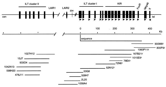
To gain insight into the genomic diversity of the KIR and ILT loci, we collected extensive mapping and sequence data from the LRC. The genomic organization of the ILT and KIR clusters was determined on two different haplotypes by using a PAC genomic library made from a single individual (18). Contigs were assembled by conventional mapping. They fell into two distinct sets, and the PACs were divided into haplotypes on the basis of additional polymorphic markers or partial sequencing (Fig. 1). PAC clones were designated haplotype 1 or 2 by a supercript number. Selected clones were sequenced to study gene arrangement after comparison by Southern blot and _KIR/ILT_-specific PCR to determine that no rearrangements or deletions had occurred. We obtained 270 kb of complete sequence of two clones (52N121 and 1060P112) spanning part of the ILT region and all of the KIR region (Fig. 1).
Figure 1.
Organization of the leukocyte receptor complex on human chromosome 19q13.4. Two contigs were deduced from PACs positive with combinations of individual probes. The complete sequences of PACs 1060P112 and 52N121 are displayed in Figs. 2 and 4, respectively. A sequence of a section of the KIRs from a haplotype similar to that of 72N61 is available (GenBank database accession no. AC011501). The KIR gene family is flanked by the ILT cluster I and the FcαR gene. The numbers of KIR genes differ depending on the haplotype. The ILT genes are grouped in two clusters separated by the LAIR genes. A link between the two clusters remains to be established. Marker 204 is based on a PCR product designed for other sequences in the database. Although all PAC sizes are to scale, distances between genes outside the sequenced region are approximate. At the telomeric end, the gene designated X is not related structurally to the Ig-like receptors. 52N121 and 72N61 represent one haplotype, and the contig containing 167B212 is part of the second haplotype. This was based on sequence polymorphism in the PCR-amplified _ILT_3 gene and other differences. Arrows point 5′ to 3′.
Gene Content.
The FcαR locus was telomeric of the KIR cluster close to another Ig-like NK gene, NKp46. Strikingly, all ILT and KIR genes in the sequenced section were in head-to-tail orientation from centromere to telomere. This, together with the conservation of gene structures and sequence homologies between the different receptor families, indicates that the LRC has evolved as a result of extensive duplication. Comparison with databases revealed a genomic clone extending telomeric of the KIR region. A gene X was identified telomeric of NKp46 but in the opposite orientation. This gene was expressed in testis and not in tissue of lymphoid origin by Northern blot analysis (data not shown), suggesting that it marks the telomeric boundary of the LRC. In total, there are at least 24 structurally and functionally related Ig-like receptors in the LRC region, spanning approximately 1 Mb of human chromosome 19q13.4.
Sequences of KIR Genes.
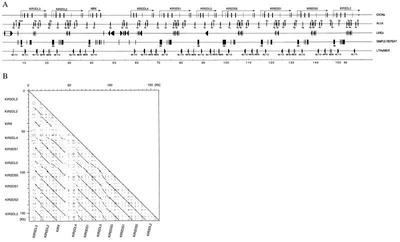
Sequencing of the ≈160-kb PAC clone 1060P112, which spans the KIR region, revealed numerous tightly clustered loci, most of which were less than 3 kb apart (Fig. 2A). Analysis of the sequence data from 1060P112 revealed 10 KIR genes, most of which have matching cDNA sequences. The exceptions are the recently described centromeric KIR locus KIR3DL3 [aka _KIRCI_ (12), named according to convention (25)], the gene _KIR_X, and a gene telomeric of KIR3DS1, which we have named _KIR2DL5. KIR_X has only exons 1–5, i.e., those encoding the matching extracellular domains of other _KIR_s. KIR2DL5 has a similar gene structure and is most homologous to KIR2DL4. KIR2DL5 has a predicted ORF that would encode potentially for a protein with two ITIM motifs in its cytoplasmic tail. We found transcripts corresponding to 2DL5 by reverse transcription–PCR using cDNA derived from PBLs. All of the two-Ig-domain _KIR_s, with the exception of KIR2DL4 and KIR2DL5, have a pseudo exon 3 that has remarkable similarity to the first Ig domain of the three Ig domain _KIR_s but are all marked by a 3-bp deletion and a nucleotide change leading to an in-frame stop codon at the same position as described for the KIR2DL3 gene (26).
Figure 2.
(A) Feature map of the complete sequence of the KIR region on PAC 1060P112. The exons of the KIR genes are shown corresponding to cDNAs or by homology with other genes (KIR3DL3, KIRX). Arrows point 5′ to 3′. The positions of Alu, LINE, and LTRs are indicated on the lines below the genes. KR indicates G+C-rich minisatellites specific to KIR genes. (B) Dot matrix analysis of the sequence from PAC 1060P112. The plot shows the 10 KIR genes on the PAC on both x and y axes. Regions of similarity are identified as a concentration of dots forming diagonal lines (22). The G+C-rich minisatellites can be visualized as boxes, missing in KIR2DL4. Insertions/deletions are visible in KIR2DL4 and KIR2DS5.
Dot-Matrix Analysis of KIR Genes.
Dot matrix analysis of the 1060P112 sequence (Fig. 2B) revealed a remarkable organization of reiterated sequences. The only unique sequences over 100 bp were: (i) upstream of KIR3DL3, (ii) outside the reiterated region, and (iii) upstream of KIR2DL4. The KIR gene sequences, including intergenic regions, are highly conserved. The sequences comprise a continuous loop that extends seamlessly from gene to gene. The reiterativeness of the loop is broken only by 14 kb upstream of the _KIR_2DL4 locus, which displays some unique features, characterized by L1 repeats. The high level of homology could facilitate exchange of exons between different KIR loci, by some form of illegitimate crossing over or gene conversion (27). Such mechanisms may be behind the variation in number of Ig exons in some members of the extended KIR family (see below).
Comparison of Two KIR Haplotypes.
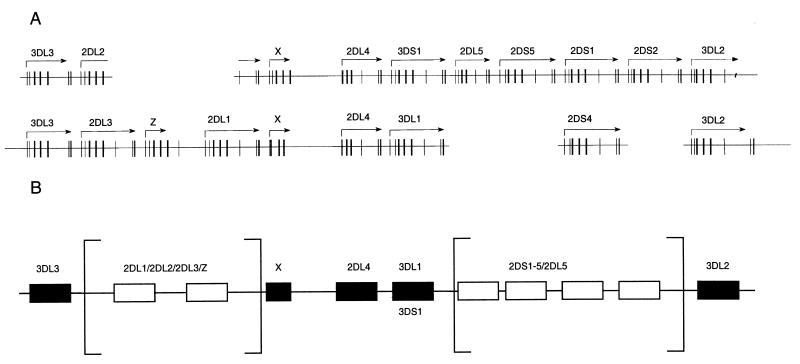
Using the sequence from 1060P112, we analyzed the other haplotype (haplotype 1) by PCR using locus-specific (16) and intergenic primer sets (Table 1). All PCR products were subcloned and completely sequenced to determine the gene arrangement of this haplotype (Fig. 3A). Genomic databases concurred with our data on this haplotype, which is the most common (16). The two haplotypes revealed a remarkable difference in organization. Certain framework loci, such as KIR3DL3 at the centromeric end, _KIR_X-KIR2DL4 in the middle, and KIR3DL2 at the telomeric end, are present on both haplotypes, consistent with their genotype frequencies of 100% in the populations studied (12, 16). The position of the KIR3DL1 locus was occupied by an activating gene, KIR3DS1 on the second haplotype. The two genes are highly homologous in both the exon and intron sequences, consistent with an allelic relationship. On haplotype 2, a set of three activating KIR genes (KIR2DS5, DS1, or DS2) was present between KIR3DS1 and 3DL2, whereas only one activating locus, 2DS4, was found at the corresponding position in haplotype 1. The sequences of these genes are very similar to each other. It was therefore difficult to determine to which locus on haplotype 2 KIR2DS4 is allelic, and it may represent a distinct locus.
Figure 3.
(A) Differences in the organization of the KIR region in two different haplotypes. On the top line is the plot of the KIR region from 1060P112 (Fig. 2A). Partial sequence was obtained from PACs 72N61, 78E41, 1015E91, and 1015 M91 for the other haplotype (Fig. 1) by using PCR as well as comparison with other data (see Fig. 1). The gene distances are not to scale and have been exaggerated to display contiguity, suggested by sequence comparisons. Thus, 2DL2 is shown as a composite of two genes on the second haplotype, namely 2DL3 and 2DL1. Overall, the genes are so similar that the precise lineup of the alleles remains uncertain, but 3DS1 and 3DL1 are shown as alleles, as supported by other data (see below and ref. 16). (B) Framework genes and variable bubbles in the KIR region. Comparison of the gene organization data in Fig. 2A, from the two haplotypes, with other data (16) is consistent with invariant framework loci, flanking regions where the gene number shows marked flexibility, indicated as open boxes flanked by square boxes. A similar model has been proposed to explain (mostly interspecies) gene expansion/contraction in the MHC, proposing independent expansion of class I genes within a framework of ancestral loci (48).
Further Centromeric, Haplotype 2 Displayed a Single KIR Gene, KIR2DL2 .
The cDNA sequence for this locus shows similarities to the extracellular domain of 2DL3 and the transmembrane and cytoplasmic part of 2DL1. Interestingly, haplotype 1 contained KIR2DL3 and 2DL1 at the position corresponding to 2DL2 (Fig. 3A). Detailed comparison of the genomic sequences revealed the precise relationship between the two haplotypes in this region, as shown in Fig. 3A. A deletion extending from exon 5 of KIR2DL3 and exon 6 of KIR2DL1 would result in the formation of a composite gene, 2DL2, with loss of the intervening KIRZ locus. This is the simplest scenario to account for the difference in organization, but such is the similarity of the sequences of all of the loci in this region, a more complex rearrangement should not be ruled out. The data suggest that there is flexibility in the presence/absence of certain KIR genes, whereas certain “framework” genes are always present, as depicted on Fig. 3B. A similar situation prevails in other variable regions, such as the MHC, where genes like C4 or DRB may be present in variable numbers on different haplotypes (28, 29), and other genes such as DRA are invariably present as singletons. In the case of the MHC framework, “orthologous” regions are conserved across species such as mice (30). KIR genes have not been identified in rodents. As mentioned above, two of the framework genes, present in all or most haplotypes, are positioned at the end of the complex (KIR3DL3, KIR3DL2) adjacent to unreiterated sequence. Similarly, the internal framework locus, KIR2DL4, also sits next to unique sequence. This property may help prevent loss of these genes by DNA looping, to which the other genes with variable presence may be prone.
Minisatellites in KIR Genes.
A feature of all of the KIR genes, with the exception of KIR2DL4, is the presence of a sequence resembling a classic moderately G+C-rich minisatellite. The repeat unit of 19–20 residues is typically GGGCCTGGAGGGAGATAT. Taking into account all of the repeats, none of the nucleotides is totally invariant at any of the positions, although the first section is generally more conserved. The number of repeat units varies from around 30–60 (≈600–1,200 bp). Apart from the residues immediately flanking the consensus-splicing signals, the first introns of the KIR genes are wholly taken up by the minisatellites. They could be connected in some way to the variation in numbers of different KIR loci. Other G+C-rich minisatellites are associated with instability via meiotic recombination processes such as gene conversion or unequal crossing over (31). Another feature of minisatellites is their association with recombination hotspots (32). Recombination in the KIR region has not been studied so far. In view of the linkage disequilibrium of various combinations of KIR alleles, this topic deserves to be explored.
Repeat Analysis Is Consistent with the Recent Origin of the KIR Region.
In addition to the microsatellites referred to above, the sequences were analyzed for repeats, including Alus, LINEs, and SINEs. Together they account for over 30% of the sequence exceeding the coding sequence ≈5-fold. The Alu family is the most frequently represented at a density of 0.49 Alu per kilobase, which is not atypical. Outstanding is the highly significant Alu S/J ratio. This ratio can be used as a measure of the age or plasticity of a given sequence. According to their evolutionary origin, _Alu_s can be divided into two main classes, J-Alu (old) and S-Alu (new), and various subclasses of S (33). Random _Alu_s in GenBank are at a universal S/J ratio of 3.00 (34). The S/J ratio over the KIR region approaches 70! This is consistent with a recent origin of the region since J-_Alu_s retrotransposed between ≈55–31 million years ago. The lack of a KIR region in mice is consistent with its recent conception. The S-_Alu_s are similarly located in all KIR genes, suggesting that all KIR loci were derived by duplication of a single primordial locus. The simplest explanation for development of the region is that of the emergence of a KIR gene in human ancestors after the mouse/human divide, followed by multiple duplications of the gene or its derivatives. This could have been followed by sequence exchange by gene conversion or nonreciprocal crossovers. In other words, the KIR region is a young region that has undergone considerable genetic turbulence. Indeed, the specificity of KIR for subsets of HLA-A, -B, or -C allotypes requires that these receptor/ligand combinations developed subsequent to the divergence of HLA loci from each other (35). Analysis of the KIR region in other primates will enable more precise dating of the origin of the region (36).
ILT Genes.
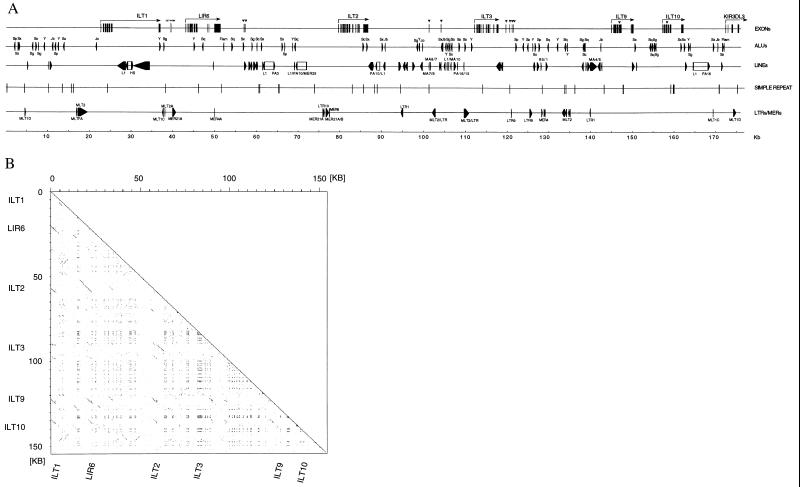
The ILT genes fell into two clusters. We sequenced a set of six ILT loci in the region proximal to the KIR loci on a 148-kb PAC clone, 52N121. We called this group, in close proximity to the _LAIR_2 gene, ILT cluster I. We performed three color fluorescence in situ hybridization analyses (not shown) using PAC clones corresponding to the FcαR/KIR border (800P9), the second ILT region (598H20), and NKG7, a marker centromeric of the ILTs, demonstrating that the order of these groups of genes, centromere to telomere, is _NKG7_-ILT cluster II-ILT cluster I-KIR. The FISH results taken together with the molecular data and information from databases show the ILT cluster II to be within 150 kb of ILT cluster I (Fig. 1). The LAIR1 locus was grouped together with the ILT cluster II (Fig. 1). ILT cluster I encoded on PAC 52N121 included the ILT1, LIR6, ILT2, and ILT3 genes (9, 37). The ILT9 and 10 genes and exons 1–4 of KIR3DL3 were present on this PAC clone (Fig. 1A). Thus, 52N121 overlaps with clone 1060P112 (Fig. 4A).
Figure 4.
(A) Organization of ILT cluster I. Sequence of the six ILT genes clustered within 150 kb centromeric of the KIR from PAC 52N121. All genes are in the same 5′ to 3′ orientation from centromere to telomere and, with exception of the ILT1 gene, are less than 6 kb in size. Fragments of ILT and LAIR exons were found throughout the entire sequence (indicated as open blocks). The exon/intron organization of the two inhibitory ILT genes, ILT2 and ILT3, is conserved, although two exons accounting for two additional Ig domains are present in the ILT2 gene. Some variation was observed between the genes encoding for putative activating _ILT_s: ILT1 contains an 11-kb intron between the fourth Ig domain and the stalk exon, and an extra stalk exon was found in the _LIR_6 gene that is not in any other activating member. In four genes, we predicted an extra 5′ untranslated region exon as seen in some LIR6 and ILT2 transcripts. (B) Dot matrix analysis of the sequence from PAC 52N121. The plot shows the region encompassing the five ILT genes and one LIR locus on both axes (see Fig. 4A for details). In contrast to the KIR loci (Fig. 2A), the sequences of the intergenic regions are not conserved.
A comparison of the exon/intron organization of the ILT genes revealed that ILT2 conformed to the prototypic structure of inhibitory ILT genes as described recently for ILT3 (12). The activating _ILT_s encoded in cluster I showed some variation. Although ILT9 and 10 have an exon/intron organization characteristic of activating ILTs, an extra exon was found in the LIR6 gene encoding for part of a stalk region that is extended in the LIR6 transcript in comparison with any other activating ILT. ILT1 had a largely extended intron 3′ of the last Ig domain exon, which results in the large gene size (15 kb). Upstream of the predicted translation start site in the LIR6 and in the ILT2 gene was an extra 5′ untranslated region exon present in matching transcripts (LIR6a and MIRcl7, respectively). Corresponding exons may be predicted in the ILT1 and ILT9 genes but not for ILT3 and ILT10.
Comparison of the ILT genes (Fig. 4A) revealed that, unlike the KIRs, their introns are not highly homologous and do not contain many repeat elements with some exceptions, such as ILT1. These data suggest an older origin of the ILT region than the KIRs, consistent with the existence of rodent counterparts, the paired Ig-like receptors (PIRs), which are in a syntenic region on mouse chromosome 7 (38–40). Analysis of the repeat composition of the ILT sequence reveals a modern–ancient S/J Alu ratio of ≈5, a value that supports the contention of a greater maturity than the KIR complex. Sequences from the two haplotypes revealed a degree of variation in the ILT coding regions, for example: ILT3 exon 12 A/G and ILT4 Ig3 T/C, both of which correspond to known polymorphic cDNA for these genes.
Haplotype-Specific Variation in ILT Cluster II.
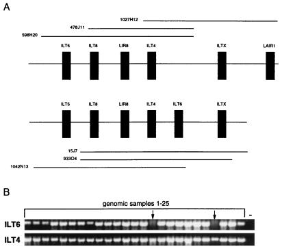
A contig of six clones was formed outside the sequenced region described above that all encoded the ILT4 locus. On the basis of a sequence polymorphism identified in the Ig3 domain of the ILT4 gene, they were grouped according to their haplotype (Fig. 5A). The link between this cluster and the two haplotypes identified in the ILT/KIR region remains to be determined. The ILT5 gene was at the centromeric end of this cluster on two PAC clones from each haplotype. ILT8 was close to _ILT_5. Telomeric of ILT8 is LIR8, which is present on PAC 478J11 and 933O4. On three PAC clones, we identified a _Bgl_II (2 kb) and a _Xba_I fragment (3 kb) by Southern blot analysis that could not be accounted for by any of the identified ILT genes. We mapped this fragment to the telomeric end of cluster II. This locus could be the ILT7 gene, which we have not been able to amplify by PCR.
Figure 5.
(A) Haplotypic variation for presence of ILT6 in ILT cluster II. The centromeric cluster of ILT genes (Fig. 1) contained six genes in most haplotypes. On the haplotype shown (Top), the ILT6 gene is missing. (B) PCR analysis for the presence of ILT6. The genotypes of 25 individuals were analyzed by PCR by using ILT6- and _ILT4_-specific primers. The two samples that were negative for ILT6 are indicated by arrows.
We amplified ILT6 from three clones (15J7, 933O4, 1042N13) that correspond to a single haplotype (Fig. 5A). Because the only overlap between these clones is between LIR8 and ILT4, the ILT6 gene must be located either between these two loci or in close proximity to one of them. Two clones of the second haplotype also span this region but do not contain the ILT6 gene. We identified a haplotype-specific 3.5-kb _Xba_I fragment by Southern blot analysis that is present only on the three _ILT6-_positive PAC clones. We postulate that the ILT6 gene is absent on one haplotype in this PAC library and therefore shows presence/absence variation similar to what has been observed for some of the KIR loci. To examine this hypothesis further, we amplified ILT6 from 25 genomic samples and found 2/25 homozygous negative for this locus (Fig. 5B). This is consistent with a gene frequency for the presence of the ILT6 gene of about 0.72, (0.51 homozygotes, 0.41 heterozygotes, and 0.08 homozygote negative, applying the Hardy–Weinberg equation).
Discussion
All genes encoded in the LRC on human chromosome 19q13.4 are members of the Ig superfamily. Parts of their gene structures are remarkably conserved, and all are in the same head-to-tail orientation, with the possible exception of ILT complex II. The many ILT/LAIR/KIR gene fragments we found in the ILT region (Fig. 4) could be the fallout of abortive rearrangements at repeated duplications throughout evolution.
Rodents have genes equivalent to NKG7 (41) and NKp46 (42) as well as the probable ILT orthologues, the paired Ig-like receptors (_PIR_s), which are located on the syntenic mouse chromosome 7 (40). However, no rodent KIR genes have been identified. There are at least 14 mouse Ly-49 genes, which fulfill a function similar to the _KIR_s (43, 44). These are encoded on mouse chromosome 6 in the NK complex. Only a single human LY49 pseudogene has been identified in the syntenic region on human chromosome 12 (45). Multiple duplication events could have led to rapid expansion of the KIR gene family in primates (2) and, conversely, the Ly49 genes in rodents. This argues for convergent evolution of function of these receptors.
Recently, a “hybrid” cDNA molecule, KIR2DL1v, was identified that appeared to comprise a 2DL1 sequence with the proximal part of the second Ig domain to the TM region being replaced by a sequence resembling KIR2DS1 (46). Our data show how the two putative parent genes are unlikely to be alleles. They are separated by over 50 kb of intervening DNA and are in different variable KIR regions. These data could be explained by some form of nonreciprocal recombination such as gene conversion, which is known to operate in the MHC (27). The arrangement of KIR genes consisting of highly related sequences in the same orientation may provide the ideal substrate for gene conversion.
Close examination of the KIR region shows that the sequences upstream of the transcribed region are remarkably similar, with the exception of the 2DL4 gene, suggesting common promoters. Different groups of 2–9 KIR genes are expressed in NK clones, but the sequences of the KIR promoter sequences are homogeneous, except for that of the KIR2DL4. Therefore, it seems likely that regulation of expression of KIR transcripts is facilitated by a stochastic process. The sequence upstream of KIR2DL4 may be significant because this gene is unique among its peers in being expressed in 100% of NK clones (15). 2DL4 is the only KIR gene lacking the repeat region in intron 1.
Examination of the LRC from two different haplotypes revealed variation in the KIR cluster. KIR genes present on all haplotypes represent framework loci that flank regions of variability. In these heterogeneous regions, there are at least 11 possible KIR loci. If we accept this model as a first approximation of the different arrangements of KIR genes, there are at least two main positions where haplotypes may differ (Fig. 3). This hypothesis would account for a large number of different haplotypes, and it concurs with the variation in the number of KIR genes observed in different genotypes (ref. 16; unpublished data). The frequencies with which certain combinations of KIR loci are found in different individuals exceeded levels expected from random association (16), indicative of linkage disequilibrium of alleles on haplotypes. The independent segregation of KIR3DL1 and KIR3DS1 suggested that these two specificities were alleles. This is the case for the two haplotypes on Fig. 3.
Taking into account further variation in sequences, particularly ILT4 and ILT5 (17), as well as the presence/absence of the ILT6 gene, the LRC is clearly highly variable. Another region of the genome that exhibits extensive variation is the MHC, the products of which are ligands for some of the KIR and ILT molecules. The MHC class I, class II, and C4 genes exhibit high levels of variability for presence/absence (28, 47). The common functional link to both sets of loci is resistance to pathogens. Like the MHC, the LRC has all the hallmarks of a dynamic genomic region. Selection for variation in KIR gene arrangement could be infection, in which case we may expect to find some haplotype frequencies skewed in different diseases.
Acknowledgments
We thank the Medical Research Council, the Wellcome Foundation, the European Economic Community (CT961105), and the Imperial Cancer Research Fund for support, and A. Ziegler, A. Volz, and A. Jeffries for helpful discussions, as well as Prof. Pieter de Jong for the genomic DNA (BACPAC Resources, Oakland, CA).
Abbreviations
LRC
leukocyte receptor complex
NK
natural killer
PAC
P1 artificial chromosome
Footnotes
This paper was submitted directly (Track II) to the PNAS office.
Article published online before print: Proc. Natl. Acad. Sci. USA, 10.1073/pnas.080588597.
Article and publication date are at www.pnas.org/cgi/doi/10.1073/pnas.080588597
References
- 1.Karre, K. & Colonna, M., eds. (1998) Curr. Top. Microbiol. Immunol. 230.
- 2.Parham, P. (1997) Immunol. Rev.155.
- 3.Lanier L L. Annu Rev Immunol. 1998;16:359–394. doi: 10.1146/annurev.immunol.16.1.359. [DOI] [PubMed] [Google Scholar]
- 4.Brown M, Scalzo A, Matsumoto K, Yokoyama W. Immunol Rev. 1997;155:53–65. doi: 10.1111/j.1600-065x.1997.tb00939.x. [DOI] [PubMed] [Google Scholar]
- 5.Plougastel B, Trowsdale J. Genomics. 1998;49:193–199. doi: 10.1006/geno.1997.5197. [DOI] [PubMed] [Google Scholar]
- 6.D'Andrea A, Chang C, Franz-bacon K, McClanahan T, Phillips J H, Lanier L L. J Immunol. 1995;155:2306–2310. [PubMed] [Google Scholar]
- 7.Suto Y, Maenaka K, Yabe T, Hirai M, Tokunaga K, Tadokoro K, Juji T. Genomics. 1996;35:270–272. doi: 10.1006/geno.1996.0355. [DOI] [PubMed] [Google Scholar]
- 8.Borges L. J Immunol. 1997;159:5192–5196. [PubMed] [Google Scholar]
- 9.Colonna M, Nakajima H, Navarro F, Lopez-Botet M. J Leukocyte Biol. 1999;66:375–381. doi: 10.1002/jlb.66.3.375. [DOI] [PubMed] [Google Scholar]
- 10.Wagtmann N, Rojo S, Eichler E, Mohrenweiser H, Long E O. Curr Biol. 1997;7:615–618. doi: 10.1016/s0960-9822(06)00263-6. [DOI] [PubMed] [Google Scholar]
- 11.Meyaard L, Adema G J, Chang C, Lanier L L, Phillips J H. Immunity. 1997;7:283–290. doi: 10.1016/s1074-7613(00)80530-0. [DOI] [PubMed] [Google Scholar]
- 12.Torkar M, Norgate Z, Colonna M, Trowsdale J, Wilson M. Eur J Immunol. 1998;28:3959–3967. doi: 10.1002/(SICI)1521-4141(199812)28:12<3959::AID-IMMU3959>3.0.CO;2-2. [DOI] [PubMed] [Google Scholar]
- 13.Wende H, Colonna M, Ziegler A, Volz A. Mamm Genome. 1998;10:154–160. doi: 10.1007/s003359900961. [DOI] [PubMed] [Google Scholar]
- 14.Selvakumar A, Steffens U, Dupont B. Immunol Rev. 1997;155:183–195. doi: 10.1111/j.1600-065x.1997.tb00951.x. [DOI] [PubMed] [Google Scholar]
- 15.Valiante N, Lienert K, Shilling H, Smits B, Parnham P. Immunol Rev. 1997;155:155–164. doi: 10.1111/j.1600-065x.1997.tb00948.x. [DOI] [PubMed] [Google Scholar]
- 16.Uhrberg M, Valiante N M, Shum B P, Shilling H G, Lienert-Weidenbach K, Corliss B, Tyan D, Lanier L L, Parham P. Immunity. 1997;7:753–763. doi: 10.1016/s1074-7613(00)80394-5. [DOI] [PubMed] [Google Scholar]
- 17.Colonna M, Navarro F, Bellon T, Llano M, Garcia P, Samaridis J, Angman J, Cella M, Lopez-Botet M. J Exp Med. 1997;186:1809–1818. doi: 10.1084/jem.186.11.1809. [DOI] [PMC free article] [PubMed] [Google Scholar]
- 18.Ioannou P A, Amemiya C T, Garnes J, Kroisel P M, Shizuya H, Batzer M A, de Jong P J. Nat Genet. 1994;6:84–89. doi: 10.1038/ng0194-84. [DOI] [PubMed] [Google Scholar]
- 19.Beck S, Alderton R P. Anal Biochem. 1993;212:498–505. doi: 10.1006/abio.1993.1359. [DOI] [PubMed] [Google Scholar]
- 20.Sanger F, Nicklen S, Coulson A R. Proc Natl Acad Sci USA. 1977;74:5463–5467. doi: 10.1073/pnas.74.12.5463. [DOI] [PMC free article] [PubMed] [Google Scholar]
- 21.Sanger F. Genome Res. 1998;8:1097–1108. [Google Scholar]
- 22.Sonnhammer E L, Durbin R. Gene. 1995;167:1–10. doi: 10.1016/0378-1119(95)00714-8. [DOI] [PubMed] [Google Scholar]
- 23.Nakai K, Horton P. Trends Biochem Sci. 1999;24:34–36. doi: 10.1016/s0968-0004(98)01336-x. [DOI] [PubMed] [Google Scholar]
- 24.Bairoch A. Prosite: A Dictionary of Protein Sites and Patterns. 5th Ed. Université de Genève, Geneva: Département de Biochimie Médicale; 1990. [Google Scholar]
- 25.Long E O, Colonna M, Lanier L L. Immunol Today. 1996;17:100. doi: 10.1016/0167-5699(96)80590-1. [DOI] [PubMed] [Google Scholar]
- 26.Wilson M J, Torkar M, Trowsdale J. Tissue Ant. 1997;49:574–579. doi: 10.1111/j.1399-0039.1997.tb02804.x. [DOI] [PubMed] [Google Scholar]
- 27.Hogstrand K, Bohme J. Immunol Rev. 1999;167:305–317. doi: 10.1111/j.1600-065x.1999.tb01400.x. [DOI] [PubMed] [Google Scholar]
- 28.Trowsdale J, Ragoussis J, Campbell R D. Immunol Today. 1991;12:443–446. doi: 10.1016/0167-5699(91)90017-n. [DOI] [PubMed] [Google Scholar]
- 29.Campbell R D, Trowsdale J. Immunol Today. 1993;14:349–352. doi: 10.1016/0167-5699(93)90234-C. [DOI] [PubMed] [Google Scholar]
- 30.Amadou C, Kumanovics A, Jones E P, Lambracht-Washington D, Yoshino M, Lindahl K F. Immunol Rev. 1999;167:211–222. doi: 10.1111/j.1600-065x.1999.tb01394.x. [DOI] [PubMed] [Google Scholar]
- 31.Jeffreys A J, Neil D L, Neumann R. EMBO J. 1998;17:4147–4157. doi: 10.1093/emboj/17.14.4147. [DOI] [PMC free article] [PubMed] [Google Scholar]
- 32.Jeffreys A J, Murray J, Neumann R. Mol Cell. 1998;2:267–273. doi: 10.1016/s1097-2765(00)80138-0. [DOI] [PubMed] [Google Scholar]
- 33.Jurka J, Milosavljevic A. J Mol Evol. 1991;32:105–121. doi: 10.1007/BF02515383. [DOI] [PubMed] [Google Scholar]
- 34.Jurka J, Smith T. Proc Natl Acad Sci USA. 1988;85:4775–4778. doi: 10.1073/pnas.85.13.4775. [DOI] [PMC free article] [PubMed] [Google Scholar]
- 35.Parham P. Semin Immunol. 1994;6:373–382. doi: 10.1006/smim.1994.1047. [DOI] [PubMed] [Google Scholar]
- 36.Zietkiewicz E, Richter C, Malalowski W, Jurka J, Labuda D. Nucleic Acids Res. 1994;22:5608–5612. doi: 10.1093/nar/22.25.5608. [DOI] [PMC free article] [PubMed] [Google Scholar]
- 37.Cosman D, Fanger N, Borges L, Kubin M, Chin W, Peterson L, Hsu M-L. Immunity. 1997;7:273–282. doi: 10.1016/s1074-7613(00)80529-4. [DOI] [PubMed] [Google Scholar]
- 38.Yamashita Y, Fukuta D, Tsuji A, Nagabukuro A, Matsuda Y, Nishikawa Y, Ohyama Y, Ohmori H, Ono M, Takai T. J Biochem. 1998;123:358–368. doi: 10.1093/oxfordjournals.jbchem.a021945. [DOI] [PubMed] [Google Scholar]
- 39.Hayami K, Fukuta D, Nishikawa Y, Yamashita Y, Inui M, Ohyama Y, Hikida M, Ohmori H, Takai T. J Biol Chem. 1997;272:7320–7327. doi: 10.1074/jbc.272.11.7320. [DOI] [PubMed] [Google Scholar]
- 40.Kubagawa H, Burrows P D, Coopers M D. Proc Natl Acad Sci USA. 1997;94:5261–5266. doi: 10.1073/pnas.94.10.5261. [DOI] [PMC free article] [PubMed] [Google Scholar]
- 41.Berg S F, Westgaard I H, Fossum S, Dissen E. Immunogenetics. 1999;49:815–818. doi: 10.1007/s002510050557. [DOI] [PubMed] [Google Scholar]
- 42.Falco M, Cantoni C, Bottino C, Moretta A, Biassoni R. Immunol Lett. 1999;68:411–414. doi: 10.1016/s0165-2478(99)00052-8. [DOI] [PubMed] [Google Scholar]
- 43.McQueen K L, Freeman J D, Takei F, Mager D L. Immunogenetics. 1998;48:174–183. doi: 10.1007/s002510050421. [DOI] [PubMed] [Google Scholar]
- 44.Brown M G, Fulmek S, Matsumoto K, Cho R, Lyons P A, Levy E R, Scalzo A A, Yokoyama W M. Genomics. 1997;42:16–25. doi: 10.1006/geno.1997.4721. [DOI] [PubMed] [Google Scholar]
- 45.Barten R, Trowsdale J. Immunogenetics. 1999;49:731–734. doi: 10.1007/s002510050675. [DOI] [PubMed] [Google Scholar]
- 46.Shilling H G, Lienert-Weidenbach K, Valiante N M, Uhrberg M, Parham P. Immunogenetics. 1998;48:413–416. doi: 10.1007/s002510050453. [DOI] [PubMed] [Google Scholar]
- 47.Campbell, R. D. & Trowsdale, J. (1997) Immunol. Today18 (Suppl.).
- 48.Amadou C. Immunogenetics. 1999;49:362–367. doi: 10.1007/s002510050507. [DOI] [PubMed] [Google Scholar]
