FKBP52 deficiency–conferred uterine progesterone resistance is genetic background and pregnancy stage specific (original) (raw)
Abstract
Immunophilin FKBP52 serves as a cochaperone to govern normal progesterone (P4) receptor (PR) function. Using Fkbp52–/– mice, we show intriguing aspects of uterine P4/PR signaling during pregnancy. Implantation failure is the major phenotype found in these null females, which is conserved on both C57BL6/129 and CD1 backgrounds. However, P4 supplementation rescued implantation and subsequent decidualization in CD1, but not C57BL6/129, null females. Surprisingly, experimentally induced decidualization in the absence of blastocysts failed in Fkbp52–/– mice on either background even with P4 supplementation, suggesting that embryonic signals complement uterine signaling for this event. Another interesting finding was that while P4 at higher than normal pregnancy levels conferred PR signaling sufficient for implantation in CD1 null females, these levels were inefficient in maintaining pregnancy to full term. However, elevating P4 levels further restored PR signaling to a level optimal for successful term pregnancy with normal litter size. Collectively, the results show that the indispensability of FKBP52 in uterine P4/PR signaling is a function of genetic disparity and is pregnancy stage specific. Since there is evidence for a correlation between P4 supplementation and reduced risks of P4-resistant recurrent miscarriages and remission of endometriosis, these findings have clinical implications for genetically diverse populations of women.
Introduction
Progesterone (P4) signaling is an absolute requirement for implantation and pregnancy maintenance in all eutherian mammals studied (1, 2). P4 acts through its nuclear P4 receptor (PR) to activate transcription of genes involved in ovulation, uterine receptivity, implantation, decidualization, and pregnancy maintenance. This is evident from the complete infertility of female mice lacking Pgr, the gene encoding PR (3). The failure of ovulation and implantation precludes using Pgr–/– mice to study potential new aspects of P4 function during pregnancy. In contrast, the targeted deletion of the Fkbp52 gene, which encodes FKBP52, an immunophilin cochaperone that optimizes PR signaling, has allowed us to address unique aspects of uterine P4/PR signaling during pregnancy in mice.
Immunophilins are so named because of their ability to bind and mediate the actions of certain immunosuppressive drugs. They are grouped into 2 families, FK506-binding proteins (FKBPs) and cyclosporin A–binding proteins (cyclophilins [CyPs]). Some FKBP and CyP family members contain a tetratricopeptide repeat (TPR) domain that targets binding to the highly conserved C terminus of Hsp90. FKBP52, FKBP51, and CyP40 are 3 such TPR-containing cochaperones that have been identified in steroid receptor complexes (4). Like other steroid receptors, PR assembles in an ordered, multistep manner for hormone binding (5, 6); the final complex involves direct association of Hsp90 with PR, which stabilizes its ability to bind P4 (7). At the mature stage, FKBPs and other TPR cochaperones dynamically exchange on Hsp90 such that there is a mixture of receptor complexes distinguished by which cochaperone is associated with Hsp90 (8, 9). Hormone binding stimulates interruption of the receptor/chaperone assembly cycle and promotes receptor activation. Recent findings show that cochaperones have unique influences on steroid receptor function. FKBP52 potentiates the responses of PR, androgen receptor, and glucocorticoid receptor to their respective ligands (10–12). In fact, only the mature PR complex bound to FKBP52 is capable of binding P4 with high affinity and efficiency, although basal PR responsiveness to P4 is still retained in the absence of FKBP52. Our recent study (12) revealed the previously unknown role of FKBP52/PR signaling in female reproduction.
The uterus consists of heterogeneous cell types that respond differentially to estrogen and P4. For successful implantation, the uterus must be transiently receptive to implantation-competent blastocysts. For example, the prereceptive uterus on days 1–3 of pregnancy or pseudopregnancy becomes receptive to implantation on day 4 under the direction of P4 and estrogen. By late day 5, the uterus becomes nonreceptive, since implantation-competent blastocysts transferred into uteri of pseudopregnant mice at this time fail to implant. Following implantation on day 4 evening (2200 to 2400 h), uterine stromal cells at the sites of blastocysts undergo extensive proliferation and differentiation, giving rise to decidual cells, a process termed decidualization (1, 13). Using proteomic analysis in Hoxa10–/– mouse uteri, we identified FKBP52 as an important signaling molecule in stromal cell function during the periimplantation period (14). Consistent with the role of FKBP52 as a PR cochaperone, uterine expression of Fkbp52 and Pgr overlaps on days 4 and 5 of pregnancy (12). More importantly, we have shown that Fkbp52–/– females on a C57BL6/129 mixed background have complete implantation failure, with normal ovulation and slightly reduced fertilization rates (12). This phenotype has recently been confirmed by another group in independently generated Fkbp52–/– mice on the same background (15). The fact that the infertile phenotype of Fkbp52–/– females is primarily due to implantation defects suggests differential sensitivity of the ovary and uterus to FKBP52/PR-mediated P4 action. This tissue-specific differential sensitivity is not noted in Pgr–/– females, in which severely compromised ovarian and uterine functions lead to complete female infertility (3, 16).
Although P4 is commonly known as the “hormone of pregnancy,” various aspects of its roles throughout pregnancy are not well understood. Therefore, _Fkbp52–/–_mice provide a unique opportunity for studying such roles of P4 signaling throughout pregnancy. Using these null mice, we show in the present investigation that P4/PR signaling is a function of genetic makeup and is pregnancy stage specific. For example, while the implantation failure phenotype was conserved in both C57BL6/129 and CD1 mice lacking Fkbp52, daily P4 supplementation rescued implantation with subsequent decidualization in CD1 Fkbp52–/– females but not in C57BL6/129 Fkbp52–/– females. Surprisingly, experimentally induced decidualization failed to occur in Fkbp52–/– mice on either background even with exogenous P4 supplementation, suggesting that embryonic signals complement the uterine signaling network. Another interesting finding is the differential requirement for P4/PR signaling at specific stages of pregnancy in P4-treated CD1 Fkbp52–/– females. For example, while P4 at higher than normal levels conferred PR signaling sufficient for uterine receptivity and implantation in Fkbp52–/– females, these levels did not maintain adequate PR signaling to sustain pregnancy, resulting in reduced litter sizes due to in utero fetal resorption and restricted growth. However, increasing P4 levels even further restored PR signaling, to a degree sufficient to maintain full-term pregnancy with normal litter size. Collectively, these findings show that FKBP52 deficiency confers uterine P4 resistance during pregnancy, since null females have normal PR and P4 levels with reduced PR activity (12, 17). This study also shows that the requirement for FKBP52 in optimizing PR activity is genetic background dependent and that levels of P4/PR signaling required to ensure successful pregnancy differ depending on pregnancy stage.
Results
Implantation failure occurs in Fkbp52–/– mice irrespective of genetic background.
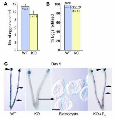
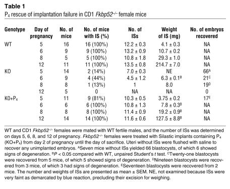
There is increasing evidence that mutation of a gene often results in altered phenotypes depending on the genetic background of mice (18). These varying phenotypes are thought to be due to differential expression and/or regulation of modifier genes (19), although identification of such modifiers remains largely unknown. There is also evidence that compensatory function of genes among the same family is genetic background dependent (20). We have recently shown that C57BL6/129 Fkbp52–/– females have complete implantation failure, although ovulation is normal (12). To determine whether this phenotype is a function of genetic background, we established Fkbp52 deletion in CD1 mice (see Methods). First, we examined ovulation and fertilization in CD1 Fkbp52–/– females after mating them with WT males, since Fkbp52–/– males, irrespective of genetic background, are infertile (10). We found ovulation and fertilization to be comparable to those of WT females (Figure 1, A and B). We then asked whether implantation occurs in these mice. Initiation of implantation accompanies an increased endometrial vascular permeability at sites of blastocysts, which are visualized as distinct blue bands after injection of a blue dye solution (13, 21). CD1 Fkbp52–/– females were examined for implantation sites (ISs) by this method on day 5 of pregnancy. We observed that only 2 of 14 CD1 Fkbp52–/– females showed very faint blue bands (Figure 1C and Table 1). Unimplanted blastocysts were recovered from uterine flushings of null mice with implantation failure (Figure 1C), suggesting that although embryos developed to blastocysts, they failed to implant. Our data indicate that optimal P4/PR signaling imparted by FKBP52 is critical to uterine receptivity and implantation in mice, a phenotype conserved across these genetic backgrounds. Because ovulation is normal in Fkbp52–/– mice, our results suggest that uterine responsiveness to PR signaling differs from ovarian responsiveness. It is possible that relatively high local P4 levels in the ovary (22), the site of its synthesis, enhance basal PR activity sufficient for ovulation and fertilization processes.
Figure 1. P4 supplementation via Silastic implants rescues implantation failure in CD1 Fkbp52–/– females.
Ovulation (A) and fertilization (B) were examined on day 2 of pregnancy. The number of ovulated eggs was not significantly different in WT and Fkbp52–/– (KO) females. Values are mean ± SEM; P > 0.05, unpaired Student’s t test. Fertilization rate was determined by counting the number of 2-cell embryos after flushing oviducts. Numbers above the bars indicate the total number of 2-cell embryos per total number of eggs recovered. The fertilization rate was comparable between WT and KO females (P > 0.05; unpaired Student’s t test). (C) Implantation fails in KO females but is rescued by P4 as examined on day 5 of pregnancy. Implants containing P4 were inserted s.c. in KO females (KO+P4) on day 2 of pregnancy. Representative photographs of uteri with or without ISs as demarcated by blue bands in WT_,_ KO, and KO+P4 mice are shown. Representative photographs of blastocysts recovered from uteri of KO females without IS. Arrowheads and short arrows indicate the location of ovaries and ISs, respectively. The long arrow indicates the uterus from which unimplanted blastocysts were recovered. Scale bar: 50 μm.
Table 1 .
P4 rescue of implantation failure in CD1 _Fkbp52_–/– female mice
P4 supplementation rescues implantation in CD1 Fkbp52–/– females.
We have shown that PR activity, but not PR or P4 levels, is compromised in C57BL6/129 Fkbp52–/– females (12, 17). It is possible that FKBP52 binding to the PR complex modulates the hormone responsiveness of PR not in an all-or-none fashion, but rather to fine-tune physiological responses to P4. This would imply that while FKBP52 is necessary for optimal PR activity, PR signaling can still operate, albeit not as efficiently, in the absence of FKBP52. Since serum P4 levels in CD1 Fkbp52–/– females are similar to those in WT mice on day 5 of pregnancy (Supplemental Figure 1; supplemental material available online with this article; doi:10.1172/JCI31622DS1), we speculated that exposing Fkbp52–/– uteri to higher-than-normal P4 levels would enhance PR activity to rescue pregnancy failure in the absence of FKBP52. This is consistent with our previous findings that PR activity in Fkbp52–/– mouse embryonic fibroblasts (MEFs) reached levels similar to those in WT MEFs exposed to higher P4 concentrations in culture (12). This may also explain why ovulation, a P4-regulated event, is normal in Fkbp52–/– females on both genetic backgrounds.
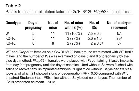
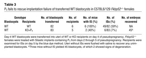
To determine whether P4 supplementation rescues implantation, we used P4-containing Silastic implants to maintain steady-state hormone levels (23). WT and Fkbp52–/– females were mated with WT males, and Silastic implants containing P4 were placed under the dorsal skin of Fkbp52–/– females from day 2 of pregnancy until the day of sacrifice. We were surprised to see that P4 supplementation not only rescued implantation in CD1 Fkbp52–/– females examined on day 5, but the number of ISs was also comparable to that in WT mice (Figure 1C and Table 1). In contrast, exogenous P4 supplementation was largely ineffective in rescuing implantation in C57BL6/129 Fkbp52–/– females; only 3 of 11 (27%) and 2 of 8 (25%) null females showed implantation when examined on days 5 and 8, respectively. In addition, the number of ISs was also remarkably low, especially on day 8, compared with that in WT females (Table 2). To circumvent any contribution arising from lower fertilization rates in C57BL6/129 Fkbp52–/– mice, we performed blastocyst transfer experiments. Day-4 WT blastocysts were transferred into uterine lumens of day-4 C57BL6/129 Fkbp52–/– pseudopregnant females carrying Silastic P4 implants from day 2. Again, we noted extremely poor implantation rates in these mice (Table 3).
Table 2 .
P4 fails to rescue implantation failure in C57BL6/129 _Fkbp52_–/– female mice
Table 3 .
P4 fails to rescue implantation failure of transferred WT blastocysts in C57BL6/129 _Fkbp52_–/– females
P4 supplementation restores P4- and implantation-regulated gene expression in CD1 Fkbp52–/– uteri.
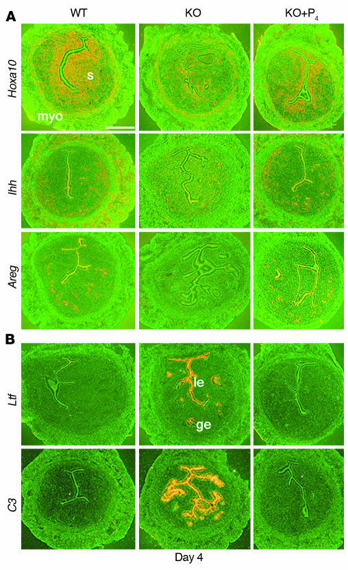
P4-regulated events combined with preimplantation estrogen secretion guide the uterus from a prereceptive to receptive state, allowing blastocyst attachment in the uterine wall on day 4 evening. To determine whether rescue of implantation by P4 is reflected in the restoration of P4-dependent uterine gene expression, we placed a Silastic P4 implant on day 2 in CD1 Fkbp52–/– females that had been mated with WT males and sacrificed them on day 4 of pregnancy. We selected P4-regulated genes that encode Hoxa10, Indian hedgehog (Ihh), and amphiregulin (Areg) because of their participation in uterine receptivity (24–27). In situ hybridization results showed that exogenous P4 treatment considerably restores expression of these genes (Figure 2A).
Figure 2. P4 supplementation via Silastic implants corrects misexpression of genes in CD1 Fkbp52–/– uteri.
(A) In situ hybridization of P4-regulated genes Hoxa10, Ihh, and Areg in WT, KO, and KO+P4 uteri on day 4 of pregnancy. (B) In situ hybridization of estrogen-target genes Ltf and complement factor 3 (C3) in WT, KO, and KO+P4 uteri on day 4 of pregnancy. Implants containing P4 were inserted s.c. in KO females on day 2 of pregnancy. ge, glandular epithelium; le, luminal epithelium; myo, myometrium; s, stroma. Scale bar: 200 μm.
Lactoferrin (Ltf) and complement factor 3 (C3) are induced to a high degree in the uterus by estrogen and antagonized by P4 (28, 29). They are abundantly expressed in the luminal epithelium on day 1 of pregnancy under the influence of a preovulatory estrogen surge but are dramatically downregulated on day 4 by rising P4 levels from newly formed corpora lutea (1, 2). However, in CD1 Fkbp52–/– mice, uterine expression of these genes was aberrantly elevated on day 4 but downregulated with exogenous P4 supplementation (Figure 2B). Together, these findings provide evidence that P4 supplementation restores the expression of P4-regulated genes and counters the expression of estrogen target genes in CD1 Fkbp52–/– uteri, shifting the uterus to a P4-dominated milieu conducive to uterine receptivity as opposed to one of estrogenic dominance that is detrimental to uterine receptivity.
The fact that exogenous P4 treatment fully rescues implantation in CD1 Fkbp52–/– females but is largely ineffective in C57BL6/129 Fkbp52–/– mice led us to examine whether P4 treatment would restore the expression of P4-regulated genes in C57BL6/129 Fkbp52–/– uteri, as was observed in CD1 Fkbp52–/– mice. We found that although exogenous P4 treatment considerably restored Hoxa10 and Ihh expression, Areg expression remained low to undetectable (Supplemental Figure 2A). More interestingly, exogenous P4, which normally inhibits estrogen-responsive Ltf expression in CD1 Fkbp52–/– uteri on day 4, was not effective in attenuating Ltf expression in C57BL6/129 Fkbp52–/– uteri (Supplemental Figure 2B). Overall, these results imply that while C57BL6/129 Fkbp52–/– uteri are somewhat responsive to P4 induction of target genes, they are less receptive to P4’s influence in antagonizing estrogen target genes. These findings are significant, since excess estrogenic influence leads to uterine nonreceptivity (30). Because implantation fails in most C57BL6/129 Fkbp52–/– mice even after P4 treatment, we performed subsequent experiments on CD1 Fkbp52–/– mice.
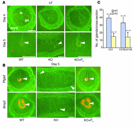
Although expression of several P4-regulated genes was considerably restored with P4 supplementation in CD1 Fkbp52–/– uteri, the expression of leukemia inhibitory factor (Lif), normally expressed in day-4 WT pregnant uterine glands, was not restored by P4 treatment in these null mice (Figure 3A). We next determined whether P4 rescue of implantation on day 5 in CD1 Fkbp52–/– mice is accompanied by correct expression of implantation-related genes, such as Lif, Ptgs2, and Bmp2 (31–33). We found that while these genes were not expressed at the site of blastocysts in CD1 Fkbp52–/– females with failed implantation in the absence of P4, P4 supplementation restored implantation with correct expression of these genes (Figure 3, A and B). The ability of P4 to rescue both implantation and Lif expression on day 5 of pregnancy in Fkbp52–/– mice without salvaging Lif expression on day 4 agrees with previous observations that the first phase of Lif expression on day 4 is not as critical as its second phase of expression in stromal cells surrounding the implanting blastocyst (33, 34).
Figure 3. Delivery of P4 via Silastic implants restores expression of implantation-related genes in CD1 Fkbp52–/– females.
(A) In situ hybridization of Lif in WT, KO, and KO+P4 uteri on days 4 and 5 of pregnancy. (B) In situ hybridization of Ptgs2 and Bmp2 in WT, KO, and KO+P4 uteri on day 5 of pregnancy. P4 implants were inserted s.c. in KO females on day 2 of pregnancy. Arrowheads in A and B indicate the location of embryos. Scale bars: 200 μm. (C) Number of glands per uterine cross-section of WT and KO uteri on CD1 and C57BL6/129 backgrounds. For each animal, glands were counted in 9–12 uterine sections. Numbers above bars indicate number of mice evaluated. Values are mean ± SEM. *P < 0.05, unpaired Student’s t test.
Upon closer examination of WT and null uterine histology, we came across an interesting observation. A significant decrease in the number of glands was noted in null uteri on both genetic backgrounds (Figure 3C), a phenotype not rescued by P4 supplementation (data not shown). This decrease in gland numbers, however, does not fully account for the altered expression of Areg, Ihh, or Lif in null uteri, since the glands that were still present failed to show normal expression patterns of these genes. There is clear evidence that both estrogen and P4 participate in uterine gland formation in sheep (35, 36). Our data also implicate a potential role for FKBP52/PR signaling in mouse uterine gland formation and function. This is an exciting finding but warrants further investigation.
Postimplantation defects in P4-treated CD1 Fkbp52–/– females.
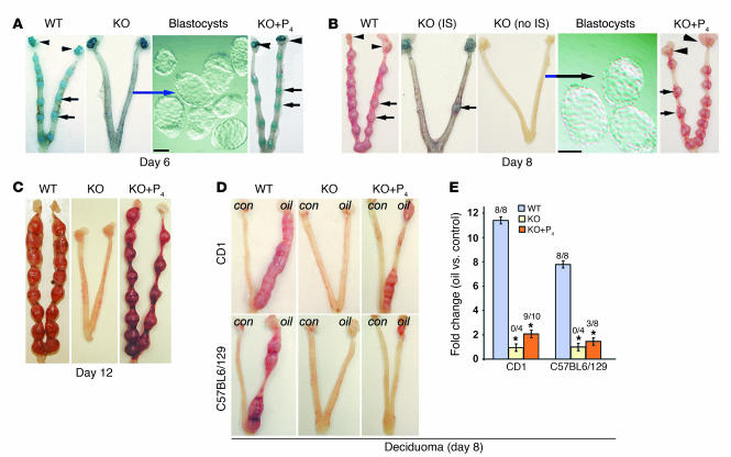
Our observation of unimplanted blastocysts recovered from CD1 Fkbp52–/– females examined on day 5 of pregnancy suggested deferral of implantation timing in these mice as is observed in cPLA2a–/– or LPA3–/– mice (37, 38). To test for this possibility, we sacrificed CD1 Fkbp52–/– mice on days 6 and 8 of pregnancy. We found that unlike in cPLA2a–/– or LPA3–/– mice, implantation timing was not altered; rather implantation drastically failed in Fkbp52–/– females (Figure 4, A and B, and Table 1). Again, blastocysts were recovered from CD1 Fkbp52–/– uteri on these days, confirming implantation as the major defect in these mice (Figure 4, A and B, and Table 1).
Figure 4. P4 delivery via Silastic implants rescues blastocyst-induced, but not oil-induced, decidualization in Fkbp52–/– females.
Representative photographs of WT, KO, and KO+P4 uteri on day 6 (A), day 8 (B), and day 12 (C) of pregnancy are shown. Arrowheads and short arrows indicate the location of ovary and IS, respectively. Representative images of recovered unimplanted blastocysts from uteri (long arrow) of KO females without ISs are shown. Scale bars: 50 μm. (D) Oil-induced decidualization fails in KO females on both CD1 and C57BL6/129 backgrounds. Representative photomicrographs of WT, KO, and KO+P4 uteri on day 8 of pseudopregnancy. On day 4 of pseudopregnancy, 25 μl of oil was infused intraluminally in one uterine horn (oil); the contralateral horn without oil infusion served as a control (con). (E) Fold changes in weight between oil-infused and noninfused (control) uterine horns. Numbers above the bars indicate the number of mice with decidual response per total number of mice examined. Fold changes are presented as mean ± SEM; *P < 0.05, unpaired Student’s t test.
We next determined whether P4 supplementation could sustain pregnancy beyond day 5. To our surprise, we found that placing Silastic P4 implants in null females allowed progression of pregnancy in 100% of CD1 Fkbp52–/– females examined on days 6, 8, and 12 of pregnancy (Figure 4, A–C, and Table 1). However, ISs in P4-treated CD1 Fkbp52–/– uteri were smaller and weighed less than those in WT uteri (Table 1), suggesting a somewhat compromised decidual response.
Experimentally induced decidualization fails in Fkbp52–/– mice irrespective of P4 treatment.
In pseudopregnant mice in the absence of embryos, the steroid hormonal milieu and responsiveness of the uterus on days 1 through 4 are similar to those of normal pregnancy. Various artificial stimuli, including intraluminal infusion of oil, can initiate many aspects of the decidual cell reaction in pseudopregnant mice if applied on day 4. Decidualization, characterized by stromal cell proliferation and differentiation into specialized types of cells with polyploidy, is critical to pregnancy establishment in many species (1). In fact, decidualization does not occur in Pgr–/– mice (3, 16), demonstrating an absolute requirement for P4/PR signaling in this process.
We asked whether experimentally induced decidualization occurs in Fkbp52–/– females on both genetic backgrounds using the model of intraluminal oil infusion and if not, whether P4 supplementation could rescue this phenotype. We observed severely compromised decidualization in both C57BL6/129 and CD1 Fkbp52–/– females when compared with WT littermates (Figure 4, D and E). Interestingly, P4 treatment could not restore decidualization in Fkbp52–/– mice on either strain, with only a few swellings noted along the oil-infused uterine horn (Figure 4, D and E). That P4 treatment rescues blastocyst implantation with decidualization in CD1 Fkbp52–/– females but not experimentally induced decidualization is remarkable. This agrees with previous observations that gene expression differs in the decidual bed induced by blastocysts from that induced experimentally (39). Differences in decidualization in these 2 models (oil-induced versus blastocyst-induced) were noted even at the ultrastructural level (40).
P4 delivery via Silastic implants partially restores full-term pregnancy in CD1 Fkbp52–/– females.
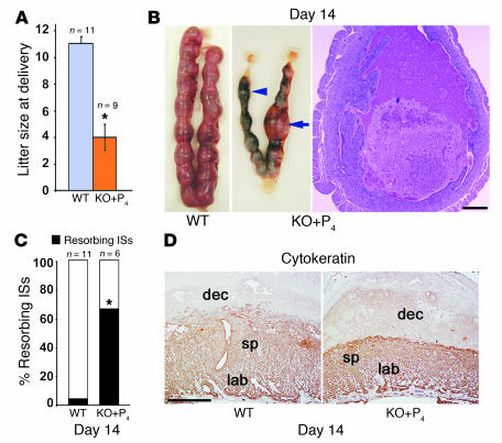
To investigate whether P4 supplementation maintains full-term pregnancy in CD1 Fkbp52–/– females, P4 implants placed on day 2 were removed on day 17, since P4 withdrawal is necessary to initiate labor (41). Although 9 of 13 Fkbp52–/– mothers delivered pups of normal weight, litter sizes were significantly smaller (Figure 5A). This led us to determine when embryonic loss occurs in null females carrying P4 implants. We observed that 70 of 106 ISs were resorbing in P4 implant-treated Fkbp52–/– uteri when examined on day 14, compared with only 9 of 194 in WT uteri (Figure 5, B and C). Resorbing ISs in P4-treated Fkbp52–/– females appeared dark blue and were infiltrated with a massive number of blood cells (Figure 5B). In addition, cytokeratin staining of sections of ISs with normal appearance from null females carrying P4 implants showed placentas with less-developed and ill-defined spongiotrophoblast and labyrinth layers compared with those of WT females (Figure 5D).
Figure 5. P4 delivery by Silastic implants fails to sustain pregnancy to full term.
(A) Average litter size of WT and KO+P4 mothers. Litter size is presented as mean ± SEM; *P < 0.05, unpaired Student’s t test. (B) Representative photomicrographs of WT and KO+P4 uteri on day 14 of pregnancy. Arrowheads and arrows indicate resorbing and normal IS, respectively. A representative H&E-stained section of resorbing IS from KO+P4 uterus shows massive infiltration of blood cells. Scale bar: 200 μm. (C) Percentage of resorption sites in WT and KO+P4 mice on day 14 of pregnancy. *P < 0.05, unpaired Student’s t test. (D) Cytokeratin staining of WT and KO+P4 IS on day 14. Scale bar: 200 μm. dec, decidua; lab, labyrinth; sp, spongiotrophoblast.
Although P4/PR signaling is absolutely required for pregnancy maintenance in all eutherians thus far studied, uterine FKBP52 expression in later days of pregnancy has not yet been examined. We found that while FKBP52 is expressed in the mesometrial decidua with high expression in the placenta on days 10–14 of pregnancy, PR is mostly expressed in the decidua (Supplemental Figure 3, A and B, and data not shown). Embryonic signals direct normal decidual functions and development (39), which in turn govern placentation and embryonic growth (42). Therefore, the expression of FKBP52 and PR in the decidua suggests that maternally derived FKBP52-mediated PR signaling contributes to fetoplacental well-being, while placental expression of FKBP52 suggests a PR-independent role for FKBP52.
Excessive estrogenic influence or complement activation does not contribute to pregnancy failure in P4-implanted Fkbp52–/– females.
We speculated that one possible explanation for the higher incidence of resorptions in Fkbp52–/– females carrying P4 implants is the tipping of the balance between estrogen and P4 signaling toward estrogenic dominance. Since high levels of estrogen are detrimental to pregnancy success (30), we examined whether combined treatment with P4 implants and ICI 182,780 (ICI; 25 μg or 125 μg/0.1 ml oil/mouse, s.c.), an estrogen receptor antagonist (Tocris Bioscience), injected on days 8–13 improves pregnancy maintenance. Combined treatment with P4 and the high dose of ICI increased the resorption rate to 100% in Fkbp52–/– mice, while treatment with a lower dose of ICI resulted in resorption rates similar to those observed in null mice receiving P4 implants alone (Supplemental Figure 4). Our observation that treatment with a higher dose of ICI results in increased resorption rates suggests that appropriate estrogen signaling is also critical to pregnancy maintenance. This agrees with a previous study showing that pregnancy maintenance under the direction of P4 is supported by low amounts of estrogen, especially on days 10 and 11 of pregnancy (43).
While the underlying causes of recurrent pregnancy failure are not well understood, one possibility is that the maternal immune response mistakenly recognizes the fetus. A recent study shows that complement activation causes growth restriction and subsequent fetal rejection, leading to pregnancy failure (44). We speculated that this pathway is activated due to reduced PR signaling from FKBP52 deficiency, especially since P4 has antiinflammatory roles within and outside the uterus (17, 45). Since low doses of heparin inhibit the complement pathway (44, 46), Fkbp52–/– mice carrying P4 implants from day 2 were given a low dose of heparin (10 U/mouse) twice a day on days 8, 10, and 12 of pregnancy. However, this treatment also failed to rescue pregnancy maintenance in these mice (Supplemental Figure 4), suggesting that activation of the complement pathway is not a major contributing factor for pregnancy failure in P4-treated Fkbp52–/– mice.
Differential P4/PR signaling is required for successful full-term pregnancy.
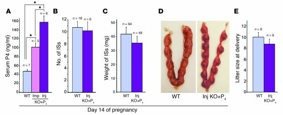
Normal serum P4 levels during pregnancy in CD1 WT mice on days 5 and 14 of pregnancy range between 40 and 47 ng/ml (Supplemental Figure 1 and Figure 6A). In our experiments, Silastic P4 implants provided an increase in serum P4 levels sufficient to induce uterine receptivity and rescue implantation in CD1 Fkbp52–/– females but failed to maintain pregnancy to full term. We speculated that further increasing P4 levels would rectify this failure. We injected P4 s.c. at a dose of 2 mg/ml per mouse daily to further increase P4 serum levels. We observed that in null mice, serum P4 levels increased to approximately 156 ng/ml by daily injection as compared with approximately 100 ng/ml in mice treated with P4 implants, as assessed on day 14 of pregnancy (Figure 6A). To our surprise, these elevated P4 levels significantly improved pregnancy maintenance in null females; the number and weights of ISs on day 14 were comparable to those in WT mothers (Figure 6, B–D). Pregnancy maintenance in WT mice exposed to a similar P4 injection regimen was normal (data not shown).
Figure 6. Daily P4 injections restore pregnancy to full term in CD1 Fkbp52–/– females.
(A) Serum P4 levels in WT and KO+P4 mice on day 14 of pregnancy. KO mice were exposed daily to P4 either via Silastic implants (Imp KO+P4) or s.c. injection (Inj KO+P4; 2 mg/ml) from day 2 of pregnancy. *P < 0.05, univariate ANOVA. (**B**) The average number of ISs was not significantly different in WT and Inj KO+P4 mice on day 14; _P_ > 0.1, unpaired Student’s t test. (C) Weights of ISs from WT and Inj KO+P4 mice on day 14 of pregnancy were not significantly different; P > 0.1, unpaired Student’s t test. (D) A representative photomicrograph of WT and Inj KO+P4 uteri on day 14 of pregnancy is shown. (E) The average litter size of WT and Inj KO+P4 mice was not significantly different; P > 0.05, unpaired Student’s t test. All values are mean ± SEM.
Our next objective was to examine whether daily P4 injection results in pregnancy to full term in null females. CD1 Fkbp52–/– females mated with WT males were injected with P4 daily from days 2 through 17 of pregnancy and monitored for term delivery on day 20. We observed that all P4-injected null females carried pups to term, and the average litter size was comparable to that of WT mothers (Figure 6E). Pup weights from null and WT mothers at weaning and during early development were also similar (data not shown).
Discussion
Although P4 signaling via PR is critical to ovulation, fertilization, implantation, postimplantation growth, and pregnancy maintenance, it is not known whether a similar P4/PR signaling mechanism determines these target and stage-specific functions or whether genetic disparity influences this signaling. By using Fkbp52–/– females with compromised PR signaling as opposed to Pgr–/– females with total infertility (3, 12), we address these issues for the first time to our knowledge. Our previous and present investigations provide clear evidence that the major reproductive phenotype in mice lacking Fkbp52 is unique to uterine deficiency in the context of implantation. The reason for the organ-specific dependence on FKBP52 for appropriate PR signaling is not clearly understood, but it is noteworthy, since ovulation that also requires P4/PR signaling is normal in null females. It is possible that relatively higher P4 levels locally in the ovary override the reduced PR signaling in the absence of FKBP52. We were also surprised to note that P4/PR-regulated mating behavior appears normal, since null females mate and produce copulatory plugs. One possibility is that the uterus requires more robust P4/PR signaling during pregnancy than other P4 targets. Alternatively, FKBP52, in addition to its role in influencing PR signaling, may have a unique PR-independent role in the uterus not observed in other tissues.
P4 signaling via PR plays major roles at essentially all stages of pregnancy, from ovulation through parturition. It is surprising to see that genetic makeup of a species alters such a fundamental signaling pathway. While FKBP52 is essential to support implantation in both strains of mice in the absence of exogenous P4, FKBP52’s role becomes less significant in CD1 mice exposed to high levels of P4, while still remaining crucial in C57BL6/129 mice under similar treatment conditions. The contrasting reproductive phenotypes of Fkbp52–/– mice on C57BL6/129 and CD1 backgrounds with respect to P4 rescue provides the first evidence to our knowledge that P4/PR/FKBP52 signaling is a function of genetic makeup. Coordinated interactions of P4 and estrogen are essential to uterine receptivity and implantation. While one aspect of P4 signaling is to correctly orchestrate P4-responsive genes, another is to appropriately constrain and/or synergize estrogen-responsive genes in the uterus. One cause of implantation failure in C57BL6/129, but not in CD1, Fkbp52–/– females supplemented with P4 could be the failure of the uterus to attain optimal receptivity arising from altered expression of Areg and/or Ltf. This inappropriate gene expression in the presence of P4 may reflect even lower basal PR activity in C57BL6/129 Fkbp52–/– uteri than in CD1 Fkbp52–/– uteri. Alternatively, differential expression of modifier genes in these 2 strains of mice could contribute to differential uterine responsiveness to P4/PR signaling in the absence of FKBP52.
There is evidence that rodent blastocysts synthesize P4 or structurally similar steroids (47–49) and that gene expression differs in embryo-induced decidua and experimentally induced deciduoma (39). Since our previous results show that Fkbp52 and Pgr are expressed in blastocysts (12), it is possible that P4 synthesized from blastocysts enhances PR/FKBP52 signaling locally at their sites of apposition in the uterus. This could explain the failure of oil-induced decidualization but not of that induced by implanting blastocysts in CD1 Fkbp52–/– mice supplemented with P4. Alternatively, signaling arising from blastocysts may influence other uterine functions. For example, the gene encoding heparin-binding epidermal growth factor–like growth factor (HB-EGF) is expressed in implantation-competent blastocysts, which can induce uterine Hbegf to initiate the implantation cascade. In fact, Affi-Gel Blue beads (Bio-Rad) presoaked in HB-EGF, when transferred into uterine lumens of pseudopregnant mice on day 4, show implantation-like responses similar to those induced by living blastocysts, including upregulation of Hbegf and Bmp2 (32, 50).
P4/PR signaling is critical throughout pregnancy until its downregulation for the onset of parturition. Indeed, ovariectomizing mice at practically any stage of pregnancy causes resorptions and/or abortion (51). However, the magnitude of this signaling during various stages of pregnancy remains unknown. Although P4 implants cannot rescue pregnancy in WT mice ovariectomized on day 8 unless they are given daily P4 injections, similar P4 injections alone maintain full-term pregnancy in mice ovariectomized on day 14 (51). The use of Fkbp52–/– females has enabled us to show that the requirement for P4/PR signaling is different for uterine receptivity, implantation, and postimplantation growth. This signaling appears to be tightly regulated, since blood levels of approximately 100 ng/ml of P4 are adequate to induce uterine receptivity and implantation, but levels above 150 ng/ml are required for full complement of pregnancy success in the absence of FKBP52. This suggests that more robust P4/PR signaling is required for pregnancy maintenance than is required for uterine receptivity, implantation, and decidualization. It is also possible that a burst of P4 levels as provided by daily injections is more amenable to pregnancy sustenance than the relatively constant levels maintained with Silastic implants.
The observation that differential P4 levels are required for various stages of pregnancy implies that signaling targets are different. We believe that P4/PR signaling for uterine preparation, implantation, and decidualization is primarily targeted to uterine epithelial and stromal cells. This is consistent with P4’s known roles in epithelial differentiation and stromal cell proliferation during the periimplantation period. On the other hand, P4/PR signaling for pregnancy maintenance is directed more toward keeping the myometrium quiescent until parturition and providing sanctuary for the growing fetus from mother’s immunological surveillance. P4/PR signaling is also known to regulate angiogenesis (52), a process integral to placental development and pregnancy maintenance. Since the events during the course of pregnancy are very dynamic, P4/PR signaling at various targets could be overlapping. We consider FKBP52’s influence on P4/PR signaling during pregnancy to be primarily of maternal origin, since both PR and FKBP52 are expressed in the decidua on days 10 and 12 of pregnancy. The high expression of FKBP52 in the placenta implies a PR-independent role for FKBP52 in placentation. This requires further investigation, but the putative PR-independent role is not critical, since pregnancies are completed to term with normal litter sizes in CD1 _Fkbp52_-null females receiving daily P4 injections.
An interesting observation is the successful nursing of pups to weaning by P4-injected CD1 Fkbp52–/– mothers. Mammary morphogenesis during pregnancy requires P4/PR signaling (16, 53), but the latter stages of lactogenesis and lactation correspond to withdrawal of this signaling (reviewed in ref. 54). It was recently reported that FKBP52 may not be critical for P4/PR signaling in the mammary gland, since exogenously provided P4 could stimulate mammary morphogenesis in Fkbp52–/– mice (15). This is similar to the findings of our studies in which Fkbp52–/– mothers were injected with exogenous P4 prior to parturition. These results suggest that exogenously supplemented P4 overcomes the P4-resistant state in the mammary gland and uterus in the absence of FKBP52.
The implanting blastocyst is the stimulus for normal decidualization in mice. However, in humans, stromal cells undergo decidualization during the receptive phase in each menstrual cycle in the absence of blastocysts. This predecidualization is thought to be critical for blastocyst implantation in the pregnant uterus (55). Our findings that P4 supplementation fails to rescue oil-induced decidualization in _Fkbp52_-null uteri may therefore have implication for predecidualization events in P4-resistant women.
P4 resistance is also a hallmark of endometriosis, a condition that affects an estimated 5 million women of childbearing age in the United States (National Women’s Health Information Center, National Institute of Child Health and Human Development [NICHD], NIH) (56–58). In fact, a recent study examining global gene expression profiles in endometria of women with or without endometriosis found dysregulation of many known P4 target genes during the window of uterine receptivity (59). While the mechanism(s) of P4 resistance remain unclear, one speculation is that downregulation of PR contributes to P4 resistance; however, other studies refute this concept (57). Whether FKBP52 expression differs in normal and endometriotic tissues has not been examined.
The finding that human FKBP52 interacts with and potentiates human PR activity in MEFs (12) and our preliminary observation on a limited number of samples showing FKBP52 and PR expression in human endometria in proliferative and secretory phases (data not shown) suggest a potential role for uterine P4/PR/FKBP52 signaling in women. In fact, 3 separate clinical trials found that P4 treatment resulted in a statistically significant decrease in miscarriages in women with a history of 3 or more consecutive pregnancy losses (60). In this respect, pregnancy rescue in CD1 Fkbp52–/– mice by daily P4 injections is clinically relevant for women who are infertile due to P4 resistance. These are exciting results, especially since no significant differences in adverse effects were found between P4 treatment and control groups. These findings in humans are similar to our findings in mice that exogenous P4 injections in WT females do not adversely affect pregnancy (data not shown). Fkbp52–/– mice with normal PR and P4 levels but reduced PR activity constitute a unique model for studying P4 resistance specifically in uterine biology, and it is hoped that our findings will encourage the development of human studies to determine whether FKBP52 status influences P4 resistance in the uterus.
Methods
Mice.
The Fkbp52 gene was disrupted in mice by homologous recombination, as previously described (10). Tail genomic DNA was used for PCR-based genotyping. Because genetic backgrounds of mice contribute to different phenotypes (18, 20), we introduced Fkbp52 deficiency in CD1 mice by crossing C57BL6/129 Fkbp52 heterozygous males to CD1 WT females producing an F1 generation. F1 Fkbp52+/– males were then back-crossed to CD1 WT females, and the process was continued for 10 generations. Crossing heterozygous females with heterozygous males of the same genetic background (CD1/F10) generated _Fkbp52_-null and WT littermates for experiments. Mice on both backgrounds were housed and used in the present investigation in accordance with NIH, and animal protocol was approved by the Vanderbilt Institutional Animal Care and Use Committee.
Ovulation, fertilization, implantation, blastocyst transfer, and experimentally induced decidualization.
Mice were examined for ovulation, fertilization, and implantation as described previously (37). To examine ovulation and fertilization, CD1 WT or Fkbp52–/– mice were mated with fertile WT males. On day 2 of pregnancy (the day the vaginal plug was first observed was considered day 1), oviducts were flushed with Whitten’s medium to recover ovulated eggs, and fertilization was assessed by the number of 2-cell embryos. ISs on days 5 and 6 of pregnancy were visualized by an i.v. injection (0.1 ml/mouse) of Chicago blue B dye solution (1% in saline), and the number of ISs demarcated by distinct blue bands was recorded. For blastocyst transfer, pseudopregnant recipients were generated by mating females with vasectomized WT males. Day-4 WT blastocysts were transferred into day-4 uteri of C57BL6/129 WT or Fkbp52–/– pseudopregnant recipients, and ISs were examined 24 hours (day 5) or 96 hours (day 8) later by the blue dye method (32).
To determine whether experimentally induced decidualization occurs in null females, WT or Fkbp52–/– females were mated with vasectomized WT males. On day 4, one uterine horn was infused with sesame oil (25 μl), while the contralateral horn served as control. Mice were sacrificed on day 8 of pseudopregnancy. Weights of infused (oil) and noninfused (control) uterine horns were recorded, and fold increase in weight was used as an index of decidualization. All mice used were between 2 and 5 months of age.
Exogenous P4 supplementation and other treatments.
To see whether P4 supplementation rescues the infertility phenotype of Fkbp52–/– females, null females were mated with WT males, and a Silastic implant (4 cm length × 0.31 cm diameter) containing P4 was placed under the dorsal skin on day 2 of pregnancy. Implants were removed upon sacrifice on days 5, 6, and 8 to examine implantation; days 12 or 14 to examine pregnancy maintenance; or day 17 to allow labor to complete full-term pregnancy. Alternatively, null female mice were given a daily injection of P4 (2 mg/0.1 ml/mouse, s.c.) from days 2 through 14 to monitor pregnancy maintenance or through day 17 to allow labor to ensue on day 20 for full-term pregnancy.
To determine the contribution of the complement pathway to pregnancy maintenance, Silastic P4 implants were placed under the dorsal skin on day 2 of pregnancy, and heparin (10 U/0.1 ml/mouse in saline) was injected s.c. twice a day on days 8, 10, and 12 of pregnancy. To determine whether the P4/estrogen ratio influences pregnancy maintenance, Silastic P4 implants were placed under the dorsal skin on day 2 of pregnancy, and ICI (25 μg or 125 μg/0.1 ml/mouse in sesame oil), an estrogen receptor antagonist, was injected once a day on days 8 through 13 of pregnancy.
To determine whether P4 supplementation rescues experimentally induced decidualization in null females, WT or Fkbp52–/– females were mated with vasectomized WT males, and P4 was either injected daily from day 2 or Silastic P4 implants placed under the dorsal skin on day 2 of pseudopregnancy. On day 4, while the uterine lumen of one horn was infused with sesame oil (25 μl), the noninfused contralateral horn served as a control. Mice were sacrificed on day 8 of pseudopregnancy. Uterine weights of oil-infused and noninfused horns were recorded, and fold increases in weight were recorded as an index of decidualization.
P4 assay.
Blood samples from mice were collected on the indicated days of pregnancy. Serum was separated by centrifugation (850 g for 15 minutes) and stored at –80°C until analysis. Serum P4 levels were measured by radioimmunoassay.
In situ hybridization.
Sense or antisense 35S-labeled cRNA probes for Areg, Ihh, Hoxa10, Ltf, C3, Lif, Bmp2, Ptgs2, Pgr, and Fkbp52 generated using appropriate polymerases from respective cDNAs were used for hybridization as described previously by us (61). Sections hybridized with sense probes showed no positive signals and served as negative controls.
Immunohistochemistry.
Immunolocalization of cytokeratin was performed using a polyclonal rabbit anti-cow antibody (Dako). A Histostain-Plus (DAB) kit (Zymed) was used to visualize antigen. Brown deposits indicated sites of positive immunostaining.
Statistics.
Statistical significance was determined as P < 0.05 by 1-tailed Student’s t test. To compare serum P4 levels in WT versus KO+P4 mice on day 14 of pregnancy, statistical significance was determined as P < 0.05 by univariate ANOVA. All values are presented as mean ± SEM.
Supplementary Material
Supplemental data
Acknowledgments
We thank Fuhua Xu for help with statistical analysis. P4 assays were performed by the University of Virginia Center for Ligand Assay and Analysis Core supported by an NICHD grant (U54 HD28934). This work was supported in part by grants from the NICHD (HD12304 and HD033994 to S.K. Dey; HD050315 to H. Wang) and NIDDK (DK48218 to D.F. Smith). S. Tranguch was supported by a National Research Service Award fellowship from the National Institute on Drug Abuse (F31 DA021062) and an NIDDK training grant (T32 DK07563).
Footnotes
Nonstandard abbreviations used: Areg, amphiregulin; FKBP, FK506-binding protein; ICI, ICI 182,780; Ihh, Indian hedgehog; IS, implantation site; Lif, leukemia inhibitory factor; Ltf, lactoferrin; MEF, mouse embryonic fibroblast; P4, progesterone; PR, P4 receptor; TPR, tetratricopeptide repeat.
Conflict of interest: The authors have declared that no conflict of interest exists.
Citation for this article: J. Clin. Invest. 117:1824–1834 (2007). doi:10.1172/JCI31622
References
- 1.Dey S.K., et al. Molecular cues to implantation. Endocr. Rev. 2004;25:341–373. doi: 10.1210/er.2003-0020. [DOI] [PubMed] [Google Scholar]
- 2.Wang H., Dey S.K. Roadmap to embryo implantation: clues from mouse models. Nat. Rev. Genet. 2006;7:185–199. doi: 10.1038/nrg1808. [DOI] [PubMed] [Google Scholar]
- 3.Lydon J.P., et al. Mice lacking progesterone receptor exhibit pleiotropic reproductive abnormalities. Genes Dev. 1995;9:2266–2278. doi: 10.1101/gad.9.18.2266. [DOI] [PubMed] [Google Scholar]
- 4.Smith D.F. Tetratricopeptide repeat cochaperones in steroid receptor complexes. Cell Stress Chaperones. 2004;9:109–121. doi: 10.1379/CSC-31.1. [DOI] [PMC free article] [PubMed] [Google Scholar]
- 5.Pratt W.B., Toft D.O. Steroid receptor interactions with heat shock protein and immunophilin chaperones. Endocr. Rev. 1997;18:306–360. doi: 10.1210/edrv.18.3.0303. [DOI] [PubMed] [Google Scholar]
- 6.Smith D.F. Chaperones in progesterone receptor complexes. Semin. Cell Dev. Biol. 2000;11:45–52. doi: 10.1006/scdb.1999.0350. [DOI] [PubMed] [Google Scholar]
- 7.Smith D.F. Dynamics of heat shock protein 90-progesterone receptor binding and the disactivation loop model for steroid receptor complexes. Mol. Endocrinol. 1993;7:1418–1429. doi: 10.1210/mend.7.11.7906860. [DOI] [PubMed] [Google Scholar]
- 8.Barent R.L., et al. Analysis of FKBP51/FKBP52 chimeras and mutants for Hsp90 binding and association with progesterone receptor complexes. Mol. Endocrinol. 1998;12:342–354. doi: 10.1210/mend.12.3.0075. [DOI] [PubMed] [Google Scholar]
- 9.Riggs D.L., et al. Functional specificity of co-chaperone interactions with Hsp90 client proteins. Crit. Rev. Biochem. Mol. Biol. 2004;39:279–295. doi: 10.1080/10409230490892513. [DOI] [PubMed] [Google Scholar]
- 10.Cheung-Flynn J., et al. Physiological role for the cochaperone FKBP52 in androgen receptor signaling. Mol. Endocrinol. 2005;19:1654–1666. doi: 10.1210/me.2005-0071. [DOI] [PubMed] [Google Scholar]
- 11.Riggs D.L., et al. The Hsp90-binding peptidylprolyl isomerase FKBP52 potentiates glucocorticoid signaling in vivo. EMBO J. 2003;22:1158–1167. doi: 10.1093/emboj/cdg108. [DOI] [PMC free article] [PubMed] [Google Scholar]
- 12.Tranguch S., et al. Cochaperone immunophilin FKBP52 is critical to uterine receptivity for embryo implantation. Proc. Natl. Acad. Sci. U. S. A. 2005;102:14326–14331. doi: 10.1073/pnas.0505775102. [DOI] [PMC free article] [PubMed] [Google Scholar]
- 13.Paria B.C., Huet-Hudson Y.M., Dey S.K. Blastocyst’s state of activity determines the “window” of implantation in the receptive mouse uterus. Proc. Natl. Acad. Sci. U. S. A. 1993;90:10159–10162. doi: 10.1073/pnas.90.21.10159. [DOI] [PMC free article] [PubMed] [Google Scholar]
- 14.Daikoku T., et al. Proteomic analysis identifies immunophilin FK506 binding protein 4 (FKBP52) as a downstream target of Hoxa10 in the periimplantation mouse uterus. Mol. Endocrinol. 2005;19:683–697. doi: 10.1210/me.2004-0332. [DOI] [PubMed] [Google Scholar]
- 15.Yang Z., et al. FK506-binding protein 52 is essential to uterine reproductive physiology controlled by the progesterone receptor A isoform. Mol. Endocrinol. 2006;20:2682–2694. doi: 10.1210/me.2006-0024. [DOI] [PMC free article] [PubMed] [Google Scholar]
- 16.Mulac-Jericevic B., Mullinax R.A., DeMayo F.J., Lydon J.P., Conneely O.M. Subgroup of reproductive functions of progesterone mediated by progesterone receptor-B isoform. Science. 2000;289:1751–1754. doi: 10.1126/science.289.5485.1751. [DOI] [PubMed] [Google Scholar]
- 17.Tranguch S., Smith D.F., Dey S.K. Progesterone receptor requires a co-chaperone for signalling in uterine biology and implantation. Reprod. Biomed. Online. 2006;13:651–660. doi: 10.1016/s1472-6483(10)60655-4. [DOI] [PubMed] [Google Scholar]
- 18.Threadgill D.W., et al. Targeted disruption of mouse EGF receptor: effect of genetic background on mutant phenotype. Science. 1995;269:230–234. doi: 10.1126/science.7618084. [DOI] [PubMed] [Google Scholar]
- 19.Bonyadi M., et al. Mapping of a major genetic modifier of embryonic lethality in TGF beta 1 knockout mice. Nat. Genet. 1997;15:207–211. doi: 10.1038/ng0297-207. [DOI] [PubMed] [Google Scholar]
- 20.Wang H., et al. Rescue of female infertility from the loss of cyclooxygenase-2 by compensatory up-regulation of cyclooxygenase-1 is a function of genetic makeup. J. Biol. Chem. 2004;279:10649–10658. doi: 10.1074/jbc.M312203200. [DOI] [PubMed] [Google Scholar]
- 21.Psychoyos A. Hormonal control of ovoimplantation. Vitam. Horm. 1973;31:201–256. doi: 10.1016/s0083-6729(08)60999-1. [DOI] [PubMed] [Google Scholar]
- 22.Pointis G., Rao B., Latreille M.T., Mignot T.M., Cedard L. Progesterone levels in the circulating blood of the ovarian and uterine veins during gestation in the mouse. Biol. Reprod. 1981;24:801–805. doi: 10.1095/biolreprod24.4.801. [DOI] [PubMed] [Google Scholar]
- 23.Milligan S.R., Cohen P.E. Silastic implants for delivering physiological concentrations of progesterone to mice. Reprod. Fertil. Dev. 1994;6:235–239. doi: 10.1071/rd9940235. [DOI] [PubMed] [Google Scholar]
- 24.Das S.K., Chakraborty I., Paria B.C., Wang X.N., Plowman G., Dey S.K. Amphiregulin is an implantation-specific and progesterone-regulated gene in the mouse uterus. Mol. Endocrinol. 1995;9:691–705. doi: 10.1210/mend.9.6.8592515. [DOI] [PubMed] [Google Scholar]
- 25.Lim H., Ma L., Ma W.G., Maas R.L., Dey S.K. Hoxa-10 regulates uterine stromal cell responsiveness to progesterone during implantation and decidualization in the mouse. Mol. Endocrinol. 1999;13:1005–1017. doi: 10.1210/mend.13.6.0284. [DOI] [PubMed] [Google Scholar]
- 26.Matsumoto H., Zhao X., Das S.K., Hogan B.L., Dey S.K. Indian hedgehog as a progesterone-responsive factor mediating epithelial-mesenchymal interactions in the mouse uterus. Dev. Biol. 2002;245:280–290. doi: 10.1006/dbio.2002.0645. [DOI] [PubMed] [Google Scholar]
- 27.Lee K., et al. Indian hedgehog is a major mediator of progesterone signaling in the mouse uterus. Nat. Genet. 2006;38:1204–1209. doi: 10.1038/ng1874. [DOI] [PubMed] [Google Scholar]
- 28.Heikaus S., Winterhager E., Traub O., Grummer R. Responsiveness of endometrial genes Connexin26, Connexin43, C3 and clusterin to primary estrogen, selective estrogen receptor modulators, phyto- and xenoestrogens. J. Mol. Endocrinol. 2002;29:239–249. doi: 10.1677/jme.0.0290239. [DOI] [PubMed] [Google Scholar]
- 29.McMaster M.T., Teng C.T., Dey S.K., Andrews G.K. Lactoferrin in the mouse uterus: analyses of the preimplantation period and regulation by ovarian steroids. Mol. Endocrinol. 1992;6:101–111. doi: 10.1210/mend.6.1.1738363. [DOI] [PubMed] [Google Scholar]
- 30.Ma W.G., Song H., Das S.K., Paria B.C., Dey S.K. Estrogen is a critical determinant that specifies the duration of the window of uterine receptivity for implantation. Proc. Natl. Acad. Sci. U. S. A. 2003;100:2963–2968. doi: 10.1073/pnas.0530162100. [DOI] [PMC free article] [PubMed] [Google Scholar]
- 31.Lim H., et al. Multiple female reproductive failures in cyclooxygenase 2-deficient mice. Cell. 1997;91:197–208. doi: 10.1016/s0092-8674(00)80402-x. [DOI] [PubMed] [Google Scholar]
- 32.Paria B.C., et al. Cellular and molecular responses of the uterus to embryo implantation can be elicited by locally applied growth factors. Proc. Natl. Acad. Sci. U. S. A. 2001;98:1047–1052. doi: 10.1073/pnas.98.3.1047. [DOI] [PMC free article] [PubMed] [Google Scholar]
- 33.Song H., Lim H., Das S.K., Paria B.C., Dey S.K. Dysregulation of EGF family of growth factors and COX-2 in the uterus during the preattachment and attachment reactions of the blastocyst with the luminal epithelium correlates with implantation failure in LIF-deficient mice. Mol. Endocrinol. 2000;14:1147–1161. doi: 10.1210/mend.14.8.0498. [DOI] [PubMed] [Google Scholar]
- 34.Song H., Lim H. Evidence for heterodimeric association of leukemia inhibitory factor (LIF) receptor and gp130 in the mouse uterus for LIF signaling during blastocyst implantation. Reproduction. 2006;131:341–349. doi: 10.1530/rep.1.00956. [DOI] [PubMed] [Google Scholar]
- 35.Carpenter K.D., Gray C.A., Bryan T.M., Welsh T.H., Jr., Spencer T.E. Estrogen and antiestrogen effects on neonatal ovine uterine development. Biol. Reprod. 2003;69:708–717. doi: 10.1095/biolreprod.103.015990. [DOI] [PubMed] [Google Scholar]
- 36.Gray C.A., Bazer F.W., Spencer T.E. Effects of neonatal progestin exposure on female reproductive tract structure and function in the adult ewe. Biol. Reprod. 2001;64:797–804. doi: 10.1095/biolreprod64.3.797. [DOI] [PubMed] [Google Scholar]
- 37.Song H., et al. Cytosolic phospholipase A2alpha is crucial [correction of A2alpha deficiency is crucial] for ‘on-time’ embryo implantation that directs subsequent development. Development. 2002;129:2879–2889. doi: 10.1242/dev.129.12.2879. [DOI] [PubMed] [Google Scholar]
- 38.Ye X., et al. LPA3-mediated lysophosphatidic acid signalling in embryo implantation and spacing. Nature. 2005;435:104–108. doi: 10.1038/nature03505. [DOI] [PMC free article] [PubMed] [Google Scholar]
- 39.Bany B.M., Cross J.C. Post-implantation mouse conceptuses produce paracrine signals that regulate the uterine endometrium undergoing decidualization. Dev. Biol. 2006;294:445–456. doi: 10.1016/j.ydbio.2006.03.006. [DOI] [PubMed] [Google Scholar]
- 40.Lundkvist O., Nilsson B.O. Endometrial ultrastructure in the early uterine response to blastocysts and artificial deciduogenic stimuli in rats. Cell Tissue Res. 1982;225:355–364. doi: 10.1007/BF00214688. [DOI] [PubMed] [Google Scholar]
- 41.McCormack J.T., Greenwald G.S. Progesterone and oestradiol-17beta concentrations in the peripheral plasma during pregnancy in the mouse. J. Endocrinol. 1974;62:101–107. doi: 10.1677/joe.0.0620101. [DOI] [PubMed] [Google Scholar]
- 42.Bilinski P., Roopenian D., Gossler A. Maternal IL-11Ralpha function is required for normal decidua and fetoplacental development in mice. Genes Dev. 1998;12:2234–2243. doi: 10.1101/gad.12.14.2234. [DOI] [PMC free article] [PubMed] [Google Scholar]
- 43.Milligan S.R., Finn C.A. Minimal progesterone support required for the maintenance of pregnancy in mice. Hum. Reprod. 1997;12:602–607. doi: 10.1093/humrep/12.3.602. [DOI] [PubMed] [Google Scholar]
- 44.Girardi G., Yarilin D., Thurman J.M., Holers V.M., Salmon J.E. Complement activation induces dysregulation of angiogenic factors and causes fetal rejection and growth restriction. . J. Exp. Med. 2006;203:2165–2175. doi: 10.1084/jem.20061022. [DOI] [PMC free article] [PubMed] [Google Scholar]
- 45.Hardy D.B., Janowski B.A., Corey D.R., Mendelson C.R. Progesterone receptor plays a major antiinflammatory role in human myometrial cells by antagonism of nuclear factor-B activation of cyclooxygenase 2 expression. Mol. Endocrinol. 2006;20:2724–2733. doi: 10.1210/me.2006-0112. [DOI] [PubMed] [Google Scholar]
- 46.Girardi G., Redecha P., Salmon J.E. Heparin prevents antiphospholipid antibody-induced fetal loss by inhibiting complement activation. Nat. Med. 2004;10:1222–1226. doi: 10.1038/nm1121. [DOI] [PubMed] [Google Scholar]
- 47.Wu J.T. Metabolism of progesterone by preimplantation mouse blastocysts in culture. Biol. Reprod. 1987;36:549–556. doi: 10.1095/biolreprod36.3.549. [DOI] [PubMed] [Google Scholar]
- 48.Wu J.T., Liu Z.H. Conversion of pregnenolone to progesterone by mouse morulae and blastocysts. J. Reprod. Fertil. 1990;88:93–98. doi: 10.1530/jrf.0.0880093. [DOI] [PubMed] [Google Scholar]
- 49.Carson D.D., Hsu Y.C., Lennarz W.J. Synthesis of steroids in postimplantation mouse embryos cultured in vitro. Dev. Biol. 1982;91:402–412. doi: 10.1016/0012-1606(82)90046-x. [DOI] [PubMed] [Google Scholar]
- 50.Hamatani T., et al. Global gene expression analysis identifies molecular pathways distinguishing blastocyst dormancy and activation. Proc. Natl. Acad. Sci. U. S. A. 2004;101:10326–10331. doi: 10.1073/pnas.0402597101. [DOI] [PMC free article] [PubMed] [Google Scholar]
- 51.Sharma R., Bulmer D. The effects of ovariectomy and subsequent progesterone replacement on the uterus of the pregnant mouse. J. Anat. 1983;137:695–703. [PMC free article] [PubMed] [Google Scholar]
- 52.Ma W., et al. Adult tissue angiogenesis: evidence for negative regulation by estrogen in the uterus. Mol. Endocrinol. 2001;15:1983–1992. doi: 10.1210/mend.15.11.0734. [DOI] [PubMed] [Google Scholar]
- 53.Mulac-Jericevic B., Lydon J.P., DeMayo F.J., Conneely O.M. Defective mammary gland morphogenesis in mice lacking the progesterone receptor B isoform. Proc. Natl. Acad. Sci. U. S. A. 2003;100:9744–9749. doi: 10.1073/pnas.1732707100. [DOI] [PMC free article] [PubMed] [Google Scholar]
- 54.Neville M.C., McFadden T.B., Forsyth I. Hormonal regulation of mammary differentiation and milk secretion. J. Mammary Gland Biol. Neoplasia. 2002;7:49–66. doi: 10.1023/a:1015770423167. [DOI] [PubMed] [Google Scholar]
- 55.Chobotova K., et al. Heparin-binding epidermal growth factor and its receptors mediate decidualization and potentiate survival of human endometrial stromal cells. J. Clin. Endocrinol. Metab. 2005;90:913–919. doi: 10.1210/jc.2004-0476. [DOI] [PMC free article] [PubMed] [Google Scholar]
- 56.Berkley K.J., Rapkin A.J., Papka R.E. The pains of endometriosis. Science. 2005;308:1587–1589. doi: 10.1126/science.1111445. [DOI] [PubMed] [Google Scholar]
- 57.Bulun S.E., et al. Progesterone resistance in endometriosis: link to failure to metabolize estradiol. Mol. Cell. Endocrinol. 2006;248:94–103. doi: 10.1016/j.mce.2005.11.041. [DOI] [PubMed] [Google Scholar]
- 58.Fang Z., et al. Intact progesterone receptors are essential to counteract the proliferative effect of estradiol in a genetically engineered mouse model of endometriosis. Fertil. Steril. 2004;82:673–678. doi: 10.1016/j.fertnstert.2004.01.048. [DOI] [PubMed] [Google Scholar]
- 59.Kao L.C., et al. Expression profiling of endometrium from women with endometriosis reveals candidate genes for disease-based implantation failure and infertility. Endocrinology. 2003;144:2870–2881. doi: 10.1210/en.2003-0043. [DOI] [PubMed] [Google Scholar]
- 60.Oates-Whitehead R.M., Haas D.M., Carrier J.A. Progestogen for preventing miscarriage. Cochrane Database Syst. Rev. 2003;2003:CD003511. doi: 10.1002/14651858.CD003511. [DOI] [PubMed] [Google Scholar]
- 61.Das S.K., et al. Heparin-binding EGF-like growth factor gene is induced in the mouse uterus temporally by the blastocyst solely at the site of its apposition: a possible ligand for interaction with blastocyst EGF-receptor in implantation. Development. 1994;120:1071–1083.. doi: 10.1242/dev.120.5.1071. [DOI] [PubMed] [Google Scholar]
Associated Data
This section collects any data citations, data availability statements, or supplementary materials included in this article.
Supplementary Materials
Supplemental data
