Xenopus Actin Depolymerizing Factor/Cofilin (XAC) Is Responsible for the Turnover of Actin Filaments in Listeria monocytogenes Tails (original) (raw)
Abstract
In contrast to the slow rate of depolymerization of pure actin in vitro, populations of actin filaments in vivo turn over rapidly. Therefore, the rate of actin depolymerization must be accelerated by one or more factors in the cell. Since the actin dynamics in Listeria monocytogenes tails bear many similarities to those in the lamellipodia of moving cells, we have used Listeria as a model system to isolate factors required for regulating the rapid actin filament turnover involved in cell migration. Using a cell-free Xenopus egg extract system to reproduce the Listeria movement seen in a cell, we depleted candidate depolymerizing proteins and analyzed the effect that their removal had on the morphology of Listeria tails. Immunodepletion of Xenopus actin depolymerizing factor (ADF)/cofilin (XAC) from Xenopus egg extracts resulted in Listeria tails that were approximately five times longer than the tails from undepleted extracts. Depletion of XAC did not affect the tail assembly rate, suggesting that the increased tail length was caused by an inhibition of actin filament depolymerization. Immunodepletion of Xenopus gelsolin had no effect on either tail length or assembly rate. Addition of recombinant wild-type XAC or chick ADF protein to XAC-depleted extracts restored the tail length to that of control extracts, while addition of mutant ADF S3E that mimics the phosphorylated, inactive form of ADF did not reduce the tail length. Addition of excess wild-type XAC to Xenopus egg extracts reduced the length of Listeria tails to a limited extent. These observations show that XAC but not gelsolin is essential for depolymerizing actin filaments that rapidly turn over in Xenopus extracts. We also show that while the depolymerizing activities of XAC and Xenopus extract are effective at depolymerizing normal filaments containing ADP, they are unable to completely depolymerize actin filaments containing AMPPNP, a slowly hydrolyzible ATP analog. This observation suggests that the substrate for XAC is the ADP-bound subunit of actin and that the lifetime of a filament is controlled by its nucleotide content.
Actin polymerization is required for many cellular movements such as protrusion of the leading edge of the cell and intracellular movement of the pathogen, Listeria monocytogenes (Cooper, 1991; Bray and White, 1988; Sanger et al., 1992; Mitchison and Cramer, 1996). To maintain continuous polymerization during such movements, actin must be depolymerized and the subunits recycled. The intrinsic disassembly rates of pure filamentous actin (F-actin)1 measured in vitro (0.044–1.14 μm/ min) (Pollard, 1986) cannot account for the depolymerization rates found in the cell (up to 9 μm/min) (Theriot and Mitchison, 1991; Zigmond, 1993; Small et al., 1995). Therefore, one or more factors must catalyze actin depolymerization in vivo. Such factors could act by increasing the dissociation rate from existing ends, by severing to increase the number of ends, or by a combination of both mechanisms. To date, severing proteins have been best characterized.
Two classes of actin-severing proteins exist in most eukaryotic cells: the gelsolin family and a family of small severing proteins closely related in sequence and function that include actin depolymerizing factor (ADF) and cofilin. Structurally, these small proteins have a remarkable similarity to a single segment of the six repeated segments of gelsolin (Hatanaka et al., 1996). Both classes of severing proteins have been studied biochemically and much is known about their in vitro behavior and regulation. The gelsolin class of proteins includes tissue-specific isoforms such as villin (Pringault et al., 1986), scinderin (Rodriguez et al., 1990), and adseverin (Sakurai et al., 1990) and species-specific forms such as fragmin (Ampe and Vandekerckhove, 1987) and severin (André et al., 1988). The molecular mass of gelsolin family members varies from 40–93 kD depending on the species or cell type. Gelsolin has strong actin-severing activity and can also cap the barbed end of actin filaments and nucleate filament formation. The activity of gelsolin is regulated positively by Ca2+ binding and inhibited by binding polyphosphoinositides (PIPs) (Janmey and Stossel, 1987).
The small actin-severing proteins include ADF (Bamburg et al., 1980) and cofilin (Nishida et al., 1984), as well as a number of species-specific isoforms (for review see Moon and Drubin, 1995). The ADFs have molecular masses ranging from 17–19 kD and the cofilins from 15–19 kD depending upon species type. The sequences of ADF and cofilin are ∼70% identical to each other. Because of their similarities in sequence and function, members of either are often termed the ADF/cofilin family of proteins. While higher eukaryotes such as mammals and chicken contain both ADF and cofilin in their genomes, it is believed that all eukaryotes contain at least one copy of an ADF/cofilin protein (Moon and Drubin, 1995). Recently two proteins have been isolated from Xenopus laevis whose amino acid sequences are 77% identical to chick cofilin, 66% identical to chick ADF, and 93% identical to each other (Abe et al., 1996). These proteins have been named Xenopus ADF/cofilin 1 and 2 (XAC 1 and 2) since their sequence is intermediate between ADF and cofilin. Because of their high sequence homology and similar patterns of temporal and spatial expression, XAC 1 and 2 are thought to be allelic variants encoded by the pseudotetraploid Xenopus laevis genome.
Thus far, the XACs exhibit the same biochemical properties as other members of the ADF/cofilin family. ADF/ cofilin family proteins can bind F-actin at pH 6.8 and depolymerize F-actin at pH 8.0 (Yonezawa et al., 1985; Hawkins et al., 1993; Hayden et al., 1993). ADF/cofilin proteins also bind monomeric actin (G-actin) (Hayden et al., 1993). However, their depolymerizing activity is thought to be derived from their ability to sever F-actin and not from their ability to bind and sequester G-actin (Maciver et al., 1991). The severing activity of ADF/cofilin proteins is much weaker than that of gelsolin in quantitative assays. The relative weakness of severing by ADF/cofilin may be explained by the fact that they preferentially sever at preexisting bends in filaments, whereas gelsolin induces bends and breaks at any point on the filament (Maciver et al., 1991). The activity of cofilin can be inhibited by tropomyosins (Bernstein and Bamburg, 1982; Bamburg and Berstein, 1991) and PIPs (Yonezawa et al., 1990). ADF/cofilin proteins in the cell are either unphosphorylated or phosphorylated on a serine near the NH2 terminus (S3 in chick ADF) (Morgan et al., 1993; Agnew et al., 1995; Moriyama et al., 1996). The phosphorylated form has greatly reduced actin binding and depolymerizing activity. Several signal transduction pathways that cause reorganization of the actin cytoskeleton also cause rapid dephosphorylation of ADF and cofilin (for review see Moon and Drubin, 1995), suggesting that ADF/cofilin dephosphorylation may be important for regulating actin depolymerization in the cell.
Although biochemical studies show that gelsolin has stronger severing activity than the ADF/cofilin proteins, genetic studies of these two families have more strongly implicated ADF/cofilin proteins in the control of the actin cytoskeleton. Cofilin is an essential protein in Saccharomyces cerevisiae (Moon et al., 1993), Drosophila melanogaster (Gunsalus et al., 1995), and Caenorhabditis elegans (McKim et al., 1994). In addition, ADF and cofilin localize to the cleavage furrow of dividing cells and have been shown to be essential for cytokinesis (Gunsalus et al., 1995; Nagaoka et al., 1995; Abe et al., 1996). In contrast, the knockout of gelsolin in Dictyostelium (André et al., 1989) produced no obvious phenotype. Fibroblasts and neutrophils from gelsolin-deficient mice migrated more slowly than those from wild-type mice (Witke et al., 1995). However, the viability of animals lacking gelsolin in the above studies may be due to compensation by other proteins that are functionally redundant to gelsolin. Despite a combination of biochemical and genetic analyses, a clear role is lacking for either the ADF/cofilin or gelsolin classes of proteins in controlling the rapid depolymerization of actin filaments essential for lamellipodial protrusion and Listeria movement.
The half-life of actin polymer in Listeria tails is similar to that observed in the lamellipodia of moving cells (Theriot et al., 1992), suggesting that the depolymerization of actin filaments in Listeria tails may be a good model for turnover in other dynamic actin arrays. Concentrated Xenopus egg extracts can support the movement of Listeria monocytogenes at rates comparable to intact cell cytoplasm (Theriot et al., 1994) and can provide a system in which to dissect biochemically the components required for actin-based motility and actin dynamics. To determine if any of the known severing proteins are responsible for rapid turnover of actin filaments in the cell, we immunodepleted gelsolin and XAC from Xenopus egg extracts and tested whether the rapid polymerization and depolymerization seen in Listeria tails were perturbed.
Materials and Methods
Preparation of Recombinant Proteins and Xenopus Egg Extracts
The COOH-terminal 1,393 bp (464 amino acids) of X. l. gelsolin was cloned by PCR from a λ-YES (Stratagene, La Jolla, CA) Xenopus egg and embryo cDNA library (gift from Jeremy Minshull) (Kinoshita et al., 1995) into a pGEX-2T vector (Pharmacia LKB Biotechnology Inc., Piscataway, NJ). XAC 2 was cloned into a pGEX expression vector as described by Abe et al. (1996). The pGEX expression plasmids were transformed into TG1, and recombinant proteins were expressed and purified on a glutathione column using standard procedures (Smith and Johnson, 1988). For use in the addback experiments or for purification of antibodies, the glutathione S–transferase (GST)-fusion proteins were cleaved with 0.4 mg/ml thrombin (Sigma Chemical Co., St. Louis, MO) in thrombin buffer (100 mM NaCl, 50 mM Tris HCl, pH 7.5, 2.5 mM CaCl2, 5 mM MgCl2, 1 mM DTT) at 37°C for 60 min. Thrombin was then removed by passing the cleaved protein over a _p_-aminobenzamidine Sepharose (Sigma Chemical Co.) column in thrombin buffer and concentrating the flow through a centriprep-10 concentrator (Amicon, Beverly, MA).
Xenopus egg extracts were made as described in Theriot et al. (1994). Briefly, after dejellying meiotically arrested Xenopus laevis eggs in 2% cysteine, pH 7.8, the eggs were washed 4× in 250 ml of XB (100 mM KCl, 10 mM Hepes, pH 7.7, 50 mM sucrose, 5 mM EGTA, 2 mM MgCl2, 0.1 mM CaCl2), transferred to 5-ml tubes containing 50 μl 0.5 M EGTA, 5 μl 1 M MgCl2, 5 μl 1,000× protease inhibitor mix (10 mg/ml leupeptin, pepstatin, and chymostatin in DMSO), and crushed at 10,000 rpm in an HB-4 rotor (Sorvall Instruments, Newtown, CT) at 15°C. The cytoplasmic layer was removed with a 20-gauge needle and syringe and 1/20 volume of energy mix (150 mM creatine phosphate, 20 mM ATP, 2 mM EGTA, pH 7.7, and 20 mM MgCl2) was added. Aliquots were frozen in liquid nitrogen and stored at −80°C for up to 6 mo.
Preparation of Anti-XAC and Antigelsolin Antibodies
GST fusion proteins with XAC 2 or gelsolin fragments expressed in Escherichia coli were used for rabbit antibody production (Berkeley Antibody Co., Berkeley, CA). The antibodies were affinity purified on pure XAC or gelsolin cleaved with thrombin from the corresponding GST- fusion proteins expressed in E. coli. Antibodies were affinity purified using published procedures (Harlow and Lane, 1988). Antibodies were eluted from the affinity column with 100 mM glycine, pH 2.5, 150 mM NaCl, neutralized, and dialyzed against 10 mM Hepes, pH 7.7, 100 mM KCl, concentrated using Aquacide II, and redialyzed against 10 mM Hepes, pH 7.7, 100 mM KCl.
Immunofluorescence
XL177 cells were grown on glass coverslips to ∼60% confluency. Cells were infected with the Listeria monocytogenes strain 10403S as described (Theriot et al., 1994) except that ∼10-fold more Listeria were used, and the cells were incubated for 8 h at 23°C (4 h before and 4 h after gentamycin addition) before processing. Coverslips were rinsed in TBS (20 mM Tris, pH 7.4, 150 mM NaCl) before fixation in 4% formaldehyde in TBS for 20 min. The coverslips were rinsed in TBS before cells were permeabilized in TBS + 0.5% Triton X-100 for 10 min. After blocking for 10 min in AbDil (TBS + 2% BSA and 0.5% Na azide), the coverslips were incubated for 30 min with either 2 μg/ml anti-XAC or 6 μg/ml antigelsolin antibodies in AbDil. Texas red–conjugated goat anti–rabbit secondary antibody was used to visualize gelsolin and XAC, and fluorescein-phalloidin (Molecular Probes, Eugene, OR) was used to visualize actin. Coverslips were mounted with FITC-guard (Testog Inc., Chicago, IL).
Immunodepletion
100 μg random rabbit IgG (Accurate Chemical and Scientific Corp., Westbury, NY), anti-XAC antibody, or antigelsolin antibody was bound to 30 μl Affiprep protein A (BioRad Labs, Hercules, CA) in TBST for 1 h at 4°C. The pellets were washed with 3× 1 ml XB and then incubated with 50 μl crude cytostatic factor–arrested Xenopus egg extracts for 1 h at 4°C on a rotator. The pellet was removed by centrifuging at 10,000 g in an Eppendorf centrifuge for 20 s. The supernatant was removed and treated as the immunodepleted extract. The pellets were washed 3× 1 ml in TBST, boiled in sample buffer, and analyzed by SDS-PAGE.
Western blots were performed by transferring SDS-PAGE gels electrophoretically to nitrocellulose in 20 mM Tris, 25 mM glycine, 20% methanol. Blots were incubated 1 h in AbDil followed by 1 h of incubation in 2.5 μg/ml antigelsolin or anti-XAC antibody in AbDil at room temperature. Alkaline phosphatase–conjugated anti-rabbit antibody was used as a secondary antibody (Promega Corp., Madison, WI). The amount of gelsolin or XAC depleted from extracts was determined using densitometry of immunoblots by comparing the band intensity of 1 μl of depleted extract to the band intensities of serially diluted undepleted extracts. Immunoblots were digitized using a scanner (model Power Look; UMAX Systems, Hsinchu, Taiwan) and analyzed using Adobe Photoshop (Adobe Systems Inc., Mountain View, CA). Purified XAC and ADF as well as the immunoprecipitates were visualized by staining with 0.25% Coomassie blue R-250 in 45% methanol and 10% acetic acid followed by destaining in 25% methanol and 7% acetic acid.
Listeria Tail Assay
Listeria monocytogenes strain SLCC-5764 (Leimeister-Wachter and Chakraborty, 1989) was grown overnight at 37°C with constant shaking to stationary phase in brain–heart infusion broth (BHI; Difco Laboratories Inc., Detroit, MI). The Listeria were killed by adding 10 mM iodoacetic acid and incubating for 20 min at room temperature (Theriot et al., 1994). The bacteria were washed once in XB, resuspended in 1/5 original volume in 20% glycerol/XB, and stored at −80°C. Rabbit skeletal muscle actin covalently labeled with _N_-hydroxysuccinimidyl 5-carboxytetramethyl rhodamine (Molecular Probes Inc.) was made as previously described (Rosenblatt et al., 1995).
Listeria tail morphology and Listeria motility were assayed by mixing 5 μl of depleted or undepleted extract with 0.5 μl each of Listeria and 1 mg/ml rhodamine-labeled actin. In experiments where XAC or ADF proteins were added, XB or proteins were added in a volume of 0.5 μl. 1 μl of this mixture was removed and squashed between a microscope slide and a 22mm2 coverslip and allowed to incubate at room temperature for 25 min. Static images or movies of tails were collected using a CCD camera (Princeton Instruments, Trenton, NJ) and fluorescence movies of bacterial motility were acquired using a video camera (model SIT; Dage-MTI, Inc., Wabash, MI), respectively, during a period of 25–60 min after transferring reactions to room temperature. The lengths of tails and total tail fluorescence were quantitated using Winview software (Princeton Instruments, Trenton, NJ). Tail lengths were measured using the program Get Curve (Princeton Instruments), and the length in pixels was converted to microns using a micrometer standard. Total fluorescence in the Listeria tails was measured by multiplying the pixel area by average pixel intensity of a selected area minus the average pixel intensity of a background selected area. The CCD camera responds linearly to fluorescence intensity in the range of 10–3,000 counts/pixel, and we used illumination levels that avoided saturating the signal.
Production and Depolymerization of AMPPNP Actin Filaments
ATP or AMPPNP actin filaments were made by diluting rhodamine- labeled actin in G-buffer (5 mM Tris HCl, pH 8.0, 0.2 mM CaCl2, 0.2 mM DTT) containing either 0.2 mM ATP or 0.2 mM AMPPNP to a final concentration of 12.8 μM in 100 μl. The mixtures were either passed by gravity or spun through 1 ml G-25 (Pharmacia LKB Biotechnology, Inc.) columns preequilibrated in G-buffer plus the 0.2 mM of the appropriate nucleotide for 1 min in a clinical centrifuge at mid-speed into tubes containing 25 μl 0.25 M KCl, 0.25 M Tris, pH 8.0, and 1 μl 0.1 M of the appropriate nucleotide and incubated for 30 min at room temperature. Filamentous actin was recovered by centrifuging the actin for 15 min at 436,000 g in a centrifuge (model TLA100; Beckman Instrs., Palo Alto, CA) and resuspending the pellet in 100 μl F-buffer (50 mM KCl, 50 mM Tris, pH 8.0, 0.2 mM DTT, 0.5 mM ATP or AMPPNP).
Nucleotide incorporation was analyzed by centrifuging the various F-actin preparations through a 600 μl 40% glycerol F-buffer (50 mM KCl, 50 mM Tris, pH 8.0, 0.2 mM DTT) cushion at 436,000 g in a table top centrifuge (model TLA100; Beckman Instrs.) for 60 min. The pellet was resuspended in 50 μl 8 M urea for 15 min at room temperature and then diluted with 100 μl of H2O and spun through a 10-kD cut-off filter (Millipore Corp., Bedford, MA). The nucleotides in the filtrate were then analyzed by HPLC on a 1 ml Mono Q column (Pharmacia LKB Biotechnology, Inc.) equilibrated in 100 mM NH4HCO3 and eluted in a 100–500 mM NH4 HCO3 gradient over 30 min at a flow rate of 1 ml/min. Peak areas were analyzed and recorded at OD254 using Gilson software (Worthington, OH).
The ATP- or AMPPNP-containing F-actin was then mixed 1:1 with F-buffer, 0.1 mg/ml recombinant XAC, or crude cytostatic factor–arrested Xenopus egg extracts. Remaining filaments from the above mixtures were visualized on the microscope and quantitated by fluorimetry. Images of the above reactions were recorded by squashing 1 μl of each reaction between a microscope slide and a 22-mm2 coverslip using a microscope (Nikon, Inc., Melville, NY), a CCD camera, and Winview software. After incubating the F-actins with buffer, XAC, or extract for 10 min at room temperature, the remaining F-actin in the mixture was pelleted at 436,000 g for 15 min at 4°C in a centrifuge (model TLA100; Beckman Instrs.). The pellets were resuspended in 0.1% SDS and the fluoresence was measured on a fluorimeter (model Aminco; SLM Instruments, Inc., Urbana, IL). Percent remaining F-actin was calculated as the fluorescence of XAC- or extract-treated pellet/fluorescence of buffer-treated pellet.
Results
Localization of XAC and Gelsolin in Listeria Actin Tails
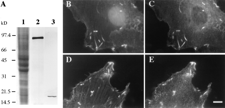
Since we suspected that severing proteins might accelerate actin turnover in Listeria tails, we examined the localization of two candidate actin-severing proteins, gelsolin and XAC, in _Listeria_-infected Xenopus tissue culture cells (XL 177). Antibodies were raised to the COOH-terminal half of Xenopus laevis gelsolin (Ankenbauer et al., 1988) and to the full-length XAC 2 protein (Abe et al., 1996) and affinity purified. Since XAC 1 and 2 differ by only four amino acids, we made only antibodies to XAC 2, which should also recognize XAC 1. Immunoblots of XL 177 lysate show that antibodies to gelsolin and XAC recognize a single band of the expected molecular mass in both cases (Fig. 1 A, lanes 2 and 3). Fluorescein-labeled phalloidin and affinity-purified antibodies to XAC or gelsolin were used to visualize the intracellular distributions of F-actin (Fig. 1, B and D), gelsolin (Fig. 1 C), and XAC (Fig. 1 E) in XL 177 cells infected with Listeria monocytogenes. Both XAC and gelsolin colocalize with the F-actin staining in the Listeria tails. Both gelsolin and XAC antibodies also give punctate staining throughout the rest of the cell. In addition, gelsolin typically stained coincidentally with F-actin in the stress fibers of the cell, whereas XAC does not.
Figure 1.
Localization of XAC and gelsolin in _Listeria_-infected XL 177 cells. (A) Western blot analysis. Lane 1, Coomassie blue–stained SDS-PAGE gel of total XL 177 cell lysate; lane 2, XL 177 lysate probed with anti-Xenopus gelsolin antibody; lane 3, XL 177 lysate probed with anti-XAC antibody. Immunostaining of _Listeria_-infected XL 177 cells with gelsolin antibody (C) or XAC antibody (E). F-actin in B and D is visualized with fluorescein-phalloidin. Both gelsolin and XAC colocalize with F-actin in Listeria tails. Bar, 10 μm.
XAC Is Required for the Depolymerization of Actin in Listeria Tails
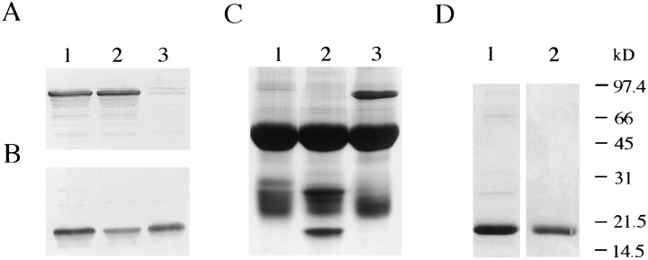
To determine the function of gelsolin and XAC in Listeria tails, we compared Listeria tails in mock-depleted Xenopus egg extracts to those in extracts depleted of XAC or gelsolin. Using our antibodies complexed to protein A beads, we were able to remove the bulk of either protein from Xenopus egg extracts. Quantitative immunoblots show that ∼75% of XAC (Fig. 2 B, lane 2) and >95% of gelsolin (Fig. 2 A, lane 3) were depleted from extracts compared to random rabbit IgG–depleted extracts (Fig. 2, A and B, lane 1). The XAC antibody precipitated XAC (19 kD) and an unknown band with an approximate molecular mass of 28 kD (Fig. 2 C, lane 2). The gelsolin antibody precipitated only gelsolin (∼93 kD) (Fig. 2 C, lane 3) when compared to the rabbit IgG control pellet (lane 1).
Figure 2.
Gels of immunodepletion of XAC and gelsolin from Xenopus laevis egg extracts and purified XAC and ADF. (A and B) Immunoblots of immunodepleted extracts using antibodies to gelsolin (A) and XAC (B). For both A and B, lane 1 is the IgG-depleted control, lane 2 is the XAC-depleted extract, and lane 3 is the gelsolin-depleted extract. Quantitation of the depletion was performed by densitometry of the bands in A and B compared to a dilution series of pure extract. (C) Coomassie blue–stained SDS-PAGE gel of immunoprecipitated complexes with XAC and gelsolin antibodies. Lane 1, the heavy and light chain of random rabbit IgG alone; lane 2, XAC (19 kD) and another band at approximately 28 kD over the IgG heavy and light chain bands; lane 3, gelsolin (∼93 kD) over the IgG bands. (D) Coomassie blue–stained SDS-PAGE shows the purity of the recombinant XAC and chicken ADF mutant that were added back to the XAC immunodepletions. Lane 1, wild-type XAC; lane 2, S3E ADF. Apparent molecular mass markers for all gels are indicated on the right.
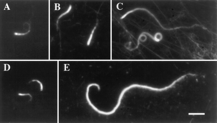
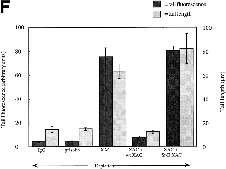
The IgG control depletion (Fig. 3 A) produced Listeria tails of the same length as untreated extract (15 ± 2.5 and 18 ± 1.5 μm, respectively). The tails in the gelsolin- depleted extracts (Fig. 3 B) were the same length and contained the same polymer mass as those of the IgG- depleted extracts (Fig. 3 A). However, the tails formed in the XAC-depleted extracts (Fig. 3 C) were on the average four to six times longer and displayed 13–21-fold more total fluorescence than those of the control extracts. Addback of pure recombinant XAC (Fig. 3 D) or chicken ADF to approximately endogenous concentrations (2.7 μM) rescued the long tail phenotype (12.3 ± 1.4 and 11.4 ± 1.6 μm, respectively). Addition of the same amount of a recombinant mutant version of ADF (S3E ADF) (Fig. 3 E), which behaves like constitutively phosphorylated ADF and has ∼10% of wild-type ADF activity on pure actin (data not shown), does not rescue the long tail phenotype. The purity of the proteins used in the addback experiment (Fig. 2 D, lanes 1 and 2) and the inability of the S3E ADF to rescue the XAC depletion phenotype strongly suggest that the observed long tail phenotype is due to the removal of XAC. The graph in Fig. 3 F shows the quantitation of the tail lengths and the amounts of tail fluorescence of the different phenotypes and provides quantitative support for our conclusion that increased tail length is due to XAC depletion, and not depletion of some other protein. These experiments demonstrate that XAC is required for the rapid depolymerization of actin filaments in Listeria tails in Xenopus extracts. While gelsolin is also concentrated within these tails, it does not appear to be essential for the depolymerization of Listeria tail actin.
Figure 3.
Listeria tail formed in XAC- or gelsolin-depleted Xenopus egg extracts. Actin tails are visualized by mixing rhodaminelabeled actin to Listeria and the following extracts: (A) IgG- depleted extracts, (B) gelsolin-depleted extracts, (C) XAC- depleted extracts, (D) XAC-depleted extracts plus 2.7 μM pure wild-type XAC, and (E) XAC-depleted extracts plus 2.7 μM pure S3E ADF. (F) Bar graph quantitating the amount of tail fluorescence (left side, ▒⃞ ▒⃞ ) and tail length (right side, ░⃞ ) using Winview software. The number of tails analyzed for IgG depletion was 33, for gelsolin depletion, 29, for XAC depletion, 43, for wildtype XAC addback, 25, and for S3E ADF addback, 11, over seven experiments using two separate extracts preps. Error bars represent standard deviation of the mean. Bar, 10 μm.
Addition of Excess XAC Decreases Listeria Tail Length
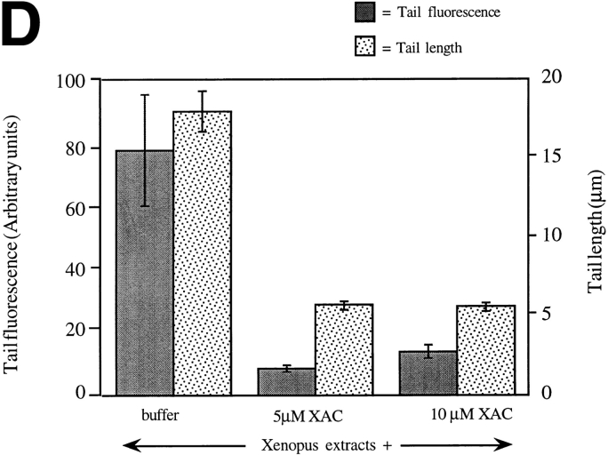
Quantitative immunoblots of Xenopus egg extracts using XAC antibodies and bacterially expressed XAC as a standard revealed that XAC is present at ∼2.1 μM in extracts (data not shown). Addition of recombinant XAC to a final concentration of 7.1 μM in the extract (Fig. 4 B) produced tails that were ∼0.33 times the length and had ninefold less total fluorescence than tails in a control extract (Fig. 4 D). Doubling the amount of XAC added to the Listeria assay to give a final concentration of 12.1 μM did not result in any further decrease in tail length (Fig. 4, C and D). When XAC was added to concentrations above 12.1 μM final concentration, few tails formed. Instead, rodlike structures containing rhodamine actin could be seen throughout the extract (data not shown). These rods appear analogous to those seen when actin and cofilin are concentrated in the nucleus upon heat shock or DMSO treatment to cells (Nishida et al., 1987; Ono et al., 1993) and probably represent a nonfilamentous coaggregate of XAC and actin. It is likely that few tails can form in such high concentrations of XAC since the actin required for Listeria tail formation may be sequestered in these XAC/actin rodlike aggregates. Thus, a Listeria tail segment of ∼5 μm is resistant to XAC depolymerization even when XAC is added up to nearly saturating concentrations.
Figure 4.
Listeria tails resulting from addition of excess XAC to Xenopus egg extracts. (A) Actin tail resulting from adding extract buffer to Xenopus egg extracts. (B) Actin tails resulting from adding wild-type XAC to extracts to 7.1 μM. (C) Actin tails resulting from adding wild-type XAC to 12.1 μM. (D) Quantitative analysis of the amount of tail fluorescence (left side, ▒⃞ ░⃞ ) and tail length (right side, ░⃞ ) using Winview software. The fluorescence units differ from those in Fig. 3 because the images were analyzed at different magnifications. The number of tails analyzed for buffer alone addition was 15, for XAC addition to 7.1 μM, 29, and for XAC addition to 12.1 μM, 33, over three experiments using two separate extract preps. Error bars represent standard deviation of the mean. Bar, 10 μm.
The Effect of XAC on Listeria Movement Rate
To determine whether the addition or depletion of XAC or gelsolin had an effect on the rate of actin polymerization, we measured the rate of Listeria movement in extracts either depleted of XAC or gelsolin, or containing additional XAC (Fig. 5). We used Listeria movement as an assay since the movement rate reflects the actin polymerization rate at the bacterial surface (Sanger et al., 1992; Theriot et al., 1992). The rate of Listeria movement in the XAC depleted extracts did not vary greatly from the mock-depleted extracts. The lack of effect that XAC-depletion had on actin assembly rates may indicate that XAC is not involved in actin polymerization. However, we cannot rule out such an involvement since only 75% of the XAC could be removed from the extracts with our reagents. Depletion of gelsolin increased the rate of Listeria movement by ∼1 μm/min (∼20%). The manipulations required for depletion slowed Listeria movement by ∼1 μm/min (compare IgG-depleted to addition of buffer). This may be due to a decrease of ATP, dilution of actin, or other factors during the depletion procedure (2–3 h at 4°C). Addition of XAC to 5.0 μM seemed to increase the rate of polymerization by ∼1 μm/min compared to when buffer alone is added, despite the shorter tails produced by this concentration (Fig. 4 B). These slight differences cannot account for the large changes in tail length and total fluorescence upon XAC depletion. Thus, the effects of XAC on actin depolymerization greatly outweigh those upon polymerization.
Figure 5.
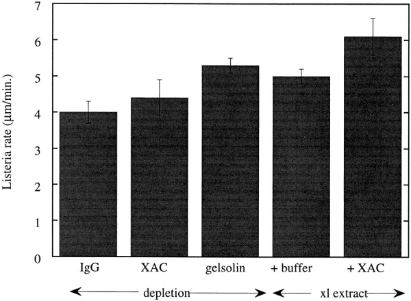
Bar graph representing analysis of Listeria movement rate during various extract treatments. The rates were measured using Image 1 software or Winview software. The number of tails measured for each treatment were: IgG depletion, 25; XAC depletion, 24; gelsolin depletion, 34; buffer addition, 27, and XAC addition (to 5.0 μM), 24, over four experiments using two separate extract preps. Error bars represent standard deviation of the mean.
XAC Depolymerization Activity Depends upon the Nucleotide Content of the Filament
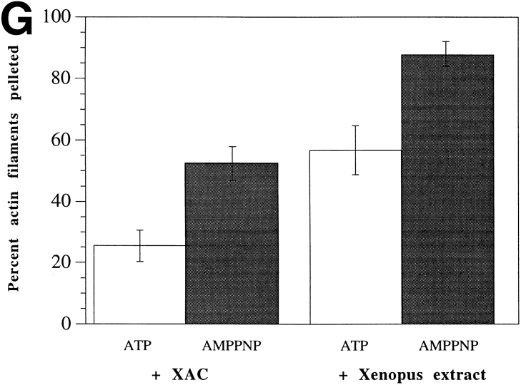
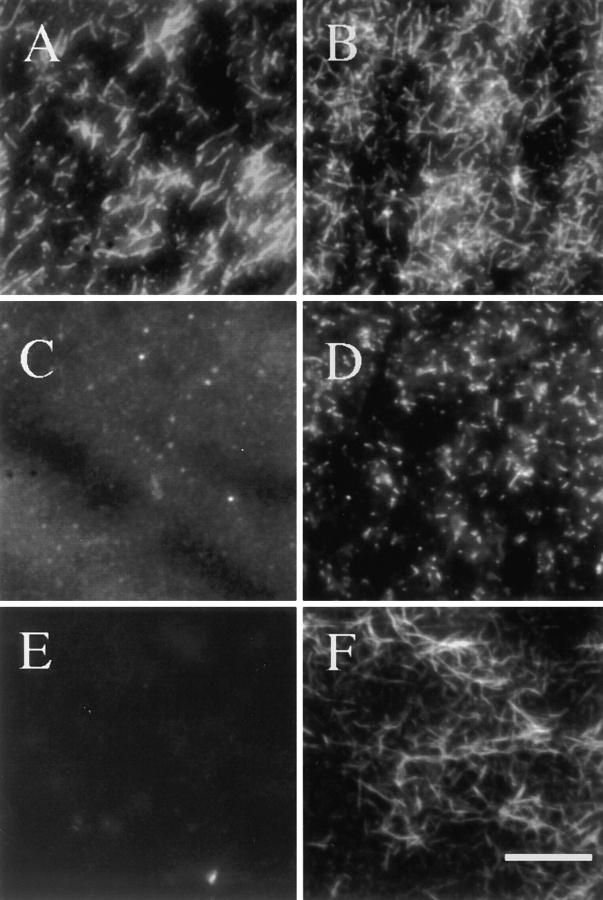
Upon polymerization, actin hydrolyzes its bound ATP and the terminal phosphate is slowly released. It has been suggested that the loss of the terminal phosphate could serve as a clock that regulates the lifetime of a filament by controlling the activity of ADF/cofilin proteins (Maciver et al., 1991; Moon and Drubin, 1995). To test this hypothesis, we made rhodamine-labeled actin filaments containing AMPPNP, a slowly hydrolyzable ATP analog, and tested their resistance to the depolymerizing activities of Xenopus egg extracts and purified XAC. Rhodamine ATP-actin filaments depolymerize rapidly in the presence of XAC (Fig. 6 C) and Xenopus egg extract (Fig. 6 E) compared to buffer (Fig. 6 A). In contrast, rhodamine-labeled AMPPNP filaments depolymerized to a limited extent in XAC (Fig. 6 D) or Xenopus egg extract (Fig. 6 F) compared to in buffer alone (Fig. 6 B). Thus, AMPPNP filaments were more stable to depolymerization in XAC or extract (Fig 6, D and F) compared to ATP filaments under the same conditions (Fig. 6, C and E). To quantitate actin filament depolymerization, rhodamine-labeled filaments were added to Xenopus extracts or purified XAC and total polymerized actin was recovered by sedimentation. The fraction of the added actin left in polymer was determined by resuspending the pellets in SDS and quantitating the fluorescence with a fluorimeter (Fig. 6 G). AMPPNP caused twice as much of the added F-actin to be recovered in the pellet fraction. Comparing Fig. 6, E and F, with G, we should note that the visual and sedimentation assays are measuring different parameters. If the labeled actin repolymerized into new filaments in the presence of extract, the filaments will not be visible to the CCD camera because they are diluted with endogenous actin (Fig. 6, E and F). However the diluted actin will still sediment, giving rise to the high fraction of label in the pellet fraction in the presence of extract (Fig. 6 G, Xenopus extract). Both types of assay demonstrate that the AMPPNP filaments are relatively resistant to depolymerization by pure XAC and total extract. Two other slowly hydrolyzable ATP analogs, ATPγS and AMPPCP, showed similar stabilizing effects (data not shown), suggesting that the actin stability is due to the state of the bound nucleotide rather than nonspecific effects on actin structure. The results from both the microscopy and the fluorimetry assays suggest that the nucleotide content of actin filaments regulates XAC.
Figure 6.
The resistance of AMPPNP–containing actin filaments to the depolymerizing activities of XAC and Xenopus egg extracts. Approximately 0.5 μM rhodamine-labeled actin polymerized with either ATP (A, C, and E) or AMPPNP (B, D, and F). Actin filaments mixed 1:1 with F-buffer (A and B), with 5.3 μM XAC (C and D), and with Xenopus extract (E and F). In both cases (C–F), XAC is in excess of rhodamine F-actin by approximately fivefold. (G) Quantitation of the amount of rhodamine- labeled ATP (□) versus AMPPNP ( ▒⃞ ░⃞ ) F-actin pelleted in the presence of XAC or Xenopus egg extracts. Percent actin filaments remaining was calculated as the fluorescence of the rhodamine F-actin pelleted in XAC or extracts/the fluorescence of rhodamine F-actin pelleted in F-buffer. The bars represent an average of four experiments and the error bars represent standard deviation of the mean. Bar, 10 μm.
Incorporation of AMPPNP into actin filaments in these experiments was modest; ∼12% of the ATP-binding sites in actin filaments incorporated AMPPNP, and the remainder contained ADP. By contrast, ATP actin filaments contained approximately only 2% ATP and 98% ADP. Despite low incorporation, ∼1.5 and 2 times as much AMPPNP-containing actin filaments as ATP-containing actin filaments pelleted in the presence of Xenopus extract or pure XAC, respectively.
Discussion
We have used the ability to reconstitute Listeria motility in Xenopus egg extracts to test directly the role of the gelsolin and ADF/cofilin proteins in the promotion of actin filament depolymerization. We have raised specific antibodies to gelsolin and to XAC, the major ADF/cofilin protein known to be present in Xenopus egg extracts. Using immunodepletions and adding back purified proteins, our studies demonstrate that ADF/cofilin is the major factor responsible for the rapid turnover of actin filaments in Listeria tails. Removal of 75% of the ADF/cofilin from extracts considerably lengthened Listeria tails and greatly increased the total actin polymer mass present in the tails. Both tail length and actin polymer mass in tails were restored to control levels by adding back pure XAC at physiological concentrations to XAC-depleted extracts. In addition, adding excess XAC shortens Listeria tails and reduces the polymer mass in the tail, and AMPPNP actin is resistant to depolymerization by both XAC and whole extracts. Taken together, these data strongly implicate XAC as a central component of the machinery responsible for rapid actin filament turnover in extracts. Although we have no direct evidence, we predict that ADF/cofilin proteins are required for the dynamic organization of populations of actin filaments within living cells that turn over rapidly, such as the filaments within lamellipodia. Three arguments support our prediction: Xenopus egg extracts can support the actin polymerization and depolymerization in Listeria tails at rates comparable to those in intact cells and suggest they reflect the actin dynamics within cell cytoplasm. XAC is required to maintain the turnover of actin filaments in these tails in extracts. Because ADF/cofilin proteins are essential in every species in which they have been found, we may infer that they are required for essential processes like rapid actin turnover in the cell. Finally, since XAC is concentrated in Listeria tails and the leading edge of cells, we suspect it is also required for rapid filament turnover in vivo.
In contrast to XAC, gelsolin depletions had no significant effect on Listeria tail length or tail polymer mass, leaving the functional role of gelsolin in the regulation of actin dynamics an open question. Since gelsolin does not affect the depolymerization of Listeria tails, its concentration in these tails is curious. Perhaps gelsolin is concentrated in these tails strictly because of its actin binding activity, or by another of its activities such as actin capping. Although the Xenopus egg extracts used in our assays contain 5 mM EGTA, it is difficult to analyze any local concentrations of Ca2+ that may be due to vesicle release. Since Ca2+ is required for gelsolin activity, it is possible that the conditions in our cell-free egg extracts are not able to support the activity of gelsolin. Therefore, we cannot rule out a role for gelsolin in tail dynamics in vivo. However, the presence of gelsolin in Listeria tails does not greatly affect actin assembly or disassembly in Listeria tails in Xenopus extracts.
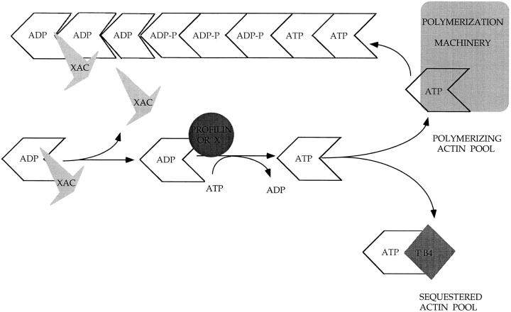
In our addition of varying amounts of excess XAC to extracts until all the actin was driven into abnormal rod structures, Listeria tails shrank to ∼5 μm but no further. The persistence of a resistant tail segment in up to sixfold the normal concentrations of XAC suggests that an additional factor may control the extent of actin depolymerization by XAC. Several investigators have suggested that the energy of ATP hydrolysis could be used to regulate the lifetime of a filament (Pollard, 1986; Carlier, 1988; Maciver et al., 1991; Moon and Drubin, 1995). In vitro studies of actin filament assembly have shown that upon polymerization of ATP-actin, the bound ATP is hydrolyzed and the terminal phosphate is slowly released (Carlier, 1987). This slow rate of phosphate loss relative to the polymerization rate should then produce filaments that contain a segment of ADP + inorganic phosphate (ADP.Pi). Since ATP- and ADP.Pi-bound actin are known to make stronger intersubunit bonds than ADP-actin (Carlier, 1991; Carlier et al., 1985), phosphate release could play a role in regulating actin filament stability. However, since in vitro filaments depolymerize very slowly, phosphate release alone is not sufficient to direct rapid disassembly of actin filaments in the cell. In vivo, perhaps the role of slow phosphate release in actin is to regulate either the binding or activity of actin-severing proteins. Severing proteins such as ADF and cofilin may preferentially sever the ADP subunits of actin filaments while the ATP and ADP.Pi subunits are resistant to severing (Fig. 7). In support of this, the depolymerizing activity of actophorin, an Acanthamoeba member of the ADF/cofilin family, can be inhibited by addition of 25 mM phosphate, which presumably mimics ADP.Pi filaments (Maciver et al., 1991). In addition, actophorin has been shown to bind tightly to ADPcontaining G-actin and weakly to ATP-actin (Maciver and Weeds, 1994). However, other studies show that chick ADF has a higher affinity for ATP-actin than for ADP-actin (Hayden et al., 1993) and have left the role of the bound nucleotide in regulating depolymerization in other species an open question. Our results showing the resistance of AMPPNP-containing actin filaments to the depolymerizing activities of both XAC and concentrated Xenopus egg extracts lend support to the idea that the nucleotide content of an actin filament controls filament lifetime by regulating the activity of ADF/cofilin family members.
Figure 7.
Model for recycling of actin by ADF/cofilin proteins. At areas of high filament turnover in the cell, ATP-bound actin is induced to polymerize by a complex of proteins. The ATP within the filament is hydrolyzed and the terminal phosphate is slowly released. The resulting ADP-containing subunits have weaker interactions with each other than those containing ATP. These subunits are now available for depolymerization by the ADF/cofilin family of proteins (XAC). Once XAC depolymerizes an actin subunit(s), it dissociates from actin. The released actin must exchange its ADP for ATP and the nucleotide exchange is probably catalyzed by profilin. The ATP-bound actin is then either repolymerized or sequestered for later use.
What is the mechanism of actin depolymerization by XAC? XAC could depolymerize by end-wise removal of subunits, by severing, or by both mechanisms. We interpret our results with AMPPNP actin as favoring the endwise mechanism. We reason that a severing protein would not be greatly affected by an actin filament with only 12% of its subunits substituted with AMPPNP. By contrast, an end-wise depolymerizing protein would be blocked whenever AMPPNP subunits were present at the filament ends. Thus, the large inhibition of depolymerization by AMPPNP filaments may be accounted for if XAC primarily depolymerized using an end-wise mechanism. The most direct way of distinguishing severing and end-wise mechanisms will come from imaging depolymerization by ADF/ cofilin proteins.
On the basis of our findings, we can postulate a model for how XAC recycles actin subunits in the Listeria tail (Fig. 7). A complex of proteins at the back of Listeria (Welch et al., 1997) induces polymerization of ATP-bound actin. Once actin polymerizes, the bound ATP is hydrolyzed and the terminal phosphate is slowly released. The resulting ADP-containing actin subunits interact more weakly within the filament than the ATP subunits and allow binding of XAC. XAC either depolymerizes single subunits or short fragments of filament. XAC is then released from the depolymerized actin subunit and recycled for another round of depolymerization. The ADP in the depolymerized actin is then exchanged for ATP by bulk mass or by catalysis from profilin, and this subunit is now available for another round of polymerization or remains unpolymerized by binding thymosin β4 or other sequestering proteins.
This model provides a framework for understanding how an actin subunit is recycled from one round of polymerization to the next in regions of the cell that rapidly turn over actin filaments. Clearly, many questions remain regarding the details of this model. Future work will need to address whether XAC works primarily by severing or end-wise mechanisms in the cell. We are currently examining how XAC is recycled for another round of depolymerization after it is bound to the actin subunit and the role that XAC phosphorylation may play in its recycling. Other studies will need to focus on how the actin subunit is recycled for repolymerization. Analysis of the nucleotide content of actin has revealed that ∼90% of unpolymerized actin in the cell is bound to ATP (Rosenblatt et al., 1995), suggesting that the actin nucleotide is exchanged early in the pathway. Information about whether the ATP- or ADP-bound actin is used for polymerization is still lacking. The 10% of actin that is ADP bound could be bound to XAC. This population of actin could undergo nucleotide exchange and be used directly for another round of polymerization, leaving the remaining 90% of actin sequestered from polymerization. However, the lack of an effect that XAC depletion has on the rate of polymerization at early time points would suggest that this is not the case. Future studies will need to address what population of actin is used for polymerization.
Acknowledgments
We thank the entire Mitchison lab for providing a stimulating and supportive environment and many great dinners. In particular, we thank Aneil Mallavarapu, Arshad Desai, and Jason Swedlow for help with computers and microscopes, Matt Welch for performing Listeria infections, Paul Peluso (University of California at San Francisco, San Francisco, CA) for help with the fluorimeter, and Claire Walczak for advice on cloning and bacterial protein expression. We are indebted to Matt Welch and especially Arshad Desai and Michael Redd (University of California at San Francisco, San Francisco, CA) for critically reading this manuscript and for stimulating conversations. We are grateful to Jeremy Minshull (Maxygen Corp., Santa Clara, CA) for giving us a Xenopus egg cDNA library and to Dr. Werner W. Franke (German Cancer Research Center, Heidelberg, Germany) for giving us X.l. gelsolin antibodies used for preliminary experiments. We also thank Tom Pollard (The Salk Institute, La Jolla, CA) who early on suggested depleting ADF/cofilin from Xenopus extracts.
This work was supported by a National Science Foundation predoctoral fellowship to J. Rosenblatt and National Institutes of Health grants (GM35126) to J.R. Bamburg and (GM48027) to T.J. Mitchison.
Abbreviations used in this paper
ADF
actin depolymerizing factor
ADP.Pi
ADP + inorganic phosphate
AMPPNP
5′adenylylamido-diphosphate
F-actin
filamentous actin
XAC
Xenopus ADF/Cofilin
Footnotes
This work is dedicated to Michael Redd and our baby, Nadja.
Address all correspondence to Jody Rosenblatt, Department of Biochemistry, University of California, San Francisco, San Francisco, CA 94143. Tel.: (415) 476-4002. Fax: (415) 476-5233.
References
- Abe H, Obinata T, Minamide LS, Bamburg JR. Xenopus laevisactin-depolymerizing factor/cofilin: a phosphorylation-regulated protein essential for development. J Cell Biol. 1996;132:871–885. doi: 10.1083/jcb.132.5.871. [DOI] [PMC free article] [PubMed] [Google Scholar]
- Agnew BJ, Minamide LS, Bamburg JR. Reactivation of phosphorylated actin depolymerizing factor and identification of the regulatory site. J Biol Chem. 1995;270:17582–17587. doi: 10.1074/jbc.270.29.17582. [DOI] [PubMed] [Google Scholar]
- Ampe C, Vandekerckhove J. The F-actin capping proteins of Physarum polycephalum: cap42(a) is very similar, if not identical, to fragmin and is structurally and functionally very homologous to gelsolin; cap42(b) is Physarum actin. EMBO (Eur Mol Biol Organ) J. 1987;6:4149–4157. doi: 10.1002/j.1460-2075.1987.tb02761.x. [DOI] [PMC free article] [PubMed] [Google Scholar]
- André E, Lottspeich F, Schleicher M, Noegel A. Severin, gelsolin, and villin share a homologous sequence in regions presumed to contain F-actin severing domains. J Biol Chem. 1988;263:722–727. [PubMed] [Google Scholar]
- André E, Brink M, Gerisch G, Isenberg G, Noegel A, Schleicher M, Segall JE, Wallraff E. A Dictyosteliummutant deficient in severin, an F-actin fragmenting protein, shows normal motility and chemotaxis. J Cell Biol. 1989;108:985–995. doi: 10.1083/jcb.108.3.985. [DOI] [PMC free article] [PubMed] [Google Scholar]
- Ankenbauer T, Kleinschmidt JA, Vandekerckhove J, Franke WW. Proteins regulating actin assembly in oogenesis and early embryogenesis of Xenopus laevis: gelsolin is the major cytoplasmic actin-binding protein. J Cell Biol. 1988;107:1489–1498. doi: 10.1083/jcb.107.4.1489. [DOI] [PMC free article] [PubMed] [Google Scholar]
- Bamburg, J.R., and B.W. Berstein. 1991. Actin and actin-binding proteins in neurons. In The Neuronal Cytoskeleton. R.D. Burgeyne, editor. Wiley-Liss, New York. 121–160.
- Bamburg JR, Harris HE, Weeds AG. Partial purification and characterization of an actin depolymerizing factor from brain. FEBS Lett. 1980;121:178–182. doi: 10.1016/0014-5793(80)81292-0. [DOI] [PubMed] [Google Scholar]
- Bernstein BW, Bamburg JR. Tropomyosin binding to F-actin protects the F-actin from disassembly by brain actin-depolymerizing factor (ADF) Cell Motil. 1982;2:1–8. doi: 10.1002/cm.970020102. [DOI] [PubMed] [Google Scholar]
- Bray D, White JG. Cortical flow in animal cells. Science (Wash DC) 1988;239:883–888. doi: 10.1126/science.3277283. [DOI] [PubMed] [Google Scholar]
- Carlier MF. Measurement of Pi dissociation from actin filaments following ATP hydrolysis using a linked enzyme assay. Biochem Biophys Res Commun. 1987;143:1069–75. doi: 10.1016/0006-291x(87)90361-5. [DOI] [PubMed] [Google Scholar]
- Carlier MF. Role of nucleotide hydrolysis in the polymerization of actin and tubulin. Cell Biophys. 1988;12:105–117. doi: 10.1007/BF02918353. [DOI] [PubMed] [Google Scholar]
- Carlier MF. Nucleotide hydrolysis in cytoskeletal assembly. Curr Opin Cell Biol. 1991;3:12–17. doi: 10.1016/0955-0674(91)90160-z. [DOI] [PubMed] [Google Scholar]
- Carlier MF, Pantaloni D, Korn ED. Polymerization of ADP-actin and ATP-actin under sonication and characteristics of the ATP-actin equilibrium polymer. J Biol Chem. 1985;260:6565–6571. [PubMed] [Google Scholar]
- Cooper JA. The role of actin polymerization in cell motility. Annu Rev Physiol. 1991;53:585–605. doi: 10.1146/annurev.ph.53.030191.003101. [DOI] [PubMed] [Google Scholar]
- Gunsalus KC, Bonaccorsi S, Williams E, Verni F, Gatti M, Goldberg ML. Mutations in twinstar, a Drosophilagene encoding a cofilin/ADF homologue, result in defects in centrosome migration and cytokinesis. J Cell Biol. 1995;131:1243–1259. doi: 10.1083/jcb.131.5.1243. [DOI] [PMC free article] [PubMed] [Google Scholar]
- Harlow, E., and D. Lane. 1988. Antibodies: A Laboratory Manual. Cold Spring Harbor Laboratory, Cold Spring Harbor, NY. 1–726.
- Hatanaka H, Ogura K, Moriyama K, Ichikawa S, Yahara I, Inagaki F. Tertiary structure of destrin and structural similarity between two actin-regulating protein families. Cell. 1996;85:1047–1055. doi: 10.1016/s0092-8674(00)81305-7. [DOI] [PubMed] [Google Scholar]
- Hawkins M, Pope B, Maciver SK, Weeds AG. Human actin depolymerizing factor mediates a pH-sensitive destruction of actin filaments. Biochemistry. 1993;32:9985–9993. doi: 10.1021/bi00089a014. [DOI] [PubMed] [Google Scholar]
- Hayden SM, Miller PS, Brauweiler A, Bamburg JR. Analysis of the interactions of actin depolymerizing factor with G- and F-actin. Biochemistry. 1993;32:9994–10004. doi: 10.1021/bi00089a015. [DOI] [PubMed] [Google Scholar]
- Janmey PA, Stossel TP. Modulation of gelsolin function by phosphatidylinositol 4,5-bisphosphate. Nature (Lond) 1987;325:362–364. doi: 10.1038/325362a0. [DOI] [PubMed] [Google Scholar]
- Kinoshita N, Minshull J, Kirschner MW. The identification of two novel ligands of the FGF receptor by a yeast screening method and their activity in Xenopusdevelopment. Cell. 1995;83:621–630. doi: 10.1016/0092-8674(95)90102-7. [DOI] [PubMed] [Google Scholar]
- Leimeister-Wachter M, Chakraborty T. Detection of listeriolysin, the thiol-dependent hemolysin in Listeria monocytogenes, Listeria ivanovii, and Listeria seeligeri. . Infect Immun. 1989;57:2350–2357. doi: 10.1128/iai.57.8.2350-2357.1989. [DOI] [PMC free article] [PubMed] [Google Scholar]
- Maciver SK, Weeds AG. Actophorin preferentially binds monomeric ADP-actin over ATP-bound actin: consequences for cell locomotion. FEBS Lett. 1994;347:251–256. doi: 10.1016/0014-5793(94)00552-4. [DOI] [PubMed] [Google Scholar]
- Maciver SK, Zot HG, Pollard TD. Characterization of actin filament severing by actophorin from Acanthamoeba castellanii. . J Cell Biol. 1991;115:1611–1620. doi: 10.1083/jcb.115.6.1611. [DOI] [PMC free article] [PubMed] [Google Scholar]
- McKim KS, Matheson C, Marra MA, Wakarchuk MF, Baillie DL. The Caenorhabditis elegansunc-60 gene encodes proteins homologous to a family of actin-binding proteins. Mol Gen Genet. 1994;242:346–357. doi: 10.1007/BF00280425. [DOI] [PubMed] [Google Scholar]
- Mitchison TJ, Cramer LP. Actin based cell motility and cell locomotion. Cell. 1996;84:371–379. doi: 10.1016/s0092-8674(00)81281-7. [DOI] [PubMed] [Google Scholar]
- Moon A, Drubin DG. The ADF/cofilin proteins: stimulus-responsive modulators of actin dynamics. Mol Biol Cell. 1995;6:1423–1431. doi: 10.1091/mbc.6.11.1423. [DOI] [PMC free article] [PubMed] [Google Scholar]
- Moon AL, Janmey PA, Louie KA, Drubin DG. Cofilin is an essential component of the yeast cortical cytoskeleton. J Cell Biol. 1993;120:421–435. doi: 10.1083/jcb.120.2.421. [DOI] [PMC free article] [PubMed] [Google Scholar]
- Morgan TE, Lockerbie RO, Minamide LS, Browning MD, Bamburg JR. Isolation and characterization of a regulated form of actin depolymerizing factor. J Cell Biol. 1993;122:623–633. doi: 10.1083/jcb.122.3.623. [DOI] [PMC free article] [PubMed] [Google Scholar]
- Moriyama K, Iida K, Yahara I. Phosphorylation of Ser-3 at cofilin regulates its essential function on actin. Genes to Cells. 1996;1:73–86. doi: 10.1046/j.1365-2443.1996.05005.x. [DOI] [PubMed] [Google Scholar]
- Nagaoka R, Abe H, Kusano K, Obinata T. Concentration of cofilin, a small actin-binding protein, at the cleavage furrow during cytokinesis. Cell Motil Cytoskel. 1995;30:1–7. doi: 10.1002/cm.970300102. [DOI] [PubMed] [Google Scholar]
- Nishida E, Maekawa S, Sakai H. Cofilin, a protein in porcine brain that binds to actin filaments and inhibits their interactions with myosin and tropomyosin. Biochemistry. 1984;23:5307–5313. doi: 10.1021/bi00317a032. [DOI] [PubMed] [Google Scholar]
- Nishida E, Iida K, Yonezawa N, Koyasu S, Yahara I, Sakai H. Cofilin is a component of intranuclear and cytoplasmic actin rods induced in cultured cells. Proc Natl Acad Sci USA. 1987;84:5262–5266. doi: 10.1073/pnas.84.15.5262. [DOI] [PMC free article] [PubMed] [Google Scholar]
- Ono S, Abe H, Nagaoka R, Obinata T. Colocalization of ADF and cofilin in intranuclear actin rods of cultured muscle cells. J Muscle Res Cell Motil. 1993;14:195–204. doi: 10.1007/BF00115454. [DOI] [PubMed] [Google Scholar]
- Pollard TD. Rate constants for the reactions of ATP- and ADP-actin with the ends of actin filaments. J Cell Biol. 1986;103:2747–2754. doi: 10.1083/jcb.103.6.2747. [DOI] [PMC free article] [PubMed] [Google Scholar]
- Pringault E, Arpin M, Garcia A, Finidori J, Louvard D. A human villin cDNA clone to investigate the differentiation of intestinal and kidney cells in vivo and in culture. EMBO (Eur Mol Biol Organ) J. 1986;5:3119–3124. doi: 10.1002/j.1460-2075.1986.tb04618.x. [DOI] [PMC free article] [PubMed] [Google Scholar]
- Rodriguez D, Castillo A, Lemaire S, Tchakarov L, Jeyapragasan M, Doucet JP, Vitale ML, Trifaró JM. Chromaffin cell scinderin, a novel calcium-dependent actin filament-severing protein. EMBO (Eur Mol Biol Organ) J. 1990;9:43–52. doi: 10.1002/j.1460-2075.1990.tb08078.x. [DOI] [PMC free article] [PubMed] [Google Scholar]
- Rosenblatt J, Peluso P, Mitchison TJ. The bulk of unpolymerized actin in Xenopusegg extracts is ATP-bound. Mol Biol Cell. 1995;6:227–236. doi: 10.1091/mbc.6.2.227. [DOI] [PMC free article] [PubMed] [Google Scholar]
- Sakurai T, Ohmi K, Kurokawa H, Nonomura Y. Distribution of a gelsolin-like 74,000 mol. wt protein in neural and endocrine tissues. Neuroscience. 1990;38:743–756. doi: 10.1016/0306-4522(90)90067-e. [DOI] [PubMed] [Google Scholar]
- Sanger JM, Sanger JW, Southwick FS. Host cell actin assembly is necessary and likely to provide the propulsive force for intracellular movement of Listeria monocytogenes. . Infect Immun. 1992;60:3609–3619. doi: 10.1128/iai.60.9.3609-3619.1992. [DOI] [PMC free article] [PubMed] [Google Scholar]
- Small JV, Herzog M, Anderson K. Actin filament organization in the fish keratocyte lamellipodium. J Cell Biol. 1995;129:1275–1286. doi: 10.1083/jcb.129.5.1275. [DOI] [PMC free article] [PubMed] [Google Scholar]
- Smith DB, Johnson KS. Single-step purification of polypeptide expressed in Escherichia colias fusions with glutathion S-transferase. Gene. 1988;67:31–40. doi: 10.1016/0378-1119(88)90005-4. [DOI] [PubMed] [Google Scholar]
- Theriot JA, Mitchison TJ. Actin microfilament dynamics in locomoting cells. Nature (Lond) 1991;352:126–131. doi: 10.1038/352126a0. [DOI] [PubMed] [Google Scholar]
- Theriot JA, Mitchison TJ, Tilney LG, Portnoy DA. The rate of actin-based motility of intracellular Listeria monocytogenesequals the rate of actin polymerization. Nature (Lond) 1992;357:257–260. doi: 10.1038/357257a0. [DOI] [PubMed] [Google Scholar]
- Theriot JA, Rosenblatt J, Portnoy DA, Goldschmidt CP, Mitchison TJ. Involvement of profilin in the actin-based motility of L. monocytogenesin cells and in cell-free extracts. Cell. 1994;76:505–517. doi: 10.1016/0092-8674(94)90114-7. [DOI] [PubMed] [Google Scholar]
- Welch MD, Iwamamatsu A, Mitchison TJ. Actin polymerization is induced by the Arp 2/3 protein complex at the surface of Listeria monocytogenes. . Nature (Lond) 1997;385:265–269. doi: 10.1038/385265a0. [DOI] [PubMed] [Google Scholar]
- Witke W, Sharpe AH, Hartwig JH, Azuma T, Stossel TP, Kwiatkowski DJ. Hemostatic, inflammatory, and fibroblast responses are blunted in mice lacking gelsolin. Cell. 1995;81:41–51. doi: 10.1016/0092-8674(95)90369-0. [DOI] [PubMed] [Google Scholar]
- Yonezawa N, Nishida E, Sakai H. pH control of actin polymerization by cofilin. J Biol Chem. 1985;260:14410–14412. [PubMed] [Google Scholar]
- Yonezawa N, Nishida E, Iida K, Yahara I, Sakai H. Inhibition of the interactions of cofilin, destrin, and deoxyribonuclease I with actin by phosphoinositides. J Biol Chem. 1990;265:8382–8386. [PubMed] [Google Scholar]
- Zigmond SH. Recent quantitative studies of actin filament turnover during cell locomotion. Cell Motil Cytoskel. 1993;25:309–316. doi: 10.1002/cm.970250402. [DOI] [PubMed] [Google Scholar]
