Cellular and Multicellular Form and Function (original) (raw)
. Author manuscript; available in PMC: 2007 Dec 12.
Published in final edited form as: Adv Drug Deliv Rev. 2007 Aug 16;59(13):1319–1328. doi: 10.1016/j.addr.2007.08.011
Abstract
Engineering artificial tissue constructs requires the appropriate spatial arrangement of cells within scaffolds. The introduction of microengineering tools to the biological community has provided a valuable set of techniques to manipulate the cellular environment, and to examine how cell structure affects cellular function. Using micropatterning techniques, investigators have found that the geometric presentation of cell-matrix adhesions are important regulators of various cell behaviors including cell growth, proliferation, differentiation, polarity and migration. Furthermore, the presence of neighboring cells in multicellular aggregates has a significant impact on the proliferative and differentiated state of cells. Using microengineering tools, it will now be possible to manipulate the various environmental factors for practical applications such as engineering tissue constructs with greater control over the physical structure and spatial arrangement of cells within their surrounding microenvironment.
Introduction
The concept of engineering artificial tissues for organ replacement was first seeded over two decades ago. Efforts in tissue engineering have typically relied on the basic notion that cells can be isolated, expanded, cultured in an environment that fosters differentiated cell function, and finally re-implanted into the body in order to replace the organ in need. One of the most difficult obstacles facing tissue engineers is defining the conditions that direct cell behavior, and recapitulating these conditions in a suitable in vitro growth environment. Initial success has been achieved primarily in tissues of relatively simple architecture, including skin and cartilage. In comparison, engineering tissues with greater complexity such as liver or pancreas has been rather unsuccessful. These tissues comprise multiple cell types within a highly structured and defined organization. Reproducing this organization in an engineered tissue constrains the juxtaposition of specific cells with various gradients of soluble factors, specific extracellular matrix scaffolds, physical forces and other cell types, and therefore is likely to be required to recapitulate the signals that drive cell and tissue function.
Many soluble factors have been used in tissue engineering to initiate signaling cascades that lead to changes in cell growth, proliferation, migration, differentiation and survival. While numerous groups have incorporated such soluble factors in tissue engineering scaffolds to drive cell function, it is now clear that in addition to soluble cues, adhesive and physical factors also need to be controlled to engineer cell function. For example, chondrocytes that are seeded into collagen type II gels form cartilage, but the same cells plated onto petri dishes lose their phenotype. Similarly, hepatocytes rapidly de-differentiate in the absence of collagen I or homotypic adhesion to other hepatocytes. In addition, physical cues such as shear stress maintain endothelial cells in a quiescent, in vivo-like phenotype, whereas cells cultured in static conditions resemble cells in an inflammatory state. It is now thought that engineering these insoluble (adhesive and physical) factors into constructs may provide the means to understand how cell function is regulated through tissue architecture.
Simply combining cells with matrix proteins in culture is not sufficient for proper tissue function. Rather, a specified spatial arrangement of cells with their surrounding proteins and neighboring cells controls the degree to which cells are in contact with matrix versus neighboring cells, and this balance could be important to drive cell and tissue behavior. Examples of cell structure in vivo demonstrate the role of structure in regulating cell behavior and tissue function. For instance, endothelial cells form tubular vessel structures that are surrounded by a basement membrane and an underlying smooth muscle layer, components which together contribute to proper blood flow. Although structure is clearly important for the function of tissues, it has traditionally been difficult to engineer structure into artificial tissues.
While controlling soluble cues is relatively straightforward, only recently have experimental systems been developed that enable investigators to control structural cues from the physical environment, including the spatial arrangement of extracellular matrix (ECM) proteins and neighboring cells. Advances in microengineered tools allow the precise control of single- and multi-cellular structures. Importantly, using these technologies to study cells has revealed several key molecular mechanisms that relay structural cues into biological outputs. In this review, we will describe classical and recently developed microengineered tools, and then discuss how these tools are used to examine the relationship between structure and function in biological systems and to create artificial tissue constructs. These studies demonstrate that cell structure, both in single cells and in multicellular assemblies, has a profound influence on intracellular signaling and cell function.
Microfabrication tools to control (single and multi) cellular structure
Pioneering efforts to engineer cell and tissue structure focused on ways to control the presentation of extracellular matrix spatially on surfaces, and then use this ‘patterning’ template to control the position of cells deposited on the surface. Cells attach to the ECM through an array of discrete micrometer-scale focal adhesions. Numerous studies have suggested that the extracellular matrix regulates intracellular signaling pathways that control cell behavior through these focal adhesion assemblies. In fact, cellular adhesions are thought to play an important role in integrating signals from both the physical and chemical environments. Adhesions not only mechanically anchor cells to the matrix, but also localize a number of soluble factor receptors and associated intracellular signaling molecules to the sites of adhesion. Thus, these adhesions serve as centers of communication between signals from the environment and intracellular signaling pathways that control cellular behaviors such as migration, proliferation and differentiation. Conventional methods to control and study cellular interactions with ECM typically involve seeding cells on flat culture dishes that are homogeneously coated with ECMs of varied composition or concentration. More recently, microengineering techniques have been developed to control the structure or geometric presentation of such extracellular matrix (and therefore adhesions) on surfaces.
Tools commonly used in the semiconductor industry to control the placement of metals for circuits were adapted to control the positioning of ECM proteins on surfaces with micrometer-scale precision. Initial approaches to pattern materials created patterned surfaces by illuminating photosensitive materials with UV radiation through a mask patterned with opaque regions- a process called photolithography [1]. This technique was used to create substrates with micron-sized features that can be adhesive or nonadhesive to cells. More recently, entirely biocompatible methods have been developed based on a soft elastomer, polydimethylsiloxane (PDMS), to deliver protein [2]. In these methods, which are generally termed soft lithography, PDMS is used to create a stamp that is cast directly from photolithographically-generated silicon master patterns containing topographic micron-sized features. Once cured, the patterned PDMS substrates are used either as cell culture surfaces or as stamps to deposit adhesive proteins onto various tissue culture surfaces [3](Figure 1A). Alternatively, nonadhesive materials such as agarose or polyacrylamide can be patterned by flowing the polymer solution between a PDMS stamps and substrate [4]. Upon removal of the stamp, the exposed regions are coated with ECM protein. Using these soft lithography-based methods, the location of one or multiple adhesive proteins can be directly positioned on the surface. Cells seeded onto these substrates adhere to and spread across the ECM-coated regions but not to the nonadhesive regions (Figure 1B). Single cells patterned onto islands spread to fill the entire islandl therefore the cell flattens and spreads on large islands, but remains rounded on small ones. Additionally, one cell may spread across many subcellular islands, where discrete adhesions are formed on each island. Using such substrates, one can vary the amount of cell-ECM contact while maintaining the degree of cell spreading [5].
Figure 1. Microfabrication methods to control cell-ECM and cell-cell adhesion.
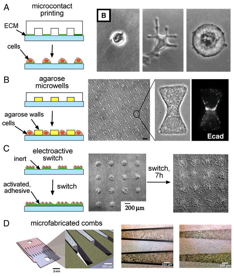
(A) Microcontact printing is used to pattern adhesive islands. Various sized islands are used to control cell-ECM adhesion and cell shape or spreading [5]. (B) Agarose microwells allow the control of both cell-ECM adhesion and cell-cell adhesion. Cells form cadherin-mediated cell-cell contacts across the center of the bowtie [42]. (C) Electroactive substrates dynamically activate inert surfaces to render them adhesive to cells. Patterned cells that are exposed to a newly adhesive surface form new adhesions and migrate away from the initial patterns [9]. (D) Microfabricated silicon combs dynamically control adhesion of cells to neighboring cells by changing from a contacting to separated configuration [10].
By creating large or small islands of ECM in various geometries and configurations, the amount of cell-cell adhesion, in addition to cell-matrix adhesion can be controlled. Cells are linked to neighboring cells through adherens junction, which like cell-matrix adhesions, bind to scaffolding proteins that link to the cytoskeleton and also associate with many signaling proteins that direct cell behavior [6]. Interestingly, contact to neighboring cells has also been shown to activate many of the same signaling pathways as cell-matrix adhesion [7]. Micropatterning tools have recently been used to control the amount of cell-cell adhesion. For example, use of increasingly larger islands elevates the number of cells present on the island, allowing for formation of progressively more interaction with neighboring cells [8]. To directly control the degree of cell spreading in addition to cell-cell contact, cells can be cultured into a bowtie-shaped microwell, where one cell lands in each side of the bowtie and contacts a neighboring cell across the middle of the bowtie. The area of each cell is controlled by the area of one half of the bowtie. In this configuration, each cell contacts a single neighboring cell, and the length of the constriction through the center of the bowtie controls the length of the contact. Hence, both cell spreading and cell-cell contact are simultaneously controlled.
Recently, techniques have been developed to control cell-matrix and cell-cell interactions dynamically. For example, self-assembled monolayers of alkanethiolates on gold can be switched from a nonadhesive to an adhesive state. Using this chemistry, cells can first be patterned onto an initially adhesive region. Upon electroactive switching of adjacent regions from nonadhesive to adhesive, the initially patterned cells form new adhesions in the surrounding regions and migrate away from the patterns (Figure 1C). Alternatively, if a second batch of cells is seeded at the time of switching, these cells will land onto the newly adhesive regions and thus two cell types can be patterned on a single substrate [9]. Dynamic manipulation of cell-cell adhesion has recently been achieved. Cells were cultured directly onto microfabricated silicon devices that were oxidized, coated with polystyrene and then plasma treated to produce a surface comparable to tissue culture plastic [10]. Using such a microfabricated device, Hui and Bhatia created a substrate consisting of two interlocking parts, which can be placed in discrete configurations—with cells contacting or separated by a micron-scaled gap (Figure 1D)[10]. By culturing cells on these substrates and adjusting placement of the interlocking components, the effects of paracrine (transmitted through soluble factors) and juxtacrine signals (transmitted through contact) was examined. This mechanically configurable device enabled controlled study of the interactions that could impact cellular phenotype.
While studies on two-dimensional surfaces have been useful in understanding basic mechanisms for how structure affects cell function, the practical goal of engineering artificial tissue constructs will require patterning within three-dimensional scaffolds. Microengineered devices for culture of cells in 3D are still in their nascent stages of development. Initial efforts have seeded cells in grooves of a PDMS substrate that are filled with an ECM protein such as collagen or fibrin. Using this method, Chobak et al. demonstrated the formation and perfusion of endothelial tubular structures within collagen gels in PDMS channels [11]. Micropatterning provides greater positional control over endothelial tube formation compared to previous methods, where endothelial cells formed randomly organized structures when cultured within a bulk gel. In another method to pattern cells in 3D gels composed of natural ECM, Nelson and colleagues used a PDMS stamp to micromold wells within collagen gels [12]. Cells seeded on these gels settled within the microwells, and then collagen was added on top to encapsulate the cells. In this method, micropatterned cells are localized within a bulk collagen gel, rather than within grooves of a PDMS substrate. PEG (polyethylene glycol)-based systems have been developed as an alternative to 3D matrices composed entirely of adhesive proteins. PEG is a nonadhesive polymer that has tunable mechanical and chemical properties. Polymer chain length is varied to regulate mechanical properties and chemical modification such as functionalization with RGD peptides varies the adhesivity [13]. Additionally, PEG can be functionalized with a photointiator group in order to render the material photopolymerizable. Cells may also be encapsulated within specific regions of a bulk gel by selective photopolymerization of PEG-based hydrogels or by encapsulating cells using dielectrophoresis within PEG gels [14]. For applications in tissue engineering, controlling the placement and the formation of cellular structures may become a valuable way to ensure appropriate geometry of vascularization or epithelialization throughout the entire construct.
Cell shape modulates signals from the ECM
One major contribution of such patterning techniques to control cell function has been to demonstrate that changes in cell shape regulate cell proliferation, differentiation, and survival. Cell adhesion to the ECM is initiated by the ligation and clustering of integrins, which leads to the subsequent recruitment of structural and signaling proteins to form focal adhesions [15]. Binding of integrins to ECM ligands causes conformational changes and subsequent binding of several scaffolding proteins. These interactions ultimately tether adhesions to the actin cytoskeleton and allow for cells to spread and flatten against their underlying substrate [16]. Integrin ligation alone is not sufficient to support some cellular functions, as early observations demonstrated that adherent cells could only proliferate if they were well spread [17]. However, since these studies used ECM ligand density to control cell spreading, and ligand density directly affects both the degree of cell spreading and integrin activation, it was not clear whether the effects of cell morphology were actually an indirect result of increased integrin activation.
In order to delineate the effects of cell spreading and integrin activation, micropatterning techniques were developed to control the presentation of ECM ligands and thus permit independent control of cell spreading and ligand density [1]. Seeding cells on progressively larger islands of a saturating density of ECM ligand increased their proliferation. Furthermore, cells that were allowed to spread across several small islands such that the total amount of ECM was comparable to that of an unspread cell also exhibited high proliferation, suggesting that cell spreading itself permits proliferation [5] (Figure 2A). Since then, it has also been shown that cell shape also plays a role in stem cell lineage commitment and differentiation. McBeath et al. demonstrated that mesenchymal stem cells tend to commit to a bone lineage when cultured on large micropatterned islands, and a fat lineage when cultured on small islands (Figure 2B)[18]. Together, these studies demonstrate that cell structure and shape play an important role in regulating multiple facets of cellular behavior.
Figure 2. Cell shape regulates proliferation and differentiation.
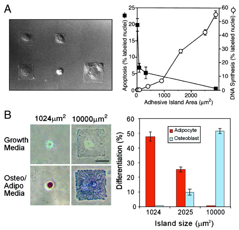
(A) Cells seeded on increasingly larger islands exhibit progressively higher rates of proliferation and lower rates of apoptosis [5]. (B) Mesenchymal stem cells seeded on large islands tend to differentiate into bone, while cells seeded on small islands differentiate into fat [18].
In addition to cell area, cell shape itself appears to regulate intracellular structure. For example, cells contrained to form square or triangular shapes appear to concentrate their adhesions in the corners [19]. Teardrop shapes appear to polarize cells so that the direction of migration is pre-defined to face toward the blunt end of the teardrop [20] (Figure 3A). Importantly, cell shape is not the only determinant for such internal organization. For example, it has been shown that when cells are organized into square shapes with asymmetric adhesive patterns, cells polarize their mitotic axis in a specific direction based on the matrix pattern [21], suggesting that spindle orientation is largely dependent on spatial distribution of ECM (Figure 3B). Using subcellular, adhesion-sized ECM islands, investigators have also revealed that controlling adhesion size, shape and density can itself feed back to control the degree of cell spreading and shape (Figure 3C) [22]. Thus there are multiple scales at which one can use patterning to control cell function.
Figure 3. Micropatterning techniques demonstrate the role of ECM geometry in regulating cell function.
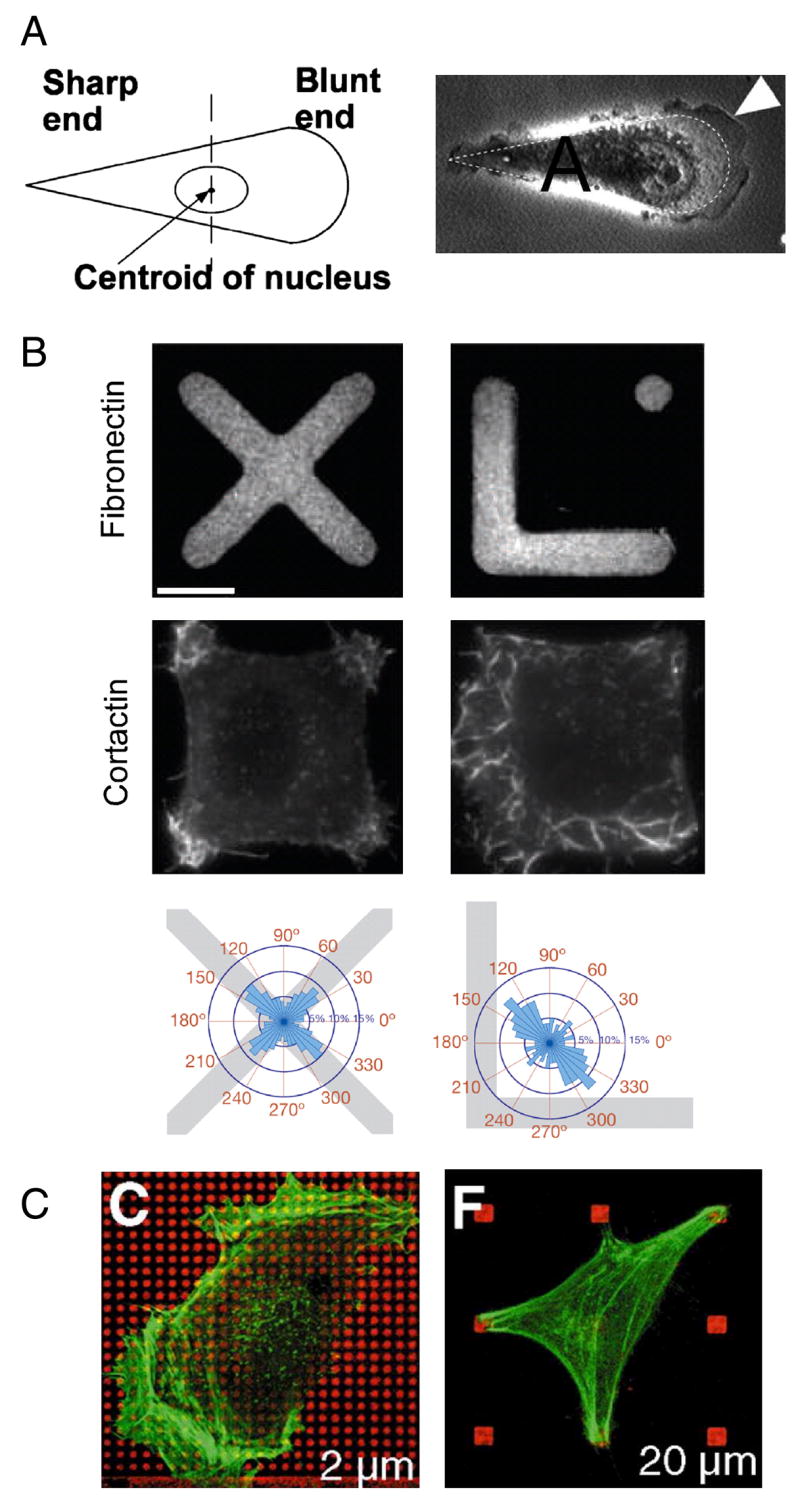
(A) Cells patterned onto a teardrop shape polarize and form lamellepodia at the blunt end of the drop [20]. (B) Cells patterned to form squares with an asymmetric underlying ECM pattern exhibit a polarized axis of division [21].
While cell shape has now been shown to regulate many cellular functions, only recently do we have some understanding of how cells translate changes in cell shape into molecular signals that regulate cell function. It appears that a class of molecules known as the Rho family of GTPases is thought to play a significant role in relaying stucture- and adhesion-mediated signals. Early studies identified a critical role for Rho GTPases in cytoskeletal organization: RhoA induces the formation of actin-myosin bundles, or stress fibers, associated with focal adhesions, Rac1 is involved in lamellipodia formation and membrane ruffling, and Cdc42 causes spike-like membrane protrusions (Nobes and Hall, 1995). Furthermore, it has since been established that Rho GTPases have numerous other cellular functions including cell proliferation, migration, and polarity [23]. It appears that cellular organization feeds back to regulate RhoA signaling and this is a key mechanism by which cell shape can regulate function. In studies of mesenchymal stem cell differentiation, it was found that Rho-mediated contractility is required for differentiation of spread cells into bone [18]. Supporting these data, it has been shown that RhoA-generated contractility is required for the high traction forces observed in well spread cells using a microfabricated force sensor array [24], and that differentiation into bone required the support of high tensional environments associated with stiff matrices [25]. RhoA signaling appears to be important not only for sensing cell shape, but also for transducing nanotopography of a surface [26] and mechanical stiffness [25] [27, 28] (Figure 4). Thus, it now appears that a feedback system between cellular and extracellular matrix structure, RhoA signaling, and adhesion signaling plays an important role in regulating cell function. Understanding how to capitalize on this control system will be an important step for moving forward in this field.
Figure 4. RhoA and cytoskeletal tension are involved in transducing extracellular signals.
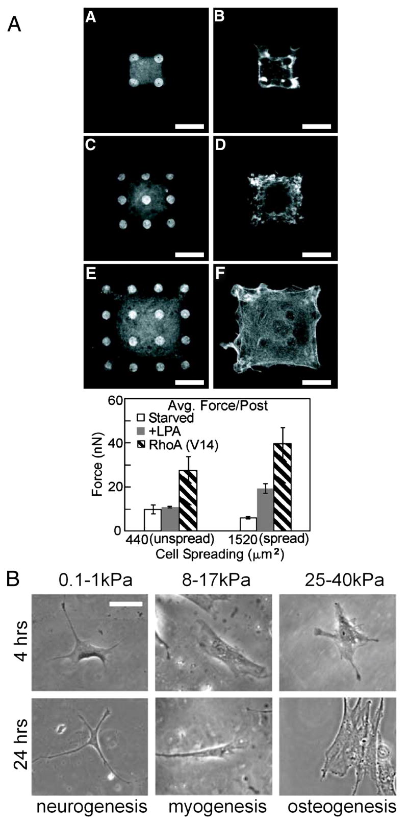
(A) Cells patterned on microfabricated post arrays exhibit higher contractility at higher levels of spreading. RhoA activation increases contractility further [24]. (B) Mesenchymal stem cells seeded on soft substrates tend to be less well spread and differentiate into neurons, while cells on stiffer matrices exhibited myogenesis, and cells even stiffer matrices exhibited osteogenesis [25].
While much has been learned about how cell structure regulates cell function on 2D substrates, we still know very little about how structure and function are linked when cells are embedded inside 3-D matrices. Studies in 3D have found that most cells lack a structured cytoskeleton and adhesions when cultured in 3D environments, although this is highly dependent on the mechanical properties of the gel. For example, fibroblasts cultured within fixed collagen gels (gels attached to the underlying dish) typically contain stress fibers and proliferate, but the same cells cultured within collagen gels that are floating within the culture dish maintain a dendritic phenotype similar to fibriblasts in vivo [29]. Similarly, it was observed long ago that epithelial cells undergo differentiation and produce milk proteins only in floating collagen gels [30] and that de-differentiated chondrocytes can re-differentiate when encapsulated within agarose gels [31]. Interestingly, it is thought that adhesion signaling to the RhoA pathway is largely responsible for these phenotypic differences [32, 33]. In sum, more detailed 3D studies will be needed to transition much of what has been learned from studies in 2D culture to help understand in vivo biology and enable design of artificial tissue constructs.
Cell-cell contact transduces signals within multicellular structures
While much about how cell structure affects function has been established from studies performed in single cells, cells in the body do not exist in isolation and are usually surrounded by neighboring cells of similar and different types. In culture, it has been observed that cellular behaviors change as they come into contact with neighboring cells. For example, endothelial and epithelial cells seeded at a low, subconfluent density typically appear spindly, fibroblastic and have high levels of proliferation, but acquire a cobblestone or cuboidal morphology and reduced proliferation rates as they become confluent. Cells interact with neighboring cells through junctional structures formed by transmembrane proteins that are bound to proteins on neighboring cells. The adherens junction is the principal adhesive type of junction and is composed primarily of cadherin molecules, which bind homophilically to cadherins on adjacent cells. Cadherins are thought to be the receptor responsible for the inhibition of proliferation by cell-cell contact at confluent densities since blocking cadherins causes a loss of contact inhibition, and this effect is rescued with exogenous expression of cadherin in cadherin null cells [34]. In addition, the engagement of VE-cadherin, the primary cadherin in vascular endothelial cells, is thought to inhibit growth factor signaling [35].
However, as the degree of cell-cell adhesion increases with increasing culture density, other structural cues such as the degree of cell spreading are also altered. Using bowtie shaped microwells to control both cell spreading and cell-cell contact, our laboratory has suggested that in endothelial cells, if the degree of spreading is controlled, cell-cell contact and engagement of VE-cadherin actually stimulates proliferation [36] (Figure 5A). Moreover, we demonstrated that VE-cadherin engagement activates a RhoA-mediated proliferative pathway. These data suggest that the effects of cell-cell adhesion depend on cell shape and cytoskeletal structure. In epithelial cells E-cadherin engagement also stimulates proliferation [37]. However, this proliferative signal was independent of cell shape, and dependent on Rac1 signaling rather than RhoA. These studies suggest that cell-cell contact and cadherin engagement can stimulate proliferation in multiple cell types, though the mechanisms underlying this proliferative response appear to be cell-type specific.
Figure 5. Multicellular structure regulates cell function.
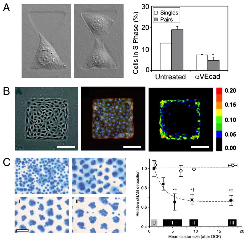
(A) Endothelial cells contacting a single neighboring cell exhibit a VE-cadherin dependent increase in proliferation when compared to single cells that are spread to the same extent [36]. (B) Groups of cells seeded on larger islands have higher levels of proliferation at the periphery compared to cells located at the interior [38]. (C) Chondrocytes patterned within three-dimensional PEG gels exhibit cluster-size dependent biosynthesis (filled circles). Unpatterned control cells did not exhibit changes in biosynthesis (open circles) [41].
Micropatterning tools can also be used to study conditions where cells are contacting multiple cells or cells of different types, rather than a single homotypic neighboring cell. Endothelial and epithelial cells cultured on larger island form discrete monolayers where cells are in contact with many neighboring cells. It was found that cells in this context tend to have high levels of proliferation when located on the edge of the monolayer (Figure 5B)[38]. This increase in proliferation required cadherins and RhoA signaling, suggesting once again that functional consequences of cell-cell interactions may be in part due to the activity of Rho GTPases. In another study, interactions between cells of different types, or heterotypic contact, restores hepatocyte function in vitro [39]. Notably, microengineered tools were used to confirm that both the amount and duration of contact are both important parameters in contact-mediated maintainance of cell function [10].
A number of studies have cultured cells in three-dimensional multicellular aggregates or spheroids in an effort to restore cellular function in vitro. To form spheroids, cells are typically cultured on a nonadhesive surface such as polystyrene or agarose, which causes cells to preferentially interact with neighboring cells. Over the course of several hours or days, cells form larger aggregates. These cultures have been shown to help preserve cellular function in multiple different cell types. For example, hepatocytes cultured in spheroids have higher levels of albumin secretion when compared to the same cells cultured on two-dimensional substrates [40]. In a more recent example where chondrocytes were patterned within PEG gels using dielectrophoresis, Albrecht et al. used micropatterning to dissect the independent roles of multicellular organization and volumetric cell density [41]. They found that chondroctye matrix biosynthesis was dependent on cell cluster size, rather than overall cell density (Figure 5C). It is thought that aggregation imparts many of the necessary structural cues required for maintaining differentiated phenotype, including proper dimensionality, shape, cell-ECM, and cell-cell interactions. The contribution of cell-cell interactions in multicellular constructs is undoubtedly important, but not completely understood. The development of more advanced methods to control multicellular structure in three-dimensional environments will be needed to investigate this further.
Conclusions and future work
The studies presented here demonstrate how the structure of single cells and simple cell-cell interactions affect cell function. With the ultimate goal of building complex tissues, the next steps in this work will involve developing tools to control multicellular interactions and understanding the interactions of multiple cell types in various configurations.
The signaling pathways relaying structural information to biochemical responses are gradually becoming elucidated. In particular, it appears that Rho GTPases are essential modulators of signals initiated by cell adhesion. However, the mechanism by which these signaling molecules are activated by adhesions and mechanical cues is still under investigation. Furthermore, how these GTPases are coordinatedly regulated by cell-ECM and cell-cell adhesion may be important in understanding how multiple extracellular cues are integrated within a cell to produce a single cellular output.
What we have learned thus far is that these engineering tools provide a necessary means to manipulate and control cell function, and therefore enable studies to support or refute our current understanding of how cell behavior is controlled. Introducing more complexity into these engineered cell cultures will ultimately enable us to not only control more complex cell and tissue functions, but also to understand their origins.
Acknowledgments
This work was supported in part by the NIH, the Army Research Office/Multidisciplinary University Research Innitiative, and the University of Pennsylvania MRSEC and NBIC Programs. W.F.L. acknowledges the NSF for financial support.
Footnotes
Publisher's Disclaimer: This is a PDF file of an unedited manuscript that has been accepted for publication. As a service to our customers we are providing this early version of the manuscript. The manuscript will undergo copyediting, typesetting, and review of the resulting proof before it is published in its final citable form. Please note that during the production process errors may be discovered which could affect the content, and all legal disclaimers that apply to the journal pertain.
References
- 1.Singhvi R, Kumar A, Lopez GP, Stephanopoulos GN, Wang DI, Whitesides GM, Ingber DE. Engineering cell shape and function. Science. 1994;264(5159):696–8. doi: 10.1126/science.8171320. [DOI] [PubMed] [Google Scholar]
- 2.Kane RS, Takayama S, Ostuni E, Ingber DE, Whitesides GM. Patterning proteins and cells using soft lithography. Biomaterials. 1999;20(23–24):2363–2376. doi: 10.1016/s0142-9612(99)00165-9. [DOI] [PubMed] [Google Scholar]
- 3.Tan JL, Liu W, Nelson CM, Raghavan S, Chen CS. Simple approach to micropattern cells on common culture substrates by tuning substrate wettability. Tissue Eng. 2004;10(5–6):865–72. doi: 10.1089/1076327041348365. [DOI] [PubMed] [Google Scholar]
- 4.Nelson CM, Raghavan S, Tan JL, Chen CS. Degradation of micropatterned surfaces by cell-dependent and -independent processes. Langmuir. 2003;19(5):1493–1499. [Google Scholar]
- 5.Chen CS, Mrksich M, Huang S, Whitesides GM, Ingber DE. Geometric control of cell life and death. Science. 1997;276(5317):1425–8. doi: 10.1126/science.276.5317.1425. [DOI] [PubMed] [Google Scholar]
- 6.Gumbiner BM. Cell adhesion: the molecular basis of tissue architecture and morphogenesis. Cell. 1996;84(3):345–57. doi: 10.1016/s0092-8674(00)81279-9. [DOI] [PubMed] [Google Scholar]
- 7.Nelson CM, Pirone DM, Tan JL, Chen CS. Vascular endothelial-cadherin regulates cytoskeletal tension, cell spreading, and focal adhesions stimulating RhoA. Molecular Biology of the Cell. 2004;15(6):2943–2953. doi: 10.1091/mbc.E03-10-0745. [DOI] [PMC free article] [PubMed] [Google Scholar]
- 8.Nelson CM, Chen CS. Cell-cell signaling by direct contact increases cell proliferation via a PI3K-dependent signal. FEBS Lett. 2002;514(2–3):238–42. doi: 10.1016/s0014-5793(02)02370-0. [DOI] [PubMed] [Google Scholar]
- 9.Yousaf MN, Houseman BT, Mrksich M. Using electroactive substrates to pattern the attachment of two different cell populations. PNAS. 2001;98(11):5992–5996. doi: 10.1073/pnas.101112898. [DOI] [PMC free article] [PubMed] [Google Scholar]
- 10.Hui EE, Bhatia SN. From the Cover: Micromechanical control of cell-cell interactions. PNAS. 2007;104(14):5722–5726. doi: 10.1073/pnas.0608660104. [DOI] [PMC free article] [PubMed] [Google Scholar]
- 11.Chrobak KM, Potter DR, Tien J. Formation of perfused functional microvascular tubes in vitro. Microvascular Research. 2006;71(3):185–196. doi: 10.1016/j.mvr.2006.02.005. [DOI] [PubMed] [Google Scholar]
- 12.Nelson CM, VanDuijn MM, Inman JL, Fletcher DA, Bissell MJ. Tissue Geometry Determines Sites of Mammary Branching Morphogenesis in Organotypic Cultures. Science. 2006;314(5797):298–300. doi: 10.1126/science.1131000. [DOI] [PMC free article] [PubMed] [Google Scholar]
- 13.Lutolf MP, Hubbell JA. Synthetic biomaterials as instructive extracellular microenvironments for morphogenesis in tissue engineering. Nat Biotechnol. 2005;23(1):47–55. doi: 10.1038/nbt1055. [DOI] [PubMed] [Google Scholar]
- 14.Albrecht DR, V, Tsang L, Sah RL, Bhatia SN. Photo- and electropatterning of hydrogel-encapsulated living cell arrays. Lab Chip. 2005;5(1):111–8. doi: 10.1039/b406953f. [DOI] [PubMed] [Google Scholar]
- 15.Schwartz MA, Ginsberg MH. Networks and crosstalk: integrin signalling spreads. Nat Cell Biol. 2002;4(4):E65–8. doi: 10.1038/ncb0402-e65. [DOI] [PubMed] [Google Scholar]
- 16.Geiger B, Bershadsky A. Assembly and mechanosensory function of focal contacts. Current Opinion in Cell Biology. 2001;13(5):584–592. doi: 10.1016/s0955-0674(00)00255-6. [DOI] [PubMed] [Google Scholar]
- 17.Folkman J, Moscona A. Role of cell shape in growth control. Nature. 1978;273(5661):345–9. doi: 10.1038/273345a0. [DOI] [PubMed] [Google Scholar]
- 18.McBeath R, Pirone DM, Nelson CM, Bhadriraju K, Chen CS. Cell shape, cytoskeletal tension, and RhoA regulate stem cell lineage commitment. Dev Cell. 2004;6(4):483–95. doi: 10.1016/s1534-5807(04)00075-9. [DOI] [PubMed] [Google Scholar]
- 19.Parker KK, Brock AL, Brangwynne C, Mannix RJ, Wang N, Ostuni E, Geisse NA, Adams JC, Whitesides GM, Ingber DE. Directional control of lamellipodia extension by constraining cell shape and orienting cell tractional forces. FASEB J. 2002;16(10):1195–1204. doi: 10.1096/fj.02-0038com. [DOI] [PubMed] [Google Scholar]
- 20.Jiang X, Bruzewicz DA, Wong AP, Piel M, Whitesides GM. Directing cell migration with asymmetric micropatterns. PNAS. 2005;102(4):975–978. doi: 10.1073/pnas.0408954102. [DOI] [PMC free article] [PubMed] [Google Scholar]
- 21.Thery M, Racine V, Pepin A, Piel M, Chen Y, Sibarita J-B, Bornens M. The extracellular matrix guides the orientation of the cell division axis. 2005;7(10):947–953. doi: 10.1038/ncb1307. [DOI] [PubMed] [Google Scholar]
- 22.Lehnert D, Wehrle-Haller B, David C, Weiland U, Ballestrem C, Imhof BA, Bastmeyer M. Cell behaviour on micropatterned substrata: limits of extracellular matrix geometry for spreading and adhesion. J Cell Sci. 2004;117(1):41–52. doi: 10.1242/jcs.00836. [DOI] [PubMed] [Google Scholar]
- 23.Etienne-Manneville S, Hall A. Rho GTPases in cell biology. Nature. 2002;420(6916):629–35. doi: 10.1038/nature01148. [DOI] [PubMed] [Google Scholar]
- 24.Tan JL, Tien J, Pirone DM, Gray DS, Bhadriraju K, Chen CS. Cells lying on a bed of microneedles: an approach to isolate mechanical force. Proc Natl Acad Sci U S A. 2003;100(4):1484–9. doi: 10.1073/pnas.0235407100. [DOI] [PMC free article] [PubMed] [Google Scholar]
- 25.Engler AJ, Sen S, Sweeney HL, Discher DE. Matrix Elasticity Directs Stem Cell Lineage Specification. Cell. 2006;126(4):677–689. doi: 10.1016/j.cell.2006.06.044. [DOI] [PubMed] [Google Scholar]
- 26.Teixeira AI, McKie GA, Foley JD, Bertics PJ, Nealey PF, Murphy CJ. The effect of environmental factors on the response of human corneal epithelial cells to nanoscale substrate topography. Biomaterials. 2006;27(21):3945–3954. doi: 10.1016/j.biomaterials.2006.01.044. [DOI] [PMC free article] [PubMed] [Google Scholar]
- 27.Wang HB, Dembo M, Hanks SK, Wang YL. Focal adhesion kinase is involved in mechanosensing during fibroblast migration. Proc Natl Acad Sci U S A. 2001;98(20):11295–11300. doi: 10.1073/pnas.201201198. [DOI] [PMC free article] [PubMed] [Google Scholar]
- 28.Wang HB, Dembo M, Wang YL. Substrate flexibility regulates growth and apoptosis of normal but not transformed cells. Am J Physiol Cell Physiol. 2000;279(5):C1345–1350. doi: 10.1152/ajpcell.2000.279.5.C1345. [DOI] [PubMed] [Google Scholar]
- 29.Fringer J, Grinnell F. Fibroblast Quiescence in Floating or Released Collagen Matrices. Contribution of the Erk signaling pathway and actin cytoskeleton organization. J Biol Chem. 2001;276(33):31047–31052. doi: 10.1074/jbc.M101898200. [DOI] [PubMed] [Google Scholar]
- 30.Emerman JT, Pitelka DR. Maintainance and induction of morphological differentiation in dissociated mammary epithalium on floating collagen membranes. In Vitro. 1977;13(5):316–328. doi: 10.1007/BF02616178. [DOI] [PubMed] [Google Scholar]
- 31.Benya PD, Shaffer JD. Dedifferentiated chondrocytes reexpress the differentiated collagen phenotype when cultured in agarose gels. Cell. 1982;30(1):215–224. doi: 10.1016/0092-8674(82)90027-7. [DOI] [PubMed] [Google Scholar]
- 32.Wozniak MA, Desai R, Solski PA, Der CJ, Keely PJ. ROCK-generated contractility regulates breast epithelial cell differentiation in response to the physical properties of a three-dimensional collagen matrix. J Cell Biol. 2003;163(3):583–595. doi: 10.1083/jcb.200305010. [DOI] [PMC free article] [PubMed] [Google Scholar]
- 33.Woods A, Beier F. RhoA/ROCK Signaling Regulates Chondrogenesis in a Context-dependent Manner. J Biol Chem. 2006;281(19):13134–13140. doi: 10.1074/jbc.M509433200. [DOI] [PubMed] [Google Scholar]
- 34.Caveda L, Martin-Padura I, Navarro P, Breviario F, Corada M, Gulino D, Lampugnani MG, Dejana E. Inhibition of cultured cell growth by vascular endothelial cadherin (cadherin-5/VE-cadherin) J Clin Invest. 1996;98(4):886–93. doi: 10.1172/JCI118870. [DOI] [PMC free article] [PubMed] [Google Scholar]
- 35.Grazia Lampugnani M, Zanetti A, Corada M, Takahashi T, Balconi G, Breviario F, Orsenigo F, Cattelino A, Kemler R, Daniel TO, Dejana E. Contact inhibition of VEGF-induced proliferation requires vascular endothelial cadherin, beta-catenin and the phosphatase DEP-1/CD148. J Cell Biol. 2003;161(4):793–804. doi: 10.1083/jcb.200209019. [DOI] [PMC free article] [PubMed] [Google Scholar]
- 36.Nelson CM, Chen CS. VE-cadherin simultaneously stimulates and inhibits cell proliferation by altering cytoskeletal structure and tension. J Cell Sci. 2003;116(Pt 17):3571–81. doi: 10.1242/jcs.00680. Epub 2003 Jul 22. [DOI] [PubMed] [Google Scholar]
- 37.Liu WF, Nelson CM, Pirone DM, Chen CS. E-cadherin engagement stimulates proliferation via Rac1. J Cell Biol. 2006;173(3):431–41. doi: 10.1083/jcb.200510087. [DOI] [PMC free article] [PubMed] [Google Scholar]
- 38.Nelson CM, Jean RP, Tan JL, Liu WF, Sniadecki NJ, Spector AA, Chen CS. From the Cover: Emergent patterns of growth controlled by multicellular form and mechanics. Proc Natl Acad Sci U S A. 2005;102(33):11594–9. doi: 10.1073/pnas.0502575102. [DOI] [PMC free article] [PubMed] [Google Scholar]
- 39.Bhatia SN, Balis UJ, Yarmush ML, Toner M. Effect of cell-cell interactions in preservation of cellular phenotype: cocultivation of hepatocytes and nonparenchymal cells. Faseb J. 1999;13(14):1883–900. doi: 10.1096/fasebj.13.14.1883. [DOI] [PubMed] [Google Scholar]
- 40.Landry J, Bernier D, Ouellet C, Goyette R, Marceau N. Spheroidal aggregate culture of rat liver cells: histotypic reorganization, biomatrix deposition, and maintenance of functional activities. J Cell Biol. 1985;101(3):914–923. doi: 10.1083/jcb.101.3.914. [DOI] [PMC free article] [PubMed] [Google Scholar]
- 41.Albrecht DR, Underhill GH, Wassermann TB, Sah RL, Bhatia SN. Probing the role of multicellular organization in three-dimensional microenvironments. 2006;3(5):369–375. doi: 10.1038/nmeth873. [DOI] [PubMed] [Google Scholar]
- 42.Nelson CM, Liu WF, Chen CS. Manipulation of Cell-Cell Adhesion Using Bowtie-Shaped Microwells. In: Coutts AS, editor. Methods in Molecular Biology: Adhesion Protein Protocols. 2. Vol. 270. Humana Press; 2007. pp. 1–9. [DOI] [PubMed] [Google Scholar]