The tandem C2 domains of synaptotagmin contain redundant Ca2+ binding sites that cooperate to engage t-SNAREs and trigger exocytosis (original) (raw)
Abstract
Real-time voltammetry measurements from cracked PC12 cells were used to analyze the role of synaptotagmin–SNARE interactions during Ca2+-triggered exocytosis. The isolated C2A domain of synaptotagmin I neither binds SNAREs nor inhibits norepinephrine secretion. In contrast, two C2 domains in tandem (either C2A-C2B or C2A-C2A) bind strongly to SNAREs, displace native synaptotagmin from SNARE complexes, and rapidly inhibit exocytosis. The tandem C2 domains of synaptotagmin cooperate via a novel mechanism in which the disruptive effects of Ca2+ ligand mutations in one C2 domain can be partially alleviated by the presence of an adjacent C2 domain. Complete disruption of Ca2+-triggered membrane and target membrane SNARE interactions required simultaneous neutralization of Ca2+ ligands in both C2 domains of the protein. We conclude that synaptotagmin–SNARE interactions regulate membrane fusion and that cooperation between synaptotagmin's C2 domains is crucial to its function.
Keywords: synaptotagmin; SNARE; membrane fusion; C2 domain; exocytosis
Introduction
Recent advances indicate that intracellular membrane fusion is catalyzed by SNARE complexes (Söllner et al., 1993; Weber et al., 1998). In neurons, the SNARE complex is composed of the synaptic vesicle protein synaptobrevin/VAMP and the plasma membrane proteins syntaxin and the synaptosome-associated protein of 25 kD (SNAP-25)*(Söllner et al., 1993). These proteins assemble to form a four-helix bundle that closely juxtaposes the vesicle and target membranes (Sutton et al., 1998). Reconstitution experiments provide direct evidence that assembly of SNARE complexes is sufficient to catalyze membrane fusion (Weber et al., 1998). However, fusion mediated by purified SNAREs exhibits slow kinetics and is not regulated by Ca2+. In contrast, exocytosis from neurons and other cell types is rapid and strictly regulated by Ca2+ (Burgoyne and Morgan, 1998; Augustine, 2001).
The Ca2+-binding synaptic vesicle protein synaptotagmin I (syt) has been proposed to serve as the Ca2+ sensor that regulates secretion (Brose et al., 1992; Geppert et al., 1994; Littleton et al., 1994, 2001; Fernández-Chacón et al., 2001), although this role has been debated (DiAntonio and Schwarz, 1994). Syt I spans the vesicle membrane once and possesses a large cytoplasmic domain largely composed of two C2 domains (Perin et al., 1990). The membrane proximal C2 domain, C2A, penetrates lipid bilayers in response to Ca2+ (Davis et al., 1999), and the membrane distal C2 domain, C2B, mediates Ca2+-triggered oligomerization of syt I (Desai et al., 2000). Interestingly, both C2 domains synergize to directly interact with the target membrane SNAREs (t-SNAREs) syntaxin and SNAP-25, and this interaction can occur at all stages of SNARE complex assembly (for review see Augustine, 2001). These findings raise the possibility that syt could regulate SNARE catalyzed membrane fusion in response to binding Ca2+.
This study investigates the biochemical and functional role of tandem C2 domains for complex formation with t-SNAREs and membranes. Our data suggest that syt–SNARE interactions are critical for Ca2+-triggered exocytosis and that the tandem C2 domains of syt exhibit unique properties and possess partially redundant Ca2+ binding sites.
Results and discussion
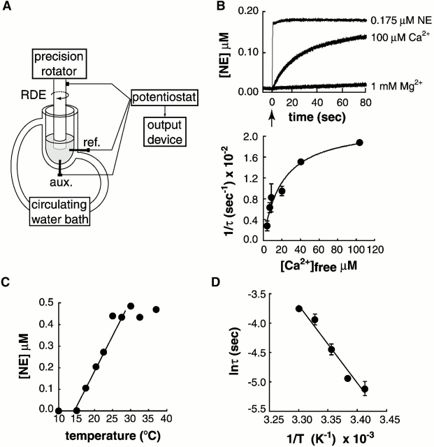
For these studies, PC12 cells were “cracked open” to gain direct access to the fusion machinery that mediates release of norepinephrine (NE) from large dense core vesicles (LDCVs). Upon addition of Ca2+, released NE was detected in real time via rotating disc electrode (RDE) voltammetry (Fig. 1 A). The timecourse of NE release in response to 100 μM Ca2+ is shown in Fig. 1 B (top); 1 mM Mg2+ failed to trigger release. The release kinetics are not limited by the diffusion of Ca2+ or the response time of the instrument as shown by the immediate electrode response upon addition of NE to the assay system. Ca2+-triggered release curves were fitted with a single exponential function to calculate the time constant (τ) for release at different Ca2+ concentrations. This made it possible to determine the Ca2+ dependence of the rate of exocytosis from these cells. In Fig. 1 B (bottom), the reciprocal of the time constant is plotted versus the free Ca2+, as measured with a Ca2+ electrode, and the [Ca2+]1/2 was 19.7 ± 4.7 μM. This value is similar to results obtained using photolysis of caged Ca2+ in intact pituitary melanotrophs and chromaffin cells (for review see Burgoyne and Morgan, 1998) and is somewhat higher than previous estimates of release from permeabilized PC12 cells that were based on static secretion measurements (<10 μM; Klenchin et al., 1998). These data indicate that LDCV exocytosis in multiple cell types is regulated via Ca2+ sensors with similar properties.
Figure 1.
Ca2 + and temperature dependence of NE release from cracked PC12 cells measured with a real-time assay. (A) Schematic diagram of the instrument used to measure exocytosis by RDE voltammetry (modified from Earles and Schenk, 1998). The Ag/AgCl reference (ref) and platinum auxiliary (aux) electrodes are indicated. (B) Ca2+ but not Mg2+ releases NE (top; arrow indicates the time of addition). Immediate response upon addition of NE to cell-free buffer demonstrates that Ca2+-triggered release is monitored in real time. Ca2+-dependent release profiles fit single exponential functions; 1/τ (τ = time constant) was plotted versus [Ca2+]free (bottom), the [Ca2+]1/2 was 19.7 ± 4.7 μM (n = 3–6, ± SD); τ = 45 s at saturating [Ca2+]. (C) Temperature dependence of exocytosis. NE was measured 80 s after addition of 100 μM Ca2+. (D) Arrhenius plot yields an activation energy (Ea) of 108 ± 7 kJ/mole (n = 3, ± SD).
Synaptic transmission has a steep temperature dependence that reflects activation energies of presynaptic voltage-activated Ca2+ channels, the fusion machinery, and postsynaptic receptors (Sabatini and Regehr, 1996). The RDE cracked cell assay eliminates the contributions from channels and postsynaptic receptors. In addition, a recent study indicates that release from cracked PC12 cells reflects the exocytosis of predocked LDCVs (Chen et al., 2001). Therefore, we used the RDE cracked cell assay to selectively examine the temperature dependence of the Ca2+-triggered release process (Fig. 1, C and D). Exocytosis was blocked at temperatures at or below 15°C and was maximal by 22.5°C (Fig. 1 C). An Arrhenius plot indicated that release exhibits an activation energy of 108 ± 7 kJ/mole (Fig. 1 D). This value is similar to the activation energies for viral-mediated fusion, GTPγS-driven fusion in mast cells, and values obtained using model systems (Lee and Lentz, 1998), suggesting that all of these fusion reactions involve common or similar mechanisms.
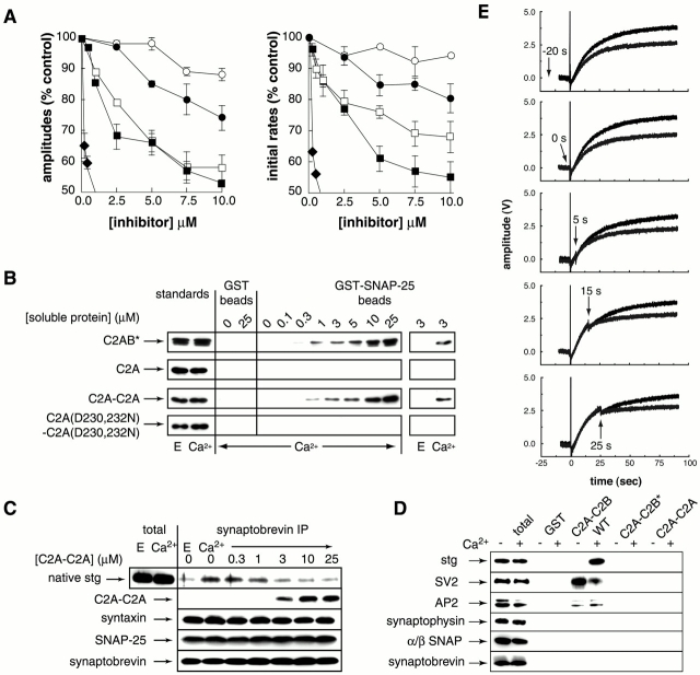
We next used this assay to examine the function of syt during exocytosis. Previously, we reported that the cytoplasmic domain of recombinant syt I (C2A-C2B) inhibits secretion from cracked PC12 cells (Desai et al., 2000) and proposed that it functions as a dominant negative reagent that blocks homo- and heterooligomerization of syt isoforms in response to Ca2+. This effect is apparent in the RDE secretion assay and serves as a reference point for the new constructs described below (Fig. 2 A). Reverse genetic and biochemical studies by Littleton et al. (2001) revealed that a mutation that disrupts the Ca2+-triggered oligomerization activity of syt I inhibits the ability of docked synaptic vesicles to fuse in response to stimulation.
Figure 2.
Tandem C2A domains (C2A-C2A) block syt–SNARE interactions and rapidly inhibit Ca2 + -triggered exocytosis. (A) Effect of syt constructs on the rate and extent of NE release. C2A-C2B (♦; G374 version) potently inhibits secretion, presumably by blocking native syt oligomerization (Desai et al., 2000; Littleton et al., 2001). C2A-C2B* (▪), harbors a K326,327A mutation that disrupts the ability of the recombinant protein to bind native syt in the cracked cells (without causing misfolding; Desai et al., 2000). Although C2A-C2B* cannot interfere with native syt oligomerization (because it does not bind endogenous syt in the cracked cells), this construct exhibits a second mode of inhibition that occurs at higher protein concentrations. C2A, which fails to bind SNAP-25, fails to inhibit secretion (○). However, tethering two C2A domains together (C2A-C2A; □) resulted in a chimeric protein with SNAP-25 binding activity (B) and inhibitory activity that were similar to C2A-C2B*. As a control, two Ca2+ ligands in each C2A domain were neutralized D230,232N, disrupting the Ca2+ binding activity of the protein (Zhang et al., 1998). C2A(D230,232N)-C2A(D230,232N) binding to SNAREs was not detectable under our assay conditions (B), and this construct exhibited reduced inhibitory activity (•). Note the potency of inhibition of C2A-C2B (♦) and C2A-C2B* (▪) using this assay differs from a previous study using primed cells in which secretion was monitored using 3H-NE (Desai et al., 2000). The reason for this difference is not clear; however, in all experiments the rank order of inhibition by different inhibitors is the same between these two assay systems. Also note that to facilitate comparisons between the proteins used in this study, the ordinate is cut-off at 50% inhibition; at 10 μM, C2A-C2B inhibited 80–90% of release. (B) Tethering two C2A domains together results in a t-SNARE binding protein. Binding assays were carried out as described in Davis et al. (1999) except that 30 μg SNAP-25 was immobilized on beads. Eight percent of bound proteins was subjected to SDS-PAGE and visualized by immunoblotting using ECL. As a positive control, C2A-C2B* was analyzed in parallel. This mutant form was used to avoid increases in bound material that occur as a result of Ca2+-triggered syt homooligomerization (that is, the interaction of additional copies of syt with the syt that is bound to the SNAP-25 beads). As a further control, C2A(D230,232N)-C2A(D230,232N) was assayed for SNAP-25 binding activity. Standards are 10 ng C2A-C2B* and C2A-C2A, and 5 ng C2A. (C) C2A-C2A displaces native syt from SNARE complexes. Experiments were carried out by immunoprecipitation of synaptobrevin. 1-ml aliquots of rat BDE (1 mg/ml; prepared as described in Chapman et al. [1996]) were incubated in either 2 mM EGTA or 1 mM Ca2+ with the indicated concentrations of C2A-C2A for 1.5 h. SNARE complexes were immunoprecipitated with an antisynaptobrevin antibody (69.1; 10 μL) for 1.5 h followed by addition of 60 μl of protein G-Sepharose Fast Flow (Amersham Pharmacia Biotech) for 1 h. Immunoprecipitates were washed four times, and 20% of bound proteins was subjected to SDS-PAGE and visualized by ECL. Total corresponds to 1 μg BDE. Polyclonal antibodies were used to detect native syt (anti-C2B) and SNAP-25, and monoclonal antibodies were used to detect syntaxin, synaptobrevin, and C2A-C2A (anti-C2A). (D) Tethering two C2A domains together does not result in additional new protein–protein interactions. These experiments were carried out as described in Chapman et al. (1998) but expanded to include SV2. To assay for SV2, 6 μl of BDE (total) and 40% of the bound material was subjected to SDS-PAGE and immunoblotted using anti-SV2 antibodies. (E) Timing of inhibition. Addition of C2A-C2B* (final concentration 4 μM) at the times indicated (arrows) inhibits release to a similar extent at all stages in the release process and does not require preincubation; addition of Ca2+ is indicated by the vertical lines. Identical results were obtained using C2A-C2A (unpublished data).
Mutations within the C2B region of the C2A-C2B construct (K326,327A; designated C2A-C2B*) largely abrogated its inhibitory activity in the cracked cell assay; however, we noted that the mutant version retained some activity, albeit at higher concentrations than C2A-C2B (2–5 μM; Fig. 1 A; Desai et al., 2000). These findings suggested that the K326,327A mutation may have unmasked a second mode of inhibition mediated by a relatively low affinity C2A-C2B*–target protein interaction. C2A-C2B* failed to bind known syt binding proteins, including other copies of syt, SV2, and AP-2 (Fig. 2 D), but retained t-SNARE binding activity (Fig. 2 B and Fig. 3 B). As shown in Fig. 2 B, C2A-C2B* bound SNAP-25 with an affinity that closely parallels its inhibitory activity in the release assay (EC50 ∼2–5 μM), consistent with a model in which C2A-C2B* blocks fusion by competitively inhibiting native syt–SNARE interactions.
Figure 3.
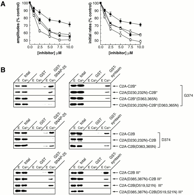
Partial functional and biochemical redundancy between Ca 2+ ligands in C2A and C2B that regulate syt–t-SNARE interactions. (A) Functional redundancy of the Ca2+ ligands in the C2 domains of syt I (G374). The effects of Ca2+ ligand mutations on the ability C2A-C2B* to inhibit release was assayed as described in the legend to Fig. 2 A. Inhibition by C2A-C2B* is indicated by ○. Inhibition was only partially abrogated by muta- tions that disrupt Ca2+ binding to C2A (D230,232N; □) or C2B (D363,365N; ▪). However, inhibitory activity was largely abolished by simultaneous disruption of the Ca2+-sensing ability of both C2A and C2B (D230,232,363,365N; •). (B) Ca2+-triggered t-SNARE binding activity of syt I (G374) and III exhibits redundancy between Ca2+ ligands in C2A and C2B. Binding assays were carried out as described in the legend to Fig. 2 B except the GST-syntaxin was 15 μg. “Total” corresponds to 1% of the binding reaction. As described in the legend to Fig. 2 B, the asterisk indicates a mutation (K326,327A in syt I or R482A,K483A in syt III) that disrupts C2B-mediated syt oligomerization (Desai et al., 2000) to avoid increases in binding to t-SNARE beads that result from syt oligomerization. (Top) In syt I (G374), disruption of the Ca2+-sensing ability of either C2 domain is partially tolerated (D230,232N or D363,365N); simultaneous neutralization of Ca2+ ligands in both C2 domains (D230,232N,D363,365N) disrupts t-SNARE binding activity. (Middle) In syt I (D374), mutations that disrupt the Ca2+-sensing ability of C2A (D230,232N) abolish SNARE binding activity, whereas mutations that disrupt the Ca2+-sensing ability of C2B domain (D363,365N) have a lesser effect (Bai et al., 2000). (Bottom) Syt III exhibits Ca2+ ligand redundancy for SNARE binding activity analogous to syt I (G374). Simultaneous neutralization of Ca2+ ligands in both C2 domains (D385,387N and D519,521N) was required to disrupt Ca2+-triggered SNARE binding activity.
To test this hypothesis and to rule out the possibility that the inhibitory activity was due to residual activity of the mutant C2B domain of C2A-C2B*, we designed a selective inhibitor of syt–SNARE interactions. Because neither of the isolated C2 domains of syt I efficiently binds SNAREs (Chapman et al., 1996; Davis et al., 1999), we reasoned that syt I acquired tandem C2 domains during evolution to regulate SNARE-mediated membrane fusion. This model predicts that a SNARE binding protein could be generated by linking two C2 domains together for use as a selective inhibitor of native syt–SNARE interactions in cracked PC12 cells. A construct comprising tandem C2B domains would be uninformative because C2B alone inhibits secretion, again presumably via blocking syt oligomerization (Desai et al., 2000). We therefore focused on C2A, which does not block secretion (Fig. 2 A; note our data contrast those of Elferink et al. [1993], reporting inhibition by this fragment). Tethering two C2A domains together (designated C2A-C2A) resulted in a protein that inhibited secretion with a potency similar to that of C2A-C2B* (Fig. 2 A). Furthermore, C2A-C2A bound to SNAP-25 with an affinity that precisely mirrored its inhibitory activity in the release assay (Fig. 2 B). As a further control, we neutralized Ca2+ ligands in both C2 domains of C2A-C2A (Sutton et al., 1995; Zhang et al., 1998), which reduced both SNAP-25 binding activity and inhibitory activity in the release assay.
These data are consistent with a model in which C2A-C2A and C2A-C2B* inhibit fusion by blocking native syt–SNARE interactions. This was tested directly by immunoprecipitating SNARE complexes from brain detergent extracts (BDEs) in the presence of increasing concentrations of C2A-C2A. SNARE complexes were isolated using an antisynaptobrevin antibody. Syt does not bind directly to synaptobrevin thus antisynaptobrevin immunoprecipitations selectively pull-down syt that is bound to SNARE complexes containing both v- and t-SNAREs. As shown in Fig. 2 C, C2A-C2A blocked the binding of native syt to SNARE complexes over precisely the same concentration range that inhibits secretion. Furthermore, maximal inhibition of binding, 50%, coincides with the extent of inhibition observed in the cracked cell assay.
These observations further suggest that inhibition by C2A-C2A is mediated by Ca2+-dependent binding to SNAREs. Although we cannot rule out that C2A-C2A inhibits exocytosis by blocking other unknown interactions, the following evidence argues against this possibility. First, in Fig. 2 D C2A-C2A fails to bind the known syt binding proteins AP-2 (Zhang et al., 1994), SV2 (Schivell et al., 1996), and other copies of syt (Desai et al., 2000); synaptophysin, α/β-SNAP, and synaptobrevin served as negative controls. Second, we have conducted an unbiased search for proteins that bind C2A-C2A but not C2A using affinity chromatography. Both proteins bind several yet-to-be characterized proteins from BDE, but the binding protein profiles for C2A and C2A-C2A were identical (unpublished data; note that C2A-C2B and C2A-C2A cannot efficiently bind SNAREs when fused to glutathione _S_-transferase and immobilized on beads [Chapman et al., 1996]). Finally, C2A binds with high affinity to acidic phospholipids (40 nM; Davis et al., 1999) as does C2A-C2A. Yet, C2A fails to inhibit secretion. This result is not unexpected; even if C2A–membrane interactions are critical for exocytosis, 10 μM C2A would not be expected to saturate all of the lipid binding sites in the cracked cells. We conclude that the most likely mode of inhibition by C2A-C2A is via blocking native syt–SNARE interactions.
If these interactions have a critical role in a late step in secretion, we would expect rapid inhibition regardless of the time at which C2A-C2A was added to the cracked cells. Addition of C2A-C2B* (Fig. 2 E) or C2A-C2A (unpublished data) before, during, or at varying times after the Ca2+ signal resulted in the rapid inhibition of exocytosis, suggesting perturbation of a late step in the fusion reaction.
The ability of tethered C2A domains to bind t-SNAREs further indicates a form of synergy between tandem C2 domains (Chapman et al., 1996; Davis et al., 1999). We further explored this synergy by disrupting the Ca2+ binding ability of each C2 domain in C2A-C2B*; disruption of either C2A or C2B only partially abrogated inhibitory activity in the cracked cell assay (Fig. 3 A). However, simultaneous disruption of both C2 domains strongly reduced inhibitory activity (Fig. 3 A). The effects of these mutations in the release assay were paralleled by their effects on SNAREs binding activity (Fig. 3 B). Titration experiments indicated that mutations within a single C2 domain reduce the affinity for SNAP-25 by approximately twofold (unpublished data) and shift the inhibitory activity of C2A-C2B* by roughly the same factor (Fig. 3 A).
We note that earlier studies demonstrated that neutralization of Ca2+ ligands in C2A blocked Ca2+-triggered t-SNARE and membrane interactions (Davis et al., 1999; Bai et al., 2000). However, previous studies made use of the initial syt I cDNA (Perin et al., 1990) that was shown recently to harbor a mutation (D374) that disrupts the activity of the C2B domain (Osborne et al., 1999; Desai et al., 2000). Because all other known syt sequences harbor a glycine at this position (G374), it is unclear whether the D374 variant is a bona fide version of syt I. In Fig. 3 B, we have confirmed that mutations in the C2A domain of D374-syt I indeed disrupt Ca2+-triggered t-SNARE binding activity.
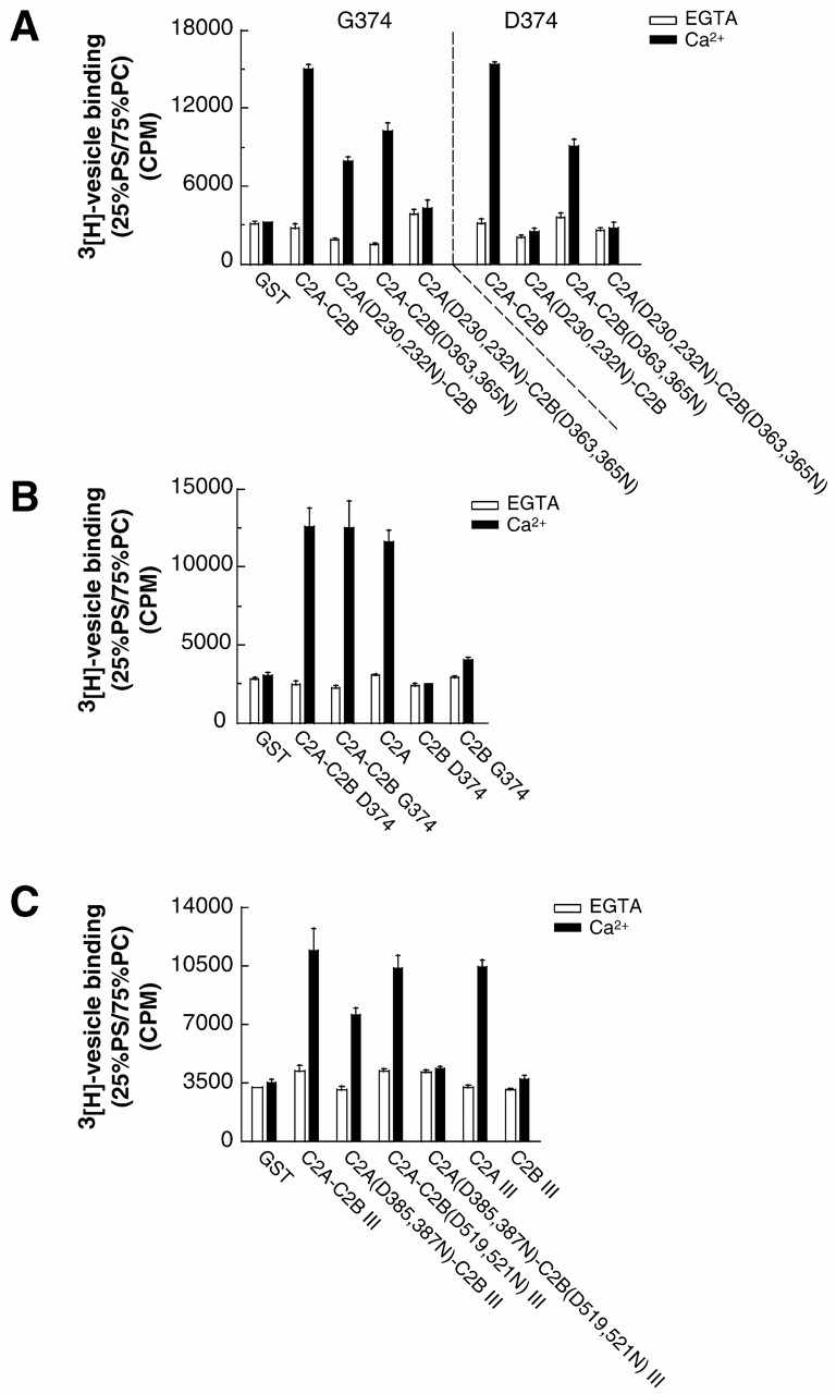
To test whether the apparent partial redundancy between the Ca2+ ligands in the C2 domains of syt I (G374) occurs with other isoforms, we generated analogous mutations in the C2 domains of syt III; again, complete disruption of Ca2+-triggered t-SNARE binding activity required mutations in both C2 domains (Fig. 3 B, bottom). We saw a similar redundancy when Ca2+-triggered liposome binding activity was measured (Fig. 4, A and C). These findings suggested that like C2A, the C2B domains of syt I (G374) and III may form autonomous Ca2+-triggered membrane binding domains. We tested this possibility and found that the isolated C2B domains fail to bind membranes in response to Ca2+ (Fig. 4 B). Thus, an inactive C2A domain when tethered to an inactive C2B domain gives rise to a protein with Ca2+-triggered lipid binding activity. These data suggest a novel form of cooperation between tandem C2 domains. In one model, C2B can compensate for Ca2+ ligand mutations in C2A such that the lipid binding activity within C2A is restored. Alternatively, an adjacent C2A domain may “activate” cryptic Ca2+-triggered membrane binding activity within C2B. Finally, as a control we confirmed that mutations in the C2A domain of the intact cytoplasmic domain of the D374 version of syt I abolished lipid binding activity (Bai et al., 2000), further establishing that the atypical aspartate residue at position 374 disrupts the function of C2B (Desai et al., 2000).
Figure 4.

Partial redundancy between Ca 2+ ligands in C2A and C2B that regulate syt–membrane interactions. Wild-type and mutant forms of syt I and III were immobilized (0.1 nmole) on glutathione–Sepharose beads. The isolated C2 domains of these isoforms were also analyzed. 3H-labeled 25% PS/75% PC liposome binding assays were carried out as described (Bai et al., 2000). (A) In the G374 form of syt I (C2A-C2B), Ca2+-triggered liposome binding activity is abolished only when Ca2+ ligands are simultaneously neutralized in both C2A and C2B (D230,232,363,365N). In the D374 form of syt I (C2A-C2B), Ca2+-triggered liposome binding activity is abolished by mutations that neutralize Ca2+ ligands in the C2A domain (D230,232N; Bai et al., 2000). (B) C2A but not C2B derived from either G374 or D374 syt I forms an autonomous Ca2+-dependent lipid-binding module. (C) Syt III exhibits the same redundancy as G374 syt I above; complete disruption of Ca2+-triggered liposome binding activity required simultaneous neutralization of Ca2+ ligands in both C2 domains (D385,387N and D519,521N); the isolated C2B domain failed to bind membranes.
The partial redundancy of the Ca2+ ligands in C2A and C2B should be taken into account in reverse genetic studies. For example, Ca2+ ligand mutations in the C2A domain of Drosophila syt I appear to be tolerated (for review see Augustine, 2001). This may be due to findings reported here; no known overall function of syt I is completely disrupted by Ca2+ ligand mutations that are restricted to the C2A domain. In contrast, disruption of the Ca2+-sensing ability of C2B is lethal (Littleton et al., 2001). These findings indicate that it will be necessary to find mutations other than Ca2+ ligand substitutions to selectively knockout Ca2+-triggered syt–SNARE interactions.
The data described here support a functional role for the interaction of syt with SNAREs during Ca2+-triggered exocytosis. We do not yet know which of the numerous isoforms of syt expressed in PC12 cells (Marqueze et al., 2000) regulate secretion. Since all syts that interact with SNAREs are likely to bind via a common mechanism, the C2A-C2A construct would be expected to compete with all endogenous syts in the same way that it blocks syt I binding.
A current goal is to determine the mechanism by which syt influences SNARE catalyzed membrane fusion. Syt binds to the membrane proximal “base” of both t-SNAREs, syntaxin, and SNAP-25 at all stages of SNARE complex assembly (Chapman et al., 1995; Davis et al., 1999; Sutton et al., 1999; Gerona et al., 2000; Littleton et al., 2001). These interactions occur in the absence of Ca2+, but Ca2+ increases their affinity by at least an order of magnitude. We propose that the Ca2+-independent mode of binding poises the release machinery for rapid responses to Ca2+ and that the conformational changes associated with Ca2+-induced increases in affinity have a role in triggering the fusion reaction, potentially by facilitating assembly of SNAREs into an active state that may involve C2B-driven multimerization of SNARE complexes (Desai et al., 2000; Littleton et al., 2001). Recent studies also point to a critical role for the penetration of the C2A domain into membranes (Davis et al., 1999; Bai et al., 2000) in excitation-secretion coupling (Fernández-Chacón et al., 2001). Thus, syt may also operate by altering the physical relationship between the base of the SNARE complex and lipid bilayers (Davis et al., 1999).
Materials and methods
Cell cracking
PC12 cells were grown and detached as described previously (Klenchin et al., 1998). Detached cells were cracked by centrifugation at 1,600 g for 3 min (∼80% of the cells were cracked as determined by trypan blue staining), and the cell pellet was resuspended in Hepes release buffer (∼1 ml per 10-cm dish). Cells were used before notable rundown and were not primed for these experiments (Klenchin et al., 1998).
RDE voltammetry release assay
Release profiles were generated by adding 350 μl containing ∼107 cracked cells to the temperature-controlled incubation chamber (containing an Ag/AgCl reference and platinum auxiliary electrode) set to 37°C. A glassy carbon RDE (Eapp = +500 mV versus Ag/AgCl reference electrode) was placed into the cell suspension and rotated at 3,000 rpm to maintain the cells in rapidly mixing conditions. Once a stable baseline was observed (usually within 2 min), a rapid pulse of Ca2+ was added to the suspension via a Hamilton constant flow rate syringe. Ca2+ releases NE from vesicular stores (∼10% of the total NE is released at 80 s in response to 100 μM Ca2+), the released NE is oxidized at the surface of the RDE, and the resulting current is converted to concentration using a standard curve. The current at an RDE is directly proportional to the concentration of the species being oxidized as defined by the Levich equation (Earles and Schenk, 1998). The sensitivity of the RDE assay made it unnecessary to preload PC12 cells with NE.
Analysis of release curves
Release curves were background subtracted to eliminate slight signal drops due to addition of Ca2+. Corrected curves were well fitted by single exponential functions, yielding the time constants and amplitudes of response (Axograph software). The reciprocal of the time constant was plotted versus Ca2+ to determine the [Ca2+]1/2. For inhibition experiments, the percent of inhibition was calculated by comparing initial rates or amplitudes of release profiles generated in the presence and absence of inhibitor. Initial rates were calculated from the slope of a tangent line fitted to the initial linear portion of the release profile. Amplitudes correspond to the cumulative amount of NE released 80 s after the addition of Ca2+.
Recombinant proteins
cDNA encoding rat syt I (D374, Perin et al., 1990; G374, Osborne et al., 1999), rat syt III (Mizuta et al., 1994), human SNAP-25B (Bark and Wilson, 1994), and rat syntaxin 1A (Bennett et al., 1992) were provided by T. Südhof (Howard Hughes Medical Institute, Dallas, TX), G. Schiavo (Imperial Cancer Research Fund, London, UK), S. Seino (Chiba University, Chiba, Japan), R. Scheller (Genentech, Inc., Stanford, CA), and M. Wilson (University of New Mexico, Albuquerque, NM), respectively.
The cytoplasmic (residues 96–421), C2A (residues 96–265), and C2B (residues 248–421) domains of wild-type and mutant versions of syt I (G374 and D374 versions) were prepared as described (Chapman et al., 1995). C2A-C2A subcloned into pGEX 4T-1 via Bam H1-Sal 1 sites is composed of the C2A domain with the linker region (96–272) (Sutton et al., 1999) followed by a two amino acid insert (Glu-Leu) and a second C2A domain (142–265). The cytoplasmic (residues 290–569), the C2A (residues 290–420), and the C2B (residues 421–569) domains of rat syt III and the indicated point mutations were generated from the sequence reported by Mizuta et al. (1994) and subcloned via EcoR1 and Xho1 sites into pGEX-4T-1. All recombinant constructs were confirmed by DNA sequencing and expressed and purified as described; soluble syt fragments were prepared by thrombin cleavage of glutathione _S_-transferase fusion proteins (Chapman et al., 1996).
Immunoprecipitation and antibodies
Mouse monoclonal antibodies directed against rat syt I (41.1 and 604.1), syntaxin (HPC-1), synaptobrevin (69.1), SV2 (7G8-IgG), synaptophysin (7.2), and α/β-SNAP (77.1) were provided by S. Engers and R. Jahn (Max-Planck Institute, Göttingen, Germany). The AP-2 antibody (anti–α-adaptin; 100/2) was from Sigma-Aldrich. Polyclonal rabbit antibodies generated to the C2B regions of syt I were provided by T.F.J. Martin (University of Wisconsin, Madison, WI); rabbit antibodies directed against the COOH-terminal SNAP25 peptide (residues 195–206) were obtained from StressGen Biotechnologies.
Acknowledgments
We thank T. Martin, J. Kowalchyck, J.O. Schenk, R. Barnard, B. Vyas, M. Jones, M. Jackson, T. Littleton, and M. Edwardson for help and discussions.
This study was supported by grants from the National Institutes of Health GM 56827-01, AHA 9750326N, and the Milwaukee Foundation. E.R. Chapman is a Pew Scholar in the Biomedical Sciences. J. Bai is supported by an American Health Association predoctoral fellowship.
C.A. Earles and J. Bai contributed equally to this work.
Footnotes
*
Abbreviations used in this paper: BDE, brain detergent extract; LDCV, large dense core vesicle; NE, norepinephrine; RDE, rotating disc electrode; SNAP-25, synaptosome-associated protein of 25 kD; syt, synaptotagmin I; t-SNARE, target membrane SNARE.
References
- Augustine, G.J. 2001. How does calcium trigger neurotransmitter release? Curr. Opin. Neuro. 11:320–326. [DOI] [PubMed] [Google Scholar]
- Bai, J., C. Earles, J. Lewis, and E.R. Chapman. 2000. Membrane-embedded synaptotagmin penetrates cis and trans target membranes and clusters via a novel mechanism. J. Biol. Chem. 275:25427–25435. [DOI] [PubMed] [Google Scholar]
- Bark, I.C., and M.C. Wilson. 1994. Human cDNA clones encoding two different isoforms of the nerve terminal protein SNAP-25. Gene. 139:291–292. [DOI] [PubMed] [Google Scholar]
- Bennett, M.K., N. Calakos, and R.H. Scheller. 1992. Syntaxin—a synaptic protein implicated in docking of synaptic vesicles at presynaptic active zones. Science. 257:255–259. [DOI] [PubMed] [Google Scholar]
- Brose, N.A., G. Petrenko, T.C. Südhof, and R. Jahn. 1992. Synaptotagmin: a Ca2+ sensor on the synaptic vesicle surface. Science. 256:1021–1025. [DOI] [PubMed] [Google Scholar]
- Burgoyne, R.D., and A. Morgan. 1998. Calcium sensors in regulated exocytosis. Cell Calcium. 24:367–376. [DOI] [PubMed] [Google Scholar]
- Chapman, E.R., P.I. Hanson, S. An, and R. Jahn. 1995. Ca2+ regulates the interaction between synaptotagmin and syntaxin. J. Biol. Chem. 270:23667–23671. [DOI] [PubMed] [Google Scholar]
- Chapman, E.R., S. An, J.M. Edwardson, and R. Jahn. 1996. A novel function for the second C2-domain of synaptotagmin: Ca2+-triggered dimerization. J. Biol. Chem. 271:5844–5849. [DOI] [PubMed] [Google Scholar]
- Chapman, E.R., R. Desai, A.F. Davis, and C. Tornhel. 1998. Delineation of the oligomerization, AP-2- and synprint-binding region of the C2B-domain of synaptotagmin. J. Biol. Chem. 273:32966–32972. [DOI] [PubMed] [Google Scholar]
- Chen, Y.A., S.J. Scales, V. Duvvuir, M. Murthy, S.M. Patel, H. Schulman, and R.H. Scheller. 2001. Calcium regulation of exocytosis in PC12 cells. J. Biol. Chem. 276:26680–26687. [DOI] [PubMed] [Google Scholar]
- Davis, A.F., J. Bai, D. Fasshauer, M.J. Wolowick, J.L. Lewis, and E.R. Chapman. 1999. Kinetics of synaptotagmin responses to Ca2+ and assembly with the core SNARE complex onto membranes. Neuron. 24:363–376. [DOI] [PubMed] [Google Scholar]
- Desai R., B. Vyas, C. Earles, J.T. Littleton, J. Kowalchyck, T.F.J. Martin, and E.R. Chapman. 2000. The C2B domain of synaptotagmin is a Ca2+ sensing module essential for exocytosis. J. Cell Biol. 150:1125–1135. [DOI] [PMC free article] [PubMed] [Google Scholar]
- DiAntonio, A., and T.L. Schwarz. 1994. The effects on synaptic physiology of synaptotagmin mutations in Drosophila. Neuron. 12:909–920. [DOI] [PubMed] [Google Scholar]
- Earles, C., and J.O. Schenk. 1998. Rotating disk electrode voltammetric measurements of dopamine transporter activity: an analytical evaluation. Anal. Biochem. 264:191–198. [DOI] [PubMed] [Google Scholar]
- Elferink, L.A., M.R. Peterson, and R.H. Scheller. 1993. A role for synaptotagmin (p65) in regulated exocytosis. Cell. 72:153–159. [DOI] [PubMed] [Google Scholar]
- Fernández-Chacón, R., A. Königstorfer, S.H. Gerber, J. Garcia, M.F. Matos, C.F. Stevens, N. Brose, J. Rizo, C. Rosenmund, and T.C. Südhof. 2001. Synaptotagmin I functions as a calcium regulator of release probability. Nature. 410:41–49. [DOI] [PubMed] [Google Scholar]
- Geppert, M., Y. Goda, R.E. Hammer, C. Li, T.W. Rosahl, C. Stevens, and T.C. Südhof. 1994. Synaptotagmin I: a major Ca2+ sensor for transmitter release at a central synapse. Cell. 79:717–727. [DOI] [PubMed] [Google Scholar]
- Gerona, R.R.L., E.C. Larsen, J.A. Kowalchyk, and T.F.J. Martin. 2000. The C terminus of SNAP25 is essential for Ca2+-dependent binding of synaptotagmin to SNARE complexes. J. Biol. Chem. 275:6328–6336. [DOI] [PubMed] [Google Scholar]
- Klenchin, V.A., J.A. Kowalchyk, and T.F.J. Martin. 1998. Large dense-core vesicle exocytosis in PC12 cells. Methods: a companion to methods in enzymology. Methods. 16:204–208. [DOI] [PubMed] [Google Scholar]
- Lee, J., and B.R. Lentz. 1998. Secretory and viral fusion may share mechanistic events with fusion between curved lipid bilayers. Proc. Natl. Acad. Sci. USA. 95:9274–9279. [DOI] [PMC free article] [PubMed] [Google Scholar]
- Littleton, J.T., M. Stern, M. Perin, and H.J. Bellen. 1994. Calcium dependence of neurotransmitter release and rate of spontaneous vesicle fusions are altered in Drosophila synaptotagmin mutants. Proc. Natl. Acad. Sci. USA. 91:10888–10892. [DOI] [PMC free article] [PubMed] [Google Scholar]
- Littleton, J.T., J. Bai, B. Vyas, R. Desai, A.E. Baltus, M.B. Garment, S.D. Carlson, B. Ganetzky, and E.R. Chapman. 2001. Synaptotagmin mutants reveal essential functions for the C2B-domain in Ca2+-triggered fusion and recycling of synaptic vesicles in vivo. J. Neurosci. 21:1421–1433. [DOI] [PMC free article] [PubMed] [Google Scholar]
- Marqueze, B., F. Berton, and M. Seagar. 2000. Synaptotagmins in membrane traffic: which vesicles do the tagmins tag? Biochimie. 82:409–420. [DOI] [PubMed] [Google Scholar]
- Mizuta, M.N., N. Inagaki, Y. Nemoto, S. Matsukura, M. Takahashi, and S. Seino. 1994. Synaptotagmin III is novel isoform of rat synaptotagmin expressed in endocrine and neuronal cells. J. Biol. Chem. 269:11675–11678. [PubMed] [Google Scholar]
- Osborne, S.L., J. Herreros, P.I.H. Bastiaens, and G. Schiavo. 1999. Calcium-dependent oligomerization of synaptotagmins I and II. J. Biol. Chem. 274:59–66. [DOI] [PubMed] [Google Scholar]
- Perin, M.S., V.A. Fried, G.A. Mignery, R. Jahn, and T.C. Südhof. 1990. Phospholipid binding by a synaptic vesicle protein homologous to the regulatory region of protein kinase C. Nature. 345:260–263. [DOI] [PubMed] [Google Scholar]
- Sabatini, B.L., and W.G. Regehr. 1996. Timing of neurotransmission at fast synapses in the mammalian brain. Nature. 384:170–172. [DOI] [PubMed] [Google Scholar]
- Schivell, A.E., R.H. Batchelor, and S.M. Bajjalieh. 1996. Isoform-specific, Ca2+-regulated interaction of the synaptic vesicle proteins SV2 and synaptotagmin. J. Biol. Chem. 271:27770–27775. [DOI] [PubMed] [Google Scholar]
- Söllner, T., S.W. Whiteheart, M. Brunner, H. Erdjument-Bromage, S. Geromanos, P. Tempst, and J.E. Rothman. 1993. SNAP receptors implicated in vesicle targeting and fusion. Nature. 362:318–324. [DOI] [PubMed] [Google Scholar]
- Sutton, R.B., B.A. Davletov, A.M. Berghuis, T.C. Südhof, and S.R. Sprang. 1995. Structure of the first C2-domain of synaptotagmin 1: a novel Ca2+/phospholipid-binding fold. Cell. 80:929–938. [DOI] [PubMed] [Google Scholar]
- Sutton, R.B., D. Fasshauer, R. Jahn, and A.T. Brunger. 1998. Crystal structure of a SNARE complex involved in synaptic exocytosis at 2.4 Å resolution. Nature. 395:347–353. [DOI] [PubMed] [Google Scholar]
- Sutton, R.B., J.A. Ernst, and A.T. Brunger. 1999. Crystal structure of the cytosolic C2A-C2B domains of synaptotagmin III: implications for Ca2+-independent SNARE complex interaction. J. Cell Biol. 147:589–598. [DOI] [PMC free article] [PubMed] [Google Scholar]
- Weber, T., B.V. Zemelman, J.A. McNew, B. Westerman, M. Gmachl, F. Parlati, T.H. Söllner, and J.E. Rothman. 1998. SNAREpins: minimal machinery for membrane fusion. Cell. 92:759–772. [DOI] [PubMed] [Google Scholar]
- Zhang, J.Z., B.A. Davletov, T.C. Südhof, and R.G.W. Anderson. 1994. Synaptotagmin I is a high affinity receptor for clathrin AP-2: implications for membrane recycling. Cell. 78:751–760. [DOI] [PubMed] [Google Scholar]
- Zhang, X.Y., J. Rizo, and T.C. Südhof. 1998. Mechanism of phospholipid binding by the C(2)A-domain of synaptotagmin I. Biochemistry. 37:12395–12403. [DOI] [PubMed] [Google Scholar]


