BCL-6 mutations in normal germinal center B cells: Evidence of somatic hypermutation acting outside Ig loci (original) (raw)
Abstract
The molecular mechanism involved in the process of antigen-driven somatic hypermutation of Ig genes is unknown, but it is commonly believed that this mechanism is restricted to the Ig loci. B cell lymphomas commonly display multiple somatic mutations clustering in the 5′-regulatory region of BCL-6, a proto-oncogene encoding for a POZ/Zinc finger transcriptional repressor expressed in germinal center (GC) B cells and required for GC formation. To determine whether BCL-6 mutations represent a tumor-associated phenomenon or reflect a physiologic mechanism, we screened single human tonsillar GC B cells for mutations occurring in the BCL-6 5′-noncoding region and in the Ig variable heavy chain sequences. Thirty percent of GC B cells, but not naive B cells, displayed mutations in the 742 bp region analyzed within the first intron of BCL-6 (overall frequency: 5 × 10⁻⁴/bp). Accordingly, an expanded survey in lymphoid malignancies showed that BCL-6 mutations are restricted to B cell tumors displaying GC or post-GC phenotype and carrying mutated Ig variable heavy chain sequences. These results indicate that the somatic hypermutation mechanism active in GC B cells physiologically targets non-Ig sequences.
Somatic hypermutation is one of the mechanisms by which Ig genes are modified in B cells to generate a large repertoire of B lymphocytes, each expressing a unique antibody molecule (1). This process is activated in germinal center (GC) B cells (2–4), where it introduces mutations in the variable region of Ig genes (IgV) at a frequency of 2–8 × 10⁻² in humans (5). The mechanism involved in IgV hypermutation is not known, although experimental evidence suggests that it requires transcription of the target sequences and the presence of the Ig enhancer but not a specific promoter (6–8).
It is generally assumed that the process of somatic hypermutation is restricted to the Ig loci including heavy and light chain variable region genes (1). However, B cell lymphomas were shown to display somatic hypermutation of the 5′-noncoding region of BCL-6 (9, 10), a proto-oncogene encoding a POZ/Zinc finger transcriptional repressor normally expressed within GC B cells and required for GC formation (11–20). In 30% of diffuse large cell lymphoma (DLCL) and 5–10% of follicular lymphoma (FL), the BCL-6 gene is altered structurally by chromosomal translocations (11). In addition, mutations of its 5′-noncoding region were frequently found in DLCL and FL even in the absence of translocations involving this locus (9, 10). In most tumor cases, mutations were multiple, often biallelic, and clustered in the 5′ regulatory sequences at frequencies (7 × 10⁻⁴ through 1.6 × 10⁻²/bp) comparable with that of IgV genes in B cells (9). These findings raised the question of whether BCL-6 mutations represent a tumor associated misfunction or the effect of the IgV hypermutation process acting on non-Ig genes.
To address this issue, we have investigated the presence of BCL-6 mutations in normal GC and naive B cells by PCR amplification and direct sequencing of DNA from single cells. This approach allows the analysis of multiple genes and alleles from a specific cell, eliminating Taq polymerase-mediated misincorporations (21). We also have analyzed BCL-6 and Ig heavy-chain V (IgVH) mutations in B cell lymphomas representative of various stages of B cell development, including GC, pre- and post-GC stages. The results demonstrate that BCL-6 mutations, like IgV mutations, are found in normal GC cells as well as in their GC and post-GC neoplastic counterparts. These findings indicate that the somatic hypermutation mechanism affecting Ig genes can physiologically target non-Ig sequences, with implications for normal B cell development and lymphomagenesis.
MATERIALS AND METHODS
Tissue Samples.
Tumor biopsies from 341 patients were collected during standard diagnostic procedures based on morphologic, immunophenotypic, and cytogenetic analysis. The fraction of neoplastic cells corresponded to >70% in non-Hodgkin lymphoma cases and at least 30% in multiple myeloma. In addition, 13 Burkitt lymphoma (BL) cell lines were included in the panel. Genomic DNA was prepared by the salting-out procedure. For the single-cell study, a reactive tonsil from a 5-year-old child and peripheral blood mononuclear cells of an unrelated donor were used.
Cell Separation and Flow Cytometry.
The protocol for purification of single tonsillar CD38+CD77+ and CD38+CD77− GC B cells has been reported (22). For the isolation of single IgD+CD27− B cells from the peripheral blood of a healthy donor, mononuclear cells were separated by Ficoll-Isopaque density centrifugation, and CD19+ B cells were purified to >98% by magnetic cell separation by using the MiniMACS system (Miltenyi Biotech, Bergisch Gladbach, Germany) as described (23). The B cell-enriched cell suspension was preincubated for 5′ with 1 mg/ml Beriglobin (Beringwerke, Marburg, Germany), a human Ig-fraction, followed by 15′ with fluorescein isothiocyanate (isomer 1)-conjugated anti-CD27 and biotinylated goat anti-human (GaH)-IgD. After washing and an additional 5′ incubation with Beriglobin, the cell suspension was stained with digoxygenated antifluorescein isothiocyanate-isomer 1, washed twice, and incubated on ice with fluorescein isothiocyanate-containing anti-Digoxigenin liposomes (24) for 30′ under constant agitation. Single IgD+CD27− B cells were sorted on a FACS 440 (Becton Dickinson) directly into PCR tubes containing 20 μl of 1X Expand High Fidelity PCR buffer (Boehringer Mannheim) and 1 ng/μl 5S rRNA.
Single-Cell PCR.
Single cells were incubated with 0.25 mg/ml proteinase K for 1 h at 50°C, followed by 10′ at 95°C for inactivation of the enzyme. A seminested multiplex PCR strategy was devised to simultaneously amplify the rearranged IgVH genes of the largest VH1, VH3, and VH4 families, along with the BCL-6 first intron (742 bp) and a 394-bp genomic region of the β-globin gene, spanning exon1/intron1/exon2. For the first round of amplification, a total number of eight gel-purified oligonucleotides (10 when the β-globin was coamplified) were used in the same reaction mix. The VH family-specific and 3′ antisense JH primers have been described (21). Sequences of the oligonucleotides for amplification of the BCL-6 and β-globin genes are available on request. The concentration of each primer pair was titrated in a PCR assay and varied considerably among the loci to allow concurrent amplification of multiple target genes (8.4 nM for the VH and JH primers; 100 nM each BCL-6 primer and 50 nM each β-globin primer). The PCR conditions for the first round of amplification have been reported (21). For the second round of amplification, a 1.5-μl aliquot of the first reaction mix was used as template in separate reactions for each of the five loci analyzed. The antisense oligonucleotides were replaced by internal primers, and the reaction was carried out in 50 μl of volume containing Expand High Fidelity buffer, 1.5 mM MgCl2, 200 μM each dNTP, 200 nM each BCL-6 or β-globin primer, 125 nM for the VH/JH pairs, and 2.5 units Taq DNA polymerase. Cycling conditions were the following: for the β-globin gene, 95°C for 5′, 65°C for 4′, 72°C for 1′, followed by 29 cycles at 95°C for 1′, 63°C for 30", and 72°C for 1′. For the BCL-6 gene, 95°C for 5′, followed by 30 cycles at 95°C for 30", 57°C for 30", and 72°C for 45". Programs ended with a final step at 72°C for 7′. The protocol for single cell reamplification of VH genes has been reported (22). To avoid contamination by DNA throughout the procedure, separate working areas were designated for pre- and post-PCR manipulations, and negative water controls were included for every cell in all the experiments (n = 200).
Cloning Procedure.
Cloning into pGEM-T vector (Promega) was required for confirmation of BCL-6 mutations in two tumor cases as well as in those cells in which the occurrence of insertions/deletions in heterozygosis induced a frameshift, preventing evaluation of the downstream sequences. Ligated PCR products were transformed into DH5α competent cells and at least four clones/reaction were sequenced.
Sequencing Analysis.
PCR products were purified by using the Wizard PCR Preps kit (Promega) and directly sequenced from both strands by using the same primers as in the second amplification reaction, with two additional internal oligonucleotides for the larger BCL-6 product (10). The procedure was accomplished by the dideoxy chain termination method on a ABI373A sequencer (Perkin–Elmer, Applied Biosystems). Sequencing analysis and alignments were performed by using thegcg package (Genetics Computer Group, Madison, WI) and the GenBank data library as well as dnaplot(www.genetik.uni-koeln.de) for comparison of the rearranged IgV genes to the most homologous germline sequences. IgVH amplicons representing either non-Ig sequences (n = 2) or double sequences (as in case both alleles are rearranged by using the same VHfamily) were not considered for analysis.
Single Strand Conformation Polymorphism (SSCP) Analysis and Sequencing of Tumor Samples.
SSCP analysis was performed as reported (9, 10). For sequencing of the BCL-6 5′-noncoding region, a unique PCR product was generated from 100 ng of genomic DNA by using the same primers and conditions as in the second round of amplification of the single-cell PCR. The protocol for amplification/sequencing of rearranged IgVH genes is described in ref. 25. All VH primers in this study hybridize to sequences in the framework region I of the respective VH families.
RESULTS
BCL-6 Mutations in Normal GC B Cells.
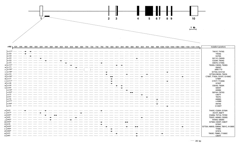
To investigate whether normal B cells display mutations in their BCL-6 gene, human B cell populations corresponding to GC centroblasts (CD38+CD77+) and centrocytes (CD38+CD77−) as well as naive B cells (sIgD+ CD27−) were isolated individually by fluorescence-activated cell sorting from a reactive tonsil and the peripheral blood of healthy donors. In these cells, we analyzed a BCL-6 742-bp genomic region previously shown to represent the major cluster of mutations in DLCL and FL (Fig.1). Using a multiplex seminested single-cell PCR approach, we generated amplicons from the BCL-6 intron 1 and the rearranged IgVH genes of the same cell and analyzed them by direct sequencing. In a subset (_n_= 36) of the cells, the nontranscribed β-globin gene was coamplified and sequenced as a control for locus specificity of the mutations.
Figure 1.
Distribution of mutations within the BCL-6 5′-noncoding sequences of 37 single GC B cells. (Upper) Schematic representation of the BCL-6 gene. Coding and noncoding exons are indicated by filled and empty boxes, respectively. The PCR fragment amplified for mutational analysis is approximately positioned below the map and blown up in the lower panel to show the distribution of mutations. Each line represents a 20-bp interval of the BCL-6 sequence amplified, and the first nucleotide of the BCL-6 cDNA is designated as position +1. Mutations included single base pair substitutions (closed ovals) and deletions (brackets). For each cell, identified by its code number, the type and exact position of the mutation are specified on the right column (Δ, deletion). Note that all of the 68 nucleotide exchanges, including deletions, were found in heterozygosis when both alleles were amplified (cells marked by an asterisk).
The results indicate that the BCL-6 gene is altered by somatic mutations in ≈30% GC lymphocytes but not in naive B cells (Table1; Fig. 1). No significant differences in the frequency of mutated cells were observed between the two GC-derived subpopulations, centroblasts and centrocytes. The percentage of mutated GC cells most likely represents an underestimate because only one allele could be amplified in 43% (50/116) of the cells analyzed. The allelic status could be assessed based on the presence of three linked polymorphisms [two previously described (9) and a G/A substitution in position +858] occurring in heterozygosis in the subject investigated (not shown). Sequencing analysis of BCL-6 amplicons in centroblasts and centrocytes revealed a frequency of mutations of 0.07 and 0.04 × 10⁻²/bp, respectively (range: 0–0.4 × 10⁻²/bp, corresponding to 0–3 mutations per allele). This frequency was significantly higher than that observed in naive B cells (0.009 × 10⁻²/bp; P = 0.01), which, in turn, was indistinguishable from the Taq misincorporation rate (see results of the β-globin analysis) (Table 1). Similarly, the average frequency of mutations in the IgVH genes from GC B cells was ≈5 × 10⁻²/bp, whereas most naive B cells displayed VH sequences in germline configuration, as expected (4, 23, 26). The 2 VH-mutated cells most likely represented accidentally sorted non-naive B cells. The BCL-6 sequences could not be analyzed in these cases because a positive PCR product was not obtained. The β-globin gene, successfully amplified in 32/36 centroblasts, was consistently found unmutated (Table 1). These results indicate that a cell-specific, locus-specific hypermutation mechanism targets BCL-6 sequences in normal GC cells. The frequency of mutations appeared to be 10- to 100-fold lower than that observed in rearranged IgVH genes from the same cells.
Table 1.
Single cell analysis of the BCL-6 and IgVHgenes in normal GC lymphocytes
| Cell phenotype | Mutated cells, %* | Mutations, %† | ||||
|---|---|---|---|---|---|---|
| BCL-6 | IgVH | β-globin | BCL-6 (742 bp) | IgVH (250 bp) | β-globin (395 bp) | |
| CD38+CD77− | 25/79 (31.6) | 11/14 (79) | nd | 40 (0.04)‡ | 141 (4.0) | nd |
| CD38+CD77+ | 12/37 (32.4) | 12/12 (100) | 1/32 (3) | 28 (0.07)§ | 184 (6.1) | 1 (0.004) |
| IgD+CD27− | 2/35 (5.7) | 2/16 (12.5) | nd | 3 (0.009)‡§ | 8 (0.2) | nd |
BCL-6 Mutations are Restricted to Neoplasms Derived from GC or Post-GC Cells and Carrying IgVH Mutations.
To corroborate the specific association among BCL-6 mutations, IgV mutations, and GC transit, we screened for BCL-6 and IgVHmutations a panel of B cell tumors representative of various stages of B cell differentiation, including pre-B cells (acute lymphoblastic leukemia: ALL), pre-GC (mantle cell lymphoma) (27), GC- (DLCL, FL, BL) and post GC- (multiple myeloma: MM) derived tumors. Thirty-three cases of chronic lymphocytic leukemia (CLL), which has been shown to harbor mutated IgV genes in up to 40% of cases (28), also were included in the panel, along with T cell-derived malignancies and non-lymphoid malignancies. Because tumors reflect the clonal history of their cell of origin, we postulated that if BCL-6 and IgV sequences were targeted by the same mechanism, the respective frequency and distribution of mutations should correlate in the various histologic categories.
A total number of 354 tumor samples was investigated for BCL-6 mutations by SSCP in the same region analyzed in normal cells. Sequence variants were found at significant frequency in lymphoid malignancies derived from GC (DLCL, FL, BL), post-GC (MM), and CD5+ B cells (CLL), but not from pre-B cells (0/19 ALL) or pre-GC B cells (1/19 mantle cell lymphoma) (Table 2). Two of 15 (13%) peripheral T cell lymphomas also displayed an altered migration pattern upon SSCP analysis; the occurrence of BCL-6 mutations in these cases may reflect the activity of the somatic hypermutation mechanism in CD4+ T cells within the GC, as suggested by the observation that these cells accumulate mutations in their T cell receptor α and β V genes (29, 30). This possibility could not be tested due to the lack of material from these particular cases. No BCL-6 mutations were found in non-lymphoid malignancies, which lack IgVH mutations.
Table 2.
Distribution of BCL-6 mutations in neoplastic diseases
| Histology | Mutated/tested | % | Normal counterpart |
|---|---|---|---|
| B cell malignancies | |||
| B-ALL | 0/19 | 0 | Pre-B cell |
| MCL | 1/19 | 5 | Pre-GC B cell |
| B-CLL | 5/33 | 15 | CD5+ B cell |
| FL | 10/27 | 37 | GC B cell |
| BL* | 11/30 | 37 | GC B cell |
| DLCL | 48/81 | 59 | GC B cell |
| MM | 19/58 | 33 | Post-GC B cell |
| Non-B cell malignancies | |||
| T cell neoplasms† | 2/35 | 6 | T cell |
| Myeloid leukemias‡ | 0/52 | 0 | Stem cell/myeloid cell |
| Solid tumors§ | 0/123 | 0 | Various |
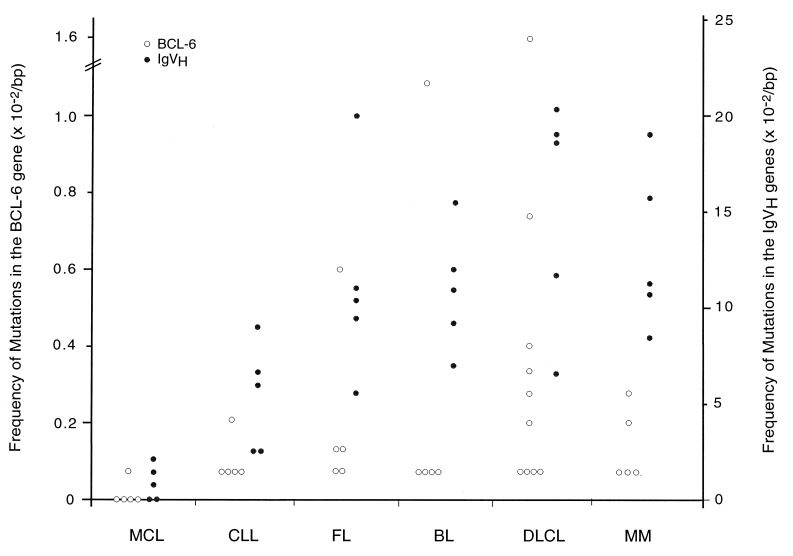
To comparatively examine the frequency and type of mutations occurring in the BCL-6 and IgVH genes, we analyzed the mutated sequences in a subset of cases representative of the main categories of B cell lymphomas, including mantle cell lymphoma, CLL, BL, FL, DLCL, and MM. The frequency of BCL-6 and IgVH mutations correlated in different tumor subtypes, with BCL-6 substitutions being consistently lower (Fig.2). In both genes, mutations were specifically associated with GC-transit; this was particularly evident in CLL cases, in which the occurrence of BCL-6 substitutions was restricted to the subset carrying IgVH mutations (28). These findings indicate that BCL-6 mutations are introduced in the same tumor types as IgV mutations, suggesting their derivation from a common mechanism.
Figure 2.
Comparative analysis of the mutation frequency in the BCL-6 5′-noncoding region and IgVH segments of B cell lymphoid malignancies. Five cases representative of the major histologic subtypes and showing BCL-6 alterations upon SSCP analysis were randomly selected, along with five mantle cell lymphomas. A 742-bp region of the BCL-6 first intron and the rearranged IgVHgenes from the same patients were amplified and directly sequenced. Filled and empty circles represent the frequency of mutations in the two loci. The majority of cases in each category harbored one to two nucleotide substitutions (0.068–0.13%); particularly high values were found in one FL (n = 9, 0.6%), one BL (n = 16, 1.1%), and various DLCL cases (n = 5–24, 0.34–1.6%).
Features of BCL-6 and IgVH Mutations in Normal and Transformed Cells.
The comparative analysis of 636 independent substitutions from the BCL-6 5′-noncoding region (n = 124) and the IgVH genes (n = 512) in normal GC cells and DLCL is summarized in Table3. For both BCL-6 and IgVH mutations, the frequency was higher in tumor than in normal GC cells. For both loci, mutations almost were exclusively represented by single base pair substitutions, and transitions were more common than transversions (BCL-6: 55% vs. 45% in normal B cells and 54.5% vs. 45.5% in DLCL, despite the potential for twice as many transversion events). In the two categories analyzed, small deletions represented 3% and 5.3% of the mutations, respectively. However, no significant differences in the frequency of mutations affecting each base were found after correction for the base composition of the BCL-6 region (in normal GC cells, C = 33%, G = 24%, T = 22%, and A = 21%; in DLCL, C, G, T, and A substitutions accounted for 25%, 27%, 23%, and 25%, respectively). Thus, the mutation pattern of BCL-6 does not indicate a preferential bias of A:N over T:N templates, suggesting the absence of strand polarity, a feature reported for IgV mutations (Table 3) (31).
Table 3.
Features of mutations in the BCL-6 5′-noncoding region and IgVH genes of normal GC cells vs. DLCL
| BCL-6 | IgVH | |||
|---|---|---|---|---|
| Normal GC | DLCL | Normal GC | DLCL | |
| Frequency × 10−2/bp (range) | 0.05 (0-0.4) | 0.23 (0-1.6)* | 5 (0-13) | 15.2 (6.3-20.5) |
| Single bp substitutions | 66 | 53 | 323 | 187 |
| Deletions | 2 | 3 | 2 | 0 |
| Transitions/transversions† | 1.2 | 1.2 | 1.4 | 1.2 |
| Strand polarity†‡ | 1 | 1.1 | 1.9 | 1.5 |
| RGYW bias§ | 0.21 (<0.05) | 1.2 (<0.05) | 21.3 (<0.001) | 53 (<0.001) |
A variety of studies have recognized specific nucleotide motifs within the Ig genes as intrinsic hotspots for somatic hypermutation: among them, the consensus RGYW (where R = purine, Y = pyrimidine, and W = A or T) appears to be the most frequently mutated one (31–33). To determine whether there is preferential targeting of the RGYW motif in the BCL-6 locus, the number of G mutations within the RGYW motif was normalized for its frequency in the region investigated and compared with the expected frequency of mutations in both normal GC B cells and DLCLs. For BCL-6, the actual occurrence of bp changes within the RGYW motif was found to differ from that expected by chance alone (P < 0.05), suggesting the existence of a preferential targeting. This bias, however, was less evident than in the IgVH sequences (P < 0.001) (Table 3). Taken together, these observations indicate that the features of BCL-6 and IgV mutations are similar, consistent with both sets of mutations being produced by the same mechanism.
DISCUSSION
This study reports that the 5′-noncoding region of the BCL-6 gene is targeted by a somatic hypermutation mechanism operating in normal GC B cells. An analysis of transformed counterparts of B cells at various differentiation stages confirms a close association among BCL-6 mutations, IgV mutations, and GC transit. These findings provide evidence that somatic hypermutation is not limited to the Ig locus in B cells, with implications for the mechanism of hypermutation as well as its role in B cell function and lymphomagenesis.
Relationship Between BCL-6 and IgV Mutations.
Several observations suggest that BCL-6 mutations may be due to the same mechanism generating IgV mutations in B cells. First, our results show that in both normal and malignant cells BCL-6 mutations selectively occur in GCs, the physiologic site of IgV hypermutation (1–4). The detection of BCL-6 mutations in a subset of CLL cases also is consistent with this notion because the same cases displayed nucleotide exchanges in their IgVH loci, indicating a GC transit of the putative precursor cell. Second, BCL-6 mutations share most of the features of IgV mutations, including the preference for single base pair substitutions with a small number of deletions, the predominance of transitions over transversions, and some degree of preferential motif (RGYW) targeting (22, 31–35). Third, both BCL-6 and IgV mutations are associated with transcribed sequences. Intriguingly, BCL-6 mutations, as IgV mutations, are scattered within 2 kb from the transcriptional initiation site (36). The frequency of BCL-6 mutations appears to be significantly lower than that of IgVH mutations in both normal and neoplastic cells. The basis for this difference is not known, but it does not seem to correlate with a lower transcription rate, because the BCL-6 gene is transcribed at high levels in GC cells (16). While this manuscript was in preparation, another study has reported the occurrence of BCL-6 mutations in normal B cells (37). The frequency, type, and distribution of nucleotide substitutions are similar in the two studies, although a strand bias was noted by Shen et al. (37) that was not detectable in our analysis. Overall, these data strongly suggest that BCL-6 and IgV mutations represent the product of the same mechanism.
Implications for the Somatic Hypermutation Mechanism.
The identification of a new locus undergoing somatic hypermutation in normal B cells prompted a reexamination of some of the known functional requirements for the IgV hypermutation mechanism. The BCL-6 5′-noncoding region and the IgV sequences do not share primary sequence homologies, consistent with the observation that somatic hypermutation is a target sequence-independent process and does not require a specific promoter (7, 8). The occurrence of BCL-6 mutations downstream of the promoter region is comparable with that observed in rearranged IgV genes, suggesting that the hypermutation process may be targeted to a specific distance from the transcription initiation site (7, 33). As IgV hypermutation requires the Ig enhancer, it is conceivable that the BCL-6 locus also contains a _cis_-acting transcriptional control element structurally or functionally similar to the Ig enhancer. The identification of such an element should provide insight into the mechanism by which hypermutation is targeted to specific loci.
Role of BCL-6 Mutations in Normal and Transformed B Cells.
The functional significance of mutations introduced into the BCL-6 5′-regulatory region of normal B cells is unknown. Because these mutations have been found at a comparable frequency in normal memory B cells (37) as well as in multiple myeloma, which represents transformed plasma cells, our study indicates that cells carrying BCL-6 mutations are neither significantly counterselected nor positively selected in the GC. The pattern of mutations is not clearly different in normal and malignant B cells, suggesting that most of the mutations found in tumors may not have any pathologic effect. However, initial studies on several tumor-derived BCL-6 alleles indicate that few mutations can significantly deregulate BCL-6 expression, whereas others are apparently functionally irrelevant or associated with silent alleles (unpublished observations). The ≈740-bp intronic sequence corresponding to the major cluster of BCL-6 mutations contains several regions of high evolutionary conservation, suggesting that some mutations may hit regulatory domains of the BCL-6 gene and influence its mode of expression. A functional dissection of the BCL-6 5′-noncoding region is needed to address these issues.
Finally, the identification of BCL-6 mutations suggests the possibility that other sequences may be subjected to the hypermutation process in GCs. Thus, it is possible that the role of somatic hypermutation is not limited to generate antibody diversity in B cells. Structural and functional similarities between BCL-6 and Ig loci may be useful to identify other possible targets of the hypermutation process.
Acknowledgments
We thank Ulla Beauchamp for expert assistance with DNA sequencing and Alexander Scheffold for a kind gift of the anti-Digoxigenin liposomes. This study was supported in part by National Institutes of Health Grants CA-44029 and CA-75553 and by a grant from the Associazione Italiana per la Ricerca sul Cancro (to A.N.). L.P. is a recipient of a Fellowship from the Associazione Italiana per la Ricerca sul Cancro.
ABBREVIATIONS
BCL
B Cell Lymphoma
GC
germinal center
IgV
variable region of Ig
IgVH
Ig heavy-chain V
SSCP
single strand conformation polymorphism
CLL
chronic lymphocytic leukemia
DLCL
diffuse large cell lymphoma
FL
follicular lymphoma
BL
Burkitt lymphoma
Footnotes
†
These authors contributed equally to this article.
References
- Rajewsky K. Nature (London) 1996;381:751–758. doi: 10.1038/381751a0. [DOI] [PubMed] [Google Scholar]
- 2.Jacob J, Kelsoe G, Rajewsky K, Weiss U. Nature (London) 1991;354:389–392. doi: 10.1038/354389a0. [DOI] [PubMed] [Google Scholar]
- 3.Berek C, Berger A, Apel M. Cell. 1991;67:1121–1129. doi: 10.1016/0092-8674(91)90289-b. [DOI] [PubMed] [Google Scholar]
- 4.Küppers R, Zhao M, Hansmann M L, Rajewsky K. EMBO J. 1993;12:4955–4967. doi: 10.1002/j.1460-2075.1993.tb06189.x. [DOI] [PMC free article] [PubMed] [Google Scholar]
- 5.Klein U, Goossens T, Fischer M, Kanzler H, Braeuninger A, Rajewsky K, Küppers R. Immunol Rev. 1998;162:261–280. doi: 10.1111/j.1600-065x.1998.tb01447.x. [DOI] [PubMed] [Google Scholar]
- 6.Betz A G, Milstein C, Gonzalez-Fernandez A, Pannell R, Larson T, Neuberger M S. Cell. 1994;77:239–248. doi: 10.1016/0092-8674(94)90316-6. [DOI] [PubMed] [Google Scholar]
- 7.Peters A, Storb U. Immunity. 1996;4:57–65. doi: 10.1016/s1074-7613(00)80298-8. [DOI] [PubMed] [Google Scholar]
- 8.Yelamos J, Klix N, Goyenechea B, Lozano F, Chui Y L, Gonzalez Fernandez A, Pannell R, Neuberger M S, Milstein C. Nature (London) 1995;376:225–229. doi: 10.1038/376225a0. [DOI] [PubMed] [Google Scholar]
- 9.Migliazza A, Martinotti S, Chen W, Fusco C, Ye B H, Knowles D M, Offit K, Chaganti R S K, Dalla-Favera R. Proc Natl Acad Sci USA. 1995;92:12520–12524. doi: 10.1073/pnas.92.26.12520. [DOI] [PMC free article] [PubMed] [Google Scholar]
- 10.Gaidano G, Carbone A, Pastore C, Capello D, Migliazza A, Gloghini A, Roncella S, Ferrarini M, Saglio G, Dalla-Favera R. Blood. 1997;89:3755–3762. [PubMed] [Google Scholar]
- 11.Ye B H, Lista F, Lo Coco F, Knowles D M, Offit K, Chaganti R S K, Dalla-Favera R. Science. 1993;262:747–750. doi: 10.1126/science.8235596. [DOI] [PubMed] [Google Scholar]
- 12.Kerckaert J P, Deweindt C, Tilly H, Quief S, Lecocq G, Bastard C. Nat Genet. 1993;5:66–70. doi: 10.1038/ng0993-66. [DOI] [PubMed] [Google Scholar]
- 13.Chang C C, Ye B H, Chaganti R S K, Dalla-Favera R. Proc Natl Acad Sci USA. 1996;93:6947–6952. doi: 10.1073/pnas.93.14.6947. [DOI] [PMC free article] [PubMed] [Google Scholar]
- 14.Seyfert V L, Allman D, He Y, Staudt L M. Oncogene. 1996;12:2331–2342. [PubMed] [Google Scholar]
- 15.Deweindt C, Albagli O, Bernardin F, Dhordain P, Quief S, Lantoine D, Kerckaert J P, Leprince D. Cell Growth Differ. 1995;6:1495–1503. [PubMed] [Google Scholar]
- 16.Cattoretti G, Chang C C, Cechova K, Zhang J, Ye B H, Falini B, Louie D C, Offit K, Chaganti R S K, Dalla-Favera R. Blood. 1995;86:45–53. [PubMed] [Google Scholar]
- 17.Onizuka T, Moriyama M, Yamochi T, Kuroda T, Kazama A, Kanazawa N, Sato K, Kato T, Ota H, Mori S. Blood. 1995;86:28–37. [PubMed] [Google Scholar]
- 18.Ye B H, Cattoretti G, Shen Q, Zhang J, Hawe N, de Waard R, Leung C, Nouri-Shirazi M, Orazi A, Chaganti R S K, et al. Nat Genet. 1997;16:161–170. doi: 10.1038/ng0697-161. [DOI] [PubMed] [Google Scholar]
- 19.Dent A L, Shaffer A L, Yu X, Allman D, Staudt L M. Science. 1997;276:589–592. doi: 10.1126/science.276.5312.589. [DOI] [PubMed] [Google Scholar]
- 20.Fukuda T, Yoshida T, Okada S, Hatano M, Miki T, Ishibashi K, Okabe S, Koseki H, Hirosawa S, Taniguchi M, et al. J Exp Med. 1997;186:439–448. doi: 10.1084/jem.186.3.439. [DOI] [PMC free article] [PubMed] [Google Scholar]
- 21.Küppers R, Hansmann M L, Rajewsky K. In: Weir’s Handbook of Experimental Immunology. Herzenberg L A, Weir D M, Blackwell D, editors. Oxford: Blackwell Scientific; 1997. pp. 206.1–206.4. [Google Scholar]
- 22.Goossens T, Klein U, Küppers R. Proc Natl Acad Sci USA. 1998;95:2463–2468. doi: 10.1073/pnas.95.5.2463. [DOI] [PMC free article] [PubMed] [Google Scholar]
- 23.Klein U, Küppers R, Rajewsky K. Eur J Immunol. 1993;23:3272–3277. doi: 10.1002/eji.1830231232. [DOI] [PubMed] [Google Scholar]
- 24.Scheffold A, Miltenyi S, Radbruch A. Immunotechnol. 1995;1:127–137. doi: 10.1016/1380-2933(95)00014-3. [DOI] [PubMed] [Google Scholar]
- 25.Küppers R, Zhao M, Rajewsky K, Hansmann M L. Am J Pathol. 1993;143:230–239. [PMC free article] [PubMed] [Google Scholar]
- 26.Klein, U., Rajewsky, K. & Küppers, R. (1998) J. Exp. Med., in press. [DOI] [PMC free article] [PubMed]
- 27.Weisenburger D D. In: Neoplastic Hematopathology. Knowles D M, editor. Baltimore: Williams & Wilkins; 1996. pp. 617–628. [Google Scholar]
- 28.Schroeder H W, Jr, Dighiero G. Immunol Today. 1994;15:288–294. doi: 10.1016/0167-5699(94)90009-4. [DOI] [PubMed] [Google Scholar]
- 29.Zheng B, Xue W, Kelsoe G. Nature (London) 1994;372:556–559. doi: 10.1038/372556a0. [DOI] [PubMed] [Google Scholar]
- 30.Cheynier R, Henrichwark S, Wain-Hobson S. Eur J Immunol. 1998;28:1604–1610. doi: 10.1002/(SICI)1521-4141(199805)28:05<1604::AID-IMMU1604>3.0.CO;2-R. [DOI] [PubMed] [Google Scholar]
- 31.Betz A G, Rada C, Panell R, Milstein C, Neuberger M S. Proc Natl Acad Sci USA. 1993;90:2385–2388. doi: 10.1073/pnas.90.6.2385. [DOI] [PMC free article] [PubMed] [Google Scholar]
- 32.Rogozin I B, Kolchanov N A. Biochim Biophys Acta. 1992;1171:11–18. doi: 10.1016/0167-4781(92)90134-l. [DOI] [PubMed] [Google Scholar]
- 33.Neuberger M S, Ehrenstein M R, Klix N, Jolly C J, Yelamos J, Rada C, Milstein C. Immunol Rev. 1998;162:107–116. doi: 10.1111/j.1600-065x.1998.tb01434.x. [DOI] [PubMed] [Google Scholar]
- 34.Golding G B, Gearhart P J, Glickman B W. Genetics. 1987;115:169–176. doi: 10.1093/genetics/115.1.169. [DOI] [PMC free article] [PubMed] [Google Scholar]
- 35.Dorner T, Brezinschek H P, Brezinschek R I, Foster S J, Domiati-Saad R, Lipsky P E. J Immunol. 1997;158:2779–2789. [PubMed] [Google Scholar]
- 36.Lebecque S G, Gearhart P J. J Exp Med. 1990;172:1717–1727. doi: 10.1084/jem.172.6.1717. [DOI] [PMC free article] [PubMed] [Google Scholar]
- 37.Shen H M, Peters A, Baron B, Zhu X, Storb U. Science. 1998;280:1750–1752. doi: 10.1126/science.280.5370.1750. [DOI] [PubMed] [Google Scholar]

