PTPα regulates integrin-stimulated FAK autophosphorylation and cytoskeletal rearrangement in cell spreading and migration (original) (raw)
Abstract
We investigated the molecular and cellular actions of receptor protein tyrosine phosphatase (PTP) α in integrin signaling using immortalized fibroblasts derived from wild-type and PTPα-deficient mouse embryos. Defects in PTPα−/− migration in a wound healing assay were associated with altered cell shape and focal adhesion kinase (FAK) phosphorylation. The reduced haptotaxis to fibronectin (FN) of PTPα−/− cells was increased by expression of active (but not inactive) PTPα. Integrin-mediated formation of src–FAK and fyn–FAK complexes was reduced or abolished in PTPα−/− cells on FN, concomitant with markedly reduced phosphorylation of FAK at Tyr397. Reintroduction of active (but not inactive) PTPα restored FAK Tyr-397 phosphorylation. FN-induced cytoskeletal rearrangement was retarded in PTPα−/− cells, with delayed filamentous actin stress fiber assembly and focal adhesion formation. This mimicked the effects of treating wild-type fibroblasts with the src family protein tyrosine kinase (Src-PTK) inhibitor PP2. These results, together with the reduced src/fyn tyrosine kinase activity in PTPα−/− fibroblasts (Ponniah et al., 1999; Su et al., 1999), suggest that PTPα functions in integrin signaling and cell migration as an Src-PTK activator. Our paper establishes that PTPα is required for early integrin-proximal events, acting upstream of FAK to affect the timely and efficient phosphorylation of FAK Tyr-397.
Keywords: Src-PTKs; protein tyrosine phosphatase; focal adhesion; actin
Introduction
The transmembrane integrins bind to ECM proteins such as fibronectin (FN)* to generate intracellular tyrosine phosphorylation–based signals that regulate cell growth, migration, and survival. Many of the signals resulting from integrin ligation are centered around the focal adhesion–associated tyrosine kinase FAK and its interactions with src family protein tyrosine kinases (Src-PTKs; for review see Schlaepfer et al., 1999; Schaller, 2001). Integrin stimulation induces FAK autophosphorylation at Tyr397, creating a binding site for the src homology 2 (SH2) domain of the Src-PTKs src or fyn (Cobb et al., 1994; Schaller et al., 1994; Xing et al., 1994; Cary et al., 1996). The recruited, active Src-PTK phosphorylates several other tyrosine residues in FAK to affect both full FAK activation and the creation of phosphotyrosine binding sites for other signaling molecules, such as Grb2 (Schlaepfer et al., 1994; Calalb et al., 1995). The Src–PTK–FAK complex mediates the phosphorylation of certain FAK-associated proteins such as p130cas and paxillin, and their recruitment of still other signaling molecules (such as phosphotyrosine-dependent Crk binding to p130cas and paxillin, and Nck binding to p130cas), leading to formation of a multi-phosphocomponent signaling complex localized at focal adhesions (Schaller and Parsons, 1995; Hamasaki et al., 1996; Vuori et al., 1996; Schlaepfer et al., 1997, 1998). Integrin-stimulated focal adhesion formation, cell spreading, and motility also involve reorganization of the actin cytoskeleton, in which Rho family GTPases play key roles (Barry et al., 1997; Clark et al., 1998; Price et al., 1998; Ren et al., 1999). The molecular mechanisms regulating the early events in the above processes and linking them to the cell surface integrins are unclear.
In addition to tyrosine kinases, several intracellular protein tyrosine phosphatases (PTPs) have been implicated as positive and negative regulators of integrin-mediated signaling (Angers-Loustau et al., 1999a). Key signaling components such as Src-PTKs, small GTPases, FAK, and p130cas appear to be regulated by more than one PTP, probably reflecting the numerous signal inputs that can be integrated by this pathway and the dynamic nature of processes such as focal adhesion formation and dissolution. Spreading and migration defects are observed in fibroblasts prepared from gene-targeted mice that are null or functionally null for the intracellular PTPs SHP-2, PTP 1B, and PTP-PEST (Yu et al., 1998; Angers-Loustau et al., 1999b; Oh et al., 1999; Cheng et al., 2001). Multiple actions reported for SHP-2 include the SHPS-1–mediated activation of Src-PTKs (Oh et al., 1999), the inactivation and activation of RhoA (Schoenwaelder et al., 2000; Lacalle et al., 2002), and the regulation of focal adhesion turnover by FAK dephosphorylation (Yu et al., 1998). PTP 1B may also function as an upstream activator of Src-PTKs (Arregui et al., 1998; Cheng et al., 2001), and like PTP-PEST, can dephosphorylate p130cas to modulate focal adhesion turnover (Liu et al., 1998; Angers-Loustau et al., 1999b; Garton and Tonks, 1999). Additionally, PTEN dephosphorylates the FAK autophosphorylation site during cell detachment, and acts as a lipid phosphatase to negatively regulate rac1 and Cdc42 GTPases and cell migration (Tamura et al., 1998, 1999; Liliental et al., 2000).
Just two receptor PTPs have been associated with integrin signaling; LAR and PTPα. LAR localizes to focal adhesions and may be integrated into cell migration pathways through its interaction with the guanine nucleotide exchange factor Trio (Serra-Pages et al., 1995; Debant et al., 1996; Seipel et al., 1999), or into cell survival pathways through its action in dephosphorylating and destabilizing p130cas (Weng et al., 1999). PTPα can also be found in focal adhesions (Lammers et al., 2000). Cells overexpressing PTPα show enhanced cell–substratum association (Harder et al., 1998), and cells lacking PTPα show defective spreading on FN and reduced tyrosine phosphorylation of FAK and p130cas (Su et al., 1999). PTPα-overexpressing cells exhibit increased Src-PTK activity, and PTPα−/− fibroblasts have reduced Src-PTK activity (Zheng et al., 1992; den Hertog et al., 1993; Bhandari et al., 1998; Harder et al., 1998; Ponniah et al., 1999; Su et al., 1999), suggesting that PTPα may exert effects on integrin signals through modulating Src-PTK activities. Also, studies with a substrate-trapping PTPα mutant have identified p130cas as a PTPα substrate (Buist et al., 2000). Collectively, the above findings show that PTPs intersect with PTKs at multiple points downstream of the integrins, and that ablated or increased PTP activity has profound effects on integrin-mediated processes.
Here, we describe studies comparing wild-type and PTPα−/− embryonic fibroblasts to define the molecular role of PTPα in integrin-stimulated events. Novel defects in migration and haptotaxis, and in Src-PTK association with FAK were found in the PTPα−/− cells. The reduced association between src/fyn and FAK was found to be due to the striking abrogation of FAK Tyr-397 phosphorylation. Reintroduction of wild-type but not catalytically inactive forms of PTPα into the cells restored FAK Tyr-397 phosphorylation, demonstrating that the phosphatase activity of PTPα is required for its function in this context. The lack of PTPα mimics the effects observed on treatment of fibroblasts with the selective Src-PTK inhibitor PP2, not only in preventing optimal phosphorylation of FAK at Tyr-397 (Salazar and Rozengurt, 2001), but also in delaying integrin-mediated rearrangement of the actin cytoskeleton and focal adhesion formation. These findings point to a site of action of PTPα proximal to receptor integrins and upstream of FAK. We propose that through its action as an Src-PTK activator, PTPα functions as a previously unidentified link between integrin engagement and the ensuing FAK autophosphorylation that promotes numerous downstream signaling events.
Results
Impaired PTPα−/− cell migration
The migration abilities of wild-type and PTPα−/− fibroblasts were examined in a cell culture wound healing assay. Confluent dishes of cells were “wounded” by scraping with a pipette tip, creating a space free of cells. A significant delay in the ability of the PTPα−/− cells to migrate into the empty space was observed (Fig. 1 A), with the wild-type cells closing the gap by 15 h, and the PTPα−/− cells still not able to completely fill the gap even after 24 h. Examination of the leading edge of the migrating cells revealed multiple protrusions and extensions in wild-type cells, but a relatively uniform flat edge on the PTPα−/− cells (Fig. 1, B and C). ECM-integrin signaling events are prominently involved in regulating cell migration, and FAK is a key mediator of this process. As our subsequent investigation of integrin-stimulated FAK phosphorylation revealed defects in FAK Tyr-397 phosphorylation in PTPα-deficient cells (see Results), we examined this in cells at the leading edge of the wound (Fig. 1 C). At a time just before wound closure (about a one-cell wide gap remaining between wild-type cells), the wild-type cells appeared to have more phospho-Tyr397 FAK than PTPα−/− cells. The phospho-Tyr397 FAK was localized within elongated, well-defined structures (likely focal adhesions) in multiple protrusions from the wild-type cells, whereas in PTPα−/− cells, the phospho-Tyr397 FAK appeared to be localized in smaller, round structures. We also analyzed haptotactic migration of the above cells and another independently derived PTPα−/− fibroblast line to the integrin ligand FN in a Transwell chamber assay. Both PTPα−/− lines of cells showed significantly impaired migration (67 and 49%) relative to the PTPα+/+ control cell line (Fig. 2 A), indicating a defect in FN-stimulated responses. To determine whether reintroduction of PTPα would restore migration ability, PTPα or a catalytically defective mutant of PTPα lacking the active site cysteine residues (C414S/C704S) were introduced into PTPα−/− cells by adenoviral infection. Expression of PTPα increased the percentage of migrating cells from 49 to 64% (Fig. 2 B). The extent of restored migration may be low because Western blotting revealed that the introduced PTPα was always less than that present in wild-type cells (unpublished data); nevertheless, this represents a significant (P < 0.005) 1.3-fold increase in haptotaxis. Expression of inactive PTPα had no effect on the number of migrating cells (Fig. 2 B). Together, the above results indicate that efficient cell migration from a wounded cell monolayer and in haptotaxis requires catalytically active PTPα, and that PTPα may function in these processes to promote FAK Tyr-397 phosphorylation and the formation of membrane extensions characteristic of migrating cells.
Figure 1.
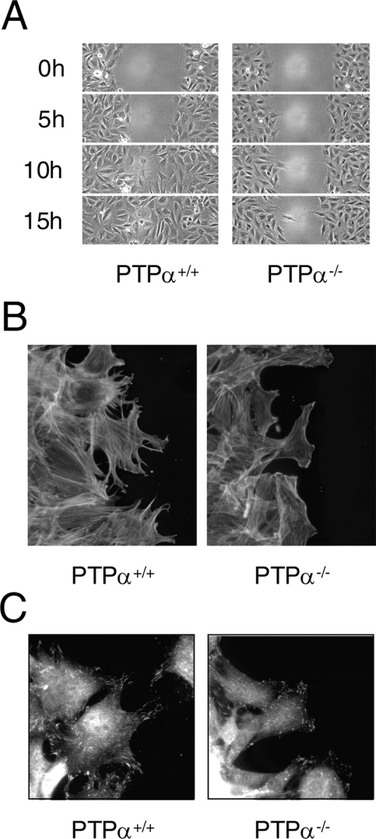
Defective migration of PTPα − / − cells. (A) Delayed migration of cells lacking PTPα. The migration of PTPα+/+ and PTPα−/− fibroblasts into an empty area of the culture dish was followed by time-lapse video microscopy. Fields at the indicated times are shown. (B) 6 h after wounding, cells at the migrating edge were stained for actin with Alexa Fluor 488–conjugated phalloidin. (C) Cells at the leading edge were stained with anti-phospho-Tyr397 FAK antibody just before wound closure of the PTPα+/+ cells.
Figure 2.
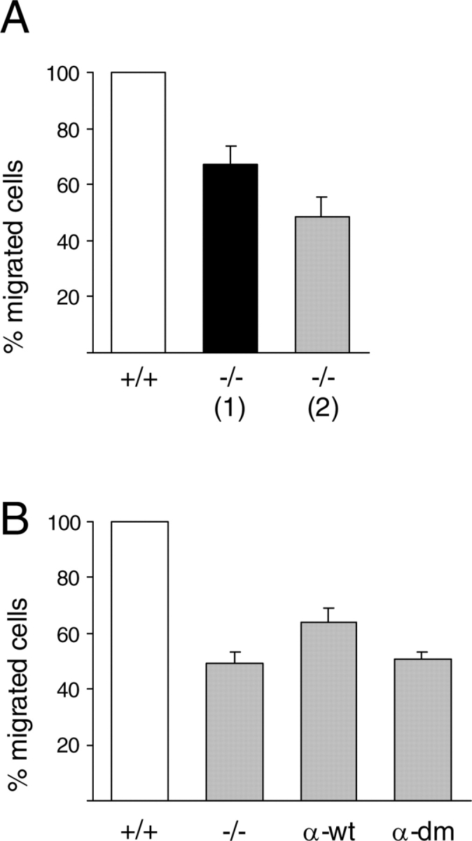
Haptotactic migration toward FN. (A) Wild-type fibroblasts (+/+) and two independently derived lines of PTPα−/− fibroblasts (−/−) were analyzed for migration toward FN as described in Materials and methods. Each bar represents the average of four independent experiments, each experiment with three wells per cell type, ± S.D. The number of migrating wild-type cells was taken as 100%, and other values were calculated relative to this. (B) In other experiments, wild-type fibroblasts (+/+), line 2 PTPα−/− fibroblasts (−/−), and line 2 PTPα−/− fibroblasts infected with adenovirus expressing PTPα (α-wt) or catalytically inactive double mutant PTPα (α-dm) were analyzed for migration toward FN. Each bar represents the average of two independent experiments, each experiment with two wells per cell type, ± S.D.
Reduced association of src and fyn with FAK
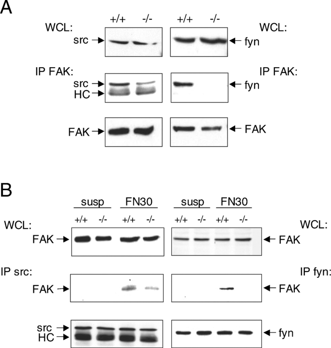
Formation of a FAK–src complex is essential for full phosphorylation and activation of FAK (Owen et al., 1999; Schlaepfer et al., 1999; Ruest et al., 2000) and phosphorylation of FAK-associated substrates such as p130cas, which are known to be required for cell motility (Vuori et al., 1996; Klemke et al., 1998). FAK has also been found to associate with the Src-PTK fyn (Cobb et al., 1994; Cary et al., 1996). Thus, we examined the association of FAK with src and fyn in adherent PTPα−/− cells. Both src and fyn were complexed with FAK in wild-type cells, as evidenced by probing FAK immunoprecipitates with anti-src or -fyn antibodies (Fig. 3 A). However, a greatly decreased amount of src was found in association with FAK in PTPα−/− cells, amounting to 38% ± 8 (n = 3) of the src detected in association with FAK in wild-type cells. Furthermore, the association of fyn with FAK was abolished in PTPα−/− cells (Fig. 3 A). FAK-src and FAK-fyn association was also examined in cells after plating on FN. This time, the src or fyn was immunoprecipitated and immunoblotted to detect associated FAK. FAK was not complexed with src or fyn in either wild-type or PTPα−/− cells held in suspension. Plating of wild-type fibroblasts on the FN-coated dishes induced the association of src and fyn with FAK, but in PTPα−/− cells there was a reduced amount of FAK, or no FAK, detected in association with src or fyn, respectively (Fig. 3 B).
Figure 3.
Reduced association of Src-PTKs and FAK in PTPα − / − cells. (A) Reduced src/fyn-FAK interaction in PTPα−/− cells cultured on plastic dishes. Lysates (WCL) of PTPα+/+ and PTPα−/− cells cultured on plastic tissues culture dishes in serum-containing medium were resolved by SDS-PAGE and immunoblotted with anti-src antibodies (top left panel) or anti-fyn antibodies (top right panel). FAK immunoprecipitates were probed with anti-src antibodies (middle left panel), anti-fyn antibodies (middle right panel), or anti-FAK antibodies (bottom panels). HC indicates the antibody heavy chain. (B) Reduced src/fyn-FAK interaction in PTPα−/− cells plated on FN-coated plastic dishes. Lysates (WCL) were prepared from PTPα+/+ and PTPα−/− cells in suspension (susp) or after plating onto FN-coated dishes for 30 min (FN30) and probed with anti-FAK antibodies (top panel). Src (middle and bottom left panels) and fyn (middle and bottom right panels) immunoprecipitates prepared from the cell lysates were probed for the presence of FAK (middle panel), src (bottom left panel), or fyn (bottom right panel). HC indicates the antibody heavy chain.
Integrin-stimulated phosphorylation of FAK Tyr-397 is reduced in PTPα−/−cells
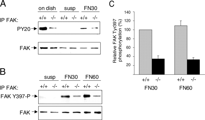
As src and fyn bind to phospho-Tyr397 of FAK, reduced FAK Tyr-397 autophosphorylation in the PTPα−/− cells could account for less src/fyn binding. The overall phosphotyrosine content of FAK was less in PTPα−/− cells than in PTPα+/+ cells, both under normal culture conditions and after plating on FN (Fig. 4 A). The phosphorylation status of FAK Tyr-397 was examined using an anti-FAK phospho-Tyr397–specific antibody. No phosphorylation of FAK Tyr-397 was detected in any cells in suspension. The phosphorylation of Tyr397 of FAK was consistently observed to be reduced in PTPα−/− cells, compared with wild-type cells, on FN-induced integrin activation (Fig. 4, B and C).
Figure 4.
Integrin-stimulated FAK Tyr-397 phosphorylation is impaired in PTPα-null cells. (A) Reduced tyrosine phosphorylation of FAK in PTPα−/− cells. FAK immunoprecipitates from PTPα+/+ and PTPα−/− cells adhering to plastic dishes (on dish), retained in suspension (susp), or plated onto FN-coated dishes for 30 min (FN30), were probed with anti-phosphotyrosine antibodies (top panel) or with anti-FAK antibodies (bottom panel). (B) Reduced FAK Tyr-397 phosphorylation in PTPα−/− cells. FAK immunoprecipitates from PTPα+/+ and PTPα−/− cells retained in suspension (susp) or plated onto FN-coated dishes for 30 min (FN30) or 60 min (FN60) were probed with anti-phospho-Tyr397–specific antibodies (top panel), or anti-FAK antibodies (bottom panel). (C) Integrin-stimulated FAK Tyr-397 phosphorylation was quantitated from at least seven independent experiments such as that presented in B and is shown as the mean ± S.D. FAK Tyr-397 phosphorylation in PTPα+/+ cells plated on FN for 30 min was taken as 100%, and the other data was calculated relative to this.
The altered FAK phosphorylation was also confirmed by visualization in cells attached to an FN substratum (Fig. 5). After plating of the cells on FN-coated dishes for 30, 60, and 120 min, the cells were processed for indirect immunofluorescent labeling with anti-vinculin and anti-FAK phosphospecific-Tyr397 antibodies. In wild-type cells, phospho-Tyr397 FAK was localized in focal adhesions present in multiple cell extensions after 30 min on FN. In contrast, PTPα−/− cells possessed membrane ruffles, but virtually no extensions or focal adhesions and mainly nuclear/perinuclear labeling with the anti-FAK Tyr-397 antibody (Fig. 5, A and B; top panels). After 60 min on FN, phospho-Tyr397 FAK was detected in thickened elongated focal adhesions in numerous well-defined cell extensions around the entire periphery of the wild-type cells, and had begun to appear in thin streaky focal adhesions in the outer periphery of PTPα−/− cells, although with less intensity than in the wild-type cells and in the absence of well-defined cell extensions (Fig. 5, A and B; middle panels). After 120 min on FN, the wild-type and PTPα−/− cells appeared similar in terms of FAK Tyr-397 staining and focal adhesion formation (Fig. 5, A and B; bottom panels). These observations suggest that relative to wild-type cells, there is an early delay or impairment of integrin-stimulated FAK Tyr-397 phosphorylation and formation of focal adhesions in the PTPα−/− cells, but that after 120 min on FN, these processes have reached approximately equivalent points in the wild-type and PTPα−/− cells.
Figure 5.
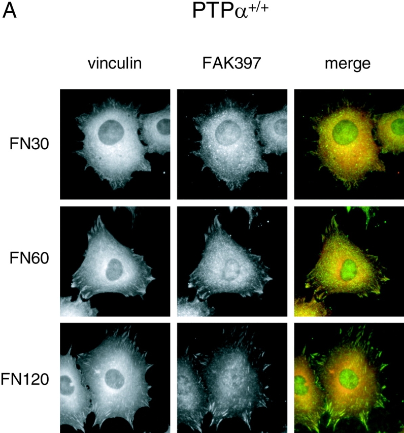
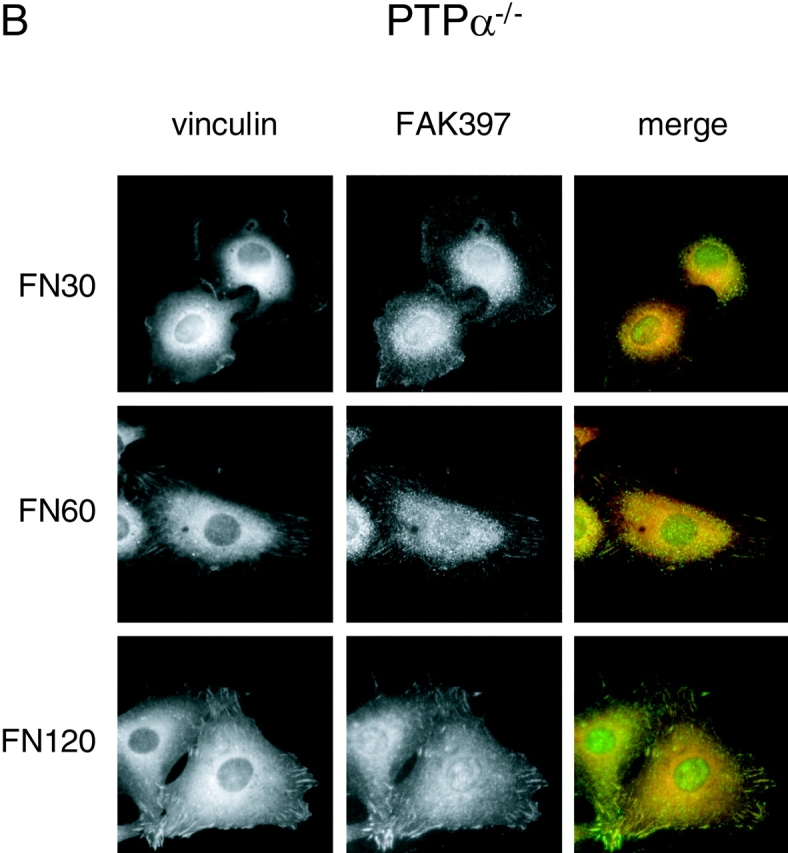
Localization of vinculin and appearance of FAK Tyr-397 phosphorylation in integrin-stimulated cells. Cells were allowed to adhere to FN-coated glass coverslips for 30 min (FN30), 60 min (FN60), and 120 min (FN120), and were then fixed and labeled with mouse monoclonal anti–vinculin antibody (red) and rabbit polyclonal antibodies specific to FAK phospho-Tyr397 (green). (A) Wild-type (PTPα+/+) cells. (B) PTPα−/− cells.
Rescue of FAK Tyr-397 phosphorylation by reintroduction of catalytically active PTPα
To confirm that the defective FAK autophosphorylation in the PTPα−/− cells was a direct result of the absence of PTPα, we examined the consequences of PTPα reexpression. We were unable to generate PTPα-expressing cells through stable transfection and selection, as reported by others and possibly due to toxic effects of high levels of the wild-type phosphatase (Lammers et al., 2000). To obtain high efficiency expression in the PTPα−/− cells, we used adenovirus-mediated expression of wild-type or a catalytically inactive mutant form of PTPα with the active site cysteine residues mutated to serine in both catalytic domains. As shown in Fig. 6 A, PTPα+/+ cells contain three immunodetectable forms of PTPα, all of which are absent in the PTPα−/− cells. The proteins with apparent molecular masses of 130 and 100 kD represent the fully glycosylated and unglycosylated forms of PTPα (Daum et al., 1994), whereas the ∼80-kD form may be a proteolyzed or processed product of 130-kD PTPα (Su et al., 1994, Lammers et al., 2000; Gill-Henn et al., 2001). After adenovirus infection of the PTPα−/− cells, we could detect low levels of the fully glycosylated 130-kD PTPα, trace amounts of the unglycosylated 100-kD PTPα, and amounts of the ∼80-kD PTPα comparable to that in the PTPα+/+ cells (Fig. 6 A). The contribution of the ∼80 kD form to PTPα-dependent integrin signaling events is unknown. In three independent experiments (Fig. 6 B), integrin-stimulation of the PTPα−/− cells induced FAK Tyr-397 phosphorylation to 56% (±2) of that quantitated in PTPα+/+ cells, and reintroduction of wild-type PTPα significantly increased FAK Tyr-397 phosphorylation to 85% (±11). However, expression of catalytically inactive PTPα in the PTPα−/− cells did not significantly affect FAK Tyr-397 phosphorylation (52% ± 17). This confirms that it is the lack of PTPα that causes this defect in integrin signaling, and that the catalytic activity of PTPα is required for FAK Tyr-397 phosphorylation to occur efficiently. The somewhat more efficient restoration of FAK Tyr-397 phosphorylation than of migration affected by reintroduction of PTPα into PTPα-deficient cells (a 1.5-fold vs. a 1.3-fold increase, respectively) may reflect PTPα expression differences, either in total PTPα or in the relative amounts of the three PTPα forms.
Figure 6.
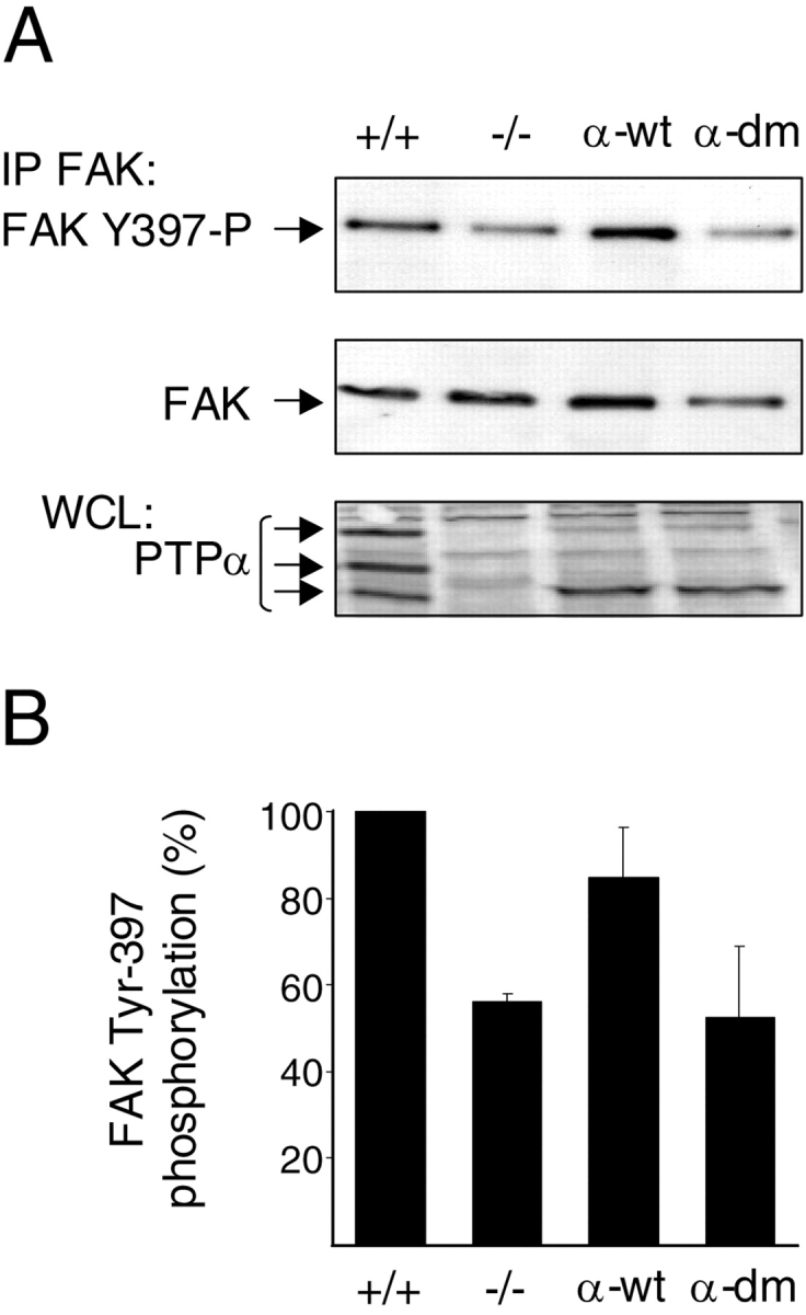
Reintroduction of PTPα into PTPα − / − cells restores integrin-stimulated FAK Tyr-397 phosphorylation. (A) PTPα−/− cells (line 2) were infected with adenovirus expressing wild-type PTPα (α-wt) or a catalytic mutant of PTPα (α-dm), and were plated on FN-coated dishes for 30 min. FAK immunoprecipitates were prepared from lysates of these cells or of uninfected PTPα+/+ and PTPα−/− cells and probed with anti-phospho-Tyr397–specific antibodies (top panel) or anti-FAK antibodies (middle panel). The expression of endogenous and introduced PTPα was examined in cell lysates (WCL) by probing with anti-PTPα antibodies (bottom panel). (B) FAK Tyr-397 phosphorylation was quantitated from at least three independent experiments such as that presented in A and is shown as the mean ± S.D. FAK Tyr-397 phosphorylation in the PTPα+/+ cells was taken as 100%, and the other data was calculated relative to this.
FN-induced morphological changes in PTPα-null cells resemble defects observed in wild-type cells after Src-PTK inhibition
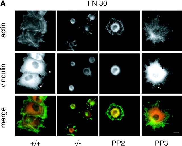
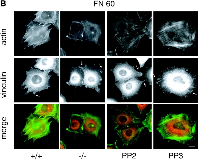
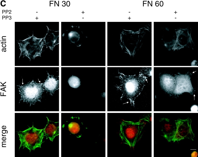
The FN-induced phosphorylation of Tyr397 of FAK is reduced on treatment of fibroblasts with the selective Src-PTK inhibitor PP2 (Salazar and Rozengurt, 2001). This suggested that defective Src-PTK function underlies the impaired FAK Tyr-397 phosphorylation in PTPα−/− cells. Integrin-stimulation is reported to activate src and promote its translocation to focal adhesions (Kaplan et al., 1995; Clark et al., 1998; Sieg et al., 1998; Oh et al., 1999), but we were unable to detect either of these events in wild-type fibroblasts and so could not determine whether they were affected by the lack of PTPα. In agreement with Su et al. (1999), we observed that FN-induced cell spreading was delayed in PTPα−/− cells. Spreading involves cytoskeletal rearrangement leading to stress fiber assembly and focal adhesion formation, and is impaired in FAK−/− cells expressing a FAK autophosphorylation site mutant (Owen et al., 1999). To determine if the parallel FAK autophosphorylation defect resulting from the absence of PTPα or the chemical inhibition of Src-PTKs extended to a similar impairment of cell spreading events, the distribution of actin, vinculin, and FAK were visualized and compared in these two situations. Plating of wild-type cells on FN for 30 min led to cell spreading and the formation of long actin stress fibers oriented in bundles at the leading edge of the polarized cells (Fig. 7 A). Putative focal adhesion sites at the end of these stress fibers were visualized by vinculin staining. In contrast, PTPα−/− cells were much less well spread and unpolarized. This was coincident with a virtual lack of actin stress fibers, and the appearance of F-actin–rich membrane ruffles and lamellipodia around the cell periphery. Furthermore, these cells had compacted cytoplasmic vinculin staining consistent with a lack of focal adhesion formation (Fig. 7 A). The PP2-treated wild-type cells appeared very similar, with reduced spreading and F-actin in membrane ruffles, no actin stress fibers, and compacted cytoplasmic vinculin staining that colocalized with what appeared to be perinuclear actin staining (Fig. 7 A). This latter colocalization was also observed in several of the PTPα−/− cells. Treatment of wild-type cells with PP3, an inactive analogue of PP2, did not alter the actin and vinculin localizations (Fig. 7 A). At 60 min on FN, actin staining defined thick longitudinally oriented stress fibers in the untreated and PP3-treated wild-type cells with vinculin present at the ends of the stress fibers in focal adhesion-like structures (Fig. 7 B). At 60 min, both the PTPα−/− cells and the PP2-treated wild-type cells had spread more and developed actin stress fibers with some vinculin colocalizing at the ends of these fibers, appearing similar to wild-type cells plated on FN for 30 min. Retarded FN-induced focal adhesion formation was also evident in PP2-treated wild-type cells stained for FAK (Fig. 7 C), as observed previously with the PTPα−/− cells (Fig. 4). Thus, the absence of PTPα leads to a temporally indistinguishable cell spreading delay and physically similar altered cell morphology as observed with the inhibition of Src-PTK activity. Delayed stress fiber assembly and focal adhesion formation were characteristic of both situations.
Figure 7.
Similar delays in FN-induced actin stress fiber assembly and focal adhesion formation in PTPα − / − cells and PP2-treated wild-type cells. Wild-type (+/+), PTPα−/− (−/−), PP2-treated (PP2), and PP3-treated (PP3) wild-type cells were plated on FN-coated coverslips for 30 (A; FN30) and 60 (B; FN60) min and stained for F-actin (green) and vinculin (red). The arrows highlight focal adhesions. Bars, 10 μm. (C) PP2- or PP3-treated wild-type fibroblasts were plated on FN-coated coverslips for 30 (FN30) and 60 (FN60) min and stained for F-actin (green) and FAK (red). The arrows highlight some putative focal adhesions visualized by high FAK immunoreactivity. Bar, 10 μm.
Discussion
The integrin-stimulated processes of migration, haptotaxis, and spreading are impaired in PTPα-deficient cells. An initial delay in integrin-stimulated cell spreading involves altered actin cytoskeletal rearrangement and retarded focal adhesion formation in the PTPα−/− cells, and this is associated with reduced FAK Tyr-397 phosphorylation and formation of an Src–PTK–FAK complex. There is a long term defect in the migration ability of PTPα-null cells even after spreading is completed, and at late times in migration there is still reduced FAK Tyr-397 phosphorylation and impairment of the cytoskeletal remodeling required to form cell extensions in PTPα−/− cells at the leading edge of the wound. FAK phosphorylation at Tyr397 is an early event in the sequence of reactions leading to full activation and function of this kinase, and is essential for SH2-dependent binding of Src-PTKs, as well as for the promotion of cell migration (Schaller et al., 1994; Owen et al., 1999; Sieg et al., 1999). We conclude that FAK Tyr-397 phosphorylation is an integrin-proximal event mediated by PTPα, and that this defective step in PTPα−/− cells is a basis for the other observed defects in Src–PTK–FAK interactions and cell migration. Additional or interrelated PTPα-mediated events also regulate cytoskeletal rearrangement in cell spreading and migration. The difference in timing between the shorter term, transient spreading defect and the long term, prolonged migration defect of PTPα−/− cells likely reflects the intrinsically different kinetics of the two processes. We speculate that it may also reflect the limited versus the continued focal adhesion turnover required for cell spreading and cell migration, respectively.
Signaling events that occur immediately after integrin stimulation and leading to autophosphorylation of FAK on Tyr397 are far from clear. Src-PTKs are not only physically, but also functionally necessary for efficient FAK autophosphorylation. Attenuated FAK autophosphorylation and/or reduced motility in response to integrin engagement is observed in triple-null fibroblasts lacking the widely expressed Src-PTKs src, fyn, and yes (SYF cells; Klinghoffer et al., 1999; Salazar and Rozengurt, 2001), and on inhibition of the catalytic activity of Src-PTKs by treatment of cells with the selective inhibitor PP2 (Salazar and Rozengurt, 2001). In these cases of defective Src-PTK function, there is a low level of integrin-stimulated FAK autophosphorylation (Salazar and Rozengurt, 2001; Cary et al., 2002), suggesting that an Src-PTK–independent response is also operable, but not sufficient to deliver maximal FAK Tyr-397 phosphorylation. The reduced (but not abolished) phosphorylation of FAK Tyr-397 and impaired migration on FN of the PTPα−/− cells is similar to the above situations, and consistent with the demonstrated action of PTPα as a positive regulator of Src-PTKs (Ponniah et al., 1999; Su et al., 1999). Cells null for src alone do not have reduced FAK Tyr-397 phosphorylation on integrin stimulation (unpublished data), indicating that the lack of src is probably compensated for by either fyn, yes, or both. Thus, the abrogated FAK Tyr-397 phosphorylation observed in the PTPα−/− cells is likely due to the impaired PTPα-dependent activity of both src and fyn (Ponniah et al., 1999; Su et al., 1999), and supported by the reduced binding of not only src to FAK, but also the complete abolition of the association of fyn with FAK, in the PTPα−/− cells. It is possible that PTPα also affects the activity of yes (Harder et al., 1998), but this has not been investigated in the PTPα-null fibroblasts.
It has been proposed that integrin ligation results in Src-PTK activation followed by binding to phospho-Tyr397, or that binding of Src-PTKs via their SH2-domain to phospho-Tyr397 of FAK disrupts autoinhibitory intramolecular interactions to result in Src-PTK activation. PTPα could function in either scenario as an Src-PTK activator (implying in the latter case that binding of Src-PTKs to phospho-Tyr397 is in itself not sufficient for Src-PTK activation) by affecting the dephosphorylation of the COOH-terminal tyrosine residue of these kinases. This predicts that in the absence of PTPα, no integrin-dependent activation of src would occur. However, we were unable to assess this because in wild-type cells we did not observe the FN-induced src activation (in vitro kinase assays; unpublished data) that has been reported by others (Clark et al., 1998; Sieg et al., 1998; Oh et al., 1999). Neither did we detect FN-induced src relocalization from the endosome/perinucleus to focal adhesions, even after transient expression of c-src. A shift of src to focal adhesions has usually been demonstrated in fibroblasts transfected with src (Kaplan et al., 1995), and even then may require coexpression of other molecules such as FAK (Schaller et al., 1999). Nevertheless, expression of an active src mutant (src Y527F) did allow its detection in focal adhesions after integrin stimulation (unpublished data), as reported (Howell and Cooper, 1994; Kaplan et al., 1994). As we found, Cary et al. (2002) were unable to detect integrin-stimulated activation of src or its relocalization to focal adhesions. Taken in conjunction, it seems likely that these are difficult to detect because they may be transient, occur with only a small population of src, and/or be dependent on additional parameters such as cell density (Kobayashi et al., 1997; Oh et al., 1999). Cary et al. (2002) propose that src activity (but not src activation) is required for integrin signaling, and that src action does not require its concentration in focal adhesions. One interpretation of our results is that if PTPα is not functioning as an integrin-responsive Src-PTK activator, then perhaps it is maintaining Src-PTKs at an activity level that permits integrin-induced Src-PTK function. In PTPα-null cells, the src and fyn activities are reduced to ∼30% of that in wild-type cells (Ponniah et al., 1999; Su et al., 1999), suggesting that this activity is insufficient to support optimal integrin signaling events. The reduction in src/fyn activity observed in the absence of PTPα was quantitated with total immunoprecipitated src and fyn, and could be even more pronounced if it is specifically associated with a subpopulation of src/fyn that participates in integrin signaling. Certainly, the phosphatase activity of PTPα is necessary for proper FAK Tyr-397 phosphorylation, as this was restored by reintroduction of wild-type PTPα into the PTPα−/− cells, but not by expression of a catalytically inactive form of PTPα. Together, these observations suggest that PTPα functions as a component in the upstream events of integrin signaling through its action as an Src-PTK phosphatase and activator.
A role for PTPα in integrin-stimulated reorganization of the actin cytoskeleton is also apparent from the defects in the PTPα−/− cells. We demonstrate that a transient delay in spreading of the PTPα−/− cells is associated with retarded formation of actin stress fibers and focal adhesions, and prolonged membrane ruffle formation. A direct comparison of FN-induced changes in the actin cytoskeleton and localization of the focal adhesion proteins vinculin and FAK of wild-type cells treated with PP2 and untreated PTPα−/− cells confirms that virtually indistinguishable delays in the above events occur in both situations, suggesting that abrogated Src-PTK activity is the common factor underlying the defective responses. The Rho family GTPases Cdc42, Rac, and Rho are all involved in regulating cell morphology and cytoskeletal events such as spreading, assembly of actin stress fibers, and focal contacts after integrin engagement (Barry et al., 1997; Clark et al., 1998; Price et al., 1998; Ren et al., 1999). Particular attention has focused on the initial transient inactivation of RhoA that is necessary for rapid focal adhesion turnover and cell spreading on the ECM (Ren et al., 1999, 2000). In addition to c-src, p190RhoGAP, the PTP SHP-2, and FAK have all been implicated in regulation of RhoA activity and may operate as c-src effector molecules in as yet unclear, but potentially linked pathways (Arthur et al., 2000; Ren et al., 2000; Schoenwaelder et al., 2000; Arthur and Burridge, 2001; Lacalle et al., 2002). Additionally, an intact actin cytoskeleton is required for Tyr397 and maximal FAK phosphorylation, as well as for full activation of c-src (Sieg et al., 1998; Oh et al., 1999), suggesting that the impaired cytoskeletal rearrangement and FAK phosphorylation defects in PTPα−/− cells are linked effects of a common and early signaling defect.
Materials and methods
Antibodies and immunological detection reagents
Anti-PTPα antiserum no. 2205 was obtained from rabbits immunized with purified recombinant PTPα-D1 (Lim et al., 1998). Antibodies toward FAK, fyn, phosphotyrosine (PY20), and Pyk2 were purchased from Transduction Laboratories. The phosphosite-specific antibody to FAK Tyr-397 was from Biosource International. Anti-fyn antibody for immunoprecipitation was purchased from Santa Cruz Biotechnology, Inc., and v-src antibody (Ab-1) was from Oncogene Research Products. Anti-actin and vinculin antibodies were purchased from Sigma-Aldrich. Alexa Fluor 594–conjugated anti–mouse IgG, Alexa Fluor 488–conjugated goat anti–rabbit IgG, and Alexa Fluor 488–conjugated phalloidin were from Molecular Probes, Inc.
Cells
Embryonic fibroblasts were prepared and genotyped as described previously (Ponniah et al., 1999). The cells were cultured in DME with 10% FCS and spontaneously immortalized by repeated passage. A PTPα+/+ and two independently derived PTPα−/− cell lines were used at passages 29–50 for experiments described here. Results shown are from PTPα−/− line 1 unless otherwise indicated to be from line 2.
Cell stimulation with FN
Cells were serum-starved overnight and harvested by 0.0625% trypsin treatment. The trypsin digestion was stopped with 0.5 mg/ml soybean trypsin inhibitor (GIBCO BRL) in DME. Cells were collected by centrifugation and washed twice with DME containing soybean trypsin inhibitor, then resuspended in DME and held in suspension for 1 h at 37°C. Cell culture dishes were precoated with 10 μg/ml FN (CHEMICON International) overnight at 4°C, and washed twice with PBS. Suspended cells were distributed onto FN-coated dishes (105 cells/ml) and incubated at 37°C for various times.
Treatment with PP2 or PP3
Subconfluent serum starved wild-type fibroblasts were incubated with 10 μM PP2 or PP3 (Calbiochem) for 15 min. The cells were trypsinized and held in suspension with PP2 or PP3 for 30 min at 37°C before plating (in DME with PP2 or PP3) onto FN-coated coverslips for the indicated times.
Cell migration
To assess cell migration using an in vitro wound healing assay, fibroblasts were grown on collagen-coated plates or coverslips in DME and 10% FCS. During the 24 h before wounding, the cells were maintained in migration medium (DME with 0.1% FCS and 0.1% ITS-X). The cell layer was scratched with a pipette tip, and the migration of cells into the wound area was followed by video microscopy. Transwell chamber (Costar) migration assays were performed as described by Sieg et al. (1999) with some modifications. In brief, the undersurface of the polycarbonate membrane of the chambers was coated with FN (10 μg/ml in PBS) for 2 h at 37°C. The membrane was washed in PBS to remove excess ligand, and the lower chamber was filled with 500 μl of migration medium (DME with 0.5% BSA). Serum-starved cells were harvested using limited trypsin treatment, and washed twice in DME containing 0.5 mg/ml soybean trypsin inhibitor and once in serum-free DME. Cells were resuspended in migration medium and 8 × 10⁴ cells in 0.1 ml were added to the upper chamber. After 1.5 h at 37°C, cells were washed, fixed in methanol for 15 min at RT, and stained with Giemsa solution. The cells on the upper surface of the membrane were removed using cotton buds. The number of migrated cells on the lower surface of the membrane was counted using a 20× objective (cells/field).
Immunofluorescence
For single or double labeling, cells on coverslips were fixed with 3.7% formaldehyde and permeabilized with 0.04% Triton X-100 for 10 min. After blocking with 1% BSA in PBS for 20 min, the cells were incubated with anti-vinculin (1:200; Sigma-Aldrich) and anti-phospho-Tyr397 FAK (1:150; Biosource International) for 60 min. This was followed by incubation with Alexa Fluor 594–conjugated goat anti–mouse IgG (1:200; Molecular Probes, Inc.) and Alexa Fluor 488–conjugated goat anti–rabbit antibody (1:200; Molecular Probes, Inc.) for 60 min. In other experiments, vinculin was labeled as above or FAK was labeled with anti-FAK antibody (1:50; Transduction Laboratories) and visualized by incubation with Alexa Fluor 594–conjugated anti–mouse IgG (1:200), and Alexa Fluor 488–conjugated phalloidin (1:250) was used to stain F-actin. Coverslips were mounted in Vectashield® mounting medium (Vector Laboratories) and viewed using a fluorescence microscope (Axioplan2; Carl Zeiss MicroImaging, Inc.). Images were captured by a CCD microcolor digital camera (CRI, Inc.) and processed by Smartcapture VP software (Digital Scientific, Ltd).
Immunoprecipitation and immunoblotting
Cells were lysed with modified RIPA buffer (50 mM Hepes, pH 7.3, 1% sodium deoxycholate, 1% Triton X-100, 0.1% SDS, 150 mM NaCl, 1 mM EDTA, 1 mM Na3VO4, 1 mM PMSF, and 10 μg/ml aprotinin). Total cell protein in lysates from serum-starved, suspended, or replated cells was determined using the Bio-Rad Laboratories protein assay reagent and standardized before further analyses. Lysates were immunoprecipitated with the indicated antibodies for 2 h at 4°C followed by incubation with protein G plus protein A agarose beads (Oncogene Research Products) for 1 h. The precipitated protein complexes were washed at 4°C in RIPA buffer without sodium deoxylcholate or SDS. For immunoblotting, proteins were resolved by SDS-PAGE and were transferred to polyvinylidene difluoride membranes. The membranes were blocked in 1% BSA and Tris-buffered saline with 0.05% Tween 20 overnight at 4°C, and were then incubated with the indicated antibodies for 1 h. Bound primary antibody was observed by ECL detection (Amersham Biosciences).
PTPα adenoviral expression system
The AdEasy™ vector system (Qbiogene, Inc.) was used to obtain PTPα expression in mouse fibroblasts. To eliminate the PacI site in PTPα cDNA and so facilitate cloning into the adenoviral vector, the codons for Leu-15 (TTA) and Ile-16 (ATT) in human PTPα cDNA were respectively altered using PCR-mediated site mutagenesis to degenerate coding sequences of CTA and ATC. A Kozak sequence of CCACC and a SalI restriction site were also added immediately 5′ to the ATG translation start code. The absence of other mutations introduced by PCR was confirmed by sequencing. The cDNAs encoding both wild-type and double mutant (C414/704S) forms of PTPα (Lim et al., 1997) were cloned into the SalI and NotI sites of the pAdTrack-CMV vector. These two resulting plasmids were linearized with PmeI and cotransformed respectively with pAdEasy-1 into Escherichia coli strain BJ5183 to generate the infective viral DNA plasmid that contains a wild-type or mutant form of PTPα by homologous recombination. Recombinants were selected by kanamycin and positive candidates were retransformed into E. coli strain DH10B to preserve the correctly recombined plasmid from further recombination. After confirmation, correct plasmid DNAs were cleaved by PacI and transfected into 293A cells using LipofectAMINE™ reagent (Life Technologies). Virus-infected cells could be visualized 24 h after transfection under the fluorescence microscope due to the integrated GFP expression in the viral vector. Viral particles were harvested from the cells by freeze/thaw and purified by continuous CsCl gradient centrifugation. To infect mouse fibroblasts, a sufficient amount of viral particles in a minimum volume of normal culture medium that can completely cover the plate was applied to the cells. Cells were then incubated at 37°C for 2 h. Thereafter, the medium was topped up to the normal level and cells were cultured for 48 h after infection before harvesting for experiments.
Acknowledgments
We thank Y.H. Tan and B.W. Sutherland for critical reading of the manuscript.
This work was supported by the National Science and Technology Board of Singapore, the Canadian Institutes for Health Research (MOP-49410), and the Johal Program in Pediatric Oncology Basic and Translational Research (to C.J. Pallen).
L. Zeng and X. Si contributed equally to this paper.
Footnotes
*
Abbreviations used in this paper: FN, fibronectin; PTP, protein tyrosine phosphatase; SH2, src homology 2; Src-PTK, src family protein tyrosine kinase.
References
- Angers-Loustau, A., J.-F. Cote, and M.L. Tremblay. 1999. a. Roles of protein tyrosine phosphatases in cell migration and adhesion. Biochem. Cell Biol. 77:493–505. [PubMed] [Google Scholar]
- Angers-Loustau, A., J.-F. Cote, A. Charest, D. Dowbenko, S. Spencer, L.A. Lasky, and M.L. Tremblay. 1999. b. Protein tyrosine phosphatase-PEST regulates focal adhesion disassembly, migration, and cytokinesis in fibroblasts. J. Cell Biol. 144:1019–1031. [DOI] [PMC free article] [PubMed] [Google Scholar]
- Arregui, C.O., J. Balsamo, and J. Lilien. 1998. Impaired integrin-mediated adhesion and signaling in fibroblasts expressing a dominant-negative mutant PTP1B. J. Cell Biol. 143:861–873. [DOI] [PMC free article] [PubMed] [Google Scholar]
- Arthur, W.T., and K. Burridge. 2001. RhoA inactivation by p190RhoGAP regulates cell spreading and migration by promoting membrane protrusion and polarity. Mol. Biol. Cell. 12:2711–2720. [DOI] [PMC free article] [PubMed] [Google Scholar]
- Arthur, W.T., L.A. Petch, and K. Burridge. 2000. Integrin engagement suppresses RhoA activity via a c-src-dependent mechanism. Curr. Biol. 10:719–722. [DOI] [PubMed] [Google Scholar]
- Barry, S.T., H.M. Flinn, M.J. Humphries, D.R. Critchley, and A.J. Ridley. 1997. Requirement for Rho in integrin signaling. Cell Adhes. Commun. 4:387–398. [DOI] [PubMed] [Google Scholar]
- Bhandari, V., K.L. Lim, and C.J. Pallen. 1998. Physical and functional interactions between receptor-like protein-tyrosine phosphatase α and p59fyn. J. Biol. Chem. 273:8691–8698. [DOI] [PubMed] [Google Scholar]
- Buist, A., C. Blanchetot, L.G. Tertoolen, and J. den Hertog. 2000. Identification of p130cas as an in vivo substrate of receptor protein-tyrosine phosphatase alpha. J. Biol. Chem. 275:20754–20761. [DOI] [PubMed] [Google Scholar]
- Calalb, M.B., T.R. Polte, and S.K. Hanks. 1995. Tyrosine phosphorylation of focal adhesion kinase at sites in the catalytic domain regulates kinase activity: a role for Src family kinases. Mol. Cell. Biol. 15:954–963. [DOI] [PMC free article] [PubMed] [Google Scholar]
- Cary, L.A., J.F. Chang, and J.-L. Guan. 1996. Stimulation of cell migration by overexpression of focal adhesion kinase and its association with src and fyn. J. Cell Sci. 109:1787–1794. [DOI] [PubMed] [Google Scholar]
- Cary, L.A., R.A. Klinghoffer, C. Sachsenmaier, and J.A. Cooper. 2002. Src catalytic but not scaffolding function is needed for integrin-regulated tyrosine phosphorylation, cell migration, and cell spreading. Mol. Cell. Biol. 22:2427–2440. [DOI] [PMC free article] [PubMed] [Google Scholar]
- Cheng, A., G.S. Bal, B.P. Kennedy, and M.L. Tremblay. 2001. Attenuation of adhesion-dependent signaling and cell spreading in transformed fibroblasts lacking protein tyrosine phosphatase-1B. J. Biol. Chem. 276:25848–25855. [DOI] [PubMed] [Google Scholar]
- Clark, E.A., W.G. King, J.S. Brugge, M. Symons, and R.O. Hynes. 1998. Integrin-mediated signals regulated by members of the Rho family of GTPases. J. Cell Biol. 142:573–586. [DOI] [PMC free article] [PubMed] [Google Scholar]
- Cobb, B.S., M.D. Schaller, T.-H. Leu, and J.T. Parsons. 1994. Stable association of pp60src and pp59fyn with the focal adhesion-associated tyrosine kinase, pp125FAK. Mol. Cell. Biol. 14:147–155. [DOI] [PMC free article] [PubMed] [Google Scholar]
- Daum, G., S. Regenass, J. Sap, J. Schlessinger, and E.H. Fischer. 1994. Multiple forms of the human tyrosine phosphatase RPTP alpha. Isozymes and differences in glycosylation. J. Biol. Chem. 269:10524–10528. [PubMed] [Google Scholar]
- Debant, A., C. Serra-Pages, K. Seipel, S. O'Brien, M. Tang, S.H. Park, and M. Streuli. 1996. The multidomain protein Trio binds the LAR transmembrane tyrosine phosphatase, contains a protein kinase domain, and has separate rac-specific and rho-specific guanine nucleotide exchange factor domains. Proc. Natl. Acad. Sci. USA. 93:5466–5471. [DOI] [PMC free article] [PubMed] [Google Scholar]
- den Hertog, J., C.E. Pals, M.P. Peppelenbosch, L.G. Tertoolen, S.W. de Laat, and W. Kruijer. 1993. Receptor protein tyrosine phosphatase alpha activates pp60c-src and is involved in neuronal differentiation. EMBO J. 12:3789–3798. [DOI] [PMC free article] [PubMed] [Google Scholar]
- Garton, A.J., and N.K. Tonks. 1999. Regulation of fibroblast motility by the protein tyrosine phosphatase PTP-PEST. J. Biol. Chem. 274:3811–3818. [DOI] [PubMed] [Google Scholar]
- Gill-Henn, H., G. Volohonsky, and A. Elson. 2001. Regulation of protein-tyrosine phosphatases α and ɛ by calpain-mediated proteolytic cleavage. J. Biol. Chem. 276:31772–31779. [DOI] [PubMed] [Google Scholar]
- Hamasaki, K., T. Mimura, N. Morino, H. Furuya, T. Nakamoto, S.I. Aizawa, C. Morimoto, Y. Yazaki, H. Hirai, and Y. Nojima. 1996. Src kinase plays an essential role in integrin-mediated tyrosine phosphorylation of Crk-associated substrate p130Cas. Biochem. Biophys. Res. Commun. 222:338–343. [DOI] [PubMed] [Google Scholar]
- Harder, K.W., N.P.H. Moller, J.W. Peacock, and F.R. Jirik. 1998. Protein-tyrosine phosphatase α regulates src family kinases and alters cell-substratum adhesion. J. Biol. Chem. 273:31890–31900. [DOI] [PubMed] [Google Scholar]
- Howell, B.W., and J.A. Cooper. 1994. Csk suppression of src involves movement of csk to sites of src activity. Mol. Cell. Biol. 14:5402–5411. [DOI] [PMC free article] [PubMed] [Google Scholar]
- Kaplan, K.B., K.B. Bibbins, J.R. Swedlow, M. Arnaud, D.O. Morgan, and H.E. Varmus. 1994. Association of the amino-terminal half of c-Src with focal adhesions alters their properties and is regulated by phosphorylation of tyrosine 527. EMBO J. 13:4745–4756. [DOI] [PMC free article] [PubMed] [Google Scholar]
- Kaplan, K.B., J.R. Swedlow, D.O. Morgan, and H.E. Varmus. 1995. c-Src enhances the spreading of src−/− fibroblasts on fibronectin by a kinase-independent mechanism. Genes Dev. 9:1505–1517. [DOI] [PubMed] [Google Scholar]
- Klemke, R.L., J. Leng, R. Molander, P.C. Brooks, K. Vuori, and D.A. Cheresh. 1998. CAS/Crk coupling serves as a “molecular switch” for induction of cell migration. J. Cell Biol. 140:961–972. [DOI] [PMC free article] [PubMed] [Google Scholar]
- Klinghoffer, R.A., C. Sachsenmaier, J.A. Cooper, and P. Soriano. 1999. Src family kinases are required for integrin but not PDGFR signal transduction. EMBO J. 18:2459–2471. [DOI] [PMC free article] [PubMed] [Google Scholar]
- Kobayashi, S., N. Okumura, T. Nakamoto, M. Okada, H. Hirai, and K. Nagai. 1997. Activation of pp60c-src depending on cell density in PC12 cells. J. Biol. Chem. 272:16262–16267. [DOI] [PubMed] [Google Scholar]
- Lacalle, R.A., E. Mira, C. Gomez-Mouton, S. Jimenez-Baranda, C. Martinez-A., and S. Manes. 2002. Specific SHP-2 partitioning in raft domains triggers integrin-mediated signaling via Rho activation. J. Cell Biol. 157:277–289. [DOI] [PMC free article] [PubMed] [Google Scholar]
- Lammers, R., M.M. Lerch, and A. Ullrich. 2000. The carboxy-terminal tyrosine residue of protein-tyrosine phosphatase α mediates association with focal adhesion plaques. J. Biol. Chem. 275:3391–3396. [DOI] [PubMed] [Google Scholar]
- Liliental, J., S.Y. Moon, R. Lesche, R. Mamillapalli, D. Li, Y. Zheng, H. Sun, and H. Wu. 2000. Genetic deletion of the Pten tumor suppressor gene promotes cell motility by activation of Rac1 and Cdc42 GTPases. Curr. Biol. 10:401–404. [DOI] [PubMed] [Google Scholar]
- Lim, K.L., D.S. Lai, M.B. Kalousek, Y. Wang, and C.J. Pallen. 1997. Kinetic analysis of two closely related receptor-like protein-tyrosine-phosphatases, PTPα and PTPɛ. Eur. J. Biochem. 245:693–700. [DOI] [PubMed] [Google Scholar]
- Lim, K.L., P.R. Kolatkar, K.P. Ng, C.H. Ng, and C.J. Pallen. 1998. Interconversion of the kinetic identities of the tandem catalytic domains of receptor-like protein-tyrosine phosphatase PTPα by two point mutations is synergistic and substrate-dependent. J. Biol. Chem. 273:28986–28993. [DOI] [PubMed] [Google Scholar]
- Liu, F., M.A. Sells, and J. Chernoff. 1998. Protein tyrosine phosphatase 1B negatively regulates integrin signaling. Curr. Biol. 8:173–176. [DOI] [PubMed] [Google Scholar]
- Oh, E.-S., H. Gu, T.M. Saxton, J.F. Timms, S. Hausdorff, E.U. Frevert, B.B. Kahn, T. Pawson, B.G. Neel, and S.M. Thomas. 1999. Regulation of early events in integrin signaling by protein tyrosine phosphatase SHP-2. Mol. Cell. Biol. 19:3205–3215. [DOI] [PMC free article] [PubMed] [Google Scholar]
- Owen, J.D., P.J. Ruest, D.W. Fry, and S.K. Hanks. 1999. Induced focal adhesion kinase (FAK) expression in FAK-null cells enhances cell spreading and migration requiring both auto- and activation loop phosphorylation sites and inhibits adhesion-dependent tyrosine phosphorylation of Pyk2. Mol. Cell Biol. 19:4806–4818. [DOI] [PMC free article] [PubMed] [Google Scholar]
- Ponniah, S., D.Z.M. Wang, K.L. Lim, and C.J. Pallen. 1999. Targeted disruption of the tyrosine phosphatase PTPα leads to constitutive downregulation of the kinases src and fyn. Curr. Biol. 9:535–538. [DOI] [PubMed] [Google Scholar]
- Price, L.S., J. Leng, M.A. Schwartz, and G.M. Bokoch. 1998. Activation of Rac and Cdc42 by integrins mediates cell spreading. Mol. Biol. Cell. 9:1863–1871. [DOI] [PMC free article] [PubMed] [Google Scholar]
- Ren, X.-D., W.B. Kiosses, and M.A. Schwartz. 1999. Regulation of the small GTP-binding protein Rho by cell adhesion and the cytoskeleton. EMBO J. 18:578–585. [DOI] [PMC free article] [PubMed] [Google Scholar]
- Ren, X.-D., W.B. Kiosses, D.J. Sieg, C.A. Otey, D.D. Schlaepfer, and M.A. Schwartz. 2000. Focal adhesion kinase suppresses Rho activity to promote focal adhesion turnover. J. Cell Sci. 113:3673–3678. [DOI] [PubMed] [Google Scholar]
- Ruest, P.J., S. Roy, E. Shi, R.L. Mernaugh, and S.K. Hanks. 2000. Phosphospecific antibodies reveal focal adhesion kinase activation loop phosphorylation in nascent and mature focal adhesions and requirement for the autophosphorylation site. Cell Growth Differ. 11:41–48. [PubMed] [Google Scholar]
- Salazar, E.P., and E. Rozengurt. 2001. Src family kinases are required for integrin-mediated but not for G protein-coupled receptor stimulation of focal adhesion kinase autophosphorylation at Tyr-397. J. Biol. Chem. 276:17788–17795. [DOI] [PubMed] [Google Scholar]
- Schaller, M.D. 2001. Biochemical signals and biological responses elicited by the focal adhesion kinase. Biochim. Biophys. Acta. 1540:1–21. [DOI] [PubMed] [Google Scholar]
- Schaller, M.D., and J.T. Parsons. 1995. pp125FAK-dependent tyrosine phosphorylation of paxillin creates a high-affinity binding site for Crk. Mol. Cell. Biol. 15:2635–2645. [DOI] [PMC free article] [PubMed] [Google Scholar]
- Schaller, M.D., J.D. Hildebrand, J.D. Shannon, J.W. Fox, R.R. Vines, and J.T. Parsons. 1994. Autophosphorylation of the focal adhesion kinase, pp125FAK, directs SH2-dependent binding of pp60src. Mol. Cell. Biol. 14:1680–1688. [DOI] [PMC free article] [PubMed] [Google Scholar]
- Schaller, M.D., J.D. Hidebrand, and J.T. Parsons. 1999. Complex formation with focal adhesion kinase: a mechanism to regulate activity and subcellular localization of src kinases. Mol. Biol. Cell. 10:3489–3505. [DOI] [PMC free article] [PubMed] [Google Scholar]
- Schlaepfer, D.D., S.K. Hanks, T. Hunter, and P. van der Greer. 1994. Integrin-mediated signal transduction linked to Ras pathway by GRB2 binding to focal adhesion kinase. Nature. 372:786–791. [DOI] [PubMed] [Google Scholar]
- Schlaepfer, D.D., M.A. Broome, and T. Hunter. 1997. Fibronectin-stimulated signaling from a focal adhesion kinase-c-Src complex: involvement of the Grb2, p130Cas and Nck adaptor proteins. Mol. Cell. Biol. 17:1702–1713. [DOI] [PMC free article] [PubMed] [Google Scholar]
- Schlaepfer, D.D., K.C. Jones, and T. Hunter. 1998. Multiple Grb2-mediated integrin-stimulated signaling pathways to ERK2/mitogen-activated protein kinase: summation of both c-Src and FAK-initiated tyrosine phosphorylation events. Mol. Cell. Biol. 18:2571–2585. [DOI] [PMC free article] [PubMed] [Google Scholar]
- Schlaepfer, D.D., C.R. Hauck, and D.J. Sieg. 1999. Signaling through focal adhesion kinase. Prog. Biophys. Mol. Biol. 71:435–478. [DOI] [PubMed] [Google Scholar]
- Schoenwaelder, S.M., L.A. Petch, D. Williamson, R. Shen, G.-S. Feng, and K. Burridge. 2000. The protein tyrosine phosphatase Shp-2 regulates RhoA activity. Curr. Biol. 10:1523–1526. [DOI] [PubMed] [Google Scholar]
- Seipel, K., Q.G. Medley, N.L. Kedersha, X.A. Zhang, S.P. O'Brien, C. Serra-Pages, M.E. Hemler, and M. Streuli. 1999. Trio amino-terminal guanine nucleotide exchange factor domain expression promotes actin cytoskeleton reorganization, cell migration and anchorage-independent cell growth. J. Cell Sci. 112:1825–1834. [DOI] [PubMed] [Google Scholar]
- Serra-Pages, C., N.L. Kedersha, L. Fazikas, Q. Medley, A. Debant, and M. Streuli. 1995. The LAR transmembrane protein tyrosine phosphatase and a coiled-coil LAR-interacting protein co-localize at focal adhesions. EMBO J. 14:2827–2838. [DOI] [PMC free article] [PubMed] [Google Scholar]
- Sieg, D.J., D. Ilic, K.C. Jones, C.H. Damsky, T. Hunter, and D.D. Schlaepfer. 1998. Pyk2 and Src-family protein-tyrosine kinases compensate for the loss of FAK in fibronectin-stimulated signaling events but Pyk2 does not fully function to enhance FAK− cell migration. EMBO J. 17:5933–5947. [DOI] [PMC free article] [PubMed] [Google Scholar]
- Sieg, D.J., C.R. Hauck, and D.D. Schlaepfer. 1999. Required role of focal adhesion kinase (FAK) for integrin-stimulated cell migration. J. Cell Sci. 112:2677–2691. [DOI] [PubMed] [Google Scholar]
- Su, J., A. Batzer, and J. Sap. 1994. Receptor tyrosine phosphatase R-PTP-α is tyrosine-phosphorylated and associated with the adaptor protein Grb2. J. Biol. Chem. 269:18731–18734. [PubMed] [Google Scholar]
- Su, J., M. Muranjan, and J. Sap. 1999. Receptor protein tyrosine phosphatase a activates src family kinases and controls integrin-mediated responses in fibroblasts. Curr. Biol. 9:505–511. [DOI] [PubMed] [Google Scholar]
- Tamura, M., J. Gu, K. Matsumoto, S. Aota, R. Parsons, and K.M. Yamada. 1998. Inhibition of cell migration, spreading, and focal adhesions by tumor suppressor PTEN. Science. 280:1614–1617. [DOI] [PubMed] [Google Scholar]
- Tamura, M., J. Gu, E.H.J. Danen, T. Takino, S. Miyamoto, and K.M. Yamada. 1999. PTEN interactions with focal adhesion kinase and suppression of the extracellular matrix-dependent phosphatidylinositol 3-kinase/Akt cell survival pathway. J. Biol. Chem. 274:20693–20703. [DOI] [PubMed] [Google Scholar]
- Vuori, K., H. Hirai, S. Aizawa, and E. Ruoslahti. 1996. Induction of p130cas signaling complex formation upon integrin-mediated cell adhesion: a role for src family kinases. Mol. Cell. Biol. 16:2606–2613. [DOI] [PMC free article] [PubMed] [Google Scholar]
- Weng, L.-P., X. Wang, and Q. Yu. 1999. Transmembrane tyrosine phosphatase LAR induces apoptosis by dephosphorylating and destabilizing p130Cas. Genes Cells. 4:185–196. [DOI] [PubMed] [Google Scholar]
- Xing, Z., H.C. Chen, J.K. Nowlen, S. Taylor, D. Shalloway, and J.L. Guan. 1994. Direct interaction of v-Src with the focal adhesion kinase mediated by the Src SH2 domain. Mol. Biol. Cell. 5:413–421. [DOI] [PMC free article] [PubMed] [Google Scholar]
- Yu, D.-H., C.K. Qu, O. Henegariu, X. Lu, and G.-S. Feng. 1998. Protein-tyrosine phosphatase Shp-2 regulates cell spreading, migration, and focal adhesion. J. Biol. Chem. 273:21125–21131. [DOI] [PubMed] [Google Scholar]
- Zheng, X.M., Y. Wang, and C.J. Pallen. 1992. Cell transformation and activation of pp60c-src by overexpression of a protein tyrosine phosphatase. Nature. 359:336–339. [DOI] [PubMed] [Google Scholar]
