Nucleolar Localization of Human Methionyl–Trna Synthetase and Its Role in Ribosomal RNA Synthesis (original) (raw)
Abstract
Human aminoacyl–tRNA synthetases (ARSs) are normally located in cytoplasm and are involved in protein synthesis. In the present work, we found that human methionyl–tRNA synthetase (MRS) was translocated to nucleolus in proliferative cells, but disappeared in quiescent cells. The nucleolar localization of MRS was triggered by various growth factors such as insulin, PDGF, and EGF. The presence of MRS in nucleoli depended on the integrity of RNA and the activity of RNA polymerase I in the nucleolus. The ribosomal RNA synthesis was specifically decreased by the treatment of anti-MRS antibody as determined by nuclear run-on assay and immunostaining with anti-Br antibody after incorporating Br-UTP into nascent RNA. Thus, human MRS plays a role in the biogenesis of rRNA in nucleoli, while it is catalytically involved in protein synthesis in cytoplasm.
Keywords: methionyl–tRNA synthetase, nucleoli, growth signal, ribosomal RNA synthesis, RNA polymerase I
Introduction
Aminoacyl–tRNA synthetases (ARSs) are enzymes decoding genetic information into amino acids. Although these enzymes normally execute their catalytic activities for protein synthesis, recent reports suggest that they are not simple enzymes, and they can play novel regulatory functions in various processes (Martinis et al. 1999). Mammalian tryptophanyl–tRNA synthetase is induced by interferon (Kisselev et al. 1993) and the same enzyme of Drosophila melanogaster is under the control of the homeotic gene, Scr, and highly expressed in salivary gland during development (Seshaiah and Andrew 1999). Mitochondrial tyrosyl–tRNA synthetase of Neurospora crassa (Akins and Lambowitz 1987) and leucyl–tRNA synthetase of Saccharomyces cerevisiae (Labouesse 1990) are involved in the splicing process. Human tyrosyl–tRNA synthetase is converted to two distinct proapoptotic cytokines (Wakasugi and Schimmel 1999) and human arginyl–tRNA synthetase (RRS) also sequesters the precursor of a proapoptotic cytokine (Park et al. 1999). Thus, we anticipated the unveiling of more diverse functions from these enzymes.
To gain an insight into the novel functions of mammalian ARSs, we investigated cellular localizations of different human ARSs using their specific antibodies. Among the tested ARSs, methionyl–tRNA synthetase (MRS) was uniquely localized in the nucleolus. Although the presence of MRS in nucleoli was previously reported (Dang et al. 1983), the functional reason for the nucleolar localization of MRS is not understood. Here, we investigated the translocational control and functional significance of nucleolar MRS.
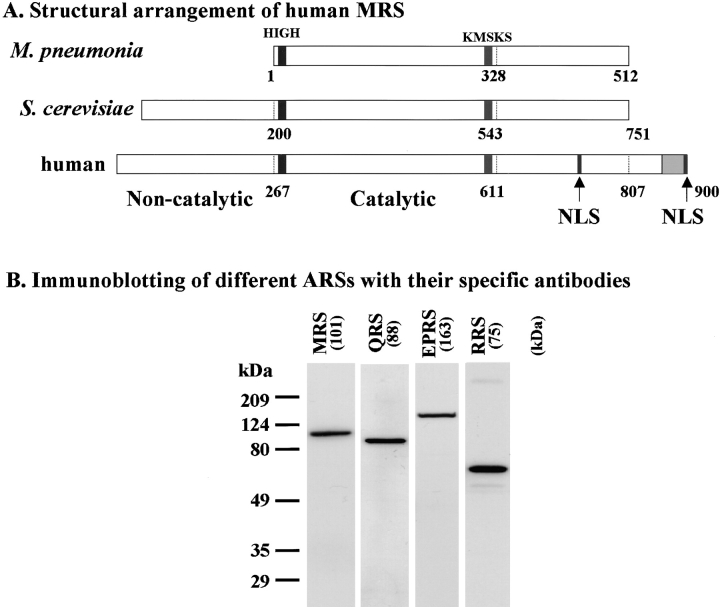
Human cytoplasmic MRS consists of 900 amino acids (Lage and Dietel 1996) and is one of the components for the multi-tRNA synthetase complex (Mirande 1991; Kisselev and Wolfson 1994; Yang 1996). The core domain is homologous to the corresponding enzymes from prokaryotes (Fig. 1 A). However, it contains the unique NH2-terminal extension of 267 amino acids that is not essential for catalytic activity (data not shown), but is involved in protein–protein interaction (Rho et al. 1999). Similarly, the NH2-terminal extension of yeast MRS is also responsible for the interaction with a nuclear pore-associated protein, Arc1p (Simos et al. 1996). Another motif of ∼40 amino acids (Fig. 1 A, gray box) is present in the COOH-terminal region (Q847-K897) that is homologous to the motifs present in other ARSs and involved in protein–protein and protein–nucleic acid interactions (Rho et al. 1996, Rho et al. 1998). In addition, putative nuclear localization signals (Schimmel and Wang 1999) are found in the COOH-terminal region as four consecutive lysines from K897 to K900 and PWKRIKG from P724 to G730 (Fig. 1 A, bars), implying that MRS may be translocated to the nucleus. Here, we report that human MRS is translocated into nucleoli by various cell proliferation signals and is involved in rRNA synthesis.
Figure 1.
Structural arrangement of human MRS and specificity of anti-ARS antibodies. A, MRSs of M. pneumoniae, S. cerevisiae, and H. sapiens are aligned schematically. The core catalytic domain is divided into the NH2- and COOH-terminal domains (marked with amino acid numbers and dotted lines). The signature sequences for class I ARSs (HIGH and KMSKS; Webster et al. 1984; Hountondji et al. 1986; Ludmerer and Schimmel 1987) are highlighted by bars. Human MRS contains the NH2- and COOH-terminal extensions that are involved in protein–protein interactions (Rho et al. 1999). An ∼40 aa peptide motif (marked as gray box) homologous to those in other ARSs is present in the COOH-terminal end (Rho et al. 1996, Rho et al. 1998). Two nuclear localization signals (NLS) are present in the COOH-terminal region. B, Polyclonal rabbit antibodies were raised against the purified polypeptides of human MRS, EPRS, RRS, and QRS (see Materials and Methods). The antigenic specificities of the prepared antibodies were determined by immunoblotting of proteins extracted from HeLa cells.
Materials and Methods
Cell Culture
HeLa, Chang, HepG-2, COS-1, and human foreskin fibroblast were grown to subconfluency on 5 × 5-mm glass coverslips in 35-mm petri dishes in DME supplemented with 10% FBS (GIBCO BRL). Confluent cells were prepared by growing 5 × 10⁵ cells on coverslips in DME/10% FBS for 4–6 d without changing the medium. Quiescent cells were also prepared by serum starvation for 5–7 d in DME. RNA polymerase I and RNA polymerase II were inhibited by the addition of cisplatin (10 μg/ml for 9 h; Jordan and Carmo-Fonseca 1998) and α-amanitin (2 μg/ml for 16 h; Kedinger et al. 1970; Lindel et al. 1970), respectively. The inhibition of RNA polymerase I and II was achieved by the treatment of α-amanitin (2 μg/ml for 16 h) and actinomycin D (0.2 μg/ml for 16 h; Perry 1963).
Antibody Preparation
The cDNA encoding the full-length human cytoplasmic MRS was isolated by PCR from pM184 (Rho et al. 1999) as a template using two specific primers. The PCR product was cleaved with EcoRI and HindIII designed into each of the primers, and inserted into the same site of pET28a (Novagen). The resulting plasmid was introduced into Escherichia coli BL21 (DE3) and induced with IPTG. Since the recombinant MRS was insoluble, it was purified as a denatured polypeptide using nickel-affinity chromatography following the manufacturer's instruction (Invitrogen). The native peptide of EPRS (bifunctional glutamyl-prolyl–tRNA synthetase) from D677 to E884, the native NH2-terminal 236 aa of human QRS (glutaminyl–tRNA synthetase) and the native NH2-terminal 72 aa of human RRS were also expressed as His-tagged proteins. All of these polypeptides were also purified using His tag. Each purified polypeptide was used to raise specific polyclonal rabbit antibodies as described previously (Park et al. 1999). The IgG from each antiserum was purified by protein A affinity chromatography according to the manufacturer's protocol (BioRad), and the antibody specificity was confirmed by immunoblotting.
Confocal Immunofluorescence Microscopy
Cellular localizations of different ARSs were investigated using confocal immunofluorescence microscopy (μ Radiance, BioRad). The cells were cultured to ∼70% confluency on 5 × 5-mm coverslips and then washed with cold PBS, fixed with 10% formaldehyde for 10 min at room temperature, washed with PBS, and then incubated in PBS containing 100 mM NH4Cl for 10 min at room temperature. After washing the fixed cells with PBS, the cells were permeabilized with 0.1% Triton-X 100 in PBS for 5 min at room temperature, washed with PBS, and blocked with 0.5% BSA in PBS for 1 h. The cells were then incubated with polyclonal rabbit antibodies raised against different ARSs in PBS containing 0.5% BSA for 2 h at 37°C. After washing the primary antibody, the FITC-conjugated secondary antibody was added and ARSs were detected using a confocal laser scanning microscope. Nucleolin was stained, as described above, using antinucleolin primary (Santa Cruz) and rhodamine-conjugated secondary antibodies.
Immunoblotting
The nuclear extract and postnuclear supernatant were prepared from the cultured HeLa cells as described previously (Neufeld and White 1997). MRS, YY1, lamin B, and heat shock protein 90 (Hsp90) were reacted with their corresponding antibodies (anti-YY1 and -lamin B antibodies from Santa Cruz; anti-Hsp90 antibody from Transduction Laboratories) and detected using the ECL system (Amersham Pharmacia Biotech).
Gel Filtration of ARSs
The subconfluent HeLa cells (three 150-mm dishes) were lysed in 1 ml of 25 mM Hepes, pH 7.4, 150 mM NaCl, 10% glycerol, and 0.5% Triton X-100. After the lysate was centrifuged at 25,000 g for 30 min, the supernatant was filtered through a 0.22-μm membrane filter and concentrated using viva spin (VIVASCIENCE) to the protein concentration of 20 mg/ml. The concentrated protein extract was then loaded into Superdex 200 HR (exclusion limit of 1,300 kD) using AKTA-FPLC (Amersham Pharmacia Biotech) and eluted at the flow rate of 0.25 ml/min. The eluted proteins in each fraction were analyzed by immunoblotting with anti-MRS, anti-p43 (Park et al. 1999), and anti-EPRS rabbit antibodies.
Nuclear Run-on Assay
Synthesis of rRNA was monitored by nuclear run-on assay as described previously (Giraudo et al. 1998). The nuclei isolated from HeLa cells were mixed with the indicated amounts of rabbit IgG or anti-MRS antibody at room temperature for 10 min. Transcription was carried out by addition of the nuclei to the reaction mixture in the presence of α[32P] UTP (3,000 Ci/mmol, New England Nuclear). The synthesized radioactive 18S and 28S rRNAs were quantified by hybridization to their respective cDNAs that were immobilized on Hybond membrane (Amersham Pharmacia Biotech.) using Bio-Dot apparatus (BioRad). The amount of the hybridized radioactive transcripts were quantified by phosphor image analyzer (FLA 3000, Fuji). The cDNA encoding β-actin was used as an internal control.
Immunostaining of Nascent rRNA with Anti-BrdU Antibody
To monitor the rRNA synthesis, Br-UTP was incorporated into nascent rRNA and detected by confocal immunofluorescence microscopy using an antibody specific to Br (Sigma Chemical Co.). HeLa cells cultured on coverglass were briefly washed twice with a PBS buffer (pH 7.4), and with permeabilization buffer (20 mM Tris-HCl, pH 7.4, 5 mM MgCl2, 0.5 mM EGTA, 0.5 mM PMSF). The cells were then permeabilized with the same buffer containing 0.05% Triton X-100 for 5 min at room temperature and washed with the permeabilization buffer without Triton X-100. The permeabilized cells were used for nuclear run-on assay. The synthesis of rRNA was initiated by adding 50 mM Tris-HCl, pH 7.4, 100 mM KCl, 5 mM MgCl2, 0.5 mM EGTA, 25 U/ml RNasin, 1 mM PMSF, 0.5 mM of ATP, CTP, and GTP, and 0.2 mM Br-UTP (Sigma Chemical Co.) in the presence of different antibodies (5 μg/ml each of anti-MRS, anti-QRS, and mock rabbit IgG), or in the presence of α-amanitin (1 μg/ml) or cisplatin (10 μg/ml). The reactions were continued for 30 min at 37°C. The cells were then washed with a PBS buffer containing 25 U/ml RNasin at room temperature. The cells were then fixed with PBS containing 10% fresh formaldehyde, 25 U/ml RNasin, and 0.1% BSA for 20 min at room temperature and washed twice with 0.1% BSA in PBS for 5 min. The cells were then treated with PBS containing 0.1% Triton X-100 and 0.1% BSA for 10 min at room temperature and washed twice with PBS containing 0.1% BSA for 5 min. An anti-Br antibody (1:100) was then reacted in PBS containing 0.5% BSA and 2% CAS for 2 h at 37°C, and washed three times with PBS containing 0.1% BSA for 5 min. Then, the FITC-conjugated secondary antibody was reacted in PBS containing 0.5% BSA and 2% CAS for 1 h at 37°C. After washing the cells, the incorporated Br-UTP was monitored by confocal immunofluorescence microscopy. The nuclei were stained with propidium iodide (10 μg/ml) as described previously (Andreassen et al. 1998).
Results
Nucleolar Localization of MRS
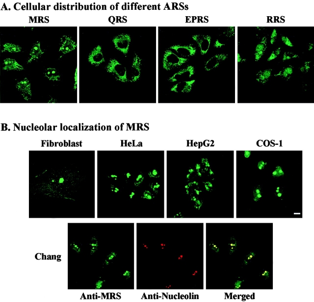
To investigate cellular distribution of different ARSs, polyclonal antibodies that were specific to MRS, EPRS, RRS, and QRS were prepared. Their antigenic specificity was determined by immunoblotting of the proteins extracted from HeLa cells. All of the antibodies showed specificity to their antigens (Fig. 1 B). Using these antibodies, cellular distribution of four different ARSs was investigated by immunostaining. Although all of these enzymes were detected in both the nucleus as well as the cytoplasm, relative partitions between the two cellular locations and staining patterns were idiosyncratic (Fig. 2 A). Among them, MRS was uniquely stained at nucleoli, implying its novel function at this site. Removal of anti-MRS IgG from anti-MRS antiserum decreased nucleolar and cytoplasmic staining and mock rabbit IgG or preimmune serum did not give any specific MRS signal (data not shown).
Figure 2.
Nucleolar localization of MRS. A, Distributions of four different ARSs in Chang cells were determined by immunostaining using confocal laser scanning microscopy. B, top, Cellular localization of MRS was monitored in different cell lines. Human foreskin fibroblast, HeLa, HpG2, and COS-1 cells were cultivated and stained with anti-MRS as described above. Bar, 10 μm. Bottom, Localization of MRS and nucleolin in Chang cells was determined using anti-MRS rabbit antibody and mouse monoclonal antinucleolin antibody (Santa Cruz) as described in Materials and Methods.
The nucleolar localization of MRS was further investigated in the different cells by confocal immunofluorescence microscopy using an anti-MRS antibody. The nucleolar MRS was detected in all of the human foreskin fibroblast, HeLa, HepG2, and COS-1 cells, indicating that the nucleolar localization of MRS is universal in mammalian cells (Fig. 2 B). The nucleolar localization of MRS was confirmed by coimmunostaining of MRS with nucleolin that was used as a marker for nucleoli. MRS was exactly colocalized with nucleolin, confirming its nucleolar localization (Fig. 2 B).
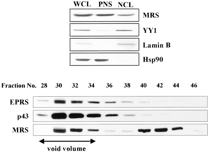
The presence of MRS at nucleoli was then investigated by immunoblotting of nuclear and postnuclear fractions of HeLa cells. The transcription factor, YY1 (Shi et al. 1991), and nuclear structural protein, lamin B (Moir et al. 1995), and HSP90 (Koyasu et al. 1986) were used as nuclear and cytoplasmic markers, respectively. The marker proteins were found in the expected fractions, indicating that the nuclei were well isolated. MRS was found both in nucleus as well as in postnuclear supernatant (Fig. 3, top). Since MRS is a component of the multi-ARS complex, the presence of MRS in nucleus implies that at least some portion of MRS should exist as a different form. This possibility was investigated by size exclusion chromatography (exclusion limit of 1,300 kD) of the proteins extracted from the HeLa cells. The multi-ARS complex would be eluted in the void volume from this column because its approximate molecular weight is 1,500 kD. The proteins eluted from the column were resolved by gel electrophoresis. MRS and two other complex-components, EPRS (Fett and Knippers 1991) and p43 (Quevillon et al. 1997), were detected by immunoblotting with their respective antibodies. The majority of the three proteins were coeluted in the void volume as expected. However, a significant amount of MRS was also detected in the following fractions in which EPRS and p43 were barely detected (Fig. 3, bottom). This result implies that MRS may be loosely associated with the multi-ARS complex or that a portion of MRS may exist unassociated from the multi-ARS complex.
Figure 3.
Determination of nuclear and free form MRS. Top, MRS was detected by immunoblotting in the whole cell lysate (WCL), nuclear extract (NCL), and postnuclear supernatant (PNS). Cell fractionation was performed as described previously (Neufeld and White 1997). YY1, lamin B, and Hsp90 were used as nuclear and cytoplasmic markers, respectively. Bottom, The proteins extracted from HeLa cells were fractionated by size exclusion chromatography. The eluted proteins in each fraction were analyzed by immunoblotting with anti-MRS, -p43 (Park et al. 1999), and -EPRS rabbit antibodies.
MRS Is Translocated to Nucleoli by Cell Proliferation Signal
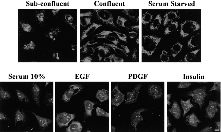
We then investigated the condition in which MRS is translocated to nucleolus. The cultured Chang cells were fixed at two different growth stages (subconfluent and confluent) and stained with an anti-MRS antibody. The nucleolar MRS was only detected in subconfluent cells, suggesting that the nucleolar localization of MRS is dependent on cell growth conditions (Fig. 4, top, left and middle). The nucleolar MRS disappeared by serum starvation (Fig. 4, top, right), but was restored by the addition of 10% serum, or mitogenic signals such as EGF, PDGF, and insulin (Fig. 4, bottom). Thus, the nucleolar localization of MRS is triggered by a cell growth signal, but is not specific as to the type of signal.
Figure 4.
MRS is translocated to nucleolus upon a mitogenic signal. Top, Cellular localization of MRS was monitored in Chang cells as described in Fig. 2. The nucleolar MRS was apparent in subconfluent (∼70% confluency) cells, but disappeared in confluent (100% confluency) or 5-d serum-starved cells. Bottom, 10% serum or EGF (40 ng/ml), PDGF (40 ng/ml), and insulin (100 μg/ml) were added to 7-d serum-starved cells and the cells were observed 24 h after the treatment. Bar, 10 μm.
Nucleolar MRS Requires rRNA and Polymerase I Activity
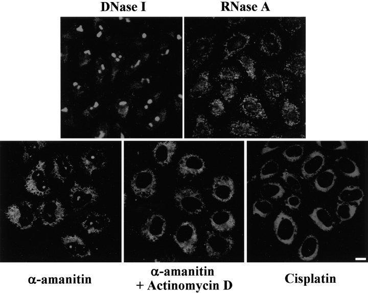
Cell proliferation would accelerate many biological processes, including ribosome biogenesis (Grummt 1999). Since the nucleolus is the site for rRNA synthesis, the nucleolar MRS that is induced by the cellular proliferation signal may be related to rRNA synthesis. To gain insight into the function of the nucleolar MRS, we first tested whether or not its nucleolar localization requires RNA in the nucleolus. HeLa cells were treated with DNase I or RNase A after fixation, and the cellular location of MRS was monitored by immunostaining. The nucleolar MRS disappeared with the treatment of RNase, but not DNase (Fig. 5, top). This result suggests that the nucleolar localization of MRS requires RNA in nucleolus.
Figure 5.
Nucleolar localization of MRS depends on rRNA synthesis. Top, HeLa cells grown on glass coverslips were permeabilized in 0.1% Triton X-100 for 5 min at room temperature. The permeabilized cells were incubated with DNase I (0.1 mg/ml) or RNase A (0.1 mg/ml) for 1 h at 37°C and fixed in 10% formaldehyde for 20 min. Digestion of nuclear DNA was confirmed by DAPI staining (data not shown). Bottom, HeLa cells were treated with α-amanitin (2 μg/ml), and α-amanitin (2 μg/ml) + actinomycin D (0.2 μg/ml), for 16 h to inhibit RNA polymerase II and RNA polymerase I + II, respectively. The specific inhibition of RNA polymerase I was performed by incubating HeLa cells with cisplatin (10 μg/ml) for 9 h. The cellular localization of MRS was monitored by immunostaining as described above. Bar, 10 μm.
We then investigated whether or not the presence of MRS is dependent on the activity of RNA polymerase I that is responsible for rRNA synthesis. The nucleolar MRS disappeared with the addition of α-amanitin and actinomycin D (Perry 1963) or cisplatin (Jordan and Carmo-Fonseca 1998) that inhibited RNA polymerase I. However, it was not affected by the treatment of the RNA polymerase II inhibitor, α-amanitin (Kedinger et al. 1970; Lindel et al. 1970; Fig. 5, bottom). These results suggest that the nucleolar MRS is related to rRNA synthesis.
Anti-MRS Antibody Blocks rRNA Synthesis
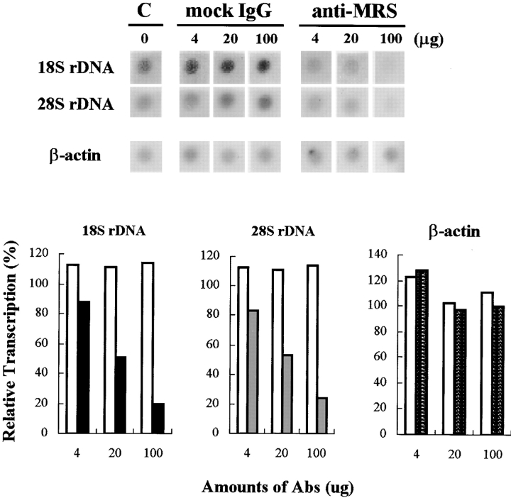
The effect of MRS on rRNA synthesis was then investigated by a nuclear run-on assay and immunostaining of nascent rRNA. The HeLa cell nuclei were isolated and the transcription was carried out in the presence of different amounts of anti-MRS antibody. The synthesized RNAs were isolated and hybridized with 18S and 28S rDNAs, as well as cDNA for β-actin that was used for internal control. The synthesis of two rRNAs was decreased by the addition of anti-MRS antibody, in a dose-dependent manner, to 20% of the control in which no antibody was added (Fig. 6). In contrast, mock rabbit IgG did not affect the synthesis of 18S and 28S rRNAs and the amounts of β-actin RNA were unchanged by the treatment of anti-MRS antibody. These results indicate that anti-MRS antibody specifically affected the synthesis of rRNA.
Figure 6.
Nuclear run-on assay for rRNA synthesis. Ribosomal RNA synthesis is blocked with anti-MRS antibody. Top, Synthesis of rRNA was monitored by nuclear run-on assay as described previously (Giraudo et al. 1998). The nuclei isolated from HeLa cells were used for the assay. The synthesized transcripts were hybridized to 18S and 28S rDNAs or cDNA for β-actin on the membrane. The amounts of the hybridized transcripts were quantified by phosphor image analyzer. Bottom, The radioactive intensities of 18S, 28S, and β-actin blots without IgG were taken as 100% and the relative intensities of other blots were shown by percentage. White bars stand for the values of the intensities of 18S and 28S treated with the indicated amounts of mock rabbit IgG. Black, gray, and lined bars represent the relative intensities of the 18S, 28S, and β-actin blots treated with the indicated amounts of anti-MRS antibody, respectively. Similar results were obtained from three independent experiments.
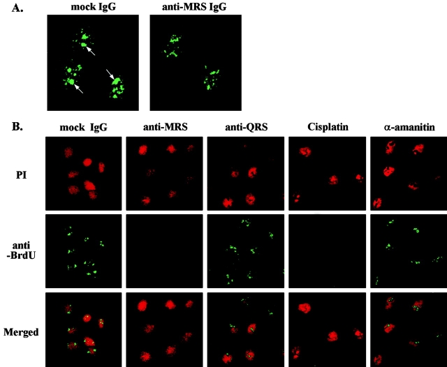
We also monitored the rRNA and mRNA synthesis by the immunostaining of Br-UTP incorporated to the nascent RNA. Ribosomal RNA synthesis, executed by RNA polymerase I, was shown as nucleolar foci, whereas synthesis of mRNA by RNA polymerase II was detected as nucleoplasmic foci. The nucleolar foci disappeared with the treatment of an anti-MRS antibody, but not by mock rabbit IgG (Fig. 7 A). The nucleoplasmic foci remained unaffected by either of the two antibodies. We then repeated these similar experiments focusing on the nucleolar rRNA synthesis in a few different conditions. Nuclei were stained using propidium iodide. Again, an anti-MRS antibody specifically inhibited rRNA synthesis, whereas mock rabbit IgG and anti-QRS antibody did not. The nucleolar Br-staining of rRNA was also confirmed by its sensitivity to the treatment of cisplatin, but not of α-amanitin (Fig. 7 B). All of these results clearly suggest that MRS is involved in the synthesis of rRNA.
Figure 7.
Br-UTP incorporation assay for rRNA synthesis. A, The nuclear rRNA synthesis was carried out in the presence of Br-UTP as described in Materials and Methods. The subconfluent HeLa cells were treated with anti-MRS or mock rabbit IgG in the presence of Br-UTP. The incorporated Br-UTP was then detected with anti-BrdU antibody. Fluorescence nucleolar foci (by RNA polymerase I, arrows) disappeared by the treatment of anti-MRS IgG, but not of mock IgG. Nucleoplasmic foci (by RNA polymerase II) were not affected by either of the two antibodies, confirming the specificity of anti-MRS antibody to the nucleolar rRNA synthesis. B, The nucleolar rRNA synthesis was monitored by immunostaining with anti-Br antibody in the presence of different antibodies and RNA synthesis inhibitors. The nuclear DNA was stained with propidium iodide (PI). The nucleolar rRNA synthesis was blocked with anti-MRS antibody, but not with mock IgG or anti-QRS antibody. The stained foci disappeared with the treatment of cisplatin that inhibits RNA polymerase I, but not with the treatment of α-amanitin that inhibits RNA polymerase II. This confirms that the stained foci resulted from the nucleolar rRNA synthesis. Nucleoplasmic Br-staining is not shown here due to the short exposure.
Discussion
Several mammalian ARSs form a macromolecular protein complex (Mirande 1991; Kisselev and Wolfson 1994; Yang 1996). However, the presence of free forms has been reported in a few different ARSs that are the components for the complex (Mirande et al. 1983; Vellekamp et al. 1983, Vellekamp et al. 1985). Here, the four complex-forming ARSs showed different patterns in immunostaining (Fig. 2 A). These results imply that at least some portions of the complex-forming ARSs are distributed differently in a cell, while most of them are present within the macromolecular protein complex.
Human MRS is also a component of this multi-ARS complex. However, MRS dissociated from the multi-ARS complex may exist based on the elution profile from size exclusion chromatography and its presence in the nucleus (Fig. 3). MRS in nuclear fraction was also eluted from the gel filtration column as a macromolecular complex (data not shown), implying that it is also associated with other nuclear factors or structure. This observation is consistent with its localization in nucleoli. Human MRS contains peptide extensions attached to the NH2- and COOH-terminal ends of the core catalytic domain (Fig. 1 A). Interestingly, these two peptide appendices are involved in protein–protein interactions (Rho et al. 1999) and phosphorylation sites for casein kinase II are heavily clustered in the NH2-terminal appendix (data not shown). Since casein kinase II is involved in the growth control and regulation of rRNA synthesis (Voit et al. 1995; Hannan et al. 1998), phosphorylation of MRS at these sites may be related to the nucleolar localization of MRS. However, we failed to determine the peptide region responsible for the nucleolar localization of MRS, because various forms of recombinant MRS were not well expressed and gave a cytotoxic effect in transfected cells (data not shown).
The nucleolus is the nuclear site in which rRNA biogenesis takes place. Interestingly, it was reported previously that nucleoli show an independent protein synthesis capability (Lamkin et al. 1973) and contain some components of protein synthesis, such as the elongation factor (Rao et al., 1998). Electron microscopic analysis also suggested the presence of a few ARSs in nucleoli (Popenko et al. 1994). However, the condition and physiological meaning for the presence of these factors in nucleolus has not been understood. Here, we report that the cytoplasmic MRS is translocated into nucleolus by various cellular proliferation signals (Fig. 4) and is involved in rRNA synthesis (Fig. 6 and Fig. 7). Interestingly, 5S rRNA is associated with MRS and enhances its activity in cytoplasm (Ogata et al. 1991). MRS may be also bound to 5S rRNA in nucleolus and regulate the biogenesis of ribosome.
The ribosome biogenesis of prokaryotes is subjected to stringent control mediated by unusual nucleotides in response to uncharged tRNA (Cozzone 1980). Amino acid starvation drops the nucleolar rRNA synthesis as well as the cytoplasmic protein synthesis in eukaryotic cells. This suggests the presence of a mechanism sensing the cellular level of the amino acid. Although eukaryotes do not seem to employ unusual nucleotide messengers analogous to those of prokaryotes for this communication (Pollard et al. 1980), many protein factors have recently been found in nucleolus and are involved in rRNA synthesis. For instance, a zinc finger protein, ZPR1, is associated with the cytoplasmic domain of EGF receptor in quiescent cells (Galcheva-Gargova et al. 1996), but translocated to nucleolus to activate rRNA synthesis by a cell proliferation signal (Galcheva-Gargova et al. 1998). In reverse, a tumor suppressor, retinoblastoma protein, is translocated to nucleolus to inhibit rRNA synthesis during cellular differentiation (Cavanaugh et al. 1995). In this sense, MRS is an ideal molecule, coordinating rRNA synthesis in the nucleolus and protein synthesis in cytoplasm.
Acknowledgments
We are grateful to Dr. K.H. Kim for sequence analysis of MRS and to Dr. I. Grummt for critical comments on the experiments.
This work was supported by a grant from National Creative Research Initiatives of the Ministry of Science and Technology of Korea.
Footnotes
Young-Gyu Ko and Young-Sun Kang contributed equally to this work.
Abbreviations used in this paper: ARSs, aminoacyl–tRNA synthetases; EPRS, bifunctional glutamyl-prolyl–tRNA synthetase; Hsp90, heat shock protein 90; MRS, methionyl–tRNA synthetase; QRS, glutaminyl–tRNA synthetase; RRS, arginyl–tRNA synthetase.
References
- Akins R.A., Lambowitz A.M. A protein required for splicing group I introns in Neurospora mitochondria is mitochondrial tyrosyl-tRNA synthetase or a derivative thereof. Cell. 1987;50:331–345. doi: 10.1016/0092-8674(87)90488-0. [DOI] [PubMed] [Google Scholar]
- Andreassen P.R., Lacroix F.B., Villa-Moruzzi E., Margolis R.L. Differential subcellular localization of protein phosphatase-1, and isoforms during both interphase and mitosis in mammalian cells. J. Cell Biol. 1998;141:1207–1215. doi: 10.1083/jcb.141.5.1207. [DOI] [PMC free article] [PubMed] [Google Scholar]
- Cavanaugh A.H., Hempel W.M., Taylor L.J., Rogalsky V., Todorov G., Rothblum L.I. Activity of RNA polymerase 1 transcription factor UBF blocked by Rb gene product. Nature. 1995;374:177–180. doi: 10.1038/374177a0. [DOI] [PubMed] [Google Scholar]
- Cozzone A.J. Stringent control and protein synthesis in bacteria. Biochimie. 1980;62:647–664. doi: 10.1016/s0300-9084(80)80022-8. [DOI] [PubMed] [Google Scholar]
- Dang C.V., Yang D.C., Pollard T.D. Association of methionyl–tRNA synthetase with detergent-insoluble components of the rough endoplasmic reticulum. J. Cell Biol. 1983;96:1138–1147. doi: 10.1083/jcb.96.4.1138. [DOI] [PMC free article] [PubMed] [Google Scholar]
- Fett R., Knippers R. The primary structure of human glutaminyl–tRNA synthetasea highly conserved core, amino acid repeat regions, and homologies with translation elongation factors. J. Biol. Chem. 1991;266:1448–1455. [PubMed] [Google Scholar]
- Galcheva-Gargova Z., Konstantinov K.N., Wu I.-H., Klier G., Barrett T., Davis R.J. Binding of zinc finger protein ZPR1 to the epidermal growth factor receptor. Science. 1996;272:1797–1802. doi: 10.1126/science.272.5269.1797. [DOI] [PubMed] [Google Scholar]
- Galcheva-Gargova Z., Gangwani L., Konstantinov K.N., Mikrut M., Theroux S.J., Enoch T., Davis R.J. The cytoplasmic zinc finger protein ZPR1 accumulates in the nucleolus of proliferating cells. Mol. Biol. Cell. 1998;9:2963–2971. doi: 10.1091/mbc.9.10.2963. [DOI] [PMC free article] [PubMed] [Google Scholar]
- Giraudo E., Primo L., Audero E., Gerber H.P., Koolwijk P., Soker S., Klagsbrun M., Ferrara N., Bussolino F. Tumor necrosis factor-α regulates expression of vascular endothelial growth factor receptor-2 and of its co-receptor neuropilin-1 in human vascular endothelial cells. J. Biol. Chem. 1998;273:22128–22135. doi: 10.1074/jbc.273.34.22128. [DOI] [PubMed] [Google Scholar]
- Grummt I. Regulation of mammalian ribosomal gene transcription by RNA polymerase I. Prog. Nucleic Acid Res. Mol. Biol. 1999;62:109–154. doi: 10.1016/s0079-6603(08)60506-1. [DOI] [PubMed] [Google Scholar]
- Hannan R.D., Hempel W.M., Cavanaugh A., Arino T., Dimitrov S.I., Moss T., Rothblum L.I. Affinity purification of mammalian RNA polymerase I. Identification of an associated kinase. J. Biol. Chem. 1998;273:1257–1267. doi: 10.1074/jbc.273.2.1257. [DOI] [PubMed] [Google Scholar]
- Hountondji C., Dessen P., Blanquet S. Sequence similarities among the family of aminoacyl–tRNA synthetases. Biochimie. 1986;68:1071–1078. doi: 10.1016/s0300-9084(86)80181-x. [DOI] [PubMed] [Google Scholar]
- Jordan P., Carmo-Fonseca C. Cisplatin inhibits synthesis of ribosomal RNA in vivo . Nucleic Acids Res. 1998;26:2831–2836. doi: 10.1093/nar/26.12.2831. [DOI] [PMC free article] [PubMed] [Google Scholar]
- Kedinger C., Gniazdowski M., Mandel J.L., Jr., Gissinger F., Chambon P. α-Amanitina specific inhibitor of one of two DNA-dependent RNA polymerase activities from calf thymus. Biochem. Biophys. Res. Commun. 1970;38:165–171. doi: 10.1016/0006-291x(70)91099-5. [DOI] [PubMed] [Google Scholar]
- Kisselev L.L., Wolfson A.D. Aminoacyl–tRNA synthetase from higher eukaryotes. Prog. Nucleic Acid Res. Mol. Biol. 1994;48:83–142. doi: 10.1016/s0079-6603(08)60854-5. [DOI] [PubMed] [Google Scholar]
- Kisselev L., Frolova L., Haenni A.L. Interferon inducibility of mammalian tryptophanyl–tRNA synthetasenew perspectives. Trends Biochem. Sci. 1993;18:263–267. doi: 10.1016/0968-0004(93)90178-p. [DOI] [PubMed] [Google Scholar]
- Koyasu S., Nishida E., Kadowaki T., Matsuzaki F., Iida K., Harada F., Kasuga M., Sakai H., Yahara I. Two mammalian heat shock proteins, HSP90 and HSP100, are actin-binding proteins. Proc. Natl. Acad. Sci. USA. 1986;83:8054–8058. doi: 10.1073/pnas.83.21.8054. [DOI] [PMC free article] [PubMed] [Google Scholar]
- Labouesse M. The yeast mitochondrial leucyl–tRNA synthetase is a splicing factor for the excision of several group I introns. Mol. Gen. Genet. 1990;224:209–221. doi: 10.1007/BF00271554. [DOI] [PubMed] [Google Scholar]
- Lage H., Dietel M. Cloning of a human cDNA encoding a protein with high homology to yeast methionyl–tRNA synthetase. Gene. 1996;178:187–189. doi: 10.1016/0378-1119(96)00313-7. [DOI] [PubMed] [Google Scholar]
- Lamkin A.F., Smith D.W., Hurlbert R.B. Independent protein synthesis in isolated rat tumor nucleoliaminoacylation of endogenous transfer ribonucleic acid. Biochemistry. 1973;12:4137–4145. doi: 10.1021/bi00745a017. [DOI] [PubMed] [Google Scholar]
- Lindel T.J., Weingerg F., Morries P.W., Roeder R.G., Rutter W.J. Specific inhibition of nuclear RNA polymerase II by α-amanitin. Science. 1970;170:447–449. doi: 10.1126/science.170.3956.447. [DOI] [PubMed] [Google Scholar]
- Ludmerer S.W., Schimmel P. Gene for yeast glutamine tRNA synthetase encodes a large amino-terminal extension and provides a strong confirmation of the signature sequence for a group of the aminoacyl–tRNA synthetases. J. Biol. Chem. 1987;262:10801–10806. [PubMed] [Google Scholar]
- Martinis S.A., Plateau P., Cavarelli J., Florentz C. Aminoacyl–tRNA synthetasesa family of expanding functions. EMBO (Eur. Mol. Biol. Organ.) J. 1999;18:4591–4596. doi: 10.1093/emboj/18.17.4591. [DOI] [PMC free article] [PubMed] [Google Scholar]
- Mirande M. Aminoacyl–tRNA synthetase family from prokaryotes and eukaryotesstructural domains and their implications. Prog. Nucleic Acid Res. Mol. Biol. 1991;40:95–142. doi: 10.1016/s0079-6603(08)60840-5. [DOI] [PubMed] [Google Scholar]
- Mirande M., Cirakoglu B., Waller J.-P. Seven mammalian aminoacyl–tRNA synthetases associated within the same complex are functionally independent. Eur. J. Biochem. 1983;131:163–170. doi: 10.1111/j.1432-1033.1983.tb07244.x. [DOI] [PubMed] [Google Scholar]
- Moir R.D., Spann T.P., Goldman R.D. The dynamic properties and possible functions of nuclear lamins. Int. Rev. Cytol. 1995;162:141–182. doi: 10.1016/s0074-7696(08)62616-9. [DOI] [PubMed] [Google Scholar]
- Neufeld K.L., White R.L. Nuclear and cytoplasmic localizations of the adenomatous polyposis coli protein. Proc. Natl. Acad. Sci. USA. 1997;94:3034–3039. doi: 10.1073/pnas.94.7.3034. [DOI] [PMC free article] [PubMed] [Google Scholar]
- Ogata K., Kurahashi A., Kenmochi N., Terao K. Role of 5S rRNA as a positive effector of some aminoacyl–tRNA synthetases in macromolecular complexes, with specific reference to methionyl–tRNA synthetase. J. Biochem. 1991;110:1037–1044. doi: 10.1093/oxfordjournals.jbchem.a123674. [DOI] [PubMed] [Google Scholar]
- Park S.G., Jung K.H., Lee J.S., Jo Y.J., Motegi H., Kim S., Shiba K. Precursor of pro-apoptotic cytokine modulates aminlacylation activity of tRNA synthetase. J. Biol. Chem. 1999;274:16673–16676. doi: 10.1074/jbc.274.24.16673. [DOI] [PubMed] [Google Scholar]
- Perry R.P. Selective effects of actinomycin D on the intracellular distribution of RNA synthesis in tissue culture cells. Exp. Cell Res. 1963;29:400–406. [Google Scholar]
- Pollard J.W., Lam T., Stanners C.P. Mammalian cells do not have a stringent response. J. Cell. Physiol. 1980;105:313–325. doi: 10.1002/jcp.1041050214. [DOI] [PubMed] [Google Scholar]
- Popenko V.I., Ivanova J.L., Cherny N.E., Filonenko V.V., Beresten S.F., Wolfson A.D., Kisselev L.L. Compartmentalization of certain components of the protein synthesis apparatus in mammalian cells. Eur. J. Cell Biol. 1994;65:60–69. [PubMed] [Google Scholar]
- Quevillon S., Agou F., Robinson J.C., Mirande M. The p43 component of the mammalian multi-synthetase complex is likely to be the precursor of the endothelial monocyte-activating polypeptide II cytokine. J. Biol. Chem. 1997;272:32573–32579. doi: 10.1074/jbc.272.51.32573. [DOI] [PubMed] [Google Scholar]
- Rao M.S., Rothblum L.I., Busch H. Presence of elongation factor 1 in nuclei and nucleoli of rat liver. Cell Biol. Int. Rep. 1978;2:25–32. doi: 10.1016/0309-1651(78)90081-4. [DOI] [PubMed] [Google Scholar]
- Rho S.B., Lee K.H., Kim J.W., Shiba K., Jo Y.J., Kim S. Interaction between human tRNA synthetases involves repeated sequence elements. Proc. Natl. Acad. Sci. USA. 1996;93:10128–10133. doi: 10.1073/pnas.93.19.10128. [DOI] [PMC free article] [PubMed] [Google Scholar]
- Rho S.B., Lee J.S., Jeong E.-J., Kim K.-S., Kim Y.G., Kim S. A multifunctional repeated motif is present in human bifunctional tRNA synthetase. J. Biol. Chem. 1998;273:11267–11273. doi: 10.1074/jbc.273.18.11267. [DOI] [PubMed] [Google Scholar]
- Rho S.B., Kim M.J., Lee J.S., Seol W., Montegi H., Kim S., Shiba K. Genetic dissection of protein–protein interactions in multi-tRNA synthetase complex. Proc. Natl. Acad. Sci. USA. 1999;96:4488–4493. doi: 10.1073/pnas.96.8.4488. [DOI] [PMC free article] [PubMed] [Google Scholar]
- Schimmel P., Wang C.C. Getting tRNA synthetase into the nucleus. Trends Biochem. Sci. 1999;24:127–128. doi: 10.1016/s0968-0004(99)01369-9. [DOI] [PubMed] [Google Scholar]
- Seshaiah P., Andrew D.J. WRS-85DA tryptophanyl–tRNA synthetase expressed to high levels in the developing Drosophila salivary gland. Mol. Biol. Cell. 1999;10:1595–1608. doi: 10.1091/mbc.10.5.1595. [DOI] [PMC free article] [PubMed] [Google Scholar]
- Shi Y., Seto E., Chang L.S., Shenk T. Transcriptional repression by YY1, a human GLI-Kruppel-related protein, and relief of repression by adenovirus E1A protein. Cell. 1991;18:377–388. doi: 10.1016/0092-8674(91)90189-6. [DOI] [PubMed] [Google Scholar]
- Simos G., Segref A., Fasiolo F., Hellmuth K., Shevchenko A., Mann M., Hurt E.C. The yeast protein Arc1p binds to tRNA and functions as a cofactor for the methionyl- and glutamyl–tRNA synthetases. EMBO (Eur. Mol. Biol. Organ.) J. 1996;15:5437–5448. [PMC free article] [PubMed] [Google Scholar]
- Vellekamp G.J., Coyle C.L., Kull F.J. Low molecular weight aspartyl–tRNA synthetase from porcine thyroidpurification, characterization, and heterogeneity. J. Biol. Chem. 1983;258:8195–8200. [PubMed] [Google Scholar]
- Vellekamp G.J., Sihag R.K., Deutscher M.P. Comparison of the complexed and free forms of rat liver arginyl–tRNA synthetase and origin of the free form. J. Biol. Chem. 1985;260:9843–9847. [PubMed] [Google Scholar]
- Voit R., Kuhn A., Sander E.E., Grummt I. Activation of mammalian ribosomal gene transcription requires phosphorylation of the nucleolar transcription factor UBF. Nucl. Acids Res. 1995;23:2593–2599. doi: 10.1093/nar/23.14.2593. [DOI] [PMC free article] [PubMed] [Google Scholar]
- Wakasugi K., Schimmel P. Two distinct cytokines released from a human aminoacyl–tRNA synthetase. Science. 1999;284:147–150. doi: 10.1126/science.284.5411.147. [DOI] [PubMed] [Google Scholar]
- Webster T.A., Tsai H., Kula M., Mackie G.A., Schimmel P. Specific sequence homology and three-dimensional structure of an aminoacyl transfer RNA synthetase. Science. 1984;226:1315–1317. doi: 10.1126/science.6390679. [DOI] [PubMed] [Google Scholar]
- Yang D.C.H. Mammalian aminoacyl–tRNA synthetases. Curr. Top. Cell. Regul. 1996;34:101–136. doi: 10.1016/s0070-2137(96)80004-5. [DOI] [PubMed] [Google Scholar]
