Ephrin-A5 Induces Collapse of Growth Cones by Activating Rho and Rho Kinase (original) (raw)
Abstract
The ephrins, ligands of Eph receptor tyrosine kinases, have been shown to act as repulsive guidance molecules and to induce collapse of neuronal growth cones. For the first time, we show that the ephrin-A5 collapse is mediated by activation of the small GTPase Rho and its downstream effector Rho kinase. In ephrin-A5–treated retinal ganglion cell cultures, Rho was activated and Rac was downregulated. Pretreatment of ganglion cell axons with C3-transferase, a specific inhibitor of the Rho GTPase, or with Y-27632, a specific inhibitor of the Rho kinase, strongly reduced the collapse rate of retinal growth cones. These results suggest that activation of Rho and its downstream effector Rho kinase are important elements of the ephrin-A5 signal transduction pathway.
Keywords: retinotectal, repulsive, Eph receptor, axon guidance, collapse assay
Introduction
Significant progress has been made recently in understanding the molecular basis of formation of the retinotectal projection (for reviews see Drescher 1997; Drescher et al. 1997; Flanagan and Vanderhaeghen 1998). However, many questions remain open at the cellular level, and signal transduction of retinal growth cones (GCs) is a field where not much is known (for review see Mueller 1999). A retinal ganglion cell (RGC) GC advancing over the tectal surface encounters many different guidance molecules simultaneously, and after signal integration, a variety of behaviors like turning, stopping, and branching is observed. Candidate molecules that might induce these behaviors in the vertebrate tectum are several repulsive guidance molecules, of which two belong to the ephrin-A family. Ephrins exert their functions by binding to receptor tyrosine kinases of the Eph family, and two groups of ligands can be distinguished. The A-ephrins are glycosylphosphatidylinositol (GPI)-anchored ligands that bind preferentially to EphA receptors, whereas transmembrane B-ephrins bind preferentially to EphB receptors (Drescher 1997). Ephrin-A5 binds to seven different EphA receptors, and at least five of them, EphA3, EphA4, EphA5, EphA6, and EphA7, are expressed by RGC axons (Connor et al. 1998; O'Leary and Wilkinson 1999). Not much is known about signal transduction induced by ephrins binding to their receptors. Ephrin-A5 and ephrin-A2 have been shown to guide retinal axons in vitro, and targeted disruption or overexpression resulted in aberrant projections in tectum and lateral geniculate nucleus (Cheng et al. 1995; Drescher et al. 1995; Nakamoto et al. 1996; Monschau et al. 1997; Feldheim et al. 1998; Frisen et al. 1998; Hornberger et al. 1999). In culture experiments, ephrin-A5 induces collapse of nasal and temporal retinal GCs, whereas ephrin-A2 induces collapse of temporal GCs but not of nasal GCs (Drescher et al. 1995; Monschau et al. 1997). A collapse-inducing activity of ephrin-A5 was also observed with rat cortical neurons (Meima et al. 1997).
To analyze the signal transduction machinery in retinal GCs underlying the ephrin-A5–induced collapse, we focused on the small GTPase Rho and its downstream effector, the serine/threonine kinase Rho kinase (ROCK). The Rho GTPases are important regulators of the actin cytoskeleton and their function is best characterized in fibroblasts (Hall 1998). Recent work has provided enough data to support the assumption that they exert similar functions in neuronal cells (Mueller 1999). In neuroblastoma cells, in neuron-like PC12 cells, and in primary neurons, activation of Rho leads to rapid GC collapse, neurite retraction, or neurite growth inhibition, and these responses are prevented by a specific inhibitor of Rho, the bacterial exoenzyme C3-transferase (Jalink and Moolenaar 1992; Jalink et al. 1994; Tigyi et al. 1996; Jin and Strittmatter 1997; Kozma et al. 1997; Hirose et al. 1998; Kranenburg et al. 1999; Lehmann et al. 1999). C3-transferase ADP ribosylates RhoA, B, and C (but not Cdc42 and Rac) at Asn-41, thereby inhibiting these GTPases (Sekine et al. 1989; Schmidt and Aktories 1998). C3-transferase dramatically reduced the fc-ephrin-A5 (f-EA5)-induced collapse of retinal GCs. Moreover, many retinal GCs continued to advance despite the presence of f-EA5. One of the downstream targets of active GTP-bound Rho is the Rho-associated kinase ROCK (for review see Redowicz 1999), which is specifically inhibited by the pyridine derivative Y-27632 (Uehata et al. 1997). Pretreatment of RGC GCs advancing on a laminin substratum, resulted in a significant decrease of the number of collapsed retinal GCs exposed to f-EA5. These results suggest that f-EA5 induces collapse of retinal GCs by activating Rho and its downstream effector ROCK.
Materials and Methods
Primary Cell Culture, Collapse Assay, and Data Analysis
Embryonic day E6 and E7 retinae from white leghorn chicken were isolated and mounted on black nitrocellulose filters as described previously (Bonhoeffer and Huf 1982; Walter et al. 1987). 275-μm-wide retinal explant strips were cut along the nasal temporal axis with a McIlwain tissue chopper. Retinal strips were placed on laminin-coated coverslips and stabilized with small metal bars. Explants were cultured at 37°C, 4% CO2 , 100% relative humidity for ∼24 h in 600 μl F12 culture medium (Gibco) with 0.4% methyl cellulose. In C3-transferase experiments, 200 μl F12 medium containing 300 ng/ml C2IN-C3 and 300 ng/ml C2II were added to the cultures 6 h before collapse induction by f-EA5. To inhibit ROCK, 10 μM Y-27632 was added to retinal cultures 2 h before performing f-EA5 collapse experiments.
Retinal explants were grown for 24 h in multi-well plates (Nunc) and were incubated with or without C3-transferase or Y-27632. Control cultures were incubated in medium without performing the collapse assay. For collapse experiments, 200 μl medium containing 1.0 μg f-EA5 (Hornberger et al. 1999) was added. 30 min later, cultures were fixed with 4% paraformaldehyde, 0.3 M sucrose in PBS.
After fixation with paraformaldehyde, the explants were stained with an F-actin stain, Alexa Fluor-Phalloidin (Molecular Probes, Inc.), or with antibodies specific for RhoA (Santa Cruz Biotechnology) or the ROCK Rock1 (Santa Cruz Biotechnology). As a secondary antibody, a Cy-3–conjugated antibody (Dianova) was used. In C2-C3 incubated cultures, immunostaining was done using a primary rabbit polyclonal antibody directed against C3-transferase, and a secondary Cy3-labeled goat anti–rabbit antibody (Dianova). The collapsed and noncollapsed GCs were counted in each culture and the data were analyzed with Microsoft Excel. Data are expressed as mean ± SD. Statistical analysis among the different groups were done by one-way analysis of variance, and in all analyses, differences were considered statistically significant at P < 0.01. Criteria for collapsed GCs were a total loss of filopodia and lamellipodia and a strong decrease in F-actin content. Images were collected with a Sony CCD camera and a PC using the analysis software (SIS).
ADP Ribosylation Assay of Rho
For the ADP ribosylation assay (Barth et al. 1999), we used highly enriched RGC cultures (Bauch et al. 1998). Cells were treated with the respective toxins as indicated. After incubation, the medium was removed and the cells were treated with 1 mg/ml trypsin for 10 min at 37°C. After blocking trypsin with serum-containing medium, the cells were harvested and lysed with cold lysis buffer (2 mM MgCl2, 0.1 mM PMSF, 20 μg/ml leupeptin, 80 μg/ml benzamidine in 50 mM Hepes, ph 7.4). Lysates were incubated with [32P]NAD and 50 ng C2IN-C3 fusion toxin at 37°C for 30 min, Laemmli buffer was added, and the samples were heated for 3 min at 95°C and run on an SDS-gel. [32P]ADP ribosylated proteins were detected by autoradiography with a PhosphorImager from Molecular Dynamics.
Glutathione S-Transferase (GST) Fusion Protein Pull-down Assay
This assay is based on the capability of GST-Rhotekin and GST-Pak to bind to the GTP-bound Rho and Rac, respectively. Both assays were performed as described (Sander et al. 1998; Ren et al. 1999). In brief, 5 × 10⁶ RGCs were plated on laminin-coated 10-cm dishes. After 24 h, f-EA5 (1 μg/ml) was added to the cultures, and after 30 min of incubation the cells were lysed. Lysates were cleared by centrifugation and aliquots were taken for determining total amount of Rac and Rho by immunodetection on Western blots using Rac- and Rho-specific antibodies (Santa Cruz Biotechnology). The remaining amount of the lysates was incubated with GST-Rho binding domain of Rhotekin or GST-Rac/Cdc42 binding domain of Pak immobilized on glutathione-coupled Sepharose beads for 30 min at 4°C. Beads were washed, eluted in Laemmli sample buffer, and analyzed by Western blotting using mouse mAb anti-Rho or rabbit polyclonal anti-Rac.
Results
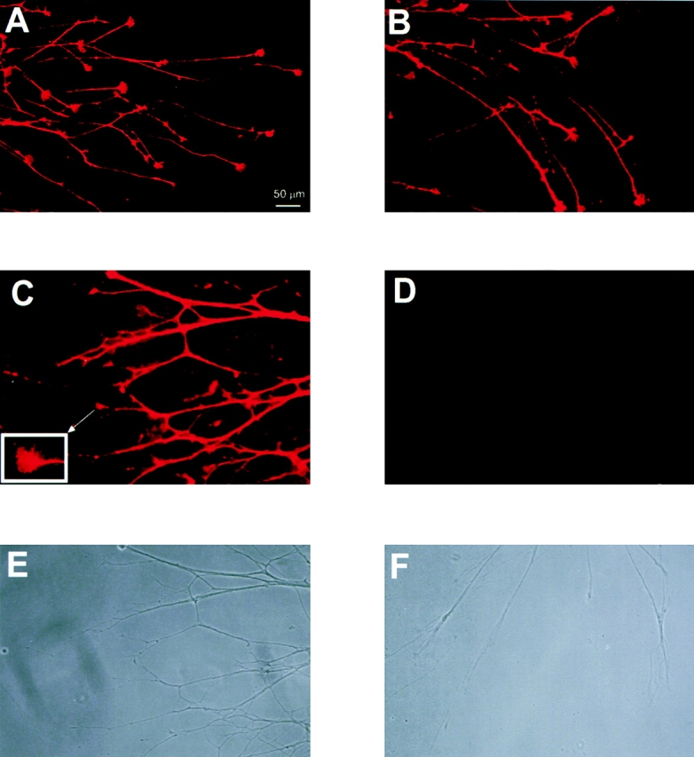
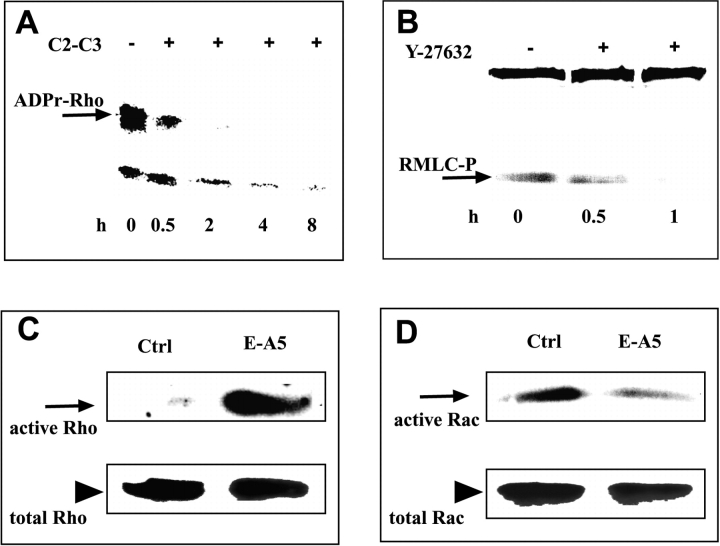
To analyze the role of the Rho GTPase in ephrin-A5–induced collapse of retinal GCs, we asked first whether RhoA and ROCK are expressed by retinal GCs. Retinal axons growing on a laminin substratum were fixed and stained with Rho- and ROCK-specific antibodies. RhoA and ROCK were detected in GCs of chick embryonic RGCs and both proteins were present in lamellipodia and filopodia (Fig. 1A and Fig. B). Inhibition of these proteins requires that a sufficient amount of the C3-like transferase permeates the plasma membrane of retinal GCs. Unfortunately, C3-transferase and its relatives do not enter cells readily (Barth et al. 1998), requiring high amounts of enzyme to be added to the culture medium. To solve this problem of low membrane permeability of the C3-transferase, a new C2-C3 fusion toxin with higher membrane permeability was used, whose activity is several hundred-fold higher when added to culture medium (Barth et al. 1998). The carrier system consists of the NH2-terminal part of the enzyme component (C2IN) of binary C. botulinum C2 toxin fused to the C3-like transferase from C. limosum. This C2IN-C3 fusion toxin interacts with the C2-binding component C2II and seems to induce uptake of both components by receptor-mediated endocytosis (Simpson 1989; Ohishi and Yanagimoto 1992; Barth et al. 1998). After adding the C2IN-C3 fusion toxin together with the C2II binding protein to RGC axons on laminin, cultures were fixed and stained with an antibody directed against the C3-like transferase. A secondary antibody, coupled to the fluorophore Cy-3, revealed clear staining of retinal axons in cultures treated with C2IN-C3 and C2II (Fig. 1 C) but no staining in control cultures (Fig. 1 D). To examine if the Rho GTPase was ADP ribosylated and inactivated, homogenates from highly enriched RGC cultures (Bauch et al. 1998) were loaded onto an SDS-gel and were subjected to an in vitro [32P]ADP ribosylation assay with C3. Labeled proteins were visualized by SDS-gel autoradiography (Barth et al. 1999). The level of Rho was significantly reduced in the cell lysate in the presence of the fusion toxin but not in the controls where no toxin was added (Fig. 2 A), or where the single components of the fusion toxin (C2I, C2II, and C2IN-C3) were added (data not shown). After 2 h of treatment with the fusion toxin, [32P]ADP ribosylation of Rho was significantly reduced, and after 4 and 8 h of treatment, no ribosylatable Rho protein was left (Fig. 2 A), suggesting that all Rho proteins have been ADP ribosylated and inactivated. These results show first that the carrier system for the C3-transferase works on retinal axons growing on laminin, and second that Rho is ADP ribosylated and inactivated.
Figure 1.

Staining of retinal axons and GCs with antibodies directed against RhoA, ROCK, and C3-transferase. (A and B) RhoA (A) and ROCK (B) are present in retinal axons and GCs. (C) RGC axons growing on laminin were treated with C2-C3 fusion toxin and were fixed and stained with an antibody directed against the C3-transferase (C, fluorescent image). Inset shows a retinal GC stained with the C3-transferase antibody. Nearly all axons are stained (E, phase-contrast image) albeit at different levels. (D) In the absence of the fusion toxin, no staining is observed with the C3-transferase antibody (D, fluorescent image; F, phase-contrast image).
Figure 2.
In RGCs, the C2-C3 fusion toxin and Y-27632 inactivate Rho and ROCK, respectively, and f-EA5 activates Rho and inactivates Rac. (A) Treatment of retinal cultures with fusion toxin reduced the level of Rho available for radioactive ADP ribosylation. (Lane 1) Control, no C2-C3 was added to RGC cultures. (Lanes 2–5) Time course of Rho ADP ribosylation in highly enriched RGC extracts. A nearly complete reduction of ADP-ribosylatable Rho is observed after 4 h (lane 4). (B) Treatment of retinal cultures with Y-27632 (10 μM) reduced the level of phosphorylated RMLC (arrow). Time course of ROCK inhibition, analyzed by immunodetection of pp2b-stained phosphorylated RMLC in highly enriched RGC extracts. No phosphorylated RMLC is detectable after a 1-h incubation in Y-27632 (lane 3). (C) Affinity precipitation of GTP-Rho using a GST fusion protein of the Rho-binding domain of Rhotekin. In f-EA5–treated RGC cultures, much higher levels of Rho are detectable (right lane) compared with control cultures (left lane). No difference is observed in the amount of total Rho (arrowhead) in lysates of control-treated and f-EA5–treated RGC cultures. (D) Affinity precipitation of GTP-Rac using the GST Pak1-CRIB domain. In f-EA5–treated RGC cultures, much lower levels of Rac are detectable (right lane) compared with control cultures (left lane). In lysates of control-treated and f-EA5–treated RGC cultures, no difference is observed in the amount of total Rac (arrowhead).
To monitor inhibition of ROCK, Y-27632 was added at several timepoints to RGC cultures before performing collapse experiments with f-EA5. Inactivation of ROCK was followed by analyzing phosphorylation of the regulatory myosin light chain (RMLC) on Western blots of gels with RGC homogenates, using the pp2b antibody specific for phosphorylated RMLC (Hirose et al. 1998; Matsumura et al. 1998). ROCK phosphorylates and inhibits myosin light chain (MLC) phosphatase, leading to an increase in phosphorylation of RMLC (Kimura et al. 1996), and incubation of RGC cultures in 10 μM Y-27632 for 1 h resulted in loss of detectable phosphorylated RMLC, suggesting complete inhibition of ROCK (Fig. 2 B).
Pull-down assays with GST fusion proteins were performed to measure activation of Rho and Rac in f-EA5–treated RGC cultures. To this aim, RGCs were treated with either f-EA5 or buffer. Cells were subsequently lysed and the level of GTP-bound Rho was determined using the Rho-binding domain of Rhotekin. Rhotekin, a downstream target of Rho, binds specifically to GTP-bound Rho (Reid et al. 1996). f-EA5 induced a dramatic increase in GTP-bound Rho (Fig. 2 C), whereas in control cultures Rho activation was hardly detectable (Fig. 2 C). In contrast to f-EA5–induced activation of Rho, affinity precipitation of GTP-bound Rac with GST-Pak-CD (Pak CRIB-domain) (Sander et al. 1998, Sander et al. 1999) resulted in significant reduction of GTP-Rac in f-EA5–treated cultures compared with control cultures (Fig. 2 D). These results suggest that treatment of RGCs with f-EA5 results in activation of Rho and in downregulation of Rac. In control and f-EA5–treated RGC cultures, no differences were observed in the levels of total Rho and Rac (Fig. 2C and Fig. D, lower panels).
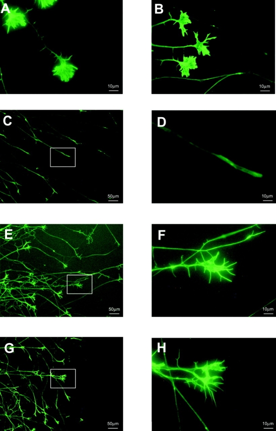
To answer the question if ADP ribosylation of Rho proteins by C2-C3 or inactivation of ROCK by Y-27632 resulted in reduction of the ephrin-A5–induced collapse, we performed collapse experiments with RGC axons growing on laminin. f-EA5 was added to RGC cultures in the presence or absence of C2-C3 or Y-27632 and cultures were fixed 30 min later. Fixed cultures were processed for Alexa Fluor-Phalloidin staining, and collapsed, intermediate, and noncollapsed retinal GCs were counted as described previously (Mueller et al. 1990). In all cultures, the percentage of intermediate GCs was ∼10%. The presence of C2-C3 or Y-27632 in RGC cultures did not significantly influence basal collapse rate or GC morphology (Fig. 3A and Fig. B). f-EA5 induces collapse of 80–90% of RGC GCs (Fig. 3C and Fig. D and Fig. 4) and collapse occurs 5–15 min after addition of 1 μg of f-EA5. In time-lapse experiments, we have never observed retinal GCs that continued to grow in the presence of f-EA5 (Wahl, S., and B.K. Mueller, data not shown). After incubating retinal cells with C2-C3, the collapse-inducing activity of 1 μg of f-EA5 was significantly reduced, whereas the single components of the fusion toxin (C2I, C2II, or C2-C3) did not affect the f-EA5–induced collapse of RGC GCs (Wahl, S., and B.K. Mueller, data not shown). The fraction of collapse-resistant retinal GCs increased from ∼10% in control cultures to >60% in C2-C3– and C2II–treated cultures (Fig. 3E and Fig. F and Fig. 4), and retinal GCs were able to advance despite the presence of f-EA5.
Figure 3.

Incubation of RGCs in C2-C3 or Y-27632 reduced the f-EA5–induced collapse of RGC GCs. (A and B) Incubation of RGC GCs in the presence of C2-C3 (A) or Y-27632 (B) did not affect GC morphology. (C and D) Addition of f-EA5 induces collapse of retinal GCs. (E and F) The f-EA5–induced collapse is reduced by pretreatment of retinal GCs with 300 ng/ml of C2-C3 fusion toxin for 4 h. (G and H) The f-EA5–induced collapse is reduced by pretreatment of retinal GCs with 10 μM of Y-27632. Left and right panels in C, E, and G and in D, F, and H correspond to low- and high-power magnification, respectively.
Figure 4.
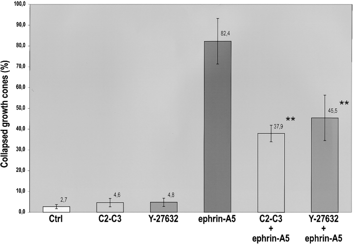
Summary of data from retinal explant cultures treated with C2-C3 fusion toxin or with Y-27632. The single components of the fusion toxin (C2I, C2II, and C2IN-C3) did not affect the f-EA5–induced collapse of RGC GCs (data not shown). Data are from six independent experiments, and in each culture ∼100 GCs at the axon front has been counted. Asterisks indicate statistical significance, P < 0.01.
Similar results were obtained after inhibition of ROCK by Y-27632. 2 h before adding f-EA5, retinal axons were treated with 10 μM Y-27632. Inhibition of ROCK reduced collapse-inducing activity of f-EA5 significantly (Fig. 3G and Fig. H) and the number of noncollapsed retinal GCs increased from ∼10% to >50% (Fig. 4).
Data are summarized in Fig. 4. In nontreated control cultures, 3% of retinal GCs are collapsed. Treatment with f-EA5 induces collapse of ∼85% of retinal GCs, and C2-C3 and Y-27632 resulted in a basal rate of collapsed GCs of ∼5%. Inhibition of Rho by C2-C3 reduced collapse rates to <40%, and inhibition of ROCK by Y-27632 reduced collapse rates to <50% (10 μM), suggesting that both proteins are involved in the ephrin-A5–induced collapse of retinal GCs.
Discussion
Repulsive guidance of RGC axons in the tectum is a crucial mechanism for establishing the initial topographic projection. To understand the signal transduction underlying repulsive guidance of RGC GCs, we performed experiments with the repulsive guidance molecule ephrin-A5.
Specific inhibitors of the Rho GTPase, the C3-transferase, and of the ROCK Y-27632, dramatically reduced the f-EA5 collapse-inducing activity of retinal GCs. The significant neutralization of the collapse activity correlates well with the extent of inactivation of the Rho proteins. A 2-h treatment of RGCs with C2-C3 and C2II reduced the level of Rho available for [32P]ADP ribosylation by ∼50%, and reduced collapse rates from 80–90% to 50%. After 6 h of treatment, the level of available Rho was further decreased to nondetectable levels, and the collapse rate was further decreased to 38%. It is possible that rapid neosynthesis of Rho prevents complete neutralization of the f-EA5 collapse activity. In rat astroglial cells, Rho-associated cytoskeletal changes were reversed when Rho neosynthesis reached ∼10% of the normal cellular content of Rho (Barth et al. 1999). An alternative explanation for incomplete inhibition might be the existence of different parallel pathways leading to GC collapse (Jin and Strittmatter 1997; Kuhn et al. 1999). Different parallel pathways are possible because ephrin-A5 interacts with high affinity with at least five different EphA receptors on RGCs (Connor et al. 1998; O'Leary and Wilkinson 1999).
In neuroblastoma cells, nerve growth factor–treated PC12 cells and NG108 cells, lysophosphatidic acid, thrombin, or sphingosine-1 phosphate induced GC collapse and neurite retraction, which was prevented by C3-transferase pretreatment (Jalink and Moolenaar 1992; Jalink et al. 1994; Postma et al. 1996; Tigyi et al. 1996; Kozma et al. 1997). Inhibition of ROCK by Y-27632 completely neutralized the collapse-inducing activity of lysophosphatidic acid in N1E-115 neuroblastoma cells (Hirose et al. 1998). Moreover, inhibition of ROCK by Y-27632 induced neurite outgrowth, which was blocked by transfection of these neuroblastoma cells with dominant-negative Cdc42 and Rac, and it was suggested that activation of Rho and ROCK transmits a negative signal to the Cdc42/Rac pathway (Hirose et al. 1998), confirming the results of Kozma et al. 1997. Mutually antagonistic effects of Rac and Rho were also observed in focal contact and focal complex formation in Swiss 3T3 cells (Rottner et al. 1999). In the same cell type, it was recently shown that Rac activation antagonized Rho activity (Sander et al. 1999).
In our experiments, we also obtained data that point to such an antagonism. The repulsive guidance molecule f-EA5 added to RGC cultures induced activation of Rho and downregulation of Rac. We currently do not know if Rac downregulation is downstream of Rho activation. Activation of the Eph receptor tyrosine kinases by f-EA5 might directly influence Rac and Rho antagonistically.
Cross-talk of the Rho and the Cdc42/Rac pathways has been described at several levels. LIM kinase, another serine/threonine kinase which in turn phosphorylates and thereby inactivates the actin-depolymerizing protein cofilin (Arber et al. 1998; Yang et al. 1998; Maekawa et al. 1999), is not only activated by ROCK but also by p21-activated kinase (Pak1), and the association of Pak1 with LIM kinase is increased by activated Cdc42 and Rac (Edwards et al. 1999). Pak is activated by GTP-bound Cdc42 and Rac (Manser et al. 1994; Knaus et al. 1995).
Another possibility of cross-talk between the Rho and the Cdc42/Rac pathway is the RMLC of myosin II. Activation of Cdc42/Rac results via Pak1 in phosphorylation and inhibition of MLC kinase (Sanders et al. 1999), in a decrease of MLC phosphorylation, and in decrease of actin–myosin filament interactions (Burridge 1999). Activation of Rho results in an increase in MLC phosphorylation indirectly by ROCK phosphorylating MLC phosphatase, thereby inhibiting it (Kimura et al. 1996), and directly by ROCK phosphorylating MLC (Amano et al. 1996). The increase in MLC phosphorylation stimulates actomyosin contractility and results in stress fiber formation or in GC collapse in neural cells. Pak and ROCK have opposing effects on MLC phosphorylation (Sanders et al. 1999) but exert similar effects on LIM kinase, which is activated by both kinases (Edwards et al. 1999; Maekawa et al. 1999).
Currently not much is known as to how ligand-induced activation of EphA receptor tyrosine kinases regulates the Rho and the Cdc42/Rac pathways. As shown for EphB2 receptors (Holland et al. 1997), EphA receptors might also interact via autophosphorylated juxtamembrane tyrosine residues, with RasGAP (Ras GTPase-activating protein), which is constitutively associated with RhoGAP. RhoGAP is a negative regulator of Rho, and the strong activation of Rho by f-EA5 in our experiments would require inactivation of RhoGAP activity. It remains to be shown if additional elements (p62 dok) of the RasGAP–RhoGAP complex are responsible for such an inhibition (Holland et al. 1997).
EphA receptors have been shown recently to interact with several different downstream elements, very often found in advancing GCs. Nonreceptor tyrosine kinases like src, fyn, yes, and abl bind directly via their SH2 domains to phosphorylated tyrosine residues of several different EphA receptors (for reviews see Brückner and Klein 1998; Kalo and Pasquale 1999). Abl was shown to be involved in GC guidance in Drosophila, and mutations in abl suggest that this cytoplasmic tyrosine kinase is required for proper axon growth and guidance (Elkins et al. 1990; Wills et al. 1999). A possible link from abl to the Rho GTPases is provided by the 3BP-1 protein, which functions as a Rac-specific GTPase-activating protein, and which could therefore counteract Rac-mediated activities (Cicchetti et al. 1992, Cicchetti et al. 1995). It is not known if 3BP-1 or a closely related protein is found in neuronal GCs but we are optimistic that the future analysis of interactions between these cytoplasmic tyrosine kinases and the Rho GTPases will be very fruitful.
Acknowledgments
The authors thank I. Just (University Freiburg/Breisgau) for his generous gift of the C3-transferase antibody and F. Matsumura (Rutgers University, Piscataway, NJ) for generously providing us with the pp2b antibody against phosphorylated RMLC. Y-27632 was a kind gift of Yoshitomi Pharmaceutical Industries, Ltd., Japan. We also thank J.G. Collard and A. Hall for GST fusion proteins and constructs. We owe many thanks to P. Monnier, C. Nobes, and A. Stoker for critically reading the manuscript, and H. Pachowsky for excellent technical help.
B.K. Mueller, H. Barth, and K. Aktories are supported by the Deutsche Forschungsgemeinschaft (DFG) (Sonderforschungsbereiche 430 and 388). The work of S. Wahl was supported by the DFG Graduiertenkolleg Neurobiologie (GKN) Tübingen.
Footnotes
Abbreviations used in this paper: f-EA5, fc-ephrin-A5; GC, growth cone; GST, glutathione _S_-transferase; MLC, myosin light chain; RGC, retinal ganglion cell; RMLC, regulatory MLC; ROCK, Rho kinase.
References
- Amano M., Ito M., Kimura K., Fukata Y., Chihara K., Nakano T., Matsuura Y., Kaibuchi K. Phosphorylation and activation of myosin by Rho-associated kinase (Rho-kinase) J. Biol. Chem. 1996;271:20246–20249. doi: 10.1074/jbc.271.34.20246. [DOI] [PubMed] [Google Scholar]
- Arber S., Barbayannis F.A., Hanser H., Schneider C., Stanyon C.A., Bernard O., Caroni P. Regulation of actin dynamics through phosphorylation of cofilin by LIM-kinase. Nature. 1998;393:805–809. doi: 10.1038/31729. [DOI] [PubMed] [Google Scholar]
- Barth H., Hofmann F., Olenik C., Just I., Aktories K. The N-terminal part of the enzyme component (C2I) of the binary Clostridium botulinum C2 toxin interacts with the binding component C2II and functions as a carrier system for a Rho ADP-ribosylating C3-like fusion toxin. Infect. Immun. 1998;66:1364–1369. doi: 10.1128/iai.66.4.1364-1369.1998. [DOI] [PMC free article] [PubMed] [Google Scholar]
- Barth H., Olenik C., Sehr P., Schmidt G., Aktories K., Meyer D.K. Neosynthesis and activation of Rho by Escherichia coli necrotizing factor (CNF1) reverse cytopathic effects of ADP-ribosylated Rho. J. Biol. Chem. 1999;274:27407–27414. doi: 10.1074/jbc.274.39.27407. [DOI] [PubMed] [Google Scholar]
- Bauch H., Stier H., Schlosshauer B. Axonal versus dendritic outgrowth is differentially affected by radial glia in discrete layers of the retina. J. Neurosci. 1998;18:1774–1785. doi: 10.1523/JNEUROSCI.18-05-01774.1998. [DOI] [PMC free article] [PubMed] [Google Scholar]
- Bonhoeffer F., Huf J. In vitro experiments on axon guidance demonstrating an anterior-posterior gradient on the tectum. EMBO (Eur. Mol. Biol. Organ.) J. 1982;1:427–431. doi: 10.1002/j.1460-2075.1982.tb01186.x. [DOI] [PMC free article] [PubMed] [Google Scholar]
- Brückner K., Klein R. Signaling by Eph receptors and their ephrin ligands. Curr. Opin. Neurobiol. 1998;8:375–382. doi: 10.1016/s0959-4388(98)80064-0. [DOI] [PubMed] [Google Scholar]
- Burridge K. Crosstalk between Rac and Rho. Science. 1999;283:2028–2029. doi: 10.1126/science.283.5410.2028. [DOI] [PubMed] [Google Scholar]
- Cheng H.J., Nakamoto M., Bergemann A.D., Flanagan J.G. Complementary gradients in expression and binding of ELF-1 and Mek4 in development of the topographic retinotectal projection map. Cell. 1995;82:371–381. doi: 10.1016/0092-8674(95)90426-3. [DOI] [PubMed] [Google Scholar]
- Cicchetti P., Mayer B.J., Thiel G., Baltimore D. Identification of a protein that binds to the SH3 region of Abl and is similar to Bcr and GAP-rho. Science. 1992;257:803–806. doi: 10.1126/science.1379745. [DOI] [PubMed] [Google Scholar]
- Cicchetti P., Ridley A.J., Zheng Y., Cerione R.A., Baltimore D. 3BP-1, an SH3 domain binding protein, has GAP activity for Rac and inhibits growth factor-induced membrane ruffling in fibroblasts. EMBO (Eur. Mol. Biol. Organ.) J. 1995;14:3127–3135. doi: 10.1002/j.1460-2075.1995.tb07315.x. [DOI] [PMC free article] [PubMed] [Google Scholar]
- Connor R.J., Menzel P., Pasquale E.B. Expression and tyrosine phosphorylation of Eph receptors suggest multiple mechanisms in patterning of the visual system. Dev. Biol. 1998;193:21–35. doi: 10.1006/dbio.1997.8786. [DOI] [PubMed] [Google Scholar]
- Drescher U. The Eph family in the patterning of neural development. Curr. Biol. 1997;7:R799–R807. doi: 10.1016/s0960-9822(06)00409-x. [DOI] [PubMed] [Google Scholar]
- Drescher U., Kremoser C., Handwerker C., Löschinger J., Noda M., Bonhoeffer F. In vitro guidance of retinal ganglion cell axons by RAGS, a 25 kDa tectal protein related to ligands for Eph receptor tyrosine kinases. Cell. 1995;82:359–370. doi: 10.1016/0092-8674(95)90425-5. [DOI] [PubMed] [Google Scholar]
- Drescher U., Bonhoeffer F., Mueller B.K. The Eph family in retinal axon guidance. Curr. Opin. Neurobiol. 1997;7:75–80. doi: 10.1016/s0959-4388(97)80123-7. [DOI] [PubMed] [Google Scholar]
- Edwards D.C., Sanders L.C., Bokoch G.M., Gill G.N. Activation of LIM-kinase by Pak1 couples Rac/Cdc42 GTPase signalling to actin cytoskeletal dynamics. Nat. Cell Biol. 1999;1:253–259. doi: 10.1038/12963. [DOI] [PubMed] [Google Scholar]
- Elkins T., Zinn K., McAllister L., Hoffmann F.M., Goodman C.S. Genetic analysis of a Drosophila neural cell adhesion moleculeinteraction of fasciclin I and Abelson tyrosine kinase mutations. Cell. 1990;60:565–575. doi: 10.1016/0092-8674(90)90660-7. [DOI] [PubMed] [Google Scholar]
- Feldheim D.A., Vanderhaeghen P., Hansen M.J., Frisen J., Lu Q., Barbacid M., Flanagan J.G. Topographic guidance labels in a sensory projection to the forebrain. Neuron. 1998;21:1303–1313. doi: 10.1016/s0896-6273(00)80650-9. [DOI] [PubMed] [Google Scholar]
- Flanagan J.G., Vanderhaeghen P. The ephrins and Eph receptors in neural development. Annu. Rev. Neurosci. 1998;21:309–345. doi: 10.1146/annurev.neuro.21.1.309. [DOI] [PubMed] [Google Scholar]
- Frisen J., Yates P.A., McLaughlin T., Friedman G.C., O'Leary D.D., Barbacid M. Ephrin-A5 (AL-1/RAGS) is essential for proper retinal axon guidance and topographic mapping in the mammalian visual system. Neuron. 1998;20:235–243. doi: 10.1016/s0896-6273(00)80452-3. [DOI] [PubMed] [Google Scholar]
- Hall A. Rho GTPases and the actin cytoskeleton. Science. 1998;279:509–514. doi: 10.1126/science.279.5350.509. [DOI] [PubMed] [Google Scholar]
- Hirose M., Ishizaki T., Watanabe N., Uehata M., Kranenburg O., Moolenaar W.H., Matsumura F., Maekawa M., Bito H., Narumiya S. Molecular dissection of the Rho-associated protein kinase (p160ROCK)-regulated neurite remodeling in neuroblastoma N1E-115 cells. J. Cell Biol. 1998;141:1625–1636. doi: 10.1083/jcb.141.7.1625. [DOI] [PMC free article] [PubMed] [Google Scholar]
- Holland S.J., Gale N.W., Gish G.D., Roth R.A., Songyang Z., Cantley L.C., Henkemeyer M., Yancopoulos G.D., Pawson T. Juxtamembrane tyrosine residues couple the Eph family receptor EphB2/Nuk to specific SH2 domain proteins in neuronal cells. EMBO (Eur. Mol. Biol. Organ.) J. 1997;16:3877–3888. doi: 10.1093/emboj/16.13.3877. [DOI] [PMC free article] [PubMed] [Google Scholar]
- Hornberger M.R., Dütting D., Ciossek T., Yamada T., Handwerker C., Lang S., Weth F., Huf J., Wessel R., Logan C. Modulation of EphA receptor function by coexpressed ephrinA ligands on retinal ganglion cell axons. Neuron. 1999;22:731–742. doi: 10.1016/s0896-6273(00)80732-1. [DOI] [PubMed] [Google Scholar]
- Jalink K., Moolenaar W.H. Thrombin receptor activation causes rapid neural cell rounding and neurite retraction independent of classic second messengers. J. Cell Biol. 1992;118:411–419. doi: 10.1083/jcb.118.2.411. [DOI] [PMC free article] [PubMed] [Google Scholar]
- Jalink K., van Corven E.J., Hengeveld T., Morii N., Narumiya S., Moolenaar W.H. Inhibition of lysophosphatidate- and thrombin-induced neurite retraction and neuronal cell rounding by ADP ribosylation of the small GTP-binding protein Rho. J. Cell Biol. 1994;126:801–810. doi: 10.1083/jcb.126.3.801. [DOI] [PMC free article] [PubMed] [Google Scholar]
- Jin Z., Strittmatter S.M. Rac1 mediates collapsin-1-induced growth cone collapse. J. Neurosci. 1997;17:6256–6263. doi: 10.1523/JNEUROSCI.17-16-06256.1997. [DOI] [PMC free article] [PubMed] [Google Scholar]
- Kalo M.S., Pasquale E.B. Signal transfer by Eph receptors. Cell Tissue Res. 1999;298:1–9. [PubMed] [Google Scholar]
- Kimura K., Ito M., Amano M., Chihara K., Fukata Y., Nakafuku M., Yamamori B., Feng J., Nakano T., Okawa K. Regulation of myosin phosphatase by Rho and Rho-associated kinase (Rho-kinase) Science. 1996;273:245–248. doi: 10.1126/science.273.5272.245. [DOI] [PubMed] [Google Scholar]
- Knaus U.G., Morris S., Dong H.J., Chernoff J., Bokoch G.M. Regulation of human leukocyte p21-activated kinases through G protein–coupled receptors. Science. 1995;269:221–223. doi: 10.1126/science.7618083. [DOI] [PubMed] [Google Scholar]
- Kozma R., Sarner S., Ahmed S., Lim L. Rho family GTPases and neuronal growth cone remodellingrelationship between increased complexity induced by Cdc42Hs, Rac1, and acetylcholine and collapse induced by RhoA and lysophosphatidic acid. Mol. Cell. Biol. 1997;17:1201–1211. doi: 10.1128/mcb.17.3.1201. [DOI] [PMC free article] [PubMed] [Google Scholar]
- Kranenburg O., Poland M., van Horck F.P., Drechsel D., Hall A., Moolenaar W.H. Activation of RhoA by lysophosphatidic acid and Galpha12/13 subunits in neuronal cellsinduction of neurite retraction. Mol. Biol. Cell. 1999;10:1851–1857. doi: 10.1091/mbc.10.6.1851. [DOI] [PMC free article] [PubMed] [Google Scholar]
- Kuhn T.B., Brown M.D., Wilcox C.L., Raper J.A., Bamburg J.R. Myelin and collapsin-1 induce motor neuron growth cone collapse through different pathwaysinhibition of collapse by opposing mutants of rac1. J. Neurosci. 1999;19:1965–1975. doi: 10.1523/JNEUROSCI.19-06-01965.1999. [DOI] [PMC free article] [PubMed] [Google Scholar]
- Lehmann M., Fournier A., Selles-Navarro I., Dergham P., Sebok A., Leclerc N., Tigyi G., McKerracher L. Inactivation of rho signaling pathway promotes CNS axon regeneration. J. Neurosci. 1999;19:7537–7547. doi: 10.1523/JNEUROSCI.19-17-07537.1999. [DOI] [PMC free article] [PubMed] [Google Scholar]
- Maekawa M., Ishizaki T., Boku S., Watanabe N., Fujita A., Iwamatsu A., Obinata T., Ohashi K., Mizuno K., Narumiya S. Signaling from Rho to the actin cytoskeleton through protein kinases ROCK and LIM-kinase. Science. 1999;285:895–898. doi: 10.1126/science.285.5429.895. [DOI] [PubMed] [Google Scholar]
- Manser E., Leung T., Salihuddin H., Zhao Z.S., Lim L. A brain serine/threonine protein kinase activated by Cdc42 and Rac1. Nature. 1994;367:40–46. doi: 10.1038/367040a0. [DOI] [PubMed] [Google Scholar]
- Matsumura F., Ono S., Yamakita Y., Totsukawa G., Yamashiro S. Specific localization of serine 19 phosphorylated myosin II during cell locomotion and mitosis of cultured cells. J. Cell Biol. 1998;140:119–129. doi: 10.1083/jcb.140.1.119. [DOI] [PMC free article] [PubMed] [Google Scholar]
- Meima L., Kljavin I.J., Moran P., Shih A., Winslow J.W., Caras I.W. AL-1-induced growth cone collapse of rat cortical neurons is correlated with REK7 expression and rearrangement of the actin cytoskeleton. Eur. J. Neurosci. 1997;9:177–188. doi: 10.1111/j.1460-9568.1997.tb01365.x. [DOI] [PubMed] [Google Scholar]
- Monschau B., Kremoser C., Ohta K., Tanaka H., Kaneko T., Yamada T., Handwerker C., Hornberger M.R., Löschinger J., Pasquale E.B. Shared and distinct functions of RAGS and ELF-1 in guiding retinal axons. EMBO (Eur. Mol. Biol. Organ.) J. 1997;16:1258–1267. doi: 10.1093/emboj/16.6.1258. [DOI] [PMC free article] [PubMed] [Google Scholar]
- Mueller B.K. Growth cone guidancefirst steps towards a deeper understanding. Annu. Rev. Neurosci. 1999;22:351–388. doi: 10.1146/annurev.neuro.22.1.351. [DOI] [PubMed] [Google Scholar]
- Mueller B.K., Stahl B., Bonhoeffer F. In vitro experiments on axonal guidance and growth-cone collapse. J. Exp. Biol. 1990;153:29–46. doi: 10.1242/jeb.153.1.29. [DOI] [PubMed] [Google Scholar]
- Nakamoto M., Cheng H.J., Friedman G.C., McLaughlin T., Hansen M.J., Yoon C.H., O'Leary D.D.M., Flanagan J.G. Topographically specific effects of ELF-1 on retinal axon guidance in vitro and retinal axon mapping in vivo. Cell. 1996;86:755–766. doi: 10.1016/s0092-8674(00)80150-6. [DOI] [PubMed] [Google Scholar]
- Ohishi I., Yanagimoto A. Visualizations of binding and internalization of two nonlinked protein components of botulinum C2 toxin in tissue culture cells. Infect. Immun. 1992;60:4648–4655. doi: 10.1128/iai.60.11.4648-4655.1992. [DOI] [PMC free article] [PubMed] [Google Scholar]
- O'Leary D.D., Wilkinson D.G. Eph receptors and ephrins in neural development. Curr. Opin. Neurobiol. 1999;9:65–73. doi: 10.1016/s0959-4388(99)80008-7. [DOI] [PubMed] [Google Scholar]
- Postma F.R., Jalink K., Hengeveld T., Moolenaar W.H. Sphingosine-1-phosphate rapidly induces Rho-dependent neurite retractionaction through a specific cell surface receptor. EMBO (Eur. Mol. Biol. Organ.) J. 1996;15:2388–2392. [PMC free article] [PubMed] [Google Scholar]
- Redowicz M.J. Rho-associated kinaseinvolvement in the cytoskeleton regulation. Arch. Biochem. Biophys. 1999;364:122–124. doi: 10.1006/abbi.1999.1112. [DOI] [PubMed] [Google Scholar]
- Reid T., Furuyashiki T., Ishizaki T., Watanabe G., Watanabe N., Fujisawa K., Mori N., Madaule P., Narumiya S. Rhotekin, a new putative target for Rho bearing homology to a serine/threonine kinase, PKN, and rhophilin in the rho-binding domain. J. Biol. Chem. 1996;271:13556–13560. doi: 10.1074/jbc.271.23.13556. [DOI] [PubMed] [Google Scholar]
- Ren X.D., Kiosses W.B., Schwartz M.A. Regulation of the small GTP-binding protein Rho by cell adhesion and the cytoskeleton. EMBO (Eur. Mol. Biol. Organ.) J. 1999;18:578–585. doi: 10.1093/emboj/18.3.578. [DOI] [PMC free article] [PubMed] [Google Scholar]
- Rottner K., Hall A., Small J.V. Interplay between Rac and Rho in the control of substrate contact dynamics. Curr. Biol. 1999;9:640–648. doi: 10.1016/s0960-9822(99)80286-3. [DOI] [PubMed] [Google Scholar]
- Sander E.E., van Delft S., ten Klooster J.P., Reid T., van der Kammen R.A., Michiels F., Collard J.G. Matrix-dependent Tiam1/Rac signaling in epithelial cells promotes either cell–cell adhesion or cell migration and is regulated by phosphatidylinositol 3-kinase. J. Cell Biol. 1998;143:1385–1398. doi: 10.1083/jcb.143.5.1385. [DOI] [PMC free article] [PubMed] [Google Scholar]
- Sander E.E., ten Klooster J.P., van Delft S., van der Kammen R.A., Collard J.G. Rac downregulates Rho activityreciprocal balance between both GTPases determines cellular morphology and migratory behavior. J. Cell Biol. 1999;147:1009–1022. doi: 10.1083/jcb.147.5.1009. [DOI] [PMC free article] [PubMed] [Google Scholar]
- Sanders L.C., Matsumura F., Bokoch G.M., de Lanerolle P. Inhibition of myosin light chain kinase by p21-activated kinase. Science. 1999;283:2083–2085. doi: 10.1126/science.283.5410.2083. [DOI] [PubMed] [Google Scholar]
- Schmidt G., Aktories K. Bacterial cytotoxins target Rho GTPases. Naturwissenschaften. 1998;85:253–261. doi: 10.1007/s001140050495. [DOI] [PubMed] [Google Scholar]
- Sekine A., Fujiwara M., Narumiya S. Asparagine residue in the rho gene product is the modification site for botulinum ADP-ribosyltransferase. J. Biol. Chem. 1989;264:8602–8605. [PubMed] [Google Scholar]
- Simpson L.L. The binary toxin produced by Clostridium botulinum enters cells by receptor-mediated endocytosis to exert its pharmacologic effects. J. Pharmacol. Exp. Ther. 1989;251:1223–1228. [PubMed] [Google Scholar]
- Tigyi G., Fischer D.J., Sebok A., Marshall F., Dyer D.L., Miledi R. Lysophosphatidic acid-induced neurite retraction in PC12 cellsneurite-protective effects of cyclic AMP signaling. J. Neurochem. 1996;66:549–558. doi: 10.1046/j.1471-4159.1996.66020549.x. [DOI] [PubMed] [Google Scholar]
- Uehata M., Ishizaki T., Satoh H., Ono T., Kawahara T., Morishita T., Tamakawa H., Yamagami K., Inui J., Maekawa M., Narumiya S. Calcium sensitization of smooth muscle mediated by a Rho-associated protein kinase in hypertension. Nature. 1997;389:990–994. doi: 10.1038/40187. [DOI] [PubMed] [Google Scholar]
- Walter J., Kern-Veits B., Huf J., Stolze B., Bonhoeffer F. Recognition of position-specific properties of tectal cell membranes by retinal axons in vitro. Development. 1987;101:685–696. doi: 10.1242/dev.101.4.685. [DOI] [PubMed] [Google Scholar]
- Wills Z., Bateman J., Korey C.A., Comer A., Van Vactor D. The tyrosine kinase Abl and its substrate enabled collaborate with the receptor phosphatase Dlar to control motor axon guidance. Neuron. 1999;22:301–312. doi: 10.1016/s0896-6273(00)81091-0. [DOI] [PubMed] [Google Scholar]
- Yang N., Higuchi O., Ohashi K., Nagata K., Wada A., Kangawa K., Nishida E., Mizuno K. Cofilin phosphorylation by LIM-kinase 1 and its role in Rac-mediated actin reorganization. Nature. 1998;393:809–812. doi: 10.1038/31735. [DOI] [PubMed] [Google Scholar]

