Perivascular Macrophages Are the Primary Cell Type Productively Infected by Simian Immunodeficiency Virus in the Brains of Macaques: Implications for the Neuropathogenesis of AIDS (original) (raw)
Abstract
The macrophage is well established as a target of HIV and simian immunodeficiency virus (SIV) infection and a major contributor to the neuropathogenesis of AIDS. However, the identification of distinct subpopulations of monocyte/macrophages that carry virus to the brain and that sustain infection within the central nervous system (CNS) has not been examined. We demonstrate that the perivascular macrophage and not the parenchymal microglia is the primary cell productively infected by SIV. We further demonstrate that although productive viral infection of the CNS occurs early, thereafter it is not easily detectable until terminal AIDS. The biology of perivascular macrophages, including their rate of turnover and replacement by peripheral blood monocytes, may explain the timing of neuroinvasion, disappearance, and reappearance of virus in the CNS, and questions the ability of the brain to function as a reservoir for productive infection by HIV/SIV.
Keywords: central nervous system, microglia, brain macrophage, neuropathogenesis, HIV
Introduction
Historically, the central nervous system (CNS) has been considered immune privileged, being devoid of cells of the peripheral immune system 1. As an extension of this notion, it has been postulated, but not demonstrated, that the CNS is a reservoir or sanctuary for HIV 2 3. Implicit with the CNS functioning as a viral sanctuary is the assumption that once virus enters it is sequestered from immune surveillance, and viral replication is maintained for long periods of time, until the development of AIDS and AIDS dementia complex (ADC 4). Evidence for this is underscored by publications that resident brain macropha-ges, known as parenchymal microglia, are the cell type in the CNS infected by HIV/simian immunodeficiency virus (SIV [5, 6]). Numerous in vitro studies with microglia support this contention 7 8 9 10 11 12 13 14. Parenchymal microglia represent a stable brain macrophage population with an extremely low level of turnover 15 16 17. Thus, if this cell population were infected, it would be expected that the CNS might serve as a reservoir or sanctuary for infection.
In addition to parenchymal microglia, the CNS contains a variety of distinct macrophage populations, including meningeal, choroid plexus, and perivascular macrophages 15 16 18 19 20 21 22. These brain macrophages are distinguished by their anatomic location within the CNS, rate of turnover, morphology, and immunophenotype 23. In HIV and SIV encephalitis (HIVE/SIVE), multinucleated giant cells (MNGCs) are also present and are a hallmark of disease. Publications describing HIV or SIV infection of the CNS refer to infection of microglia or macrophages without attempting to further define the cell type(s). In most studies, it appears that the data refer to parenchymal microglia or perivascular macrophages as the main cell infected 5 12 13 24 25 26 27 28 29 30 31 32. Meningeal and choroid plexus macrophages are rarely considered as major targets of HIV/SIV, although both populations are infected 33 34. The biology and kinetics of turnover of perivascular macrophages versus parenchymal microglia are very different and may have a major influence on whether the brain functions as a retroviral reservoir. In contrast to parenchymal microglia which turn over very slowly 15 16 17 35, ∼30% of perivascular macrophages turn over every 2–3 mo 15 16.
Both perivascular macrophages and parenchymal microglia are intimately juxtaposed with CNS endothelial cells 20. Perivascular macrophages wrap around CNS vessels. Parenchymal microglia maintain a reticular network throughout the CNS and also comprise part of the blood–brain barrier with 5–13% of foot processes belonging to parenchymal microglia 15 17. Thus, both perivascular macrophages and parenchymal microglia are positioned to encounter agents entering the CNS via the vasculature and are likely targets of HIV/SIV infection. Additionally, perivascular macrophages and parenchymal microglia are bone marrow derived and have the ability to express CD4 and chemokine receptors, including CC chemokine receptor (CCR)3, CCR5, and CXC chemokine receptor (CXCR)4, which are coreceptors for HIV and SIV 14 36 37. Although both populations of brain macrophages could be HIV/SIV infected in vivo, this has not been critically studied. Given the markedly different biology of these cell populations, it would be expected that they function differently with regard to their ability to support viral replication and in their contribution to viral pathogenesis. To address the role of different populations of brain macrophages in neuroinvasion and the development of encephalitis, we have used the SIV macaque model of AIDS.
We have differentiated between perivascular macrophages, parenchymal microglia, and MNGCs (when present) based on the expression of myeloid markers 38 39 40 41 42. We hypothesized that an immunophenotypically distinct subpopulation of brain macrophage is the major cell infected in the CNS. Herein we report that perivascular macrophages and not resident parenchymal microglia are the major cell population productively infected in the CNS during peak viremia (2 wk postinfection [p.i.]) and in animals with terminal AIDS and SIVE. Furthermore, using CNS tissues from serial sacrifice studies, we demonstrate that viral DNA once detected in the CNS of infected animals is present throughout the course of infection. In contrast, viral RNA and protein are found early, within 7 to 14 d p.i., but then become difficult to detect until terminal AIDS and the development of SIVE 28 29 30 43 44 45. Our finding that perivascular macrophages are the major cell type productively infected at peak viremia and in animals with SIVE may in part account for the observed timing of neuroinvasion, disappearance, and reappearance of viral RNA and protein in the CNS. The biology of perivascular macrophages, including their fairly rapid turnover and replacement by peripheral blood monocyte/macrophages, has important implications for understanding viral neuroinvasion and clearance. These data question whether the CNS functions as a reservoir for productive infection in the face of long-term, successful, highly active antiretroviral therapy (HAART).
Materials and Methods
Animals and Viral Infection.
A total of 34 animals were used in two related studies. In the first study, 14 juvenile rhesus macaques (Macaca mulatta) were used to identify the cell types in the CNS infected at peak viremia and in animals with terminal AIDS and SIVE. These animals were infected with SIVmac251 (20 ng of SIV p27) by intravenous injection and killed at peak viremia or terminal AIDS. The 14 animals examined included 2 normal uninfected controls, 5 SIV-infected animals at peak viremia and neuroinvasion (2 wk p.i.), and 7 SIV-infected macaques with terminal AIDS. Of the animals with terminal AIDS, five had SIVE. We define SIVE as perivascular aggregates of mononuclear cells and MNGCs containing virus 29 30 31 46. Animals with scattered perivascular cuffs (typically found early in infection) have encephalitis but are not characterized as SIVE because they lack MNGCs.
CNS tissues from an additional 20 juvenile macaques that had been infected with SIVmac239 (20 ng of SIV p27 intravenously; n = 10) or SIVmac251 (20 ng of SIV p27 intravenously; n = 10) and killed at 3, 7, 14, 21, and 50 d p.i. (two animals/time point for each virus) were used to assess SIV DNA, RNA, and protein (p28 and gp120) during disease progression. In addition to tissues from these 20 animals, CNS tissue from the 5 animals with SIVE described above were also used for this analysis. All animals were housed at Harvard University's New England Regional Primate Research Center in accordance with standards of the American Association for Accreditation of Laboratory Animal Care.
Tissue Collection and Processing.
Animals were anesthetized with Ketamine HCl, killed by intravenous pentobarbital overdose, and exsanguinated. CNS tissues were collected in 10% neutral buffered formalin, embedded in paraffin, sectioned at 6 μm, and stained with hematoxylin and eosin by routine histologic techniques. Adjacent tissues were snap-frozen in optimum cutting temperature compound (OCT; Miles Inc.), by immersion in 2-methylbutane in dry ice.
Immunophenotype of Brain Macrophage Subpopulations.
Routine, single-label immunohistochemistry was performed on frozen sections from animals killed at peak viremia (2 wk p.i.) and animals with terminal AIDS and SIVE. Markers used included CD4 (Nu Th/I; from M.M. Yokoyama and Y. Matuso, Nicheri Res Institute, Tokyo, Japan), CD11b (Bear1; Immunotech), CD14 (AML-2-23; Medarex), CD16 (2H7; Novocastra), CD40 (FUN-1; Ancell), CD45/CD45RB (PD7/26; Dako), CD68 (EBM-11; Dako), CD69 (FN50; Dako), and HLA-DR (CR3/43; Dako).
Immunophenotype of Infected Cells.
To examine the immunophenotype of infected cells we performed either: (a) in situ hybridization for SIV on frozen sections followed by immunohistochemistry for macrophage-specific markers on adjacent serial sections; or (b) double-label immunohistochemistry. For in situ hybridization followed by immunohistochemistry on serial sections, frozen sections were cut at 6 μM with the first section subjected to in situ hybridization followed by immunohistochemistry on the adjacent serial section for CD14 or CD11b. The probe for in situ hybridization consists of the entire SIVmac239 genome. The probe was labeled with digoxigenin (DIG)-11-dUTP by random priming (Boehringer) and used as described previously under conditions to detect both viral RNA and DNA 47.
Double-label immunohistochemistry on the same tissue sections was used to localize virus using Senv71.1 (SmithKline Becham) that recognizes SIVgp120 followed by antibodies against CD14 as described previously 48 49.
Confocal Microscopy.
Immunofluorescence confocal microscopy was used to identify subpopulations of brain macrophages, determine their anatomic location relative to CNS endothelium, and define which populations were infected by SIV. CNS tissues embedded in optimum cutting temperature compound were selected from animals that were infected and killed 2 wk p.i., with SIV but no CNS disease, and animals with SIVE. Sections 20-μm thick were fixed with 2% paraformaldehyde for 15 min on ice. Thereafter, sections were washed with PBS/FSG (PBS diluted to 1× with ddH20, 0.2% fish skin gelatin) and permeabilized by treatment with PBS/FSG and 0.1% Triton X-100 (Sigma-Aldrich) for 1 h. Sections were then washed with PBS/FSG at room temperature and blocked with 10% normal goat serum diluted in PBS/FSG. Primary antibodies were diluted in 10% normal goat serum incubated overnight or for 1 h at 4°C. CD11b-FITC (Bear1; Immunotech), CD14-allophycocyanin (APC; M5E2; BD PharMingen) and human glial fibrillary acidic protein (GFAP)-Cy3 (G-A-5; Sigma-Aldrich) were used as directly labeled antibodies. Glucose transporter 1 (Glut-1) (rabbit polyclonal; Chemicon) and anti-SIV P28 (3F7; American Biotech) were used followed by anti–rabbit Alexa 568 or anti–mouse 488 (Molecular Probes). Sections were washed with PBS/FSG before the addition of the next primary or secondary antibody. Secondary antibodies were also diluted in 10% normal goat serum and incubated for 30 min. After antibody treatment, sections were rinsed in PBS/FSG, then placed in ddH20 for 1 min. To quench endogenous autofluorescence by lipofuscin in CNS macrophages, sections were treated with a 50 mM CU3SO4 in ammonium acetate buffer for 45 min 50. The sections were then rinsed in ddH2O and mounted with aqueous medium (Vectashield; Vector Laboratories).
Confocal microscopy was performed using a Leica TCS SP laser scanning microscope equipped with three lasers (Leica). Individual optical slices represent 0.2 μm, and 32 to 62 optical slices were collected at 512 × 512 pixel resolution. The fluorescence of individual fluorochromes was captured separately in sequential mode, after optimization to reduce bleed-through between channels (photomultiplier tubes) using Leica software. NIH-image v1.62 and Adobe Photoshop v4 software were used to assign correct colors of up to five channels collected including four fluorochromes: FITC or Alexa 488 (green), Cy3 (yellow), Alexa 568 (red), and CY5 (blue), and the differential interference contrast image (gray scale). Colocalization of antigens is indicated by the addition of colors as indicated in the figure legends.
Determination of Productive Infection of the CNS by SIV.
Assessment of SIV DNA, RNA, and protein in the CNS from 25 monkeys infected with SIV was done using PCR, in situ hybridization with riboprobes, and immunohistochemistry, respectively. Nested PCR for viral DNA was performed using sequences for SIVgag. DNA was isolated from tissues using QIAGEN DNA isolation kit as per the manufacturer's instructions with an additional overnight incubation with proteinase K at 55°C. 200 ng of genomic DNA was amplified using nested primers (10 pmol/50 μl reaction) for SIVgag (outer 5′, 5′-CTA CGA CCC AAC GGC AAG-3′; outer 3′, 5′-TTG CTT CCT CAG TGT GTT TC-3′; inner 5′, 5′-GAA AGC CTG TTG GAG AAC AAA GAA GGA-3′; inner 3′, 5′-AGT GTG TTT CAC TTT CTC TTC TGC GTG-3′) with 2 mmol/liter MgGl2 and denatured at 92°C for 2 min. The amplification profile consisted of 30 s at 92°C, 30 s at 61°C, and 30 s at 72°C for 30 cycles followed by an extension time of 10 min at 72°C. PCR products were visualized on an ethidium bromide–impregnated agarose gel.
In situ hybridization for SIV RNA was performed using DIG-labeled antisense riboprobes obtained from Drs. V. Hirsch and C. Brown (National Institutes of Health, Rockville, MD; reference 51). Tissues were subjected to microwave pretreatment and hybridized overnight at 45°C with 10 ng of antisense riboprobe. Hybridized probe was detected with alkaline phosphatase–conjugated anti-DIG antibody using standard immunological methods. Control slides consisted of serial sections hybridized with DIG-labeled sense probe. Viral protein was detected by immunohistochemistry as described above.
Results
Perivascular Macrophages Are Immunophenotypically Distinct from Parenchymal Microglia.
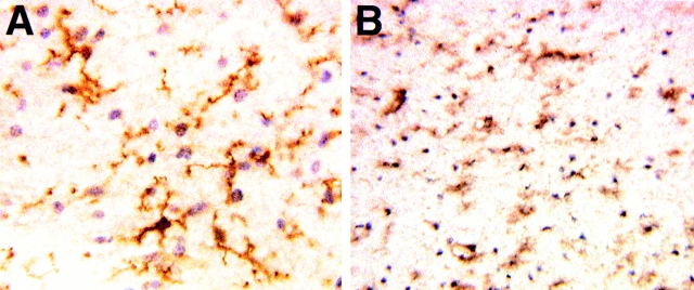
Several studies have used myeloid markers to distinguish between resident parenchymal microglia and perivascular macrophages in rodents and humans by flow cytometry and immunohistochemistry where CD45 expression can identify perivascular macrophages in mice, and CD14 and CD45 expression can distinguish between perivascular macrophages and parenchymal microglia in humans 38 39 42. We applied this same approach to brains of normal noninfected rhesus macaques (n = 2) and macaques infected with SIVmac251 and killed at peak viremia (2 wk p.i.; n = 5) or terminal AIDS (n = 7). Of the animals with terminal AIDS, five had SIVE and two did not. Brain tissue from these animals was screened with a panel of monoclonal antibodies against myeloid antigens including CD4, CD11b (CR3), CD14, CD45, and CD68. All brain macrophage populations were positive for CD11b and CD68. The CD11b+ parenchymal microglia formed a reticular network of finely branched processes throughout the brain. This reticular network was maintained in animals with SIVE, although the cells were rounded rather than ramified, indicative of cellular activation associated with SIV infection (Fig. 1). CD14 and CD45 were not detected on microglia in the CNS parenchyma. These observations are consistent with the lack of CD14 and CD45 detected on parenchymal microglia within inflamed human tissues 38 39 52 and the lack of CD14 on immediately ex vivo human microglial cells 41 53. CD14 and CD45 expression was restricted to scattered cells with a perivascular location (perivascular macrophages) in normal brain and on macrophages within meninges and the choroid plexus. CD14 and CD45 were readily detected on perivascular macrophages within the CNS parenchyma of animals 2 wk p.i., at the time of peak viremia and SIV neuroinvasion 28 29 30 43. The number of CD14+ and CD45+ cells was greater in animals with SIVE than in animals at 2 wk p.i. resulting from the accumulation and recruitment of perivascular macrophages (31; Fig. 2). Low to nondetectable levels of CD4 were observed on perivascular macrophages and microglia in the normal brain (data not shown). 2 wk p.i., CD4 immunoreactivity was slightly more prevalent, whereas in animals with SIVE, CD4 was readily detected on perivascular macrophages and scattered parenchymal microglia (data not shown). The MNGCs, thought to consist of fused macrophages or microglia, were strongly CD14+ and CD45+ with variable but low expression of CD4. These data provide a basis for differentiating in vivo between perivascular macrophages, MNGCs, and parenchymal microglia based on the expression of CD11b, CD14, and CD45 (Table ). On initial inspection, it appeared that the viral protein (gp120)-positive cells in the CNS of animals with SIVE had a distribution that overlapped with CD11b+, CD14+, and CD45+ perivascular macrophages (Fig. 2). Thus, further studies were performed to determine whether the perivascular macrophage was preferentially infected by SIV in the CNS of macaques.
Figure 1.
CD11b immunoreactivity in the CNS of normal noninfected and SIV-infected macaques. (A) CD11b immunoreactivity on nonactivated parenchymal microglia with branched and crenulated processes in a normal noninfected animal. These cells makeup a reticular branching network throughout the white matter in the CNS. (B) CD11b immunoreactivity on parenchymal microglia in the CNS of an animal with SIVE. Microglia maintain a reticular network in the CNS but morphologically become more rounded with fewer processes, consistent with cellular activation.
Figure 2.
Immunophenotype of parenchymal microglia and perivascular macrophages in macaques with SIVE. (A) CD11b immunoreactivity on brain macrophages. Immunoreactivity is demonstrated on parenchymal microglia evenly spaced throughout the white matter, and on expansive aggregates of perivascular macrophages and MNGCs typical of SIVE. (B) CD14 immunoreactivity on perivascular macrophages within a SIVE lesion. CD14 is not detected on parenchymal microglia. (C) CD45 immunoreactivity on perivascular macrophages and lymphocytes within an SIVE lesion. CD45 is not detected on parenchymal microglia. (D) Viral envelope protein (gp120) in SIVE. The distribution of SIV-gp120 appears to overlap with the distribution of CD11b+CD14+CD45+ perivascular macrophages (A–C). Data presented here are representative of immunohistochemical studies performed on two to three CNS regions of n = 5 animals with SIVE.
Table 1.
Immunophenotypes of Perivascular Macrophages, Parenchymal Microglia, and MNGCs
| Perivascular macrophage | Parenchymal microglia | MNGC |
|---|---|---|
| CD11b | CD11b | CD11b |
| CD68 | CD68 | CD68 |
| CD14 | − | CD14 |
| CD45 | − | CD45 |
Perivascular Macrophages and Not Parenchymal Microglia Are Infected by SIV.
The immunophenotypic differences between perivascular macrophages and parenchymal microglia described above were used to examine the cell types that accumulate and are infected in the CNS of SIV-infected macaques. This was approached in three ways: (a) in situ hybridization for SIV followed by immunohistochemistry for CD14 on serial sections, (b) double-label immunohistochemistry for CD14 and gp120, and (c) multilabel confocal microscopy for viral protein and brain macrophage, astrocyte, and endothelial cell markers.
In situ hybridization for viral nucleic acid followed by CD14 immunohistochemistry on serial sections demonstrated scattered individual positive cells in perivascular cuffs of animals at peak viremia (2 wk p.i.; Fig. 3). The immunophenotype of these cells was consistent with perivascular macrophages. In all sections examined, these SIV RNA-positive CD14+ cells were also gp120+. These data clearly demonstrate that CD14+ perivascular macrophages are productively infected by SIV within 14 d of infection (Fig. 3).
Figure 3.
Perivascular macrophages are infected in the CNS of animals at peak viremia. (A) SIV in situ hybridization–positive cells in the CNS in a perivascular location. (B) CD14 immunoreactivity on a serial section demonstrating that the in situ–positive cells shown in A are CD14-positive perivascular macrophages. (C) Double-label immunohistochemistry for detection of CD14 (red) and viral protein (gp120; blue). Essentially all of the cells in this perivascular cuff are CD14+gp120+ (purple) perivascular macrophages. (D) Double-label immunohistochemistry on a tissue section from the thymus of an SIV-infected animal demonstrating single-color controls using a macrophage marker (red) and anti-SIV–gp120 (blue). Thymus tissues as single color controls were performed in parallel with all double-label immunohistochemistry experiments. Results are representative of immunohistochemical studies performed on two to three CNS regions of n = 5 animals killed at peak viremia.
Double-label immunohistochemistry on animals with terminal AIDS and SIVE revealed that essentially all of the gp120+ cells were CD14+ perivascular macrophages. Additionally, all MNGCs were strongly gp120+ and CD14+, suggesting that these cells arise from the fusion of perivascular macrophages and not fused parenchymal microglia (Fig. 4). MNGCs were also CD11b+ and CD68+ but CD3−, consistent with their lineage as myeloid cells (data not shown; references 5 and 25). Quantitation of the percentage of macrophages that were CD14+/gp120+ and CD14+/gp120− in the CNS of animals with SIVE revealed that noninfected perivascular macrophages (CD14+/gp120−) were the most numerous cell type within SIVE lesions, followed by infected perivascular macrophages (CD14+/gp120+; Table ). Less than 1% of the cells were CD14−/gp120+. These rare virally infected cells that were not CD14+ perivascular macrophages are likely to be either CD14− parenchymal microglia or CD4+ T lymphocytes (Fig. 4). Our finding that the major cell type in SIVE lesions is the noninfected perivascular macrophages is consistent with previous data 31 and observations in humans of a correlation between numbers of inflammatory macrophages, as opposed to viral load, and the development of ADC 54. These data are also consistent with the hypothesis that viral infection of the CNS occurs as a result of recruitment of monocyte/macrophages from the periphery.
Figure 4.
Perivascular macrophages are the major target of SIV in animals with SIVE. Double-label immunohistochemistry for CD14 (red) and viral protein (gp120; blue). Colocalization results in purple cells. (A) Numerous CD14+ perivascular macrophages within an SIVE lesion. Few of the CD14+ cells (red) in this lesion are infected as demonstrated by the small number of purple cells. (B) Numerous CD14+ perivascular macrophages are viral infected (purple) in a different lesion in the CNS of the same animal. (C) A MNGC that is CD14+ and gp120+ (purple) suggesting that MNGCs arise from the fusion of CD14+ perivascular macrophages and not CD14− parenchymal microglia. Results are representative of immunohistochemical studies performed on two to three CNS regions of n = 5 animals with SIVE.
Table 2.
Percentage of CD14 and SIV-gp120 Single- and Double-positive Cells in the CNS of Animals with SIVE
| Animal | CD14+gp120− | CD14+gp120+ | CD14−gp120+ |
|---|---|---|---|
| A97-6 | 63 | 36 | 0.6 |
| A95-346 | 84 | 16 | 0.0 |
| A95-383 | 76 | 24 | 0.0 |
Confocal Microscopy Studies.
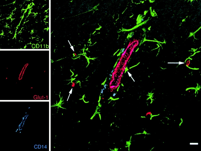
Although it has not been demonstrated in vivo, it is possible that parenchymal microglia can upregulate CD14 as has been demonstrated in vitro 53. For this reason, and to further investigate the cell types infected in the CNS, we performed multilabel confocal microscopy of normal CNS and CNS from SIV-infected animals with SIVE. Confocal microscopy for CD11b and CD14 combined with Glut-1 for CNS endothelium confirmed that these markers are differentially expressed on brain macrophage populations in vivo: CD11b expression was found on parenchymal microglia and perivascular macrophages whereas CD14 expression was limited to perivascular macrophages (Fig. 5). CD11b expression on parenchymal microglia revealed a reticular network of cells throughout the brain. Parenchymal microglia in the brain of animals with SIVE have morphology consistent with activated cells and express activation antigens including HLA-DR, CD16, and CD69 (data not shown). However, these activated parenchymal microglia maintain their reticular network in the CNS white matter and do not express detectable CD14 (Fig. 5 and Fig. 6). Thus, they can be differentiated from perivascular macrophages 38 39 55 56 57. In agreement with the immunohistochemical observations, the number of perivascular macrophages was low in the normal CNS and increased in the CNS of animals with SIVE (Fig. 6). These confocal microscopy studies also clearly show the intimate association of both parenchymal microglia and perivascular macrophages with Glut-1+ CNS endothelial cells (Fig. 5). Perivascular macrophages wrap around the vessels while parenchymal microglia have foot processes that contact vessels forming part of the blood–brain barrier 15 17.
Figure 5.
Triple-label confocal microscopy of the brain of an SIV-infected macaque. Images for individual channels (CD11b, green; Glut-1, red; and CD14, blue) are shown on the left and a larger merged image contain all three channels plus the differential interference contrast (DIC) image are shown on the right. Parenchymal microglia (CD11b, green) maintain a reticular network in the white matter of SIV-infected macaques. Perivascular macrophages (CD11b+CD14+, blue-green) are situated around CNS endothelium (Glut-1, red). Both parenchymal microglia (green) and perivascular macrophages (blue-green) are in contact with CNS endothelium where parenchymal microglia have foot processes on endothelium (arrows) and perivascular macrophages are in contact with and wrap around CNS vessels. Bar, 10 μM.
Figure 6.
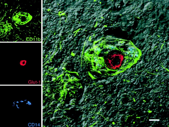
Accumulation of perivascular macrophages in the CNS of SIV-infected macaques. Images for individual channels (CD11b, green; Glut-1, red; and CD14, blue) are shown on the left and the merged image combining all three channels plus the DIC image are shown on the right. CD11b+ (green) and CD14+ (blue) perivascular macrophages (blue-green) accumulate around a CNS vessel (Glut-1, red) in an SIVE lesion. Parenchymal microglia that are CD11b+CD14− (green) outside of the lesion area maintain a reticular network, although their morphology has changed as a result of activation. Bar, 10 μM.
To further examine the cell type infected, multilabel confocal microscopy with anti-SIV p28, CD14, and Glut-1 was performed (Fig. 7). Similar to double-label immunohistochemistry and in situ hybridization followed by immunohistochemistry on serial sections, we found in all cases that the SIV-p28+ cells were CD14+ perivascular macrophages (Fig. 7). These data clearly demonstrate that perivascular macrophages are productively infected in vivo. A number of other reports suggest that other cell types can be infected 14 26 58 59 60 61. However, most of these studies have relied on anatomic location or morphology to identify the infected cell types, making it difficult to differentiate between trafficking mononuclear cells, CNS endothelium, and processes of parenchymal microglia and astrocytes. For this reason, we undertook additional multilabel confocal microscopy studies to more rigorously identify perivascular macrophages (CD11b+CD14+), parenchymal microglia (CD11b+CD14−), astrocytes (GFAP+), and CNS endothelium (Glut-1+) to determine if cells other than perivascular macrophages were infected in the CNS of animals with SIVE (Fig. 7 and Fig. 8). In our extensive analysis of tissue sections from animals with SIVE, we did not detect Glut-1+ endothelium, GFAP+ astrocytes, or CD11b+CD14− parenchymal microglia that were also SIV-p28+.
Figure 7.
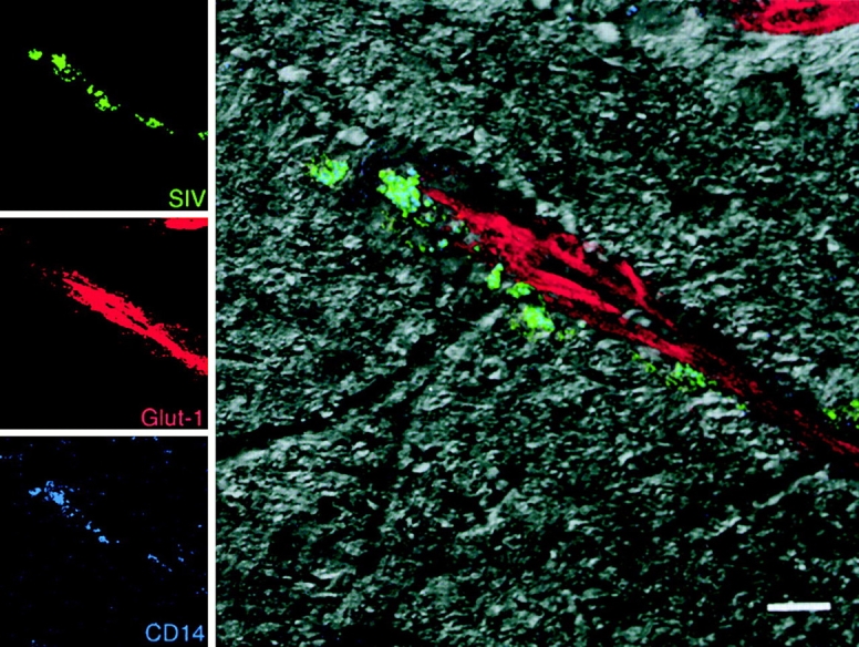
SIV-infected perivascular macrophages. Images for individual channels (SIV-p28, green; Glut-1, red; and CD14, blue) are shown on the left, and the merged image combining all three channels plus the DIC image is shown on the right. Perivascular macrophages (CD14, blue) that are viral protein positive (SIV-p28, green) appear blue-green near a CNS vessel (Glut-1, red). Bar, 10 μM.
Figure 8.
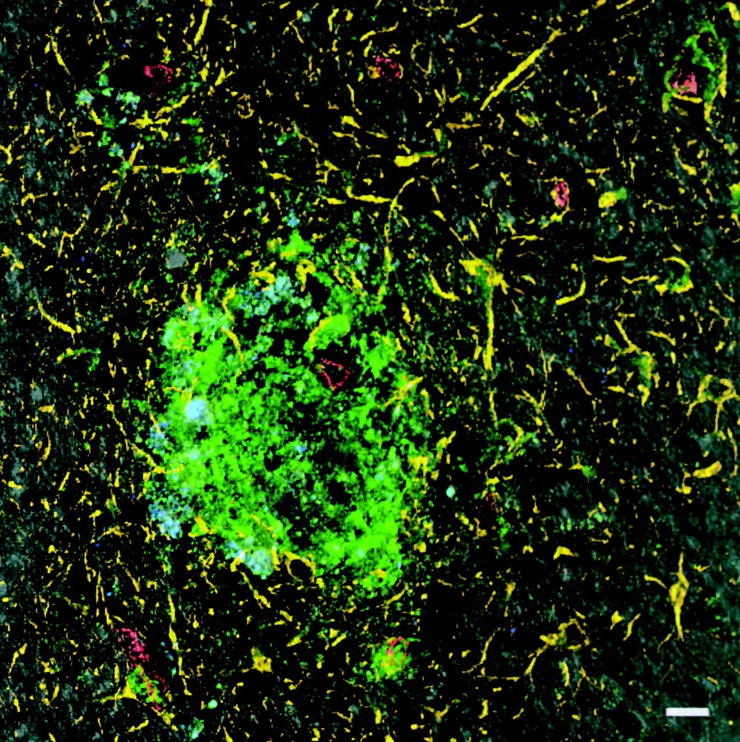
CNS macrophages are the target of SIV infection in animals with SIVE. CNS macrophages (CD11b, green), astrocytes (GFAP, yellow), CNS endothelial cells (Glut-1, red), and viral protein (SIV-p28, blue) were examined to assess whether cells other than macrophages were productively infected. The image shows a combination of the four channels plus the DIC image. Extensive analysis of multiple brain regions from n = 5 animals with SIVE demonstrated p28-positive infected cells were exclusively CD11b+ macrophages. Bar, 10 μM.
Analysis of SIV DNA, RNA, and Protein throughout Infection.
There are numerous reports of SIV or HIV in the CNS of macaques and humans that die with AIDS 27 32 46. In addition, several groups have reported SIV or HIV in the CNS soon after infection 28 29 30 43. However, it is not clear whether virus in the CNS early after infection results in persistent productive infection or in productive infection that is not sustained and requires repeated seeding of the brain with virus from the periphery. To address this question, groups of animals were infected with either the molecular clone SIVmac239 or uncloned SIVmac251. Both SIVmac239 and SIVmac251 are pathogenic viruses. Infection of macaques with SIVmac239 or SIVmac251 results in similar viral loads and these viruses have similar chemokine receptor usage. Both of these viruses cause AIDS and SIVE in a similar time frame and percentage of animals 46. Frontal cortex and brain stem were collected from animals at 3, 7, 14, 21, and 50 d p.i., and from animals with AIDS and SIVE and analyzed for viral DNA, RNA, and protein. Nested PCR for viral DNA (gag) demonstrated that SIV was detectable in the CNS as early as 7 d p.i. and consistently by 14 d p.i. Once detected in the CNS, viral DNA was detected at all later time points including animals with terminal AIDS and SIVE (Table ). In contrast, studies on the presence of viral RNA by in situ hybridization or viral protein (SIV-gp120 or SIV-p28) by immunohistochemistry revealed scattered positive cells associated with vessels 7–14 d p.i., which were absent at later time points until terminal AIDS. These data are similar to those in humans where HIV-infected cells have been found in the CNS within days after infection 43 and at terminal AIDS 6 24 25 26 27, but viral RNA- and protein-positive cells cannot be found in the CNS of HIV-infected, asymptomatic patients 62 63. In our study and in studies of HIV-infected, asymptomatic humans, although viral RNA and protein were not detected, viral DNA was. This DNA might be from latently infected cells or from contaminating cells that were trapped in the CNS vasculature at the time of death 62 63.
Table 3.
Assessment of Viral DNA, RNA, and Protein in the CNS As a Function of Time After Infection
| Day 3 | Day 7 | Day 14 | Day 21 | Day 50 | SIVE | |
|---|---|---|---|---|---|---|
| SIV DNA | ||||||
| SIVmac239 | − | +/− | + | + | + | + |
| SIVmac251 | − | +/− | + | + | + | + |
| SIV RNA | ||||||
| SIVmac239 | − | +/− | +/− | − | − | + |
| SIVmac251 | − | +/− | +/− | − | − | + |
| SIV protein | ||||||
| SIVmac239 | − | +/− | +/− | − | − | + |
| SIVmac251 | − | +/− | +/− | − | − | + |
In terminal animals with AIDS and SIVE, there were numerous cells with abundant viral RNA and protein (Table ). These results are similar to previous observations in macaques and humans 5 26 27 29 30. These data confirm that SIV neuroinvasion occurs very rapidly but suggests that the initial productive infection of the brain parenchyma is not maintained or occurs at a very low level after 14 d of infection until terminal AIDS. The preferential infection of perivascular macrophages and not parenchymal microglia may account for the early productive viral infection, disappearance, and reappearance of SIV in the CNS.
Discussion
In this study we immunophenotypically differentiate subpopulations of brain macrophages into perivascular macrophages and parenchymal microglia and demonstrate that perivascular macrophages are the major cell productively infected by SIV in the CNS of macaques. Preferential infection of perivascular macrophages in the CNS may account for several important observations concerning infection of the CNS, viral dynamics in the CNS, and the role of the CNS as a viral sanctuary or reservoir.
Although it has not been directly demonstrated, it is generally assumed that lentiviruses enter the CNS by the traffic of infected monocyte/macrophages 64. Our data showing that perivascular macrophages are the major cell type, infected in the brain, support this hypothesis. Studies in chimeric rodents and humans receiving bone marrow indicate that perivascular macrophages are continuously replaced from the circulation 15 16 17 43. The immunophenotype described for perivascular macrophages, CD11b+CD14+CD45+, is consistent with circulating peripheral blood monocytes, some of which may be destined to traffic to the CNS 38 39 41 42 53 65. Whether infected or not, our data suggest that it is this population that comprises the majority of the lesions in the CNS of animals with SIVE. Cells of this immunophenotype have been previously noted in the blood of HIV-infected individuals where their percentages are increased in response to infection 66 67 68. Pulliam and Gartner have described a population of circulating blood monocytes that express CD14, CD16, and CD69 which may be HIV infected and whose presence appears to correlate with the development of ADC 69 70. The perivascular macrophages that accumulate in the CNS- of SIV-infected macaques have essentially the same immunophenotype.
Our data on viral DNA, RNA, and protein supports the observations by others that virus enters the CNS early in infection 28 29 30 43. Our data further suggests that productive infection of the CNS occurs for a limited amount of time or at very low levels beyond 2 wk p.i. until terminal AIDS. Similar observations where viral DNA can be found in the absence of viral RNA or protein have been made in the CNS of HIV-infected humans who died from non-AIDS causes during the asymptomatic period 62 63. Recent reports have noted similar observations in human 44 and monkey 45 cerebral spinal fluid where there is early detection of virus in these compartments that subsides, then reappears with the progression to AIDS. The demonstrated kinetics of perivascular macrophage turnover in the CNS may in part account for appearance of virus early in the CNS after infection and then disappearance of virus until terminal AIDS. Perivascular macrophages in rodents and humans are repopulated at a constant rate that is accelerated in inflammatory conditions such as viral infection or autoimmune reactions 15 16 71. The early presence of virus in the CNS appears to occur by the traffic of monocytes that are potentially programmed to become perivascular macrophages in the CNS. Furthermore, the traffic of these cells, as demonstrated by the presence of viral DNA, RNA, and protein in the CNS, coincides with peak viremia 28 29. Once viremia subsides in the clinically latent period, it is possible that the monocytes that continue to traffic to the CNS are no longer infected or that the number of infected cells is greatly diminished by the host immune response, making it difficult to detect active viral replication. Later in disease, when individuals develop AIDS and the immune system has failed, once again the number of infected monocytes that traffic to the CNS may be increased. It has been demonstrated that perivascular macrophages can leave the CNS 72. Whether virally infected perivascular macrophages also can leave the CNS is unknown. The mechanism for retention and accumulation of infected and noninfected perivascular macrophages at terminal AIDS is undefined, but appears to correlate with dysfunction of the cellular immune system.
Numerous reports indicate that HIV and SIV infect brain macrophages/microglia; however, these studies have not distinguished between perivascular macrophages and parenchymal microglia 5 7 8 10 14 25 26 27 28 29 30 31 58 73. Other cell types that have been reported to be infected include astrocytes and endothelial cells 26 58 59 60 61. Our double-label immunohistochemistry and multilabel confocal microscopy data failed to demonstrate productive infection of parenchymal microglia, astrocytes, or endothelial cells in vivo. One difficulty in interpretation of earlier reports of the infection of astrocytes, endothelium, and microglia in adults is the extensive use of cell lines or primary cultures for these infection studies. Another difficulty is the lack of double labels for the identification of the infected cell type, and the use of imaging techniques which do not allow for sufficient resolution to differentiate two closely opposed cells (e.g., endothelium and perivascular macrophages). At the light microscopic level, it is difficult to distinguish between endothelial cells, perivascular macrophages, or leukocytes migrating into the brain 26 59. With regard to macrophage subpopulations and distinguishing between perivascular macrophages and parenchymal microglia, few double-label immunohistochemistry studies have been performed. The double-label studies that have been done to date on CNS macrophage populations have used CD68 or Ricinus comminis agglutinin 1 (RCA-1) that identify all brain macrophages, including perivascular macrophages and microglia 5 28.
Most in vitro studies of brain macrophage infection as it might relate to the neuropathogenesis of AIDS have relied on parenchymal microglia 5 7 8 10 11 14. However, it is known that in vitro parenchymal microglia become highly activated within hours and are quite different from their in vivo or immediately ex vivo counterparts 41 53. Although supernatant from infected microglia can cause neuronal injury and death 7 8 10 11 74 75, it is difficult to appreciate the significance of such observations in light of the difference between the biology of perivascular macrophages and parenchymal microglia.
Our studies demonstrate that perivascular macrophages are immunophenotypically distinct from the resident parenchymal microglia. We further demonstrate that perivascular macrophages are the major cell type productively infected in the CNS of macaques at peak viremia and with SIVE. Within SIVE lesions, noninfected perivascular macrophages are the major cell population followed by infected perivascular macrophages. These data potentially point to cells in the blood that may function as a trojan horse carrying virus to the CNS. Our data suggest that the accumulation of perivascular macrophages, which is coincident with CNS pathology and development of ADC, occur late in infection, with the development of terminal AIDS.
Acknowledgments
We thank the veterinary staff at the New England Regional Primate Research Center for animal care, pathology residents and staff for assisting with necropsies and tissue collection, and Kristen Toohey for graphic services.
This work was supported in part by Public Health Service grants NS37654, NS40237, NS35732, NS30769, and RR00168, and a grant from the National Multiple Sclerosis Society RG 2856-A-1. A.A. Lackner is the recipient of an Elizabeth Glaser Scientist Award.
Footnotes
Abbreviations used in this paper: ADC, AIDS dementia complex; CNS, central nervous system; DIC, differential interference contrast; DIG, digoxigenin; GFAP, glial fibrillary acidic protein; Glut-1, glucose transporter 1; HIVE/SIVE, HIV/SIV encephalitis; MNGC; multinucleated giant cell; p.i., postinfection; RCA-1, Ricinus comminis agglutinin 1; SIV, simian immunodeficiency virus.
References
- Barker C.F., Billingham R.E. Immunologically privileged sites. Adv. Immunol. 1977;25:1–6. [PubMed] [Google Scholar]
- Balter M. AIDS ResearchHIV's other immune-system targets: macrophages. Science. 1996;274:1464–1465. doi: 10.1126/science.274.5292.1464. [DOI] [PubMed] [Google Scholar]
- Cohen J. Exploring how to get at — and eradicate — hidden HIV. Science. 1998;279:1854–1855. doi: 10.1126/science.279.5358.1854. [DOI] [PubMed] [Google Scholar]
- Hughes E.S., Bell J.E., Simmonds P. Investigation of the dynamics of the spread of human immunodeficiency virus to brain and other tissues by evolutionary analysis of sequences from the p17gag and env genes. J. Virol. 1997;71:1272–1280. doi: 10.1128/jvi.71.2.1272-1280.1997. [DOI] [PMC free article] [PubMed] [Google Scholar]
- Dickson D.W., Mattiace L.A., Kure K., Hutchins K., Lyman W.D., Brosnan C.F. Microglia in human disease, with an emphasis on acquired immune deficiency syndrome. Lab. Invest. 1991;64:135–156. [PubMed] [Google Scholar]
- Kure K., Weidenheim K.M., Lyman W.D., Dickson D.W. Morphology and distribution of HIV-1 gp41-positive microglia in subacute AIDS encephalitis. Pattern of involvement resembling a multisystem degeneration. Acta Neuropathol. 1990;80:393–400. doi: 10.1007/BF00307693. [DOI] [PubMed] [Google Scholar]
- Watkins B.A., Dorn H.H., Kelly W.B., Armstrong R.C., Potts B.J., Michaels F., Kufta C.V., Dubois-Dalcq M. Specific tropism of HIV-1 for microglial cells in primary human brain cultures. Science. 1990;249:549–553. doi: 10.1126/science.2200125. [DOI] [PubMed] [Google Scholar]
- Sharpless N., Gilbert D., Vandercam B., Zhou J.M., Verdin E., Ronnett G., Friedman E., Dubois-Dalcq M. The restricted nature of HIV-1 tropism for cultured neural cells. Virology. 1992;191:813–825. doi: 10.1016/0042-6822(92)90257-p. [DOI] [PubMed] [Google Scholar]
- Strizki J.M., Albright A.V., O'Conner M., Perrin L., Gonzalez-Scarano F. Infection of primary human microglia and monocyte-derived macrophages with human immunodeficiency virus type 1 isolatesevidence of differential tropism. J. Virol. 1996;70:7654–7662. doi: 10.1128/jvi.70.11.7654-7662.1996. [DOI] [PMC free article] [PubMed] [Google Scholar]
- Jordan C.A., Watkins B.A., Kufta C., Dubois-Dalcq M. Infection of brain microglial cells by human immunodeficiency virus type 1 is CD4 dependent. J. Virol. 1991;65:736–742. doi: 10.1128/jvi.65.2.736-742.1991. [DOI] [PMC free article] [PubMed] [Google Scholar]
- Lee S.C., Hatch W.C., Liu W., Kress Y., Lyman W.D., Dickson D.W. Productive infection of human fetal microglia by HIV-1. Am. J. Pathol. 1993;143:1032–1039. [PMC free article] [PubMed] [Google Scholar]
- Watry D., Lane T.E., Streb M., Fox H.S. Transfer of neuropathogenic simian immunodeficiency virus with naturally infected microglia. Am. J. Pathol. 1995;146:914–923. [PMC free article] [PubMed] [Google Scholar]
- Prospero-Garcia O., Gold L.H., Fox H.S., Polis I., Koob G.F., Bloom F.E., Henriksen S.J. Microglia-passaged simian immunodeficiency virus induces neurophysiological abnormalities in monkeys. Proc. Natl. Acad. Sci. USA. 1996;93:14158–14163. doi: 10.1073/pnas.93.24.14158. [DOI] [PMC free article] [PubMed] [Google Scholar]
- He J., Chen Y., Farzan M., Choe H., Ohagen A., Gartner S., Busciglio J., Yang X., Hofmann W., Newman W. CCR3 and CCR5 are co-receptors for HIV-1 infection of microglia. Nature. 1997;385:645–649. doi: 10.1038/385645a0. [DOI] [PubMed] [Google Scholar]
- Hickey W.F., Vass K., Lassmann H. Bone marrow derived elements in the central nervous systeman immunohistochemical and ultrastructural survey of rat chimeras. J. Neuropathol. Exp. Neurol. 1992;5:246–256. doi: 10.1097/00005072-199205000-00002. [DOI] [PubMed] [Google Scholar]
- Hickey W.F., Kimura H. Perivascular microglial cells of the CNS are bone marrow-derived and present antigen. Science. 1988;239:290–292. doi: 10.1126/science.3276004. [DOI] [PubMed] [Google Scholar]
- Lassmann H., Schmied M., Vass K., Hickey W.F. Bone marrow derived elements and resident microglia in brain inflammation. Glia. 1993;7:19–24. doi: 10.1002/glia.440070106. [DOI] [PubMed] [Google Scholar]
- Williams K., Hickey W.F. Traffic of lymphocytes into the CNS during inflammation and HIV infection. J. Neuro-AIDS. 1996;1:31–55. doi: 10.1300/j128v01n01_02. [DOI] [PubMed] [Google Scholar]
- Mato M., Sakamoto A., Ookawara S., Takeuchi K., Suzuki K. Ultrastructural and immunohistochemical changes of fluorescent granular perithelial cells and the interaction of FGP cells to microglia after lipopolysaccharide administration. Anat. Rec. 1998;251:330–338. doi: 10.1002/(SICI)1097-0185(199807)251:3<330::AID-AR8>3.0.CO;2-Z. [DOI] [PubMed] [Google Scholar]
- Lassmann H., Zimprich F., Vass K., Hickey W.F. Microglial cells are a component of the perivascular glia limitans. J. Neurosci. Res. 1991;28:236–243. doi: 10.1002/jnr.490280211. [DOI] [PubMed] [Google Scholar]
- Graeber M.B., Streit W.J., Kreutzberg G.W. Identity of ED2-positive perivascular cells in rat brain. J. Neurosci. Res. 1989;22:103–106. doi: 10.1002/jnr.490220114. [DOI] [PubMed] [Google Scholar]
- Perry V.H., Gordon S. Macrophages and microglia in the nervous system. Trends Neurosci. 1988;11:273–275. doi: 10.1016/0166-2236(88)90110-5. [DOI] [PubMed] [Google Scholar]
- Williams K.C., Hickey W.F. Traffic of hematogenous cells through the central nervous system. Curr. Top. Microbiol. Immunol. 1995;202:221–245. doi: 10.1007/978-3-642-79657-9_15. [DOI] [PubMed] [Google Scholar]
- Kure K., Lyman W.D., Weidenheim K.M., Dickson D.W. Cellular localization of an HIV-1 antigen in subacute AIDS encephalitis using an improved double-labeling immunohistochemical method. Am. J. Pathol. 1990;136:1085–1092. [PMC free article] [PubMed] [Google Scholar]
- Koenig S., Gendelman H.E., Orenstein J.M., Dal Canto M.C., Pezeshkpour G.H., Yungbluth P., Janotta F., Aksamit A., Martin M., Fauci A.S. Detection of AIDS virus in macrophages in brain tissue from AIDS patients with encephalopathy. Science. 1986;233:1089–1093. doi: 10.1126/science.3016903. [DOI] [PubMed] [Google Scholar]
- Wiley C.A., Schrier R.D., Nelson J.A., Lampert P.W., Oldstone M.B.A. Cellular localization of human immunodeficiency virus infection within the brains of acquired immunodeficiency syndrome patients. Proc. Natl. Acad. Sci. USA. 1986;83:7089–7093. doi: 10.1073/pnas.83.18.7089. [DOI] [PMC free article] [PubMed] [Google Scholar]
- Price R.W., Brew B., Sidtis J., Rosenblum M., Scheck A.C., Cleary P. The brain in AIDScentral nervous system HIV-1 infection and AIDS dementia complex. Science. 1988;239:586–592. doi: 10.1126/science.3277272. [DOI] [PubMed] [Google Scholar]
- Chakrabarti L., Hurtrel M., Maire M., Vazeux R., Dormont D., Montagnier L., Hurtrel B. Early viral replication in the brain of SIV-infected rhesus monkeys. Am. J. Pathol. 1991;139:1273–1280. [PMC free article] [PubMed] [Google Scholar]
- Lackner A.A., Vogel P., Ramos R.A., Kluge J.D., Marthas M. Early events in tissues during infection with pathogenic (SIVmac239) and nonpathogenic (SIVmac1A11) molecular clones of simian immunodeficiency virus. Am. J. Pathol. 1994;145:428–439. [PMC free article] [PubMed] [Google Scholar]
- Smith M.O., Heyes M.P., Lackner A.A. Early intrathecal events in rhesus macaques (Macaca mulatta) infected with pathogenic or nonpathogenic molecular clones of simian immunodeficiency virus. Lab. Invest. 1995;72:547–558. [PubMed] [Google Scholar]
- Lane J.H., Sasseville V.G., Smith M.O., Vogel P., Pauley D.R., Heyes M.P., Lackner A.A. Neuroinvasion by simian immunodeficiency virus coincides with increased numbers of perivascular macrophages/microglia and intrathecal immune activation. J. Neurovirol. 1996;2:423–432. doi: 10.3109/13550289609146909. [DOI] [PubMed] [Google Scholar]
- Lackner A.A., Smith M.O., Munn R.J., Martfeld D.J., Gardner M.B., Marx P.A., Dandekar S. Localization of simian immunodeficiency virus in the central nervous system of rhesus monkeys. Am. J. Pathol. 1991;139:609–621. [PMC free article] [PubMed] [Google Scholar]
- Falangola M.F., Hanly A., Galvao-Castro B., Petito C.K. HIV infection of human choroid plexusa possible mechanism of viral entry into the CNS. J. Neuropathol. Exp. Neurol. 1995;54:497–503. doi: 10.1097/00005072-199507000-00003. [DOI] [PubMed] [Google Scholar]
- Horouse J.M., Wroblewska Z., Laughlin M.A., Hickey W.F., Schonwetter B.S., Gonzalez-Scarano F. Human choroid plexus cells can be latently infected with human immunodeficiency virus. Ann. Neurol. 1989;25:406–411. doi: 10.1002/ana.410250414. [DOI] [PubMed] [Google Scholar]
- Unger E.R., Sung J.H., Manivel J.C., Chenggis M.L., Blazar B.R., Krivit W. Male donor-derived cells in the brains of female sex-mismatched bone marrow transplant recipientsa Y chromosome specific in situ hybridization study. J. Neuropathol. Exp. Neurol. 1993;52:460–470. doi: 10.1097/00005072-199309000-00004. [DOI] [PubMed] [Google Scholar]
- Luster A.D. Chemokineschemotactic cytokines that mediate inflammation. N. Engl. J. Med. 1998;338:436–445. doi: 10.1056/NEJM199802123380706. [DOI] [PubMed] [Google Scholar]
- Lavi E., Strizki J.M., Ulrich A.M., Zhang W., Fu L., Wang Q., O'Connor M., Hoxie J.A., González-Scarano F. CXCR-4 (fusin), a co-receptor for the type 1 human immunodeficiency virus (HIV-1), is expressed in the human brain in a variety of cell types, including microglia and neurons. Am. J. Pathol. 1997;151:1035–1042. [PMC free article] [PubMed] [Google Scholar]
- Williams K., Barr-Or A., Ulvestad E., Olivier A., Antl J.P., Yong W.V. Biology of adult human microglia in culturecomparisons with peripheral blood monocytes and astrocytes. J. Neuropathol. Exp. Neurol. 1992;51:538–549. doi: 10.1097/00005072-199209000-00009. [DOI] [PubMed] [Google Scholar]
- Ulvestad E., Williams K., Mork S., Antel J., Nyland H. Phenotypic differences between human monocytes/macrophages and microglial cells studied in situ and in vitro. J. Neuropathol. Exp. Neurol. 1994;53:492–501. doi: 10.1097/00005072-199409000-00008. [DOI] [PubMed] [Google Scholar]
- Ulvestad E., Williams K., Bjerkvig R., Tiekotter K., Antel J., Matre R. Human microglial cells have phenotypic and functional characteristics in common with both macrophages and dendritic antigen presenting cells. J. Leukoc. Bio. 1994;52:331–342. doi: 10.1002/jlb.56.6.732. [DOI] [PubMed] [Google Scholar]
- Becher B., Antel J. Comparison of phenotypic and functional properties of immediately ex vivo and cultured human adult microglia. Glia. 1996;18:1–10. doi: 10.1002/(SICI)1098-1136(199609)18:1<1::AID-GLIA1>3.0.CO;2-6. [DOI] [PubMed] [Google Scholar]
- Sedgwick J.D., Schender S., Imrich H., Dorries R., Butcher G.W., ter Meulen V. Isolation and direct characterization of resident microglial cells from the normal and inflamed central nervous system. Proc. Natl. Acad. Sci. USA. 1991;88:7438–7442. doi: 10.1073/pnas.88.16.7438. [DOI] [PMC free article] [PubMed] [Google Scholar]
- Davis L.E., Hjelle B.L., Miller V.E., Palmer D.L., Llewellyn A.L., Merlin T.L., Young S.A., Mills R.G., Wachsman W., Wiley C.A. Early viral brain invasion in iatrogenic human immunodeficiency virus infection. Neurology. 1992;42:1736–1739. doi: 10.1212/wnl.42.9.1736. [DOI] [PubMed] [Google Scholar]
- Price R.W. The two faces of HIV infection of cerebrospinal fluid. Trends Microbiol. 2000;8:387–391. doi: 10.1016/s0966-842x(00)01821-7. [DOI] [PubMed] [Google Scholar]
- Zink M.C., Clements J.E. The two faces of HIV infection of cerebrospinal fluid. Trends Microbiol. 2000;8:390. doi: 10.1016/s0966-842x(00)01821-7. [DOI] [PubMed] [Google Scholar]
- Westmoreland S.V., Halpern E., Lackner A.A. Simian immunodeficiency virus encephalitis in rhesus macaques is associated with rapid disease progression. J. Neurovirol. 1998;4:260–268. doi: 10.3109/13550289809114527. [DOI] [PubMed] [Google Scholar]
- Gibbs J.S., Lackner A.A., Lang S.M., Simon M.A., Sehgal P.K., Daniel M.D., Desrosiers R.C. Progression to AIDS in the absence of a gene for vpr or vpx . J. Virol. 1995;69:2378–2383. doi: 10.1128/jvi.69.4.2378-2383.1995. [DOI] [PMC free article] [PubMed] [Google Scholar]
- Wykrzykowska J.J., Rosenzweig M., Veazey R.S., Simon M.A., Halvorsen K., Desrosiers R.C., Johnson R.P., Lackner A.A. Early regeneration of thymic progenitors in rhesus macaques infected with simian immunodeficiency virus. J. Exp. Med. 1998;187:1767–1778. doi: 10.1084/jem.187.11.1767. [DOI] [PMC free article] [PubMed] [Google Scholar]
- Ringler D.J., Walsh D.G., Chalifoux L.V., MacKey J.J., Daniel M.D., Desrosiers R.C., King N.W., Hancock W.W. Soluble and membrane-associated interleukin 2 receptor-alpha expression in rhesus monkeys infected with simian immunodeficiency virus. Lab. Invest. 1990;62:435–443. [PubMed] [Google Scholar]
- Schnell A.S., Staines W.A., Wessendorf M.W. Reduction of lipofuscin-like autofluorescence in fluorescently labeled tissue. J. Histochem. Cytochem. 1999;47:719–730. doi: 10.1177/002215549904700601. [DOI] [PubMed] [Google Scholar]
- Hirsch V.M., Adger-Johnson D., Campbell B., Goldstein S., Brown C., Elkins W.R., Montefiori D.C. A molecularly cloned, pathogenic, neutralization-resistant simian immunodeficiency virus, SIVsmE543-3. J. Virol. 1997;71:1608–1620. doi: 10.1128/jvi.71.2.1608-1620.1997. [DOI] [PMC free article] [PubMed] [Google Scholar]
- Ulvestad E., Williams K., Vedeler C., Antel J., Nyland H., Mork S., Matre R. Reactive microglia in multiple sclerosis lesions have an increased expression of receptors for the Fc part of IgG. J. Neurol. Sci. 1994;121:125–131. doi: 10.1016/0022-510x(94)90340-9. [DOI] [PubMed] [Google Scholar]
- Becher B., Fedorowicz V., Antel J.P. Regulation of CD14 expression on human adult central nervous system-derived microglia. J. Neurosci. Res. 1996;45:375–381. doi: 10.1002/(SICI)1097-4547(19960815)45:4<375::AID-JNR6>3.0.CO;2-6. [DOI] [PubMed] [Google Scholar]
- Glass J.D., Fedor H., Wesselingh S.L., McArthur J.C. Immunocytochemical quantitation of human immunodeficiency virus in the braincorrelations with dementia. Ann. Neurol. 1995;38:755–762. doi: 10.1002/ana.410380510. [DOI] [PubMed] [Google Scholar]
- Bauer J., Siminia T., Wouterlood F.G., Dijkstra C.D. Phagocytic activity of macrophages and microglial cells during the course of allergic encephalymyelitis. J. Immunol. 1994;126:365–375. doi: 10.1002/jnr.490380402. [DOI] [PubMed] [Google Scholar]
- Flaris N.A, Densmore T.L., Molleston M.C., Hickey W.F. Characterization of microglia and macrophages in the central nervous system of ratsdefinition of the differential expression of molecules using standard and novel monoclonal antibodies in normal CNS and in four models of parenchymal reaction. Glia. 1993;7:34–40. doi: 10.1002/glia.440070108. [DOI] [PubMed] [Google Scholar]
- Rinner W.A., Bauer J., Schmidts M., Lassmann H., Hickey W.F. Resident microglia and hematogenous macrophages as phagocytes in adoptively transferred EAEan investigation using rat radiation bone marrow chimeras. Glia. 1995;14:257–266. doi: 10.1002/glia.440140403. [DOI] [PubMed] [Google Scholar]
- Moses A.V., Bloom F.E., Pauza C.D., Nelson J.A. Human immunodeficiency virus infection of human brain capillary endothelial cells occurs via a CD4/galactosylceramide-independent mechanism. Proc. Natl. Acad. Sci. USA. 1993;90:10474–10478. doi: 10.1073/pnas.90.22.10474. [DOI] [PMC free article] [PubMed] [Google Scholar]
- Mankowski J.L., Spelman J.P., Ressetar H.G., Strandberg J.D., Laterra J., Carter D.L., Clements J.E., Zink M.C. Neurovirulent simian immunodeficiency virus replicates productively in endothelial cells of the central nervous system in vivo and in vitro. J. Virol. 1994;68:8202–8208. doi: 10.1128/jvi.68.12.8202-8208.1994. [DOI] [PMC free article] [PubMed] [Google Scholar]
- Tornatore C., Chandra R., Berger J.R., Major E.O. HIV-1 infection of subcortical astrocytes in the pediatric central nervous system. Neurology. 1994;44:481–487. doi: 10.1212/wnl.44.3_part_1.481. [DOI] [PubMed] [Google Scholar]
- Ranki A., Nyberg M., Ovod V., Haltia M., Elovaara I., Raininko R., Haapasalo H., Krohn K. Abundant expression of HIV Nef and Rev proteins in brain astrocytes in vivo is associated with dementia. AIDS. 1995;9:1001–1008. doi: 10.1097/00002030-199509000-00004. [DOI] [PubMed] [Google Scholar]
- Gosztonyi G., Artigas J., Lamperth L., Webster H.D. Human immunodeficiency virus (HIV) distribution in HIV encephalitisstudy of 19 cases with combined use of in situ hybridization and immunocytochemistry. J. Neuropathol. Exp. Neurol. 1994;53:521–534. doi: 10.1097/00005072-199409000-00012. [DOI] [PubMed] [Google Scholar]
- Sinclair E., Gray F., Ciardi A., Scaravilli F. Immunohistochemical changes and PCR detection of HIV provirus DNA in brains of asymptomatic HIV-positive patients. J. Neuropathol. Exp. Neurol. 1994;53:43–50. doi: 10.1097/00005072-199401000-00006. [DOI] [PubMed] [Google Scholar]
- Peluso R., Haase A., Stowring L., Edwards M., Ventura P. A trojan horse mechanism for the spread of visna virus in monocytes. Virology. 1985;147:231–236. doi: 10.1016/0042-6822(85)90246-6. [DOI] [PubMed] [Google Scholar]
- Ford A.L., Goodsall A.L., Hickey W.F., Sedgwick J.D. Normal adult microglia separated from other CNS macrophages by flow cytometric sortingphenotypic differences defined and direct ex vivo antigen presentation to myelin basic protein reactive CD4 cells compared. J. Immunol. 1995;154:4309–4321. [PubMed] [Google Scholar]
- Ziegler-Heitbrock H.W., Fingerle G., Strobel M., Schraut W., Stelter F., Schutt C., Passlick B., Pforte A. The novel subset of CD14+/CD16+ blood monocytes exhibits features of tissue macrophages. Eur. J. Immunol. 1993;23:2053–2058. doi: 10.1002/eji.1830230902. [DOI] [PubMed] [Google Scholar]
- Passlick B., Flieger D., Ziegler-Heitbrock H.W. Identification and characterization of a novel monocyte subpopulation in human peripheral blood. Blood. 1989;74:2527–2534. [PubMed] [Google Scholar]
- Thieblemont N., Weiss L., Sadeghi H.M., Estcourt C., Haeffner-Cavaillon N. CD14lowCD16higha cytokine-producing monocyte subset which expands during human immunodeficiency virus infection. Eur. J. Immunol. 1995;25:3418–3424. doi: 10.1002/eji.1830251232. [DOI] [PubMed] [Google Scholar]
- Pulliam L., Gascon R., Stubblebine M., McGuire D., McGrath M.S. Unique monocyte subset in patients with AIDS dementia. Lancet. 1997;349:692–695. doi: 10.1016/S0140-6736(96)10178-1. [DOI] [PubMed] [Google Scholar]
- Gartner S. HIV infection and dementia. Science. 2000;287:602–604. doi: 10.1126/science.287.5453.602. [DOI] [PubMed] [Google Scholar]
- Lassmann H., Hickey W.F. Radiation bone marrow chimeras as a tool to study microglia turnover in normal brain and inflammation. Clin. Neuropathol. 1993;12:284–285. [PubMed] [Google Scholar]
- Broadwell R.D., Baker B.J., Ebert P.S., Hickey W.F. Allografts of CNS tissue possess a blood-brain barrierIII. Neuropathological, methodological, and immunological considerations. Microsc. Res. Tech. 1994;27:471–494. doi: 10.1002/jemt.1070270603. [DOI] [PubMed] [Google Scholar]
- Brinkman R., Schwinn A., Narayan O., Zink C., Kreth H.W., Roggendorf W., Dorries R., Schwender S., Imrich H., ter Meulen V. Human immunodeficiency virus infection in microgliacorrelation between cells infected in the brain and cells cultured from infectious brain tissue. Ann. Neurol. 1992;31:361–365. doi: 10.1002/ana.410310403. [DOI] [PubMed] [Google Scholar]
- Giulian D., Vaca K., Noonan C.A. Secretion of neurotoxins by mononuclear phagocytes infected with HIV-1. Science. 1990;250:1593–1596. doi: 10.1126/science.2148832. [DOI] [PubMed] [Google Scholar]
- Lee S.C., Liu W., Dickson D.W., Brosnan C.F., Berman J.W. Cytokine production by human fetal microglia and astrocytes. J. Immunol. 1993;150:2659–2667. [PubMed] [Google Scholar]
