Yersinia V–Antigen Exploits Toll-like Receptor 2 and CD14 for Interleukin 10–mediated Immunosuppression (original) (raw)
Abstract
A characteristic of the three human-pathogenic Yersinia spp. (the plague agent Yersinia pestis and the enteropathogenic Yersinia pseudotuberculosis and Yersinia enterocolitica) is the expression of the virulence (V)-antigen (LcrV). LcrV is a released protein which is involved in contact-induced secretion of yersinia antihost proteins and in evasion of the host's innate immune response. Here we report that recombinant LcrV signals in a CD14- and toll-like receptor 2 (TLR2)-dependent fashion leading to immunosuppression by interleukin 10 induction. The impact of this immunosuppressive effect for yersinia pathogenesis is underlined by the observation that TLR2-deficient mice are less susceptible to oral Y. enterocolitica infection than isogenic wild-type animals. In summary, these data demonstrate a new ligand specificity of TLR2, as LcrV is the first known secreted and nonlipidated virulence-associated protein of a Gram-negative bacterium using TLR2 for cell activation. We conclude that yersiniae might exploit host innate pattern recognition molecules and defense mechanisms to evade the host immune response.
Keywords: immunity, monocytes/macrophages, inflammation, bacterial proteins, immunosuppression
Introduction
Yersinia pestis, the etiologic agent of plague, and the enteropathogenic species Yersinia pseudotuberculosis and Yersinia enterocolitica have a 70-kb conserved virulence plasmid (pYV) in common which enables these obligate extracellular Gram-negative bacteria to resist the immune system of their host (1). It encodes several virulence-associated components including a type III protein secretion and translocation system (TTS),* the surface exposed adhesin YadA, anti-host effector proteins, which are probably injected by a needle-like mechanism into the cytosol of target cells (2), and proteins which are released into the environment (1, 3). The function of several Yops have been elucidated, e.g., YopH impairs focal adhesion complexes by its protein tyrosine phosphatase activity, YopE and YopT inactivate the small GTPases Rac and Rho, respectively, and YopP/YopJ blocks activation of nuclear factor (NF)-κB and mitogen-activated protein (MAP) kinases (1, 3). The concerted action of injected Yops results in paralysis of those professional phagocytes which have contacted yersinae bacilli (“paralytical kiss”).
The virulence- (V-) antigen is a released multifunctional protein (4, 5). It was originally described as the major virulence marker of Y. pestis (6). Later, it was shown that the V-antigen (denoted as LcrV) is encoded by pYV and is expressed and released into the environment by all human pathogenic Yersinia species. It is required for regulation of Yop production and, together with YopB and YopD, for polarized translocation of Yops into host cells; furthermore, LcrV is capable to form channels in artificial bilayer membranes (7, 8). Additionally, LcrV exhibits immunomodulatory features: treatment of mice with _r_LcrV results in suppression of TNF-α and IFN-γ and amplification of IL-10 in spleen homogenates (9, 10). There is a large body of evidence that LcrV has also a protective capacity as vaccine, probably because antibodies against LcrV result in neutralization of the immunosuppressive effect and/or inhibition of Yop translocation (9, 11, 12). Recently, we could demonstrate that _r_LcrV treatment of murine toll-like receptor 4 (TLR4)-defective C3H/HeJ macrophages and human MonoMac6 monocytic cells leads to TNF-α suppression via IL-10 induction in a dose-dependent manner (range of activity: 50 ng/ml to 5 μg/ml, corresponding to 1.35 to 135 nM; 13). Boiling of LcrV, digestion with proteinase K, and use of anti-LcrV antibodies completely abolished the immunomodulating capacity of LcrV. Strikingly, _r_LcrV-treated proteose-peptone elicited macrophages (PPMs) from IL-10-deficient (IL-10−/−) mice exhibit no TNF-α suppression upon subsequent zymosan A stimulation. Interestingly, despite sharing several features with LcrV (8, 14), the LcrV-homologue protein of Pseudomonas aeruginosa, PcrV, elicits no comparable immunosuppressive capacity (13). The signaling pathway leading to IL-10 production by LcrV is unknown. Due to its resemblance with the effects of endotoxin (LPS) tolerance we wondered whether LPS and LcrV might share common signaling pathways.
Materials and Methods
Mice.
CD14−/− and C57/BL6 mice were purchased from The Jackson Laboratory. Mice deficient in TLR2 generated by gene targeting as described previously (15) were provided by Tularik Inc. All mice were bred under specific pathogen-free conditions. Female mice were used at 6–8 wk of age.
Materials.
_r_LcrV and _r_PcrV were produced as described previously (13). The molecular mass of _r_LcrV (39.661 kD) was confirmed by mass spectrometry using matrix-assisted laser desorption-ionization mass spectrometry (MALDI-MS) analysis (Toplab). Thus, a putative protein modification, e.g., acetylation or lipidation, could be excluded. Moreover, _r_LcrV was found to be LPS-free as measured by limulus amebocyte assay (Pyroquant). Peptides were provided by Dieter Palm, Physiologische Chemie I, Würzburg, Germany.
Preparation and Stimulation of Murine Peritoneal Macrophages In Vitro.
PPMs were prepared as described previously (13). Briefly, 106 cells/ml were treated for 2 h with 5 μg/ml _r_LcrV. Supernatants were collected for IL-10 measurements by ELISA.
Transfections.
Cells of a subclone of the human embryonic kidney (HEK) 293 cell line (Tularik Inc.) were transfected with DNA constructs for CD14, TLR 1, TLR 2, TLR 4, endothelial cell-leukocyte adhesion molecule (ELAM-1) luciferase, and Rous sarcoma virus (RSV-)-β-galactosidase as described previously (16). The DNA constructs for TLR1, TLR2, and TLR4 were FLAG-epitope-tagged as described (16). MD2 plasmid was a kind gift from Kensuke Miyake, University of Tokyo, Japan. Transfected cells were stimulated for 6 h with _r_LcrV in the presence or absence of 10 μg/ml polymyxin B (Sigma-Aldrich). To rule out any bacterial contaminant known so far to signal via CD14/TLR2 to be present in our _r_LcrV preparation, _r_LcrV was boiled for 1 h before use to serve as negative control. After _r_LcrV-treatment cells were lysed for measurement of luciferase activity using reagents from Boehringer. Luciferase activity was measured in a darkbox with a microtiter plate chemiluminometer (Hamamatsu Phototonics). An RSV-β-galactosidase control plasmid was used for normalizing transfection efficiencies.
MonoMac-6 Cell Procedures.
Cells of the human monocytic cell line MonoMac-6 grown in RPMI 1640 supplemented with 10% FCS for 3 d at a cell density of 2 × 10⁵ cells/ml were used for experiments testing responsiveness to LcrV as described previously (13). After 18 h of pretreatment with 5 μg/ml _r_LcrV, cells were stimulated with 1 mg/ml zymosan A (Sigma-Aldrich). 6 h later supernatants were collected for measurement of TNF-α levels. To evaluate the influence of CD14 on the immunomodulating effect of _r_LcrV, MonoMac-6 cells were preincubated for 1 h before _r_LcrV-treatment with monoclonal anti-CD14 antibodies bIG 10 (biometec) or MEM-18 (a kind gift from Dr. Václav Horejší, Institute of Molecular Genetics, Praha, Czech Republic) which inhibit LPS-binding to CD14. As control served the corresponding isotype anti-CD14 mAbs bIG 4 and MEM-15 which do not block LPS effects. mAb concentrations of 10 μg/ml were used.
Assays for TNF-α and IL-10.
TNF-α and IL-10 levels were measured by commercially available ELISAs as described previously (13).
Coating of rLcrV and rPcvV to Latex Beads.
Non-covalent coating of latex beads (4-μm diameter beads, sulfate microspheres; Molecular Probes) with _r_LcrV and _r_PcrV was performed as described (17). Briefly, purified recombinant proteins were dialyzed against PBS, pH 7.0. Approximately 109 latex beads were washed with 1 ml of PBS and resuspended in 500 μl of PBS. Recombinant LcrV and PcrV (1 mg) were coincubated with the latex beads for 3 h at room temperature to allow absorption to the beads. After adding 500 μl of 20 mg/ml BSA, the solution was incubated at room temperature for 1 h. Subsequently, beads were washed with PBS containing 1 mg/ml BSA and stored at 4°C in 500 μl of PBS containing 0.2 mg/ml BSA. The coupling efficiency was tested by immunofluorescence staining of the coated beads with specific anti-_r_LcrV and anti-_r_PcrV antibodies, respectively, and a FITC-labeled secondary goat anti-rabbit antibody (Molecular Probes; unpublished data).
Immunofluorescence Procedures.
Immunofluorescence staining was performed as described previously (17). Briefly, cells were washed three times with serum-free medium. Latex beads coated with _r_LcrV or _r_PcrV were centrifuged onto the cells (800 r.p.m., 3 min, 4°C). After an incubation period of 30 min at 37°C in a humidified atmosphere of 5% CO2, cells were fixed with 3.7% formaldehyde (Sigma-Aldrich) and permeabilized with 0.1% triton-PBS (Sigma-Aldrich). Before staining, cells were blocked with 10% human serum. For staining, polyclonal rabbit anti-murine TLR2 (from a purified polyclonal rabbit antiserum raised against a 28-mer peptide H2N-FNPSESDVVSELGKVETVTIRRLHIPQC-CONH2 representing a portion of the mouse TLR2 extracellular domain, purchased from Eurogentec Inc.; the antibodies were found not to stain TLR2−/− PPMs) or polyclonal rabbit anti-FLAG (Sigma-Aldrich) were used as primary antibodies, while Alexa 568- or 350-labeled goat anti–rabbit antibodies or donkey anti–goat (Molecular Probes) served as secondary antibodies. Fluorescence was evaluated using a confocal laser microscope.
Experimental Infection of Mice.
Y. enterocolitica O8 strain WA-314 was grown in Luria-Bertani medium at 27°C overnight, sedimented, resuspended in 20% glycerol, and frozen at −80°C. For infection of mice, aliquots of glycerol-stock cultures were thawed, washed in sterile PBS (pH 7.4), and diluted to the appropriate bacterial concentrations (13). Mice were orally infected with 5 × 10⁷ CFU after fasting for 4 h. Mice were killed after 5 d of oral infection.
Determination of the Number of Yersiniae in Spleen and Liver.
Spleens and livers were dissected and homogenized as described previously (13). Yersiniae CFU were determined by plating serial dilutions on Yersinia selective agar (CIN agar; Becton Dickinson) and counting the CFU after an incubation period of 40 h at 27°C.
Statistical Analysis.
In vitro experiments were performed at least five times. Results are presented as means ± SD. Animal experiments were performed at least twice using 5–10 animals per group. Statistical analysis was performed using Student's two-sided t test. Differences were considered statistically significant at P values < 0.05.
Results
CD14 Is Required for LcrV-induced Immunomodulation.
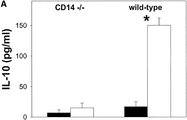
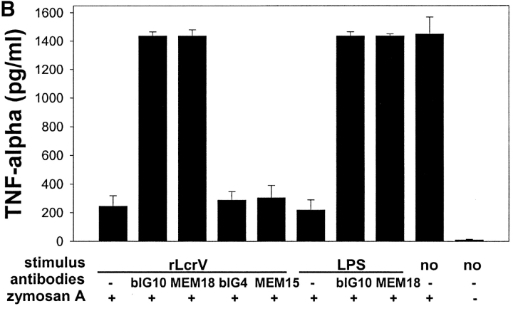
Initially, we investigated whether CD14, the accessory surface protein of TLR4-mediated LPS-signaling (18), is required for LcrV-induced IL-10 production. PPMs from CD14−/− and isogenic CD14+/+ mice were treated with _r_LcrV for 2 h. Then IL-10 was determined in the culture supernatant. In contrast to wild-type PPMs, CD14−/− macrophages did virtually not respond with IL-10 production (Fig. 1 A). This result showing CD14 dependency of LcrV-induced immunomodulation was confirmed with the human macrophage cell line MonoMac-6 using CD14-blocking antibodies: _r_LcrV-mediated suppression of TNF-α could be abrogated by pretreatment of MonoMac-6 with monoclonal anti-CD14-blocking antibodies bIG10 or MEM18, but not with anti-CD14-nonblocking antibodies blG4 or MEM15, respectively (Fig. 1 B). Taken together, these results indicate that CD14 is required for LcrV-caused IL-10–mediated TNF-α suppression.
Figure 1.
CD14 is required for LcrV-induced immunomodulation. (A) IL-10 induction by rLcrV is virtually absent in CD14 −/− PPMs. PPMs were treated for 2 h with 5 μg/ml _r_LcrV (white bars) or left untreated (black bars). IL-10 was measured in the supernatants by ELISA. *P < 0.001. (B) _r_LcrV-induced immunomodulation is CD14-dependent in MonoMac-6 cells. MonoMac-6 cells were treated for 1 h with monoclonal anti-CD14 blocking antibodies bIG 10 and MEM-18 or the corresponding isotype nonblocking anti-CD14 mAbs bIG 4 and MEM-15 before 18 h stimulation with _r_LcrV (5 μg/ml) or LPS (1 ng/ml), respectively. TNF-α production upon stimulation with zymosan A (1 mg/ml) for 6 h was determined in the cell supernatants by ELISA.
LcrV Signals in a CD14/TLR2-dependent Manner.
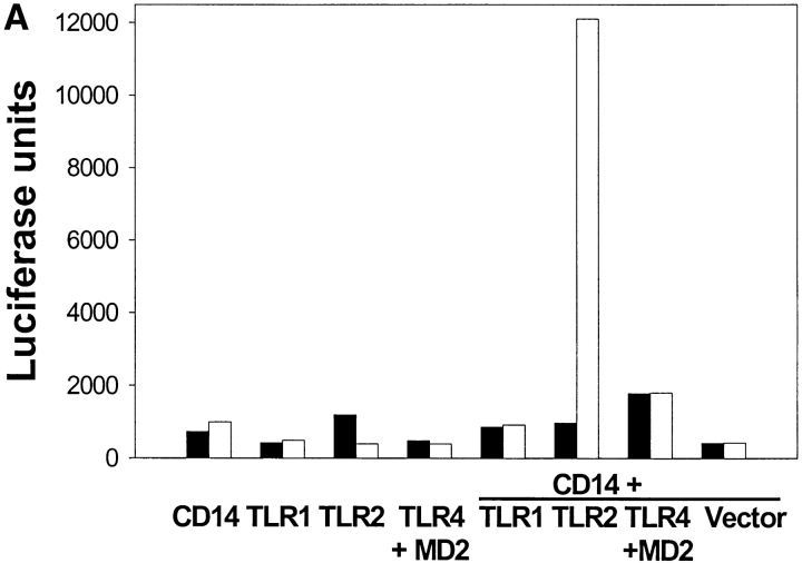
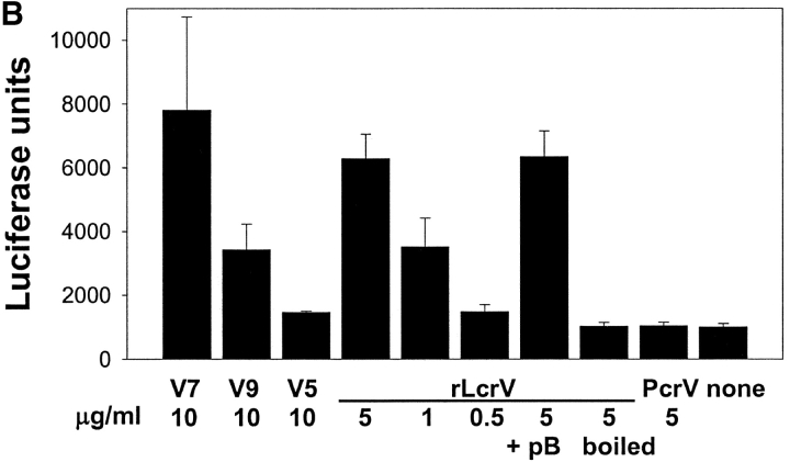
The innate immune system orchestrates the inflammatory response to pathogens by receptors recognizing microbial molecules (19). The activation of members of the mammalian TLR family, which are homologous to the Toll system of Drosophila, by microbial products triggers intracellular signaling pathways finally leading to the transcription of genes relevant for the immune response (20–23). As previous data using C3H/HeJ PPMs defective in tlr4 (24) showed no requirement for TLR4 in LcrV-induced immunomodulation (13), we asked if a different TLR might be involved in LcrV signaling. We transfected HEK 293 cells with FLAG-tagged TLR1, TLR2, and TLR4/MD2, respectively, either in the presence or absence of cotransfected CD14 and tested for the ability to respond to _r_LcrV in an NF-κB–dependent ELAM-1 promoter luciferase reporter gene assay (16). Transfected cells expressing CD14, TLR1, TLR2, or TLR4/MD2 alone did not respond to LcrV treatment (Fig. 2 A). However, reporter gene activation was seen in HEK 293 cells cotransfected with CD14 and TLR2, but not with any of the other TLRs mentioned indicating that LcrV signaling requires CD14/TLR2 complex. Moreover, treatment with polymyxin B (pB), a potent LPS inhibitor, did not influence LcrV signaling via CD14/TLR2 (Fig. 2 B). In contrast, boiling of our LcrV preparation which does not impair TLR2-activating bacterial components known so far (including lipopeptides, peptidoglycan, carbohydrates, and LPS) completely abolished LcrV-signaling via CD14/TLR2 (Fig. 2 B). This finding of a requirement for CD14 in transfection experiments is also consistent with the reported results using murine CD14−/− macrophages and human MonoMac-6 cells
Figure 2.
LcrV signals in a CD14/TLR2-dependent manner. (A) CD14/TLR2-cotransfected HEK 293 cells respond to _r_LcrV. HEK 293 cells were transiently cotransfected with plasmids encoding an ELAM-1 luciferase reporter, a β-galactosidase gene (for normalizing transfection efficiencies) and the indicated TLRs (plus MD2 where depicted) or an empty vector plasmid without or with a CD14 plasmid for 24 h. Cells were left untreated (black bars) or stimulated with 5 μg/ml _r_LcrV (white bars). After 6 h luciferase activities were determined. (B) A synthetic oligopeptide derived from the NH2-terminal region of LcrV is capable to induce CD14/TLR2 signaling. CD14-TLR2 cotransfected HEK 293 cells were stimulated with the peptides V7, V9, and V5 (10 μg/ml), _r_LcrV (5, 1, and 0.5 μg/ml) and _r_PcrV (5 μg/ml), respectively, or left untreated. Addition of polymyxin B (+pB; 10 μg/ml) and boiling of LcrV for 1 h before stimulation served as controls. After 6 h luciferase activities were determined. (C) Amino acid sequences of the NH2-terminal region of PcrV (aa1-aa39), LcrV (aa1-aa56), and the synthetic polypeptides V5, V7, and V9.
Synthetic Peptides Derived from LcrV Are Capable to Signal via TLR2 and CD14.
Next, we were interested to localize the stimulatory region of LcrV responsible for CD14/TLR2 signaling. We found that _r_LcrV constructs (25) lacking different amino acid (aa) regions in the COOH terminus of the protein (aa190–278) and _r_LcrV constructs comprising the first 130 and 100 NH2-terminal aa (_r_V130 and _r_V100) were similarly active as _r_LcrV regarding their capacity to induce IL-10–mediated TNF-α suppression in C3H/HeJ PPMs or MonoMac-6 cells and to signal via CD14/TLR2 (unpublished data). Therefore, we concluded that the COOH-terminal region comprising aa101-aa334 is not needed for IL-10–mediated immunosuppression and CD14/TLR2-dependent signaling. In a next approach, overlapping synthetic peptides of 36 and 37 aa in size covering stepwise the NH2 terminus of LcrV (V1: aa2–aa39, V2: aa31–aa70, V3: aa60–aa98, and V4: aa94–aa131) were generated and tested in CD14/TLR2-cotransfected HEK 293 cells indicating that the active region of LcrV was presumably located between aa 31 and aa70 (unpublished data). By comparing the aa sequence of LcrV with that of PcrV, which had been found to be unable to cause TNF-α suppression via IL-10 induction in murine and human macrophages (13), a gap in the aa sequence of PcrV was recognized corresponding to aa41 to aa59 of LcrV (Fig. 2 C). Thus, we designed three synthetic polypeptides derived from LcrV aa sequence: V5: aa27 – aa43, V7: aa31 – aa49, and V9: aa39-aa57. In CD14/TLR2 cotransfected HEK 293 cells, peptide V7 (10 μg/ml) induced luciferase activity in our reporter assay to a similar degree as _r_LcrV (5 μg/ml), whereas treatment with peptide V5 (10 μg/ml) or the pseudomonas homologue _r_PcrV (5 μg/ml) were not stimulating (Fig. 2 B). Peptide V9 (10 μg/ml) was less active than V7. These results demonstrate that a synthetic oligopeptide derived from the NH2-terminal region of LcrV is capable to induce CD14/TLR2 signaling.
TLR2 Colocalizes with LcrV, but Not with PcrV.
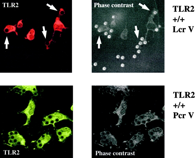
Recently, it was shown that TLR2 accumulation at the macrophage membrane–zymosan particle contact site and phagocytosis precede zymosan-mediated TLR2 signaling (26). Therefore, we wondered whether latex beads coated with rLcrV could show similar characteristics as zymosan particles with respect to TLR2 recruitment. Latex beads of 4 μm in diameter were coated with _r_LcrV (LcrV-beads) and _r_PcrV (PcrV-beads), respectively, and incubated with wild-type PPMs. Immunofluorescence-staining with anti-TLR2 antibodies revealed the enrichment of TLR2 around _r_LcrV-, but not around _r_PcrV-coated particles in so-called “phagocytic cups” (Fig. 3) . Corresponding results were obtained with LcrV- and PcrV-beads in FLAG-tagged TLR2 transfected HEK 293 cells (unpublished data). Thus, these results support the hypothesis that LcrV, but not PcrV is capable to induce signaling by interaction with TLR2 and suggest that both murine and human TLR2 discriminate between the closely related proteins _r_LcrV and _r_PcrV.
Figure 3.
TLR2 colocalizes with LcrV, but not with PcrV. TLR2 is enriched around _r_LcrV- but not _r_PcrV-coated latex beads in murine PPMs. Murine TLR2+/+ PPMs were incubated with latex beads coated with _r_LcrV or _r_PcrV. After fixation, permeabilization and blocking cells were stained with polyclonal anti–mouse TLR2 antibodies and a second immunofluorescent antibody. White arrows indicate enrichment of TLR2 around LcrV beads.
TLR2 Is Involved in LcrV-induced Immunomodulation and Is Required for Susceptibility to Y. enterocolitica Infection.
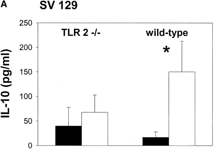
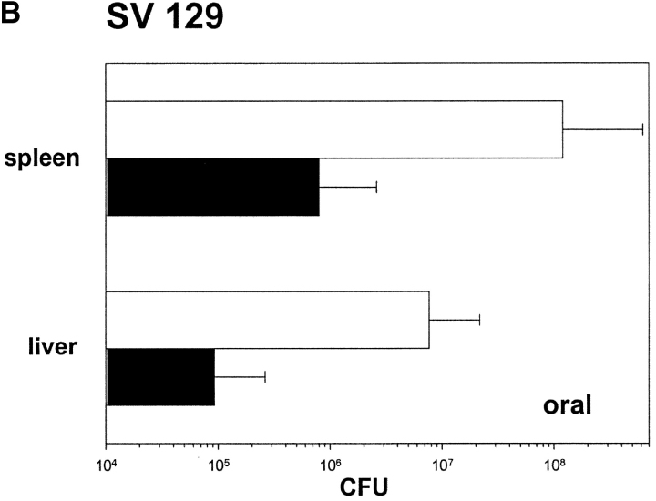
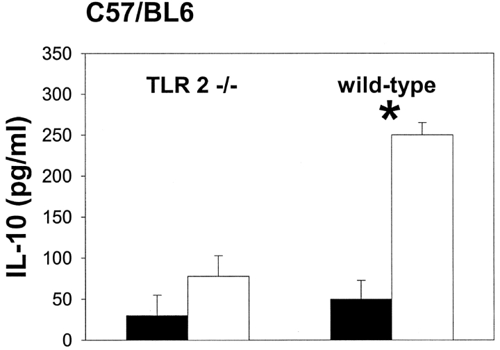
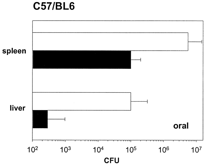
Further evidence for recognition of LcrV by TLR2 eventually leading to IL-10 induction was obtained by comparison of PPMs from TLR2−/− and congenic TLR2+/+ mice of SV129xB57BL/6 (denoted SV129) or C57/BL6 background, respectively. _r_LcrV-treatment of PPMs derived from the respective wild-type mice resulted in a markedly enhanced IL-10 response when compared with PPMs from the corresponding TLR2−/− mice (Fig. 4 A). Based on previous data showing that IL-10−/− mice are highly resistant to yersiniae infection (13) and assuming that LcrV-mediated immunosuppression via IL-10 which is transmitted in a CD14/TLR2-dependent manner could play an important role in yersinae infection we speculated that TLR2−/− mice might be less susceptible to an oral challenge with the enteropathogenic Y. enterocolitica strain WA-314. 5 d after oral inoculation with 5 × 10⁷ CFU Y. enterocolitica, bacterial counts in spleen and liver of TLR2−/− SV129xB57BL/6 and TLR2−/− C57/BL6 mice were about two logs lower than those in the respective organs of congenic TLR2+/+ mice (Fig. 4 B).
Figure 4.
TLR2 is involved in LcrV-induced immunomodulation and is required for susceptibility to Y. enterocolitica infection. (A) LcrV-induced IL-10 production is highly impaired in TLR2-deficient PPMs. PPMs of different TLR2-deficient and corresponding wild-type mice (SV129xB57BL/6 and C57BL/6 background) were treated with _r_LcrV (5 μg/ml) for 2 h (white bars) or left untreated (black bars). IL-10 was measured in the supernatant by ELISA. *P < 0.001. (B) TLR2-deficient mice are highly resistant to oral Y. enterocolitica infection. TLR2−/− (black bars) and corresponding wild-type (white bars) mice of the indicated genetic backgrounds were orally infected with 5 × 10⁷ CFU Y. enterocolitica O8. After 5 d the number of yersiniae in spleen and liver was determined. Data represent results obtained by using 5–10 mice in at least two independent experiments (P values < 0.05).
Discussion
In this study we focused on the signaling of V antigen–induced innate immunity responses. For the first time we demonstrate that a bacterial nonlipidated protein associated with virulence and derived synthetic oligopeptides are capable to induce IL-10 production via TLR2/CD14 signaling. In contrast to the pseudomonas _r_PcrV, _r_LcrV (range of activity: 10–100 nM) transmits signaling via TLR2 in a CD14-dependent manner leading to IL-10 induction which finally causes TNF-α suppression thus probably enabling yersiniae to evade the host innate immune system. Several lines of evidence obtained in vitro both in murine and human cell systems support this conclusion: (a) Only CD14-TLR2-cotransfected HEK 293 cells, but not cells transfected with TLR2 alone responded with NF-κB–dependent ELAM-1 promoter luciferase activity upon _r_LcrV stimulation. (b) Blocking anti-CD14 monoclonal antibodies, but not nonblocking isotype anti-CD14 antibodies completely abolished TNF-α suppression in LcrV-treated MonoMac-6 cells suggesting that CD14 is needed for recognition of V antigen and transmission of V antigen-caused cellular effects. (c) In contrast to wild-type or TLR4-deficient (13) murine macrophages of different genetic backgrounds, CD14- as well as TLR2-deficient PPMs of different genetic backgrounds do not respond to _r_LcrV by IL-10 induction. (d) Colocalization of _r_LcrV, but not _r_PcrV with TLR2 was visualized both in murine PPMs and TLR2-transfected human HEK 293 cells; the enrichment of TLR2 around _r_LcrV-, but not around _r_PcrV-coated particles in so-called “phagocytic cups” suggests that both murine and human TLR2 discriminate between the closely related proteins LcrV and PcrV.
Previously we could clearly attribute TNF-α suppression and IL-10 induction to the protein component of our rLcrV preparation (13), as denaturation by boiling or proteinase K degradation completely abolished the observed immunomodulation. Moreover, antibodies against _r_LcrV greatly impaired LcrV-induced immunomodulating effects (13). In the present study we chose further strategies to rule out an involvement of contaminants possibly present in our _r_LcrV preparation in CD14-TLR2–dependent signaling by _r_LcrV: (a) addition of polymyxin B to LcrV-treated cotransfected cells which did not influence CD14-TLR2–dependent signaling, (b) boiling of _r_LcrV before use which completely abrogated CD14-TLR2–dependent signaling, (c) comparison of _r_LcrV-signaling with that of the similarly produced _r_PcrV which is not able to signal via CD14/TLR2, and (d) the successful use of synthetic peptides. Finally, we could exclude _r_LcrV modification by MALDI-MS analysis. All these approaches indicate that any bacterial contaminants known so far to use TLR2 for signaling are unlikely to be responsible for CD14-TLR2–dependent signaling by _r_LcrV. Moreover, the active region of LcrV for CD14-TLR2-mediated recognition could be located to the first 100 NH2-terminal aa of LcrV, since truncated constructs of LcrV carrying different deletions in the region of aa190–278 (25) or lacking two thirds of the COOH-terminal region of LcrV (_r_V130 and _r_V100) exhibited similar TNF-α suppressive effects in MonoMac-6 cells and a NF-kB–inducing capacity in CD14/TLR2-cotransfected HEK 293 cells as _r_LcrV. Furthermore, we could define an active region of LcrV which comprises aa31–49 using different synthetic peptides derived from LcrV aa-sequence. Interestingly, a protective antigenic region located between aa 2 and 135 of V-antigen had previously been identified besides a protective antigenic region between aa 135 and 275 by vaccination experiments (27).
In summary, our data clearly demonstrate a new ligand specificity of TLR2. In striking contrast to all bacterial components known so far to signal via TLR2, LcrV is a chemically nonmodified bacterial virulence-associated protein.
The TLR2 dependency of _r_LcrV-induced IL-10 production raised the question whether TLR2 contributes to susceptibility of mice to yersinia infection. In striking contrast to a protective role of TLR2 in Borrelia burgdorferi (28) and Staphylococcus aureus (29) infected mice, however, the absence of TLR2 renders mice more resistant to an oral Y. enterocolitica infection. The strategy of yersiniae to exploit an innate immunity effector, i.e., host macrophage-derived IL-10, might therefore be connected to the use of a host pattern recognition receptor (PRR), TLR2, to evade the host immune response. As TLR2-deficient PPMs show a greatly impaired IL-10 production upon LcrV stimulation, one might speculate that LcrV is a key player in this novel pathogenicity mechanism by which yersiniae exploit a host PRR for subverting the host immune system. Unfortunately, this key role of LcrV cannot be demonstrated by simple insertional inactivation of the encoding gene lcrV, because this leads to impairment of the TTS and conversion to avirulence of yersiniae (1). Therefore, sophisticated site-directed mutagenesis of the NH2-terminal encoding region of the lcrV gene by maintaining the TTS function would be required.
Mammals are endowed with a large set of PRRs for surveillance of microbial invaders and for immediate immune response. This system is efficient for defense of apathogenic and probably of facultative pathogenic microbes (“immune homeostasis”). Presumably apathogenic invaders elicit a short-lived inflammatory response followed by IL-10–mediated down-regulation which is sufficient for elimination of harmless microbes. However, a characteristic feature of pathogenic microbes is the capability to break through the first line of defense represented by innate immunity. Thus, IL-10–mediated down-regulation of the innate immunoresponse can be harmful for the host infected with pathogenic microbes, in particular with yersiniae, which have developed a short distance anti-host weapon mediated by the TTS/Yop system and a long distance weapon mediated by the released LcrV bacteriokine (30).
Acknowledgments
We thank Anna M. Geiger for rPcrV, Susanne Bierschenk for excellent technical assistance, and Andreas Schroeder for help with digital imaging.
This work was supported in part by the Deutsche Forschungsgemeinschaft (GRK 303 and SPP 1089).
D. Rost and N. Tvardovskaia contributed equally to this work.
Footnotes
*
Abbreviations used in this paper: aa, amino acid; ELAM-1, endothelial cell-leukocyte adhesion molecule; NF, nuclear factor; PRR, pattern recognition receptor; TLR, toll-like receptor; TTS, type III protein secretion and translocation system.
References
- 1.Cornelis, G.R., A. Boland, A.P. Boyd, C. Geuijen, M. Iriarte, C. Neyt, M.P. Sory, and I. Stainier. 1998. The virulence plasmid of Yersinia, an antihost genome. Microbiol. Mol. Biol. Rev. 62:1315–1352. [DOI] [PMC free article] [PubMed] [Google Scholar]
- 2.Hoiczyk, E., and G. Blobel. 2001. Polymerization of a single protein of the pathogen Yersinia enterocolitica into needles punctures eukaryotic cells. Proc. Natl. Acad. Sci. USA. 98:4669–4674. [DOI] [PMC free article] [PubMed] [Google Scholar]
- 3.Aepfelbacher, M., R. Zumbihl, K. Ruckdeschel, C.A. Jacobi, C. Barz, and J. Heesemann. 1999. The tranquilizing injection of Yersinia proteins: a pathogen's strategy to resist host defence. Biol. Chem. 380:795–802. [DOI] [PubMed] [Google Scholar]
- 4.Lee, V.T., C. Tam, and O. Schneewind. 2000. LcrV, a substrate for Yersinia enterocolitica type III secretion, is required for toxin targeting into the cytosol of HeLa cells. J. Biol. Chem. 275:36869–36875. [DOI] [PubMed] [Google Scholar]
- 5.Fields, K.A., M.L. Nilles, C. Cowan, and S.C. Straley. 1999. Virulence role of V antigen of Yersinia pestis at the bacterial surface. Infect. Immun. 67:5395–5408. [DOI] [PMC free article] [PubMed] [Google Scholar]
- 6.Burrows, T.W. 1956. An antigen determining virulence in _Pasteurella pestis_Nature. 177:426–427. [DOI] [PubMed] [Google Scholar]
- 7.Price, S.B., C. Cowan, R.D. Perry, and S.C. Straley. 1991. The Yersinia pestis V antigen is a regulatory protein necessary for Ca2(+)-dependent growth and maximal expression of low-Ca2+ response virulence genes. J. Bacteriol. 173:2649–2657. [DOI] [PMC free article] [PubMed] [Google Scholar]
- 8.Holmström, A., J. Olsson, P. Cherepanov, E. Maier, R. Nordfelth, J. Pettersson, R. Benz, H. Wolf-Watz, and A. Forsberg. 2001. LcrV is a channel size-determining component of the Yop effector translocon of Yersinia. Mol. Microbiol. 39:620–632. [DOI] [PubMed] [Google Scholar]
- 9.Motin, V.L., R. Nakajima, G.B. Smirnov, and R.R. Brubaker. 1994. Passive immunity to yersiniae mediated by anti-recombinant V antigen and protein A-V antigen fusion peptide. Infect. Immun. 62:4192–4201. [DOI] [PMC free article] [PubMed] [Google Scholar]
- 10.Nedialkov, Y.A., V.L. Motin, and R.R. Brubaker. 1997. Resistance to lipopolysaccharide mediated by the Yersinia pestis V antigen-polyhistidine fusion peptide: amplification of interleukin-10. Infect. Immun. 65:1196–1203. [DOI] [PMC free article] [PubMed] [Google Scholar]
- 11.Roggenkamp, A., A.M. Geiger, L. Leitritz, A. Kessler, and J. Heesemann. 1997. Passive immunity to infection with Yersinia spp. mediated by anti-recombinant V antigen is dependent on polymorphism of V antigen. Infect. Immun. 65:446–451. [DOI] [PMC free article] [PubMed] [Google Scholar]
- 12.Pettersson, J., A. Holmström, J. Hill, S. Leary, E. Frithz-Lindsten, A. von Euler-Matell, E. Carlsson, R. Titball, A. Forsberg, and H. Wolf-Watz. 1999. The V-antigen of Yersinia is surface exposed before target cell contact and involved in virulence protein translocation. Mol. Microbiol. 32:961–976. [DOI] [PubMed] [Google Scholar]
- 13.Sing, A., A. Roggenkamp, A.M. Geiger, and J. Heesemann. 2002. Yersinia enterocolitica evasion of the host innate immune response by V antigen-induced IL-10 production of macrophages is abrogated in IL-10-deficient mice. J. Immunol. 168:1315–1321. [DOI] [PubMed] [Google Scholar]
- 14.Sawa, T., T.L. Yahr, M. Ohara, K. Kurahashi, M.A. Gropper, J.P. Wiener-Kronish, and D.W. Frank. 1999. Active and passive immunization with the Pseudomonas V antigen protects against type III intoxication and lung injury. Nat. Med. 5:392–398. [DOI] [PubMed] [Google Scholar]
- 15.Werts, C., R.I. Tapping, J.C. Mathison, T.H. Chuang, V. Kravchenko, I. Saint Girons, D.A. Haake, P.J. Godowski, F. Hayashi, A. Ozinsky, et al. 2001. Leptospiral lipopolysaccharide activates cells through a TLR2-dependent mechanism. Nat. Immunol. 2:346–352. [DOI] [PubMed] [Google Scholar]
- 16.Kirschning, C.J., H. Wesche, T. Merrill Ayres, and M. Rothe. 1998. Human toll-like receptor 2 confers responsiveness to bacterial lipopolysaccharide. J. Exp. Med. 188:2091–2097. [DOI] [PMC free article] [PubMed] [Google Scholar]
- 17.Wiedemann, A., S. Linder, G. Grassl, M. Albert, I. Autenrieth, and M. Aepfelbacher. 2001. Yersinia enterocolitica invasin triggers phagocytosis via beta1 integrins, CDC42Hs and WASp in macrophages. Cell. Microbiol. 3:693–702. [DOI] [PubMed] [Google Scholar]
- 18.Ulevitch, R.J., and P.S. Tobias. 1999. Recognition of gram-negative bacteria and endotoxin by the innate immune system. Curr. Opin. Immunol. 11:19–22. [DOI] [PubMed] [Google Scholar]
- 19.Medzhitov, R., and C.A. Janeway, Jr. 1997. Innate immunity: impact on the adaptive immune response. Curr. Opin. Immunol. 9:4–9. [DOI] [PubMed] [Google Scholar]
- 20.Underhill, D.M., and A. Ozinsky. 2002. Toll-like receptors: key mediators of microbe detection. Curr. Opin. Immunol. 14:103–110. [DOI] [PubMed] [Google Scholar]
- 21.Akira, S., K. Takeda, and T. Kaisho. 2001. Toll-like receptors: critical proteins linking innate and acquired immunity. Nat. Immunol. 2:675–680. [DOI] [PubMed] [Google Scholar]
- 22.Aderem, A., and R.J. Ulevitch. 2000. Toll-like receptors in the induction of the innate immune response. Nature. 406:782–788. [DOI] [PubMed] [Google Scholar]
- 23.Lien, E., T.J. Sellati, A. Yoshimura, T.H. Flo, G. Rawadi, R.W. Finberg, J.D. Carroll, T. Espevik, R.R. Ingalls, J.D. Radolf, and D.T. Golenbock. 1999. Toll-like receptor 2 functions as a pattern recognition receptor for diverse bacterial products. J. Biol. Chem. 274:33419–33425. [DOI] [PubMed] [Google Scholar]
- 24.Poltorak, A., X. He, I. Smirnova, M.Y. Liu, C.V. Huffel, X. Du, D. Birdwell, E. Alejos, M. Silva, C. Galanos, et al. 1998. Defective LPS signaling in C3H/HeJ and C57BL/10ScCr mice: mutations in Tlr4 gene. Science. 282:2085–2088. [DOI] [PubMed] [Google Scholar]
- 25.Roggenkamp, A., L. Leitritz, A. Sing, V.A. Kempf, K. Baus, and J. Heesemann. 1999. Anti-recombinant V antigen serum promotes uptake of Yersinia enterocolitica serotype 08 by macrophages. Med. Microbiol. Immunol. 188:151–159. [DOI] [PubMed] [Google Scholar]
- 26.Underhill, D.M., A. Ozinsky, A.M. Hajjar, A. Stevens, C.B. Wilson, M. Bassetti, and A. Aderem. 1999. The toll-like receptor 2 is recruited to macrophage phagosomes and discriminates between pathogens. Nature. 401:811–815. [DOI] [PubMed] [Google Scholar]
- 27.Hill, J., S.E. Leary, K.F. Griffin, E.D. Williamson, and R.W. Titball. 1997. Regions of Yersinia pestis V antigen that contribute to protection against plague identified by passive and active immunization. Infect. Immun. 65:4476–4482. [DOI] [PMC free article] [PubMed] [Google Scholar]
- 28.Wooten, R.M., Y. Ma, R.A. Yoder, J.P. Brown, J.H. Weis, J.F. Zachary, C.J. Kirschning, and J.J. Weis. 2002. Toll-like receptor 2 is required for innate, but not acquired, host defense to Borrelia burgdorferi. J. Immunol. 168:348–355. [DOI] [PubMed] [Google Scholar]
- 29.Takeuchi, O., K. Hoshino, and S. Akira. 2000. Cutting edge: TLR2-deficient and MyD88-deficient mice are highly susceptible to Staphylococcus aureus infection. J. Immunol. 165:5392–5396. [DOI] [PubMed] [Google Scholar]
- 30.Wilson, M., R. Seymour, and B. Henderson. 1998. Bacterial perturbation of cytokine networks. Infect. Immun. 66:2401–2409. [DOI] [PMC free article] [PubMed] [Google Scholar]
