Signal transducer and activator of transcription (Stat) 5 controls the proliferation and differentiation of mammary alveolar epithelium (original) (raw)
Abstract
Functional development of mammary epithelium during pregnancy depends on prolactin signaling. However, the underlying molecular and cellular events are not fully understood. We examined the specific contributions of the prolactin receptor (PrlR) and the signal transducers and activators of transcription 5a and 5b (referred to as Stat5) in the formation and differentiation of mammary alveolar epithelium. PrlR- and Stat5-null mammary epithelia were transplanted into wild-type hosts, and pregnancy-mediated development was investigated at a histological and molecular level. Stat5-null mammary epithelium developed ducts but failed to form alveoli, and no milk protein gene expression was observed. In contrast, PrlR-null epithelium formed alveoli-like structures with small open lumina. Electron microscopy revealed undifferentiated features of organelles and a perturbation of cell–cell contacts in PrlR- and Stat5-null epithelia. Expression of NKCC1, an Na-K-Cl cotransporter characteristic for ductal epithelia, and ZO-1, a protein associated with tight junction, were maintained in the alveoli-like structures of PrlR- and Stat5-null epithelia. In contrast, the Na-Pi cotransporter Npt2b, and the gap junction component connexin 32, usually expressed in secretory epithelia, were undetectable in PrlR- and Stat5-null mice. These data demonstrate that signaling via the PrlR and Stat5 is critical for the proliferation and differentiation of mammary alveoli during pregnancy.
Keywords: prolactin receptor; Stat5; mammary gland; cell specification; epithelia
Introduction
Cytokines, such as prolactin (Prl),* growth hormone (GH), interleukin (IL)-2, and erythropoietin (Epo) elicit a wide range of cell-specific responses including cell proliferation, survival, differentiation, and death. Upon binding of these cytokines to their respective receptors, the receptor-associated kinase Jak2 phosphorylates the latent transcription factors signal transducer and activator of transcription (Stat)5a and Stat5b at tyrosines 694/699, respectively. Upon activation, Stat5a and Stat5b form homo- and heterodimers that translocate to the nucleus and induce cell-specific genetic programs. In erythroid cells, Epo-induced Stat5 activation may lead to the transcriptional activation of the _bcl_-x gene and thus promote cell survival (Socolovsky et al., 1999), and in T cells, IL-2–mediated cell proliferation is severely impaired in the absence of Stat5 (Teglund et al., 1998; Moriggl et al., 1999).
Development of the mammary gland occurs predominantly in the postnatal animal and is controlled by steroid and peptide hormones (Hennighausen and Robinson, 1998, 2001). Proliferation and differentiation of mammary alveolar epithelia occurs during pregnancy through prolactin and its receptor (PrlR) (Ormandy et al., 1997b). Stat5a-null mice fail to develop functional mammary tissue during pregnancy, mainly as a result of impaired functional differentiation and not due to the lack of lobulo-alveolar units (Liu et al., 1997). After multiple pregnancies, functional mammary development was attained in Stat5a-null mice (Liu et al., 1998). This phenotype was accompanied by increased levels of active Stat5b, suggesting that Stat5b can partially compensate for the absence of Stat5a. However, Stat5b itself is not required for lactation (Teglund et al., 1998). Because mice carrying inactivated Stat5a and 5b (referred to as Stat5 throughout the text) genes are infertile, the combined function of both Stat5a and 5b during pregnancy had not been investigated.
Components of regulatory pathways are in many cases not exclusive, and may participate in several signaling cascades. In mammary epithelia, Stat5 is activated through the PrlR, but also by GH and the epidermal growth factor receptors (Gallego et al., 2001), and possibly the Src pathway (Kazansky et al., 1999). In addition, the PrlR not only activates Stat5 via Jak2, but also stimulates the mitogen-activated protein kinase and phosphoinositide 3-kinase (PI3K) pathways (Bole-Feysot et al., 1998; Kim and Cochran, 2001). If mammary alveolar epithelial development depends on a pathway that includes the PrlR and Stat5, a similar phenotype should be expected in the respective gene deletion mice. However, if Prl-independent Stat5 activation controls some steps of development, a different phenotype would be expected in the absence of the two components (i.e., PrlR and Stat5).
We have now investigated the relative contributions of Stat5 and the PrlR in pregnancy-induced mammary epithelial development. Although gross morphological analyses have linked the PrlR (Ormandy et al., 1997a) and Stat5a (Liu et al., 1997) to cell proliferation and differentiation, the molecular and cellular mechanisms of prolactin and Stat5 signaling are not understood. Specifically, the combined roles of Stat5a and 5b during pregnancy-induced mammopoiesis are not known. It remains unclear whether the Prl pathway is actually required for the acquisition of a particular cell fate, or whether it controls the differentiation of an already specified cell type. We have used molecular markers that can distinguish between different mammary epithelial cell types in the developing mammary gland to investigate the role of PrlR and Stat5 in the development of alveolar epithelium.
Results
Development of mammary tissue in PrlR- and Stat5-null mice
Although the PrlR is required for development of mammary epithelium during pregnancy (Ormandy et al., 1997b), the contribution of Stat5 in this process is not known. Stat5 is downstream of the PrlR, and it can be hypothesized that the loss of either signaling component might lead to a comparable phenotype. To investigate this, we compared the pregnancy-induced development of PrlR- and Stat5-null epithelia. Because Stat5- and PrlR-null mice are infertile, mammary epithelia from mature virgins was transplanted into the cleared fat pad of recipients to expose these epithelia to pregnancy hormones. Whole mount analyses demonstrated that ductal development during puberty was not affected in PrlR- and Stat5-null mice (Fig. 1 C). In contrast, pregnancy-mediated alveolar development was severely impaired (Fig. 1 A). Whereas wild-type ducts were decorated with expanded alveoli, the epithelium was greatly reduced in Stat5-null transplants and did not have the appearance of true lobulo-alveolar units. Histological sections demonstrated that the majority of Stat5-null alveoli-like structures did not have lumina (Fig. 1 B, right panel). Furthermore, individual Stat5-null epithelial cells exhibited abnormal columnar shapes and the epithelial architecture appeared disorganized (Fig. 1 B, right panel, white arrow). On the other hand, small open lumina were evident in the more organized alveoli-like structures in the PrlR-null transplants (Fig. 1 B, middle panel). To further evaluate the differences between Stat5- and PrlR-null alveoli-like structures, we analyzed serial sections. Whereas PrlR-null tissue exhibited consistently open lumina, they were not apparent in Stat5-null tissue (unpublished data).
Figure 1.
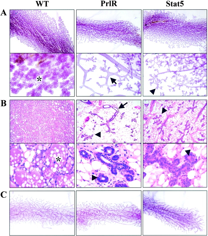
Pregnancy-mediated mammary gland development depends on the PrlR and Stat5. (A) Whole mount analyses of wild-type, PrlR-, and Stat5-null mammary epithelia at parturition. The lower panel represents a higher magnification. Wild-type epithelium filled the fat pad and developed lobuloalveolar structures (*). PrlR- and Stat5-null mammary epithelia were severely underdeveloped. PrlR-null epithelium exhibited wide ducts (black arrow), short branches, and few alveoli-like structures. Stat5-null epithelium displayed normal branches with small decorations (black arrowhead). (B) Histological sections of the whole mounts shown in A. The lower panel represents a 6× higher magnification. Wild-type epithelium was fully expanded, filled the fat pad and alveolar lumina contained milk (*). However, all null epithelia were sparse, and the alveoli-like structures (black arrowhead) did not contain milk. The epithelial cells are cuboidal in shape (white arrow). Note the presence of open lumina in the alveoli-like structures in PrlR-, but not Stat5-null epithelium. (C) Whole mount analyses of wild-type and PrlR- and Stat5-null virgin mammary epithelia 8 wk after transplantation. All transplanted epithelia completely filled the fat pad.
Proliferation of PrlR- and Stat5-null mammary epithelia
We investigated potential causes for the lack of functional alveolar development in PrlR- and Stat5-null epithelia, and examined estrogen- and progesterone-mediated proliferation (Fig. 2). Previous studies have demonstrated that acute treatment with estrogen and progesterone for 2 d resulted in ∼15% of the wild-type ductal epithelial cells entering the cell cycle, as assessed by BrdU labeling (Seagroves et al., 2000). Maximal proliferation appears to occur in the alveolar progenitors in the ducts during the first few days of pregnancy. The estrogen and progesterone treatment of virgin transplants that have filled the fat pad is designed to mimic the effect of early pregnancy on alveolar development. In comparison to the thoracic #3 controls from the host animal, steroid hormone–induced proliferation of PrlR-null epithelium was reduced by ∼60%, whereas proliferation of Stat5-null epithelium was reduced by only 30% (Fig. 2 B). Thus, whereas in both cases there was a significant reduction in proliferation relative to the wild-type control, PrlR-null epithelium exhibited a twofold greater decrease than Stat5-null epithelium. These differences were statistically significant (P < 0.001).
Figure 2.
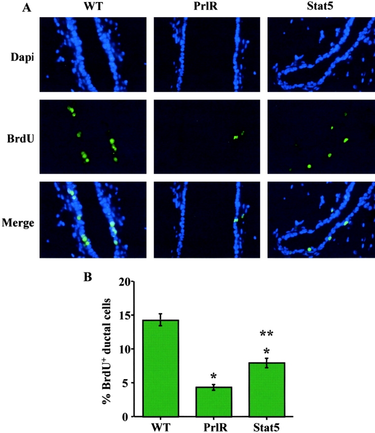
Progesterone- and estrogen-induced proliferation is more severely impaired in PrlR-null than in Stat5a/b–null mammary epithelium. 9 wk after transplantation, mice were given an acute, 2-d E+P treatment. Mammary glands were removed and proliferation was evaluated by BrdU immunostaining. (A) Green staining represents BrdU-positive cells and DAPI-stained nuclei are blue. Merging the images shows the proliferation occurring mainly in the ductal epithelium. The number of BrdU-positive cells was clearly decreased in both PrlR- and Stat5-null transplants, as compared with the control #3 gland. (B) Quantitation of BrdU-positive ductal cells (mean percentage ± SEM) is shown in the bar graph. The P values comparing PrlR- or Stat5-null to control (*) and Stat5-null to PrlR-null (**) were <0.001 as determined by Mann-Whitney paired t test.
Differentiation of PrlR- and Stat5-null mammary epithelia
There were no morphological and histological signs of milk secretion in PrlR- and Stat5-null epithelia (Fig. 1 B). To determine the differentiation status of PrlR- and Stat5-null epithelia, we examined the expression of milk protein genes (β-casein, whey acidic protein [WAP], and WDNM1). Steady-state levels of these mRNAs increased in wild-type (Fig. 3) and Stat5b-null (unpublished data) mammary tissue during pregnancy. Expression of WAP, but not β-casein, mRNA was decreased in Stat5a-null mice (Fig. 3) as shown previously (Liu et al., 1997). β-casein and WAP mRNAs were not detected in PrlR- and Stat5-null epithelia by total RNA Northern blot. On the other hand, WDNM1 mRNA was detected at lower levels in the PrlR- and Stat5-null epithelia. Using more sensitive assays, low levels of WAP and β-casein were detected in Stat5-null epithelia (unpublished data). These results suggest that PrlR- and Stat5-null epithelia failed to undergo functional differentiation.
Figure 3.
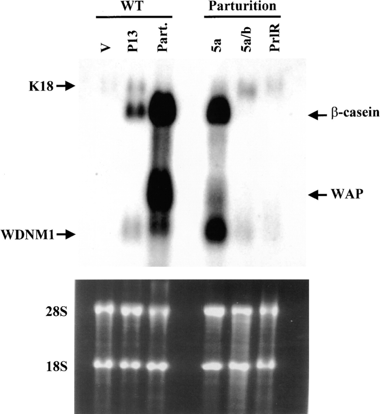
Milk protein expression is impaired in Stat5- and PrlR-null mammary epithelia. Northern blot analysis of milk protein mRNAs (β-casein, WAP, WDNM1) and keratin 18 (K18) mRNA in mammary tissue of wild-type mice (WT) and Stat5a-, 5a/b-, and PrlR-null epithelial transplants. V, virgin (5 wk); P13, pregnancy day 13; Part, after parturition.
Stat5-null epithelium have impaired cell–cell contacts and lack connexin 32
We evaluated the ultrastructure of wild-type, Stat5-, and PrlR-null mammary epithelia at parturition by electron microscopy (Fig. 4). Mammary tissue from lactating control mice contained secretory epithelia with features indicative of a fully differentiated phenotype: the alveolar lumina were expanded and contained casein micelles and lipid droplets (Fig. 4, A and D). Alveolar cells contained a well-developed Golgi apparatus and a rough endoplasmic reticulum (Fig. 4 D). In contrast, PrlR- (Fig. 4, B and E) and Stat5-null (Fig. 4, C and F) epithelial structures were highly disorganized. Most of the alveoli-like structures in Stat5-null epithelium had no lumina (Fig. 4, C and F). Occasionally, the lumina that were present that were small, and did not contain casein micelles and lipid droplets (Fig. 4 F). Often, there were several layers of epithelia so that the lumina seemed congested with cells (Fig. 4 C). We frequently observed several pseudo-lumina within one structure, suggesting the inability to form a single lumen surrounded by a single layer of epithelial cells (unpublished data). Interestingly, the cells near the basement membrane, thought to be myoepithelia, contained lipid droplets reminiscent of secretory cells (Fig. 4 C). In contrast, PrlR-null epithelia were more organized and the alveoli-like structures contained small but open lumina (Fig. 4, B and E). Neither the rough endoplasmic reticulum nor the active Golgi apparatus were prominent in PrlR- or Stat5-null epithelial cells (Fig. 4, E and F). We observed that the epithelial cells furthest from the basement membrane, and therefore the most “luminal,” contained centrosomes located close to their apical membrane as determined by the presence of microvilli (Fig. 4 E). This centrosomal orientation suggested that these cells had recently divided and that their cleavage plane was perpendicular to the basement membrane potentially resulting in the epithelial layering and the crowding of the luminal space.
Figure 4.
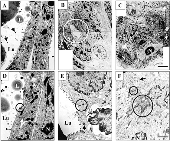
Disorganized structures of Stat5- and PrlR-null epithelia. Stat5- and PrlR-null mammary epithelia at parturition were analyzed by electron microscopy. (A and D) Control mammary epithelium at lactation day 1 was fully differentiated and contained secreted milk proteins and lipid droplets. Golgi apparatus (white long arrow) and RER (white long arrow) were detected. Lu, lumen; L, lipid droplet; N, nucleus; black short arrow, β-casein micelles; black circle, tight junction. (B and E) Alveoli-like structures of PrlR-null epithelium were more organized than Stat5-null epithelia. Lumina were detected in alveolar-like structure (white circle). The centrosome was located close to the apical membrane (black arrow). Tight junctions were maintained (black circle). (C and F) Alveoli-like structures in Stat5-null epithelia were disorganized and cell–cell contacts were aberrant (black arrow). Frequently, two or more pseudo-lumina were detected in one alveolar-like structure (black circles). The cells near the basement membrane contained lipid droplet-like structures (white arrow). Active Golgi apparatus and a RER in Stat5- and PrlR-null epithelia were not apparent. Bars: (A–C) 2.4 μm; (D–F) 1.6 μm.
Epithelial cells contact each other via tight and adherens junctions (Cereijido et al., 1998; Borrmann et al., 2000; Vasioukhin and Fuchs, 2001), which stabilize epithelial structures and determine their integrity. Further, these junctions are necessary to establish and maintain cell polarity (Knust, 2000; Vicente-Manzanares and Sanchez-Madrid, 2000) that permits vectorial secretion (Barcellos-Hoff et al., 1989). Fig. 4 D shows the morphological appearance of a tight junction complex between the apical poles of two individual secretory cells in wild-type epithelium. One of the most conspicuous features of the Stat5-null epithelium was the lack of organized cell contacts. To identify possible causes for the lack of organized cell contacts we investigated the expression of zonula occludens (ZO)-1, a component of tight junctions. Tight junctions were identified in PrlR- and Stat5-null epithelia (Fig. 4, E and F) and visualized by ZO-1 staining (Fig. 5, A and B). We further investigated the expression of connexin, a protein in the gap junction complex, in PrlR- and Stat5-null epithelia. It has been demonstrated previously (Pozzi et al., 1995; Locke et al., 2000) that mouse mammary tissue expresses three connexin isoforms (Cx 43, Cx 26, and Cx 32), which we confirmed using reverse transcription (RT)-PCR analyses. Whereas Cx 32 mRNA was detected in wild-type epithelia at day 1 of lactation, we were unable to detect expression in PrlR- and Stat5-null epithelia at parturition (Fig. 5 D).
Figure 5.
Maintenance of ZO-1 expression but loss of Cx 32 expression. Immunohistochemical staining of ZO-1 (green) and E-cadherin (red) at parturition (A–C). Tight junctions (green dots) were present in PrlR- (A) and Stat5-null (B) the same as in wild-type epithelium (C). Arrows point to alveoli-like structures of PrlR- and Stat5-null epithelia. (D) RT-PCR analysis of Cx 32 mRNA. Total RNA from wild-type, Stat5-, and PrlR-null transplanted epithelia at parturition was reverse transcribed, and the cDNA was subjected to PCR. Cx 32 cDNA was detected in wild-type but not in PrlR- or Stat5-null samples. GAPDH levels were similar in all samples. M, marker.
To further examine the cell adhesion defect apparent in Stat5-null epithelial cells, we investigated the expression of additional molecules involved in cell adhesion. Cadherins mediate cell–cell adhesion and also contribute to the maintenance of apical–basal polarity (Tepass et al., 2000). Indeed, E-cadherin has been shown to play a role in the morphogenesis and growth of the mammary gland (Daniel et al., 1995; Delmas et al., 1999). On this basis, we hypothesized that E-cadherin expression might be altered in Stat5-null mammary epithelial cells and thus contribute to the observed alterations in cell adhesion. However, E-cadherin expression along cell–cell borders did not appear to be significantly perturbed when comparing Stat5-null and wild-type epithelia (Figs. 5 and 7).
Figure 7.
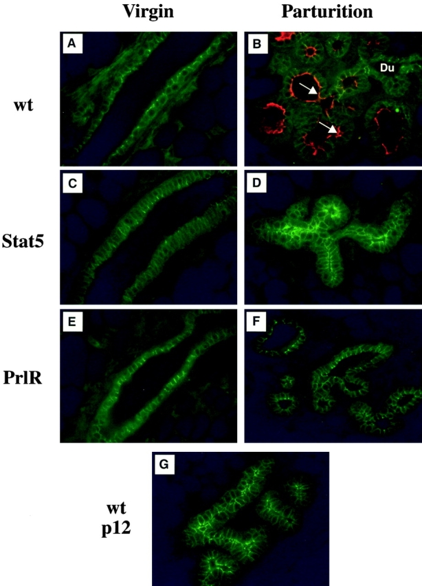
Npt2b expression was not detected in PrlR- and Stat5-null alveoli-like structures at parturition. Immunohistochemical staining of Npt2b (red) and E-cadherin (green) in mammary epithelia of virgin mice (A, C, and E) and after parturition (B, D, and F). Npt2b was detected in apical membranes of secreting epithelia from wild-type tissue (B, white arrow). PrlR- and Stat5-null epithelia at parturition did not contain Npt2b (D and F). E-cadherin was detected in the sub-apical/basolateral membrane of all samples (A–F). (G) Wild-type epithelia at pregnancy day 12 did not express Npt2b. Du, duct.
PrlR- and Stat5-null epithelia maintain virgin-like ductal features during pregnancy
Whole mount and histological analyses have demonstrated that the formation of mammary alveolar epithelium was severely impaired in the absence of the PrlR or Stat5. However, these types of analyses do not allow the identification of the epithelial cells as ductal or alveolar. To further determine the histological identity of these cells, we investigated the expression of proteins that characterize either ductal or secretory alveolar epithelium. We have observed that the Na-K-Cl cotransporter (NKCCl) is expressed at high levels in virgin mice and is located on the basolateral membrane of ductal epithelial cells, and unpublished data). Further, NKCC1 expression levels decreased in developing alveoli but are maintained in some cells of the ductal epithelia during pregnancy. Therefore, we examined the expression of NKCC1 by immunohistochemistry in transplanted PrlR- and Stat5-null epithelia in virgin mice and at parturition. In virgin mice, NKCC1 was detected in endogenous epithelium and PrlR- and Stat5-null transplants (Fig. 6, A, C, and E). At parturition, the levels of NKCC1 were sharply reduced in wild-type secretory alveolar cells (Fig. 6 B). In contrast, NKCC1 expression was consistently higher in PrlR- and Stat5-null epithelia at parturition (Fig. 6, D and F), and the alveoli-like structures stained strongly for NKCC1. At pregnant day 12, wild-type alveolar cells already had reduced NKCC1 levels (Fig. 6 G). These results suggest that the cells forming alveoli-like structures still maintain features of ductal epithelia. Smooth muscle actin, which characterizes myoepithelial cells, was found in PrlR- and Stat5-null, and control epithelia from virgin mice (Fig. 6, A, C, and E). At parturition, smooth muscle actin staining demonstrated a contiguous layer of thin myoepithelial cells surrounding the fully expanded wild-type alveoli (Fig. 6 B). On the other hand, PrlR- and Stat5-null (Fig. 6, D and F) epithelia exhibited a staining pattern very similar to that seen in virgin wild-type epithelium (Fig. 6 A). The expression pattern of smooth muscle actin in alveoli-like structures was identical to that observed in ducts (Fig. 6, A, E, and F).
Figure 6.
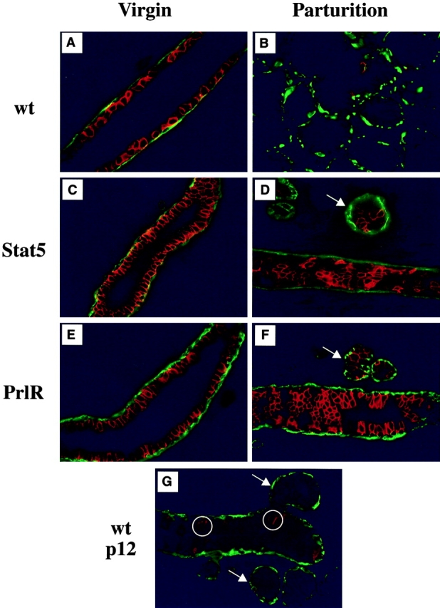
NKCC1 and smooth muscle actin are expressed in PrlR- and Stat5-null alveoli-like structures at parturition, but not in wild-type mice. Immunohistochemical staining of NKCC1 (red) and smooth muscle actin (green) in mammary epithelia of virgin mice (A, C, and E) and after parturition (B, D, and F). NKCC1 levels were high in ductal epithelium of wild-type virgin mice (A) and sharply reduced in alveoli by pregnancy day 12 (G, circle) and at parturition (B). PrlR- and Stat5-null epithelia maintained high levels of NKCC1 at parturition (D and F, compare white arrows with white arrow in G).
In an attempt to identify potential markers of secretory function, we searched the mouse EST database for genes preferentially expressed in the lactating mammary library, NMLMG. Using this approach, we discovered that a sodium phosphate cotransporter isoform, Npt2b, was highly expressed in the mouse mammary gland library derived from lactating tissue, but not virgin tissue (unpublished data). Next, we examined the expression of Npt2b during normal mammary gland development. RT-PCR analyses demonstrated that Npt2b mRNA was not present in virgin or early (day 13) pregnancy, but was evident during late (day 16) pregnancy through mid (day 5) lactation (unpublished data). The cellular localization of Npt2b protein was addressed by immunohistochemistry. We detected expression of Npt2b protein in mammary tissue from day 18 pregnant mice and at day one of lactation (Fig. 7), but not in virgin or early pregnant mice (unpublished data). Interestingly, Npt2b protein was expressed on the apical membrane of the mammary secretory cells (unpublished data). Taken together, these results suggest that Npt2b is a potential marker of secretory function. As mentioned above, PrlR- and Stat5-null epithelia fail to secrete milk proteins suggesting they were devoid of normal secretory function. Therefore, we examined the expression of Npt2b protein in these samples (Fig. 7). Whereas the mammary secretory cells present in wild-type epithelia showed expression of Npt2b in the apical membrane (Fig. 7 D), Npt2b was not detected in PrlR- and Stat5-null epithelia (Fig. 7, E and F). In contrast, E-cadherin (Fig. 7) was expressed in the basolateral membrane of all samples examined.
Epidermal growth factor and GH can activate Stat5 in PrlR-null epithelium
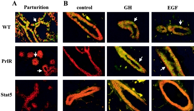
Based on histological and electron microscopy studies, Stat5- and PrlR-null epithelia exhibited differences. Whereas Stat5-null epithelium was highly disorganized and did not form open lumina, PrlR-null epithelium formed small open lumina. The observation that Stat5-null epithelium exhibited a more severe phenotype than PrlR-null epithelium, suggested that Stat5 might be activated to some extent by other cytokines in the absence of the PrlR. We have recently demonstrated by Western blot analysis that EGF and GH can activate Stat5 in mammary tissue (Gallego et al., 2001). However, their respective receptors within the epithelial compartment are not required for functional development (Gallego et al., 2001). We now investigated whether Stat5 could be activated in PrlR-null epithelium using immunohistochemistry (Fig. 8). Because Stat5a is more abundant than Stat5b, and Stat5b is not critical for alveolar development, we examined Stat5a activation. Stat5-null epithelia served as a negative control. At parturition, Stat5a was localized within nuclei of wild-type alveolar and ductal cells, which is indicative of its active state (Fig. 8 A). In contrast, only a few cells in PrlR-null epithelia exhibited nuclear, and thus activated Stat5a. However, upon injection with EGF or GH, extensive nuclear translocation of Stat5 was observed in PrlR-null epithelium (Fig. 8 B) indicative of Prl-independent activation of Stat5a.
Figure 8.
Epidermal growth factor and GH can activate Stat5a in PrlR-null epithelium. Immunohistochemical staining of Stat5a (green) and E-cadherin (red) in mammary epithelium at parturition (A) and in virgin tissue after hormone injection (B). Arrows show Stat5a nuclear staining. (A) In PrlR-null epithelium, some Stat5a nuclear staining was detected. In contrast, almost all cells in wild-type epithelia had Stat5a nuclear translocation. (B) Virgin mice carrying PrlR- and Stat5-null epithelia were injected with EGF or GH. Extensive nuclear localization of Stat5a was observed in PrlR-null epithelium but not in Stat5-null epithelium.
Discussion
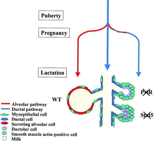
Here we demonstrate that Stat5 controls the establishment of functionally differentiated and secreting alveoli during pregnancy (Fig. 9). This pathway is activated to a large extent, but not exclusively, through the PrlR. We propose that activation of the Stat5 pathway by prolactin, GH and epidermal growth factor transduces signals that instruct cells at the branch points to proliferate and adopt alveolar characteristics. Further, we suggest that Stat5 determines cell fate through the establishment of cell–cell adhesion.
Figure 9.
PrlR and Stat5 control the cell fate of mammary alveolar epithelium. Wild-type mammary epithelium differentiates into functional alveoli during pregnancy. However, PrlR- and Stat5-null epithelia do not undergo alveolar development. The null epithelia maintain ductal features and cell proliferation at the branching points results in the development of “ductoli” (ductal feature but alveoli-like structure). In contrast to Stat5-null ductoli, PrlR-null ductoli contain open lumina. In the absence of Stat5 ductolar cells have impaired cell–cell contacts. The ductoli do not differentiate into functional alveoli. Blue line, ductal development; red line, alveolar development. The myoepithelial cells surrounding the alveoli are flat. The outer cell layer of ductoli is positive for smooth muscle actin and can thus be considered to be of myoepithelial nature.
The PrlR–Stat5 pathway is obligatory for mammary alveolar cell fate
The transcription factor Stat5 is central to several signaling pathways, and is activated by various cytokines through their respective receptors and Jak2 (Ihle, 2001). Because Stat5-null mice are infertile (Teglund et al., 1998), their mammary development had not been studied during pregnancy. We avoided the problem of infertility through the transplantation of knockout mammary epithelia into wild-type hosts. Although a ductal tree developed in the absence of Stat5, no alveolar development was apparent after one pregnancy. Whereas Stat5-null mammary epithelium exhibited a similar phenotype, it was distinct from that observed in the absence of the PrlR (Fig. 9). Ductal branching occurred in the absence of Stat5, but the alveoli-like structures did not form lumina as were observed in the absence of the PrlR. Our results suggest that Stat5 plays a role in regulating cell organization through cell–cell contacts independent of PrlR activation. Based on molecular markers that characterize ductal epithelia and secretory alveolar epithelia, we suggest that the PrlR- and Stat5-null alveoli-like structures have retained ductal-like characteristics. Thus, we propose that an early function of the prolactin pathway in mammary epithelia is the specification and determination of alveolar epithelia. Two separate and distinct lineage-limited mammary epithelial progenitors have been identified in the mouse mammary gland (Kordon and Smith, 1998; Smith, 1996). The failure to develop secretory alveoli may be either due to a failure to generate the alveoli-limited progenitor or the inability of the alveoli-limited progenitor and its progeny to respond to prolactin signals. It is apparent that proliferation of ductal epithelia can occur in the absence of a functional PrlR–Stat5 pathway. However, inactivation of the PrlR or Stat5 results in the formation of small alveoli-like structures with ductal features that we propose to name “ductoli.” Even after several pregnancies, we did not detect functional alveoli in PrlR- and Stat5-null transplants (unpublished data). This suggests that if compensatory pathways are activated, they are unable to elicit functional alveolar development.
In addition to the mammary gland phenotype observed in the transplant studies described herein, the Stat5-null mice exhibit phenotypes in other tissues and cell types consistent with the inactivation of several cytokine signaling pathways (Teglund et al., 1998). Loss of Stat5 disrupts IL-2 signaling and results in impaired T cell proliferation and a failure to express genes controlling cell cycle progression (Moriggl et al., 1999). In addition, Stat5 has also been linked to cell survival (Humphreys and Hennighausen, 1999; Socolovsky et al., 1999; Schwaller et al., 2000; Ihle, 2001). Lastly, Stat5 has been linked to B cell differentiation induced by IL-4 and IL-7 (Sexl et al., 2000) and Stat5a is required for functional differentiation, but not proliferation, of mammary epithelial cells (Liu et al., 1997).
Cell–cell adhesion defects in PrlR- and Stat5-null mammary epithelia
The absence of Stat5 resulted in defective cell–cell adhesion as assessed by electron microscopy. Thus, in control samples the basolateral membranes of neighboring cells were in close contact with each other. Conversely, there was evidence of gaps between adjacent cells in the Stat5-null samples. This suggests that Stat5 mediates signals that promote cell–cell adhesion. The expression of E-cadherin and ZO-1, proteins known to be involved in cell–cell adhesion and associated with tight junctions, respectively (Gumbiner, 1996), revealed normal staining patterns in both the PrlR- and Stat5-null samples. However, it is possible that whereas adjacent cells express detectable E-cadherin, they may not physically contact each other. In addition to E-cadherin, the process of cell adhesion involves additional proteins and it cannot be ruled out that any one of these are responsible for the cell adhesion defects that we observed.
The establishment of cell–cell adhesion is not only important for epithelial organization, but also for effective communication between individual cells. Such intercellular communication allows neighboring cells within a defined structural unit to respond in unison to a given signal. For example, the gap junction subunit Cx 43 is expressed in the myoepithelial cell compartment, and it has been suggested that this may help coordinate myoepithelial contraction and milk ejection upon oxytocin stimulation (Plum et al., 2000). The lack of appropriate gap junction protein expression, and by extension intercellular communication, could disrupt the cellular unit (i.e., alveoli) whose formation may be required for their full functional development. It has previously been shown that the mouse mammary gland expresses three connexin isoforms, Cx 26, Cx 32, and Cx 43 (Pozzi et al., 1995). Whereas Cx 43 is expressed in myoepithelial cells, both Cx 26 and Cx 32 are expressed in the epithelial compartment. Interestingly, Cx 32 expression is induced at lactation and cannot readily be detected at other time points, suggesting that it may contribute to the attainment of a secretory phenotype (Locke et al., 2000). Furthermore, Cx 32 has been shown to interact with proteins in the tight junction complex and determine cell polarization in hepatocytes (Kojima et al., 2001). Although we were able to detect Cx 32 expression in control samples at parturition, neither PrlR- nor Stat5-null epithelia expressed detectable levels of Cx 32. It is possible that the lack of Cx 32 expression in PrlR- and Stat5-null epithelia is the result of a lack of secretory differentiation. Alternatively, it is possible that Cx 32 is a Stat5 target gene. In fact, the mouse Cx 32 gene promoter contains an interferon γ–activated sequence site at position –800, suggesting that this promoter may be under direct prolactin control.
We have provided experimental evidence on the level of histology and electron microscopy that cell-cell adhesion and organization is impaired in the absence of Stat5, and to a lesser extent in the absence of the PrlR. Although we have shown apparent normal localization of E-cadherin and ZO-1, this cannot be a true measure for their functional integrity, which needs to be addressed in future studies. There is now widespread interest in the regulation of mammary epithelial cells by cell–cell adhesion molecules. It is likely that many proteins, including the transcription factor Stat5, control these processes.
Pathway redundancy
There were notable differences when comparing the individual phenotypes on the histological level. In particular, epithelial development in the absence of the PrlR was more inhibited than in the absence of Stat5. This could in part be explained by a reduction in the proliferative capacity of PrlR-null epithelium relative to Stat5-null epithelium. Such differences may be due to the activation of compensatory signaling pathways. For example, mitogen-activated protein kinase and PI3K can be activated upon Prl stimulation. It is also possible that other Stats (i.e., Stat1 and/or Stat3) may be recruited to Jak2 in the absence of Stat5, thus resulting in additional stimulation of epithelial development.
There was evidence of open lumina in the PrlR-, but not in the Stat5-null, transplants. Furthermore, the ductoli present in the PrlR-null epithelia were more organized compared with Stat5-null epithelia, suggesting that Stat5 is necessary for appropriate organization of individual cells into cohesive cellular structures. Whereas Prl is probably the key cytokine responsible for the activation of Stat5, we demonstrated that Stat5 has some residual activity in the absence of the PrlR. Because both GH and EGF activate Stat5 in the absence of the PrlR, it is likely that these two cytokines contribute to the formation of cell–cell contacts and thus a lumen.
Pathways controlling alveolar epithelial development
We have established that the PrlR and Stat5 are each essential for the attainment of functional alveologenesis. Several other genes and signaling pathways that control alveolar development have been identified, including ErbB2 (Jones and Stern, 1999) and ErbB4 (Jones et al., 1999), cyclin D1 (Fantl et al., 1995; Sicinski et al., 1995), C/EBPβ (Robinson et al., 1998; Seagroves et al., 1998), the osteoclast differentiation factor RANKL and its receptor RANK (Fata et al., 2000), and the helix-loop-helix protein Id2 (Mori et al., 2000). Data from these mouse models suggests that alveologenesis is a complex process requiring the functional cooperation of numerous molecules. Interestingly, comparable phenotypes were observed in some of these mice, i.e., lack of alveolar development. Developmental roles for ErbB2 and 4 have been suggested based on transgenic mice that express dominant negative forms under control of a mouse mamary tumor virus long terminal repeat. Expression of a dominant negative ErbB2 resulted in condensed alveoli and reduced luminal secretion at parturition (Jones and Stern, 1999). ErbB4-dominant negative epithelium formed condensed alveoli and failed to expand at mid lactation, which correlated with reduce expression of α-lactalbumin and WAP and a loss of Stat5 activity (Jones et al., 1999).
Similar to the models described here, C/EBPβ-null mice possess undifferentiated alveolar epithelium; in contrast, branching morphogenesis was also impaired. C/EBPβ mRNA levels in PrlR- and Stat5-null transplanted epithelia at parturition were similar to those seen in wild-type tissue (unpublished). These results suggest that C/EBPβ expression is essentially independent of the PrlR–Stat5 pathway, although they may converge at the β-casein promoter (Wyszomierski and Rosen, 2001). In the absence of RANKL, a growth factor produced by mammary epithelia in the second half of pregnancy, mammary epithelia also fail to develop (Fata et al., 2000). Because Prl can activate RANKL expression (Fata et al., 2000), it may be downstream of Stat5. However, both RANKL and RANK are expressed at high levels in PrlR- and Stat5-null transplanted epithelia at parturition (unpublished data), suggesting that these pathways are parallel and not dependent on each other. Id2-deficient mice also show severely impaired mammary gland development (Mori et al., 2000). Furthermore, Id2-deficient mammary epithelia exhibit reduced phosphorylation of Stat5. Normal levels of Id2 mRNA were detected in PrlR- and Stat5-null epithelia at parturition (unpublished data), suggesting that Id-2 is not downstream of Stat5. The presence of several, and apparently parallel, pathways controlling mammary alveolar development further emphasizes that distinct signals contribute to alveologenesis. At this point it is not clear whether these pathways have unique molecular targets leading to the formation of functional alveoli. The understanding of signaling pathways that are required for the formation of mammary epithelia but are dispensable for life of the organism itself provides a unique opportunity to develop molecular interventions and prevention for breast cancer.
Materials and methods
Animals
The Stat5a/b–null mice (Teglund et al., 1998) were bred into the C57BL/6 background. PrlR-null mice (Ormandy et al., 1997b) were in a C57BL/6 background, and Stat5a-null mice (Liu et al., 1997) were in a C57BL/6 and 129 mixed background. For the transplantation studies of PrlR- and Stat5-null mammary epithelia, athymic NCr-nu/nu mice were used as hosts. Wild-type littermates were used as controls. More than 30 PrlR- and Stat5-null transplants each were analyzed. For hormone injection studies, the transplanted virgin mice (12 wk after transplantation) were used. The mice were injected by intraperitoneally with either murine GH (5 μg/g of body weight), or human EGF (10 μg/g of body weight). Mammary glands were harvested 15 min later, fixed in 4% paraformaldehyde for 4 h, and processed for paraffin embedding and sectioning by standard procedures.
Antibodies
Polyclonal anti–rabbit Stat5a antibodies have been described previously (Liu et al., 1996). Mouse monoclonal E-cadherin and smooth muscle actin antibodies were obtained from Transduction Laboratories, polyclonal anti–rabbit ZO-1 antibodies were purchased from Zymed Laboratories, and polyclonal anti–rabbit NKCC1 antibodies (Moore-Hoon and Turner, 1998) were a gift from Dr. Jim Turner (National Institute of Dental and Craniofacial Research, National Institutes of Health, Bethesda, MD). The polyclonal anti–rabbit Npt2b antibodies (Hilfiker et al., 1998) were a gift from Dr. Jurg Biber (Department of Physiology, University of Zurich, Zurich, Switzerland).
Transplantation of adult mammary epithelia into the cleared fat pad of nude mice
The transplantation was performed as previously described (DeOme et al., 1959). In brief, small pieces of mammary tissue were excised from mature virgin female wild-type, Stat5-, or PrlR-null mice. Athymic nude mice (3-wk-old) were anesthetized with an intraperitoneally injection of avertin and the proximal part of the inguinal gland containing the mammary epithelium was excised. Pieces of mammary tissue from a PrlR- and a Stat5-null mouse were grafted into contralateral cleared fat pads of recipients. The other combinations of transplants were Stat5:wild type and PrlR:wild type. To assess the completeness of clearing, the excised endogenous glands were processed for whole mount staining according to standard protocols. 8 wk after transplantation, fat pads containing transplants were harvested from virgin hosts. Alternatively, the hosts were bred and tissue was harvested on the day of parturition. For whole mounts, mammary glands were removed, fixed in Carnoy's fixative overnight and stained in carmine alum.
Immunofluorescence
After fixation in Tellyesniczky's fixative for 4 h at room temperature, tissues were embedded in paraffin and sectioned at 5 μm. Sections were cleared in xylene and rehydrated. Antigen retrieval was performed by heat treatment using an antigen unmasking solution (Vector Laboratories) and tissue sections were blocked for 30 min in PBST containing 10% goat or horse serum. For ZO-1 detection, antigen retrieval was performed by protease treatment (Auto/Zyme Reagent set; Biomeda Corp.) at 37°C for 10 min. Sections were incubated with E-cadherin (1:1,000) and ZO-1 (1:500) antibodies, E-cadherin (1:1,000) and Npt2b (1:100) antibodies, smooth muscle actin (1:1,000) and NKCC1 (1:1,000) antibodies or E-cadherin (1:1,000), and Stat5a (1:250) antibodies. The primary antibodies were allowed to bind for 60 min at 37°C except for Stat5a:E-cadherin and ZO-1:E-cadherin (4°C, overnight). Nonspecifically bound antibody was removed by rinsing in PBST before the addition of both anti–mouse FITC-conjugated (1:250) and anti–rabbit Texas red–conjugated (1:250) secondary antibodies. Sections were incubated in the dark for 30 min, washed in two changes of PBST, and mounted in Vectashield (Vector Laboratories, Inc.). Fluorescence was visualized with a Zeiss Axioscop microscope equipped with FITC, TRITC, and FITC:TRITC filters. Images were captured using a Sony DKC5000 digital camera.
Analysis of cellular proliferation
After 9 wk, the transplant recipients were treated for 48 h with 1 μg β-estradiol (E) (Sigma-Aldrich) and 1 mg progesterone (P) (Sigma-Aldrich) in 100 μl sesame oil via a single interscapular subcutaneous injection behind the neck. After acute hormone treatment, both of the transplanted number 4 inguinal mammary glands and an endogenous number 3 gland (control) were removed. 2 h before sacrifice, mice were injected with 0.3 mg BrdU per 10 g body weight (Amersham Pharmacia Biotech). Tissue was fixed in 4% paraformaldehyde in PBS for 2 h at 4°C. Immunofluorescence and BrdU-positive cell counting were performed as previously described (Seagroves et al., 2000). In brief, paraffin sections (5–7 μm) were dewaxed and subjected to microwave antigen retrieval in 10 mM citrate buffer, pH 6.0. After blocking in 5% BSA/0.5% Tween 20 for 4 h at room temperature, sections were incubated with anti–BrdU-FITC–conjugated antibody (1:5; Becton Dickinson) in blocking solution overnight at room temperature. After PBS washes, slides were mounted in Vectashield + DAPI medium (Vector Laboratories). At least four glands per genotype were used for each experiment (endogenous control #3, n = 5; PrlR-null epithelia transplanted gland, n = 5; Stat5-null epithelia transplanted gland, n = 4). Cells from 16 fields at 60× magnification were counted from each sample. The number of BrdU-positive cells in a given field was expressed as a percentage of the total number of DAPI-stained cells. Statistical significance was determined by Mann-Whitney paired t test.
Gene expression analysis
Total RNA was isolated from fresh or frozen tissues and Northern blots were performed as described previously (Robinson et al., 1995). In brief, 10 μg of total RNA was loaded in each lane. Membranes were hybridized with random-primed [α-32P] dCTP-labeled probes in QuickHyb solution for 3 h at 65°C. Washes were performed in 0.1 × SSC/0.1% SDS at 65°C. At first, the membrane was hybridized with WAP and β-casein probes together and exposed to x-ray films. After stripping, membranes were rehybridized with WDNM1 and keratin 18 probes together. A 415-bp WAP-specific probe was generated by RT-PCR with primers 5′-GTA-CCA-TGC-GTT-GCC-TCA-TC-3′ and 5′-GCT-GCT-CAC-TGA-AGG-GTT-ATC-3′. A 577-bp β-casein–-specific probe was generated by RT-PCR with primers 5′-CTA-AAG-TTC-ACT-CCA-GCA-TCC-3′ and 5′-CAT-TTC-CAG-TTT-CAG-TCA-GTT-C-3′. A full-length cDNA for WDNM1 was used (Robinson et al., 1995). The keratin 18–specific probe was a 1.1-kb EcoRI cDNA fragment (Singer et al., 1986).
Electron microscopy
Small pieces of mammary tissue were cut, minced into 1-mm cubes, and fixed in a 0.025% solution of glutaraldehyde and 3.8% paraformaldehyde in PBS, pH 7.2, for 2 h. The tissue was rinsed in PBS and postfixed in 2% osmium tetroxide in 0.5 M sodium cacodylate buffer for 2 h, and dehydrated in a graded series of acetone solutions (Blanchette-Mackie and Scow, 1971). Temperature was maintained at 4°C from excision through dehydration and tissues were embedded in epon at room temperature (Luft, 1961). Sections were cut on a Reichert Om U2 ultramicrotome. Thick sections were stained with Toluidine blue in 1% sodium borate (pH 8.3) (Trump et al., 1961). Thin sections were stained with Karnovsky's lead hydroxide (Karnovsky, 1961) and uranyl acetate (Zobel and Beer, 1961) and examined with a JEOL 1010 electron microscope.
RT-PCR assays
Total RNA (1 μg) was transcribed into cDNA using Thermoscript reverse transcriptase (Life Technologies, Inc.) according to the manufacturer's protocol. Total RNA (1 μg) was first incubated with dNTPs and an oligo-dT(12–18) primer at 65°C for 5 min. All components were added except the reverse transcriptase, and the reaction was incubated at 42°C for 2 min. Thermoscript RT (50 units) was added to each reaction and incubated for a further 50 min. For controls, the samples without RT reactions were amplified. Single-stranded RNA was degraded by treating the reaction with Escherichia coli RNase H for 20 min at 37°C. PCR assays were performed for Cx 32 and GAPDH cDNA. Cx 32 gene-specific primers were 5′-GTT-GCA-ACC-AGG-TGT-GGC-AGT-G-3′ and 5′-CGG-AGG-CTG-CGA-GCA-TAA-AGA-C-3′. GAPDH gene-specific primers were 5′-CAA-CGG-GAA-GGG-CCC-CCA-TAC-CAT-C-3′ and 5′-ACG-ACG-GAC-ACA-TTG-GGG-GTA-G-3′. The template was first denatured at 94°C for 2 min followed by 35 cycles (Cx 32) or 25 cycles (GAPDH) of denaturation (94°C, 40 s), annealing (65°C, 40 s), and extention (72°C, 1 min). A final extention at 72°C for 10 min was performed.
Acknowledgments
We are grateful to Drs. Paul Kelly (INSERM, Paris, France) and James Ihle (St. Jude's Children's Research Hospital, Memphis, TN) for providing the PrlR- and Stat5a/b–null mice, respectively. We thank Drs. Jim Turner and Jurg Biber for anti-NKCC1 and anti-Npt2b antibodies, respectively. We thank the National Institute of Diabetes and Digestive and Kidney Diseases's National Hormone and Pituitary Program, and its Scientific Director, Dr. A.F. Parlow (National Institutes of Health, Bethesda, MD) for providing us with murine GH. We thank Dr. Jorcano (CIEMAT, Madrid, Spain) for providing us with human epidermal growth factor. We acknowledge Dr. Elaine Neale (National Institute of Dental and Craniofacial Research, National Institutes of Health, Bethesda, MD) who generously allowed us to use her electron microscope facility. K. Miyoshi is grateful to Professor Hideo Inoue for his continued encouragement.
J. Shillingford is funded by Department of Defense fellowship (17-00-1-0246). S.L. Grimm is supported by Department of Defense fellowship 17-00-1-0138.
Footnotes
*
Abbreviations used in this paper: Epo, erythropoietin; GH, growth hormone; IL, interleukin; PI3K, phosphoinositide 3-kinase; PrlR, prolactin receptor; RT, reverse transcription; Stat, signal transducer and activator of transcription; WAP, whey acidic protein; ZO, zonula occludens.
References
- Barcellos-Hoff, M.H., J. Aggeler, T.G. Ram, and M.J. Bissell. 1989. Functional differentiation and alveolar morphogenesis of primary mammary cultures on reconstituted basement membrane. Development. 105:223–235. [DOI] [PMC free article] [PubMed] [Google Scholar]
- Blanchette-Mackie, E.J., and R.O. Scow. 1971. Sites of lipoprotein lipase activity in adipose tissue perfused with chylomicrons. Electron microscope cytochemical study. J. Cell Biol. 51:1–25. [DOI] [PMC free article] [PubMed] [Google Scholar]
- Bole-Feysot, C., V. Goffin, M. Edery, N. Binart, and P.A. Kelly. 1998. Prolactin (PRL) and its receptor: actions, signal transduction pathways and phenotypes observed in PRL receptor knockout mice. Endocr. Rev. 19:225–268. [DOI] [PubMed] [Google Scholar]
- Borrmann, C.M., C. Mertens, A. Schmidt, L. Langbein, C. Kuhn, and W.W. Franke. 2000. Molecular diversity of plaques of epithelial-adhering junctions. Ann. NY. Acad. Sci. 915:144–150. [DOI] [PubMed] [Google Scholar]
- Cereijido, M., J. Valdes, L. Shoshani, and R.G. Contreras. 1998. Role of tight junctions in establishing and maintaining cell polarity. Annu. Rev. Physiol. 60:161–177. [DOI] [PubMed] [Google Scholar]
- Daniel, C.W., P. Strickland, and Y. Friedmann. 1995. Expression and functional role of E- and P-cadherins in mouse mammary ductal morphogenesis and growth. Dev. Biol. 169:511–519. [DOI] [PubMed] [Google Scholar]
- Delmas, V., P. Pla, H. Feracci, J.P. Thiery, R. Kemler, and L. Larue. 1999. Expression of the cytoplasmic domain of E-cadherin induces precocious mammary epithelial alveolar formation and affects cell polarity and cell-matrix integrity. Dev. Biol. 216:491–506. [DOI] [PubMed] [Google Scholar]
- DeOme, K.B., L.J. Faulkin, Jr., H.A. Bern, and P.E. Blair. 1959. Development of mammary tumors from hyperplastic alveolar nodules transplanted into gland-free mammary fat pads of female C3H mice. Cancer Res. 19:515–520. [PubMed] [Google Scholar]
- Fantl, V., G. Stamp, A. Andrews, I. Rosewell, and C. Dickson. 1995. Mice lacking cyclin D1 are small and show defects in eye and mammary gland development. Genes Dev. 9:2364–2372. [DOI] [PubMed] [Google Scholar]
- Fata, J.E., Y.Y. Kong, J. Li, T. Sasaki, J. Irie-Sasaki, R.A. Moorehead, R. Elliott, S. Scully, E.B. Voura, D.L. Lacey, et al. 2000. The osteoclast differentiation factor osteoprotegerin-ligand is essential for mammary gland development. Cell. 103:41–50. [DOI] [PubMed] [Google Scholar]
- Gallego, M.I., N. Binart, G.W. Robinson, R. Okagaki, K.T. Coschigano, J. Perry, J.J. Kopchick, T. Oka, P.A. Kelly, and L. Hennighausen. 2001. Prolactin, growth hormone, and epidermal growth factor activate Stat5 in different compartments of mammary tissue and exert different and overlapping developmental effects. Dev. Biol. 229:163–175. [DOI] [PubMed] [Google Scholar]
- Gumbiner, B.M. 1996. Cell adhesion: the molecular basis of tissue architecture and morphogenesis. Cell. 84:345–357. [DOI] [PubMed] [Google Scholar]
- Hennighausen, L., and G.W. Robinson. 1998. Think globally, act locally: the making of a mouse mammary gland. Genes Dev. 12:449–455. [DOI] [PubMed] [Google Scholar]
- Hennighausen, L., and G.W. Robinson. 2001. Signaling pathways in mammary gland development. Developmental Cell. 1:467–475. [DOI] [PubMed] [Google Scholar]
- Hilfiker, H., O. Hattenhauer, M. Traebert, I. Forster, H. Murer, and J. Biber. 1998. Characterization of a murine type II sodium-phosphate cotransporter expressed in mammalian small intestine. Proc. Natl. Acad. Sci. USA. 95:14564–14569. [DOI] [PMC free article] [PubMed] [Google Scholar]
- Humphreys, R.C., and L. Hennighausen. 1999. Signal transducer and activator of transcription 5a influences mammary epithelial cell survival and tumorigenesis. Cell Growth Differ. 10:685–694. [PubMed] [Google Scholar]
- Ihle, J.N. 2001. The Stat family in cytokine signaling. Curr. Opin. Cell Biol. 13:211–217. [DOI] [PubMed] [Google Scholar]
- Jones, F.E., and D.F. Stern. 1999. Expression of dominant-negative ErbB2 in the mammary gland of transgenic mice reveals a role in lobuloalveolar development and lactation. Oncogene. 18:3481–3490. [DOI] [PubMed] [Google Scholar]
- Jones, F.E., T. Welte, X.Y. Fu, and D.F. Stern. 1999. ErbB4 signaling in the mammary gland is required for lobuloalveolar development and Stat5 activation during lactation. J. Cell Biol. 147:77–88. [DOI] [PMC free article] [PubMed] [Google Scholar]
- Karnovsky, M.J. 1961. Simple methods for “staining with lead” at high pH in electron microscopy. J. Biophys. Biochem. Cytol. 11:729–732. [DOI] [PMC free article] [PubMed] [Google Scholar]
- Kazansky, A.V., E.B. Kabotyanski, S.L. Wyszomierski, M.A. Mancini, and J.M. Rosen. 1999. Differential effects of prolactin and src/abl kinases on the nuclear translocation of STAT5B and STAT5A. J. Biol. Chem. 274:22484–22492. [DOI] [PubMed] [Google Scholar]
- Kim, D.W., and B.H. Cochran. 2001. Jak2 activates tfii-i and regulates its interaction with extracellular signal-regulated kinase. Mol. Cell Biol. 21:3387–3397. [DOI] [PMC free article] [PubMed] [Google Scholar]
- Knust, E. 2000. Control of epithelial cell shape and polarity. Curr. Opin. Genet. Dev. 10:471–475. [DOI] [PubMed] [Google Scholar]
- Kojima, T., Y. Kokai, H. Chiba, M. Yamamoto, Y. Mochizuki, and N. Sawada. 2001. Cx32 but not Cx26 is associated with tight junctions in primary cultures of rat hepatocytes. Exp. Cell Res. 263:193–201. [DOI] [PubMed] [Google Scholar]
- Kordon, E.C., and G.H. Smith. 1998. An entire functional mammary gland may comprise the progeny from a single cell. Development. 125:1921–1930. [DOI] [PubMed] [Google Scholar]
- Liu, X., G.W. Robinson, and L. Hennighausen. 1996. Activation of Stat5a and Stat5b by tyrosine phosphorylation is tightly linked to mammary gland differentiation. Mol. Endocrinol. 10:1496–1506. [DOI] [PubMed] [Google Scholar]
- Liu, X., G.W. Robinson, K.U. Wagner, L. Garrett, A. Wynshaw-Boris, and L. Hennighausen. 1997. Stat5a is mandatory for adult mammary gland development and lactogenesis. Genes Dev. 11:179–186. [DOI] [PubMed] [Google Scholar]
- Liu, X., M.I. Gallego, G.H. Smith, G.W. Robinson, and L. Hennighausen. 1998. Functional release of Stat5a-null mammary tissue through the activation of compensating signals including Stat5b. Cell Growth Differ. 9:795–803. [PubMed] [Google Scholar]
- Locke, D., N. Perusinghe, T. Newman, H. Jayatilake, W.H. Evans, and P. Monaghan. 2000. Developmental expression and assembly of connexins into homomeric and heteromeric gap junction hemichannels in the mouse mammary gland. J. Cell. Physiol. 183:228–237. [DOI] [PubMed] [Google Scholar]
- Luft, J.H. 1961. Improvements in epoxy resin embedding methods. J. Biophys. Biochem. Cytol. 9:409–414. [DOI] [PMC free article] [PubMed] [Google Scholar]
- Moore-Hoon, M.L., and R.J. Turner. 1998. Molecular and topological characterization of the rat parotid Na+-K+-2Cl- cotransporter1. Biochim. Biophys. Acta. 1373:261–269. [DOI] [PubMed] [Google Scholar]
- Mori, S., S.I. Nishikawa, and Y. Yokota. 2000. Lactation defect in mice lacking the helix-loop-helix inhibitor Id2. EMBO J. 19:5772–5781. [DOI] [PMC free article] [PubMed] [Google Scholar]
- Moriggl, R., V. Sexl, R. Piekorz, D. Topham, and J.N. Ihle. 1999. Stat5 activation is uniquely associated with cytokine signaling in peripheral T cells. Immunity. 11:225–230. [DOI] [PubMed] [Google Scholar]
- Ormandy, C.J., N. Binart, and P.A. Kelly. 1997. a. Mammary gland development in prolactin receptor knockout mice. J. Mammary Gland Biol. Neoplasia. 2:355–364. [DOI] [PubMed] [Google Scholar]
- Ormandy, C.J., A. Camus, J. Barra, D. Damotte, B. Lucas, H. Buteau, M. Edery, N. Brousse, C. Babinet, N. Binart, and P.A. Kelly. 1997. b. Null mutation of the prolactin receptor gene produces multiple reproductive defects in the mouse. Genes Dev. 11:167–178. [DOI] [PubMed] [Google Scholar]
- Plum, A., G. Hallas, T. Magin, F. Dombrowski, A. Hagendorff, B. Schumacher, C. Wolpert, J. Kim, W.H. Lamers, M. Evert, et al. 2000. Unique and shared functions of different connexins in mice. Curr. Biol. 10:1083–1091. [DOI] [PubMed] [Google Scholar]
- Pozzi, A., B. Risek, D.T. Kiang, N.B. Gilula, and N.M. Kumar. 1995. Analysis of multiple gap junction gene products in the rodent and human mammary gland. Exp. Cell Res. 220:212–219. [DOI] [PubMed] [Google Scholar]
- Robinson, G.W., P.F. Johnson, L. Hennighausen, and E. Sterneck. 1998. The C/EBPbeta transcription factor regulates epithelial cell proliferation and differentiation in the mammary gland. Genes Dev. 12:1907–1916. [DOI] [PMC free article] [PubMed] [Google Scholar]
- Robinson, G.W., R.A. McKnight, G.H. Smith, and L. Hennighausen. 1995. Mammary epithelial cells undergo secretory differentiation in cycling virgins but require pregnancy for the establishment of terminal differentiation. Development. 121:2079–2090. [DOI] [PubMed] [Google Scholar]
- Schwaller, J., E. Parganas, D. Wang, D. Cain, J.C. Aster, I.R. Williams, C.K. Lee, R. Gerthner, T. Kitamura, J. Frantsve, et al. 2000. Stat5 is essential for the myelo- and lymphoproliferative disease induced by TEL/JAK2. Mol. Cell. 6:693–704. [DOI] [PubMed] [Google Scholar]
- Seagroves, T.N., S. Krnacik, B. Raught, J. Gay, B. Burgess-Beusse, G.J. Darlington, and J.M. Rosen. 1998. C/EBPbeta, but not C/EBPalpha, is essential for ductal morphogenesis, lobuloalveolar proliferation, and functional differentiation in the mouse mammary gland. Genes Dev. 12:1917–1928. [DOI] [PMC free article] [PubMed] [Google Scholar]
- Seagroves, T.N., J.P. Lydon, R.C. Hovey, B.K. Vonderhaar, and J.M. Rosen. 2000. C/EBPbeta (CCAAT/enhancer binding protein) controls cell fate determination during mammary gland development. Mol. Endocrinol. 14:359–368. [DOI] [PubMed] [Google Scholar]
- Sexl, V., R. Piekorz, R. Moriggl, J. Rohrer, M.P. Brown, K.D. Bunting, K. Rothammer, M.F. Roussel, and J.N. Ihle. 2000. Stat5a/b contribute to interleukin 7-induced B-cell precursor expansion, but abl- and bcr/abl-induced transformation are independent of stat5. Blood. 96:2277–2283. [PubMed] [Google Scholar]
- Sicinski, P., J.L. Donaher, S.B. Parker, T. Li, A. Fazeli, H. Gardner, S.Z. Haslam, R.T. Bronson, S.J. Elledge, and R.A. Weinberg. 1995. Cyclin D1 provides a link between development and oncogenesis in the retina and breast. Cell. 82:621–630. [DOI] [PubMed] [Google Scholar]
- Singer, P.A., K. Trevor, and R.G. Oshima. 1986. Molecular cloning and characterization of the Endo B cytokeratin expressed in preimplantation mouse embryos. J. Biol. Chem. 261:538–547. [PubMed] [Google Scholar]
- Smith, G.H. 1996. Experimental mammary epithelial morphogenesis in an in vivo model: evidence for distinct cellular progenitors of the ductal and lobular phenotype. Breast Cancer Res. Treat. 39:21–31. [DOI] [PubMed] [Google Scholar]
- Socolovsky, M., A.E. Fallon, S. Wang, C. Brugnara, and H.F. Lodish. 1999. Fetal anemia and apoptosis of red cell progenitors in Stat5a-/-5b-/- mice: a direct role for Stat5 in Bcl-X(L) induction. Cell. 98:181–191. [DOI] [PubMed] [Google Scholar]
- Teglund, S., C. McKay, E. Schuetz, J.M. van Deursen, D. Stravopodis, D. Wang, M. Brown, S. Bodner, G. Grosveld, and J.N. Ihle. 1998. Stat5a and Stat5b proteins have essential and nonessential, or redundant, roles in cytokine responses. Cell. 93:841–850. [DOI] [PubMed] [Google Scholar]
- Tepass, U., K. Truong, D. Godt, M. Ikura, and M. Peifer. 2000. Cadherins in embryonic and neural morphogenesis. Natl. Rev. Mol. Cell Biol. 1:91–100. [DOI] [PubMed] [Google Scholar]
- Trump, B.F., E.A. Smuckler, and E.P. Benditt. 1961. A method for staining epoxy sections for light microscopy. J. Ultrastruct. Res. 5:343–348. [DOI] [PubMed] [Google Scholar]
- Vasioukhin, V., and E. Fuchs. 2001. Actin dynamics and cell-cell adhesion in epithelia. Curr. Opin. Cell Biol. 13:76–84. [DOI] [PubMed] [Google Scholar]
- Vicente-Manzanares, M., and F. Sanchez-Madrid. 2000. Cell polarization: a comparative cell biology and immunological view. Dev. Immunol. 7:51–65. [DOI] [PMC free article] [PubMed] [Google Scholar]
- Wyszomierski, S.L., and J.M. Rosen. 2001. Cooperative effects of STAT5 (signal transducer and activator of transcription 5) and C/EBPbeta (CCAAT/enhancer-binding protein-beta) on beta-casein gene transcription are mediated by the glucocorticoid receptor. Mol. Endocrinol. 15:228–240. [DOI] [PubMed] [Google Scholar]
- Zobel, R.C., and M. Beer. 1961. Electron stains. Chemical studies on the interactions of DNA with uranyl salts. J. Biophys. Biochem. Cytol. 10:335–346. [DOI] [PMC free article] [PubMed] [Google Scholar]

