Disruption of Nrf2 enhances susceptibility to severe airway inflammation and asthma in mice (original) (raw)
Abstract
Oxidative stress has been postulated to play an important role in the pathogenesis of asthma; although a defect in antioxidant responses has been speculated to exacerbate asthma severity, this has been difficult to demonstrate with certainty. Nuclear erythroid 2 p45-related factor 2 (Nrf2) is a redox-sensitive basic leucine zipper transcription factor that is involved in the transcriptional regulation of many antioxidant genes. We show that disruption of the Nrf2 gene leads to severe allergen-driven airway inflammation and hyperresponsiveness in mice. Enhanced asthmatic response as a result of ovalbumin sensitization and challenge in _Nrf2_-disrupted mice was associated with more pronounced mucus cell hyperplasia and infiltration of eosinophils into the lungs than seen in wild-type littermates. Nrf2 disruption resulted in an increased expression of the T helper type 2 cytokines interleukin (IL)-4 and IL-13 in bronchoalveolar lavage fluid and in splenocytes after allergen challenge. The enhanced severity of the asthmatic response from disruption of the Nrf2 pathway was a result of a lowered antioxidant status of the lungs caused by lower basal expression, as well as marked attenuation, of the transcriptional induction of multiple antioxidant genes. Our studies suggest that the responsiveness of _Nrf2_-directed antioxidant pathways may act as a major determinant of susceptibility to allergen-mediated asthma.
Asthma is a complex inflammatory disorder characterized by airway inflammation, intermittent reversible airway obstruction, airway hyperreactivity (AHR), excessive mucus production, and elevated levels of immunoglobulin E and Th2 cytokines (1–3). Studies have shown that increases in reactive oxygen species (ROS) that occur during asthma are associated with damage to a wide range of biologic molecules in the lung, and many observations suggest that oxidative stress plays an important role in the pathogenesis of asthma (4, 5).
Inflammatory cells in the airways and alveolar spaces can release ROS/reactive nitrogen species after phagocytosis of inhaled particles or after their functional activation by various stimuli. Granulocytic cells in bronchoalveolar lavage (BAL), particularly eosinophils, are the major source of ROS after antigen challenge in allergic subjects (6). ROS-mediated activation of NF-κB can induce the expression of proinflammatory factors such as IL-8, TNF-α, regulated on activation, normal T cells expressed and secreted (RANTES), and eotaxin that further promotes inflammatory cell infiltration into the lungs (7–9). The direct mechanism by which ROS exacerbates asthma might include effects on airway smooth muscle and mucin secretion. ROS decreases β-adrenergic function in lungs (10) and also sensitizes airway muscle to acetylcholine-induced contraction (11). The levels of nitric oxide (NO) are elevated in the exhaled air of patients with asthma and may contribute to airway edema and inflammation (12). The recent finding that NO reduces the effect of β-adrenergic signaling pathways may be a deleterious effect of the elevated reactive nitrogen species in asthma (13). A reaction between NO and O2 − resulting in the formation of peroxinitrite anions (ONOO−) induces airway hyperesponsiveness in guinea pigs, inhibits pulmonary surfactant, and leads to membrane lipid peroxidation and tyrosine- and mitogen-activated protein kinase activation, ultimately damaging pulmonary epithelial cells (13–16).
An excess production of oxidants is kept to a minimum by a well coordinated and efficient endogenous antioxidant defense system that is both enzymatic and nonenzymatic in nature. The enzymatic antioxidants include the families of superoxide dismutase (SOD), catalase, glutathione peroxidase, glutathione _S_-transferase (GST), and thioredoxin. The nonenzymatic category of antioxidant defenses includes glutathione, ascorbate, α-tocopherol, urate, bilirubin, and lipoic acid (4, 17, 18). Individuals with asthma demonstrate depressed levels of ascorbate and α-tocopherol in BAL, diminished activities of superoxide dismutase, and elevated oxidized GSH disulfide (GSSG)/GSH ratios, suggesting both increased ROS/reactive nitrogen species and decreased antioxidant capacity (19). Interestingly, a recent study has indicated that higher levels of serum antioxidants such as vitamin C and β-carotene were associated with a lower risk of asthma (20).
Although oxidative stress, which originates from infiltrating inflammatory cells, is suspected to be involved in the pathogenesis of the disease, there is no conclusive experimental evidence to support the idea that defective antioxidant responses in lungs may lead to worsened asthma or exacerbate airway inflammation and AHR. Critical host factors that protect the lungs against oxidative stress may either directly determine susceptibility to asthma or act as risk modifiers by inhibiting associated inflammation. Nuclear erythroid 2 p45 related factor 2 (Nrf2) encodes a cap “n” collar basic leucine zipper (b-ZIP) transcription factor, which detaches from its cytosolic inhibitor (keap1), translocates to the nucleus, and binds to the antioxidant response element (ARE) in the promoter of target genes upon activation in response to oxidative stress, leading to their transcriptional induction (21). The Nrf2-regulated genes in the lungs include almost all of the relevant antioxidants, such as heme oxygenase 1 (HO-1), γ-glutamyl cysteine synthase, and several members of the GST family (21).
To test the hypothesis that deficiency in _Nrf2_-mediated antioxidant defenses plays a central role in pathogenesis of asthma, we studied allergen-driven airway inflammation in wild-type and _Nrf2_-deficient mice. Our results show that disruption of the Nrf2 gene in mice leads to increased airway inflammation, AHR, goblet cell hyperplasia, and an elevated level of Th2 cytokines in the lungs in a model of allergen-mediated asthma using OVA challenge.
Results
BAL cell counts
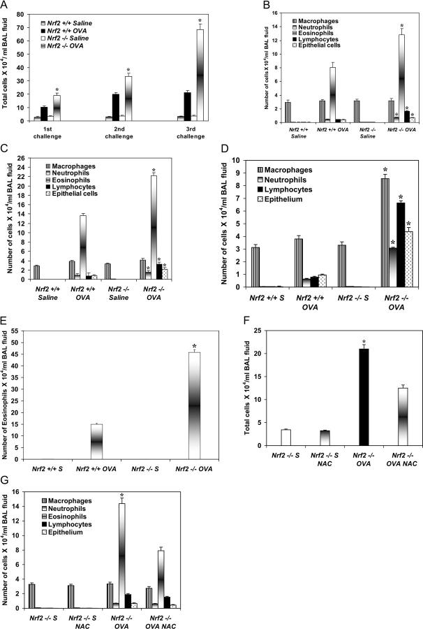
The total number of inflammatory cells in the BAL fluid of all OVA-challenged (first to third) _Nrf2_-deficient mice (Nrf2 − / − OVA mice) was significantly higher than OVA-challenged Nrf2 wild-type mice (Nrf2 + / + OVA mice) (P ≤ 0.05; Fig. 1 A). The number of inflammatory cells in the BAL fluid of Nrf2 − / − OVA mice (third challenge) was 2.9-fold higher (6.7 × 10⁶ cells/ml BAL fluid) than its level (2.3 × 10⁶ cells/ml BAL fluid) in Nrf2 + / + OVA mice. The increase in inflammation was progressive from the first to the third OVA challenge. A differential cell count analysis showed a significantly higher number of eosinophils, lymphocytes, and neutrophils, as well as epithelial cells, in the BAL fluid of Nrf2 − / − OVA mice (P ≤ 0.05; Fig. 1, B–E). 72 h after the third challenge, there were 2.3-, 3-, 4.5-, 4.8-, and 8.5-fold more macrophages, eosinophils, epithelial cells, neutrophils, and lymphocytes, respectively, in the BAL fluid of Nrf2 − / − OVA than Nrf2 + / + OVA mice (Fig. 1, D and E). Among the inflammatory cell populations, eosinophils were the predominant cell population, followed by macrophages, lymphocytes, and neutrophils, at each time point (Fig. 1, B–E).
Figure 1.
Increased allergen-driven asthmatic inflammation in OVA-challenged Nrf2 −/− mice. (A) Total and differential inflammatory cell populations in the BAL fluid of OVA- and saline-challenged Nrf2 + / + and Nrf2 − / − mice (n = 8). (B) First challenge with OVA. (C) Second challenge with OVA. (D and E) Third challenge with OVA. There was a progressive increase in the total number of inflammatory cells in the BAL fluid of both OVA-challenged Nrf2 + / + and Nrf2 − / − mice from the first to third challenges. However, the number of inflammatory cells in the BAL fluid of Nrf2−/ − OVA mice was significantly higher than in the BAL fluid of Nrf2 + / + OVA mice, as well as the respective saline-challenged mice. The number of eosinophils, lymphocytes, neutrophils, and epithelial cells were significantly (*) higher in the BAL fluid of Nrf2 − / − OVA mice compared with Nrf2 + / + OVA mice. Pretreatment with NAC significantly (*) reduced the inflammatory cells (F), predominantly eosinophils (G), in the BAL fluid of Nrf2 − / − OVA mice (n = 6). Data are mean ± SEM. *, P ≤ 0.05.
Lung histopathology
There was a marked extravasation of inflammatory cells into the lungs of Nrf2 − / − OVA mice (third challenge) relative to the mild cellular infiltration in the lungs of Nrf2 + / + OVA mice, as determined by staining of the lung sections with hematoxylin and eosin (H&E). Higher numbers of inflammatory cells were observed in the perivascular, peribronchial, and parenchymal tissues of the Nrf2 − / − OVA mice compared with a few inflammatory cell infiltrates in the Nrf2 + / + OVA mice (Fig. 2 A). Immunohistochemical staining with anti–major basophilic protein (MBP) antibody showed numerous eosinophils around the blood vessels and airways (Fig. 2 B) and in the parenchymal tissues (Fig. 2 C) of Nrf2 − / − OVA mice compared with Nrf2 + / + OVA mice. These histological data are consistent with the differential cell counts in the BAL fluid obtained from the OVA-challenged Nrf2 + / + and Nrf2 − / − mice.
Figure 2.
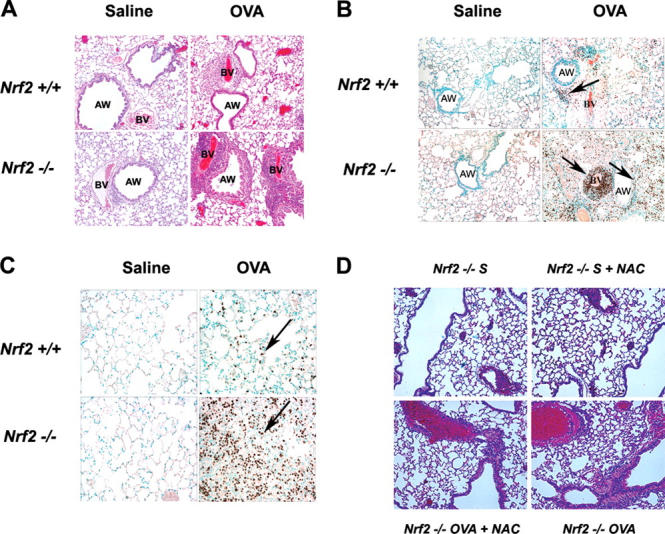
Increased infiltration of inflammatory cells into the lungs of OVA-challenged Nrf2 −/− mice. Lung tissues from the saline- and OVA-challenged (third challenge) Nrf2 + / + and Nrf2 − / − mice (n = 6) were stained with H&E and examined by light microscope (20×). (A) OVA challenge caused a marked infiltration of inflammatory cells into the lungs of Nrf2 − / − OVA compared with Nrf2 + / + OVA mice. Immunohistochemical staining showed the presence of numerous eosinophils around the blood vessels and airways (B) and in the parenchyma (C) of OVA-challenged (third challenge) Nrf2 − / − compared with Nrf2 + / + mice. The figure is representative of three experiments (n = 6). AW, airways; BV, blood vessels. H&E staining of the lung sections from the saline-or NAC-treated (7 d before first OVA challenge) _Nrf2_-deficient mice. The arrows indicate cells staining positively with anti-MBP antibody. (D) Widespread peribronchial and perivascular inflammatory infiltrates were observed in OVA-sensitized mice after antigen provocation (bottom right). Pretreatment of _Nrf2_-deficient mice with NAC resulted in substantial reduction in the infiltration of inflammatory cells in the peribronchial and perivascular region (bottom left).
Intervention with _N_-acetyl l-cysteine (NAC)
To determine whether reducing the oxidative burden would attenuate airway inflammation, we treated mice for 7 d with NAC before the first OVA challenge. Histological analysis showed widespread peribronchial and perivascular inflammatory infiltrates in the OVA-challenged (first challenge) Nrf2 − / − mice when compared with the saline-challenged control mice. NAC-pretreated mice showed a marked reduction in the infiltration of inflammatory cells in the peribronchiolar and perivascular region (Fig. 2 D). Concomitant with histological assessment, airway inflammation was evaluated in the BAL fluid. As expected, antigen-challenged Nrf2 − / − mice showed a marked increase in the total number of inflammatory cells (21 × 104 cells/ml BAL fluid vs. 3.2 × 10⁴ cells/ml BAL fluid in saline group) in the BAL fluid 24 h after OVA challenge (Fig. 1 F). Among the inflammatory cell population, eosinophils were the predominant cells in the BAL fluid (14.38 × 10⁴ cells/ml BAL fluid) and were significantly diminished (7.8 × 10⁴ cells/ml BAL fluid) by treatment with NAC (P ≤ 0.05; Fig. 1 G) in the OVA-challenged _Nrf2_-deficient mice. NAC treatment did not have any significant inhibitory effect on other cell types such as macrophages, neutrophils, lymphocytes, and epithelial cells 24 h after the first OVA challenge. The total and differential cell counts observed in saline-challenged mice treated with NAC did not differ from counts obtained in saline-challenged untreated mice.
Increased level of oxidative stress markers, eotaxin, and enhanced activation of NF-κB in the lungs of Nrf2 −/− OVA mice
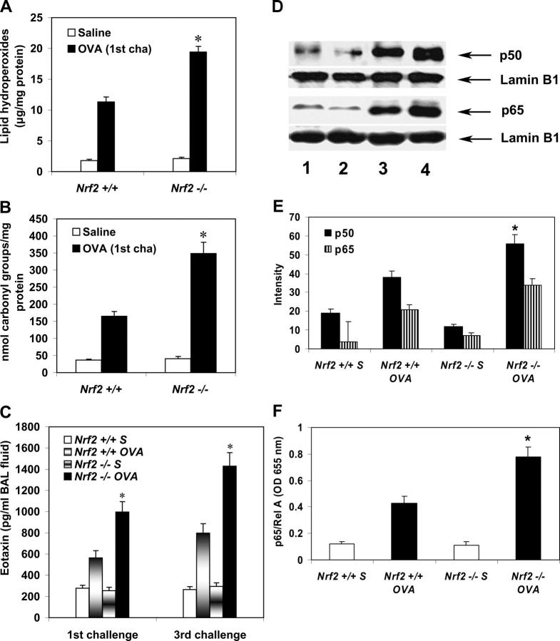
We measured the levels of lipid hydroperoxides and protein carbonyls in the lungs as markers of oxidative stress. When compared with OVA-challenged Nrf2 wild-type mice, there was a significantly increased amount of lipid hydroperoxides (11.3 μg/mg protein vs. 19.4 μg/mg protein; Fig. 3 A) and protein carbonyls (165 nM/mg protein vs. 349 nM/mg protein; Fig. 3 B) in the lungs of Nrf2 − / − OVA mice, suggesting the occurrence of excessive oxidative stress in response to allergen challenge (P ≤ 0.05).There was a significant increase in the GSH level and GSH/GSSG ratio in the lungs of Nrf2 + / + OVA mice (first and third challenge) when compared with the lungs of Nrf2 − / − OVA mice (P ≤ 0.05; Fig. S1, A and B, available at http://www.jem.org/cgi/content/full/jem.20050538/DC1).
Figure 3.
Increased oxidative stress markers, eotaxin, and enhanced activation of NF-κB in the lungs of Nrf2 −/− OVA mice. Increased levels of lipid hydroperoxides (A) and protein carbonyls (B) in the lungs of OVA-challenged Nrf2 − / − mice. Values are mean ± SEM. *, Significantly higher than the Nrf2 + / + OVA mice (n = 6). (C) Eotaxin level in the BAL fluid. When compared with OVA-challenged Nrf2 + / + mice, the BAL eotaxin level was markedly higher in OVA-challenged (both first and third challenge) Nrf2 − / − mice (P ≤ 0.05; n = 6). (D–F) Activation of NF-κB in the lungs. Western blot was used to determine the activation of p50 and p65 subunits of NF-κB in the lungs (D). (lanes 1 and 2) Saline-challenged Nrf2 + / + and Nrf2 –/– mice, respectively; (lanes 3 and 4) OVA-challenged Nrf2 + / + and Nrf2 –/– mice, respectively. (E) Quantification of p50 and p65 subunits of NF-κB obtained in Western blots. Values are mean ± SEM of three experiments. (F) ELISA measurement of p65/Rel A subunit of NF-κB using Mercury TransFactor kit. *, P ≤ 0.05 versus OVA-challenged Nrf2 wild-type mice. Data are mean ± SEM of three experiments.
The eosinophil chemottractant eotaxin level in the BAL fluid of first and third OVA-challenged _Nrf2_-deficient mice was significantly higher when compared with its wild-type counterpart (P ≤ 0.05; Fig. 3 C). A substantial increase in the level of eotaxin was observed in the BAL fluid of third OVA-challenged animals, which was concomitant with the increased infiltration of eosinophils in the lungs (Fig. 2, B and C).
NF-κB has been reported to be activated by oxidative stress and also regulate eotaxin production (22). Therefore, we determined the activation of NF-κB in the lungs of Nrf2 + / + and Nrf2 − / − mice by Western blot analysis with anti–NF-κB p65 and anti–NF-κB p50 antibodies. Immunoblot analysis showed significantly higher levels of both p65 and p50 NF-κB subunits in the lung nuclear extracts of Nrf2 − / − OVA mice than in the lung nuclear extracts of Nrf2 + / + OVA mice (P ≤ 0.05; Fig. 3, D and E). DNA binding activity assay with a Mercury TransFactor ELISA kit showed the increased binding of p65/Rel A subunit from the lung nuclear extracts of Nrf2 − / − OVA mice to an NF-κB binding sequence compared with its wild-type counterpart (Fig. 3 F).
Mucus cell hyperplasia
Periodic acid-Schiff (PAS) staining of lung sections showed a marked increase in the mucus-producing granular goblet cells in the proximal airways of Nrf2 − / − OVA mice relative to fewer purple-staining goblet cells in the Nrf2 + / + OVA mice after the third challenge (Fig. 4 A). There were no or few PAS-positive cells in the proximal airways of saline-challenged mice or in the distal airways (not depicted) of both Nrf2 + / + OVA and Nrf2 − / − OVA mice. The percentage of airway epithelial cells staining for mucus glycoproteins by PAS was significantly higher in the proximal airways of Nrf2 − / − OVA mice than in those of the Nrf2 + / + OVA and the respective saline-challenged mice (P ≤ 0.05; Fig. 4 B).
Figure 4.
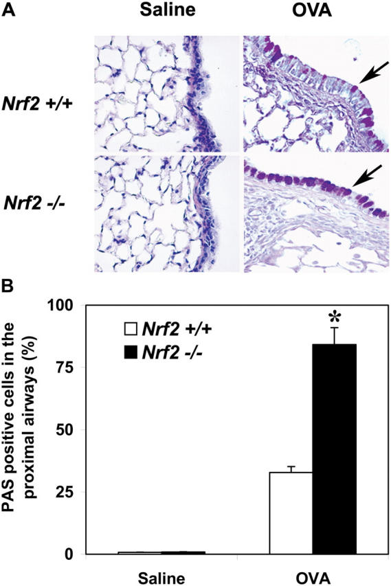
Nrf2_-deficient mice show increased mucus cell hyperplasia in response to allergen challenge. (A) Lung sections (72 h after the final OVA challenge) stained with PAS. Shown are the purple staining epithelial cells (arrows) in the proximal airways of OVA-challenged mice and pronounced mucus cell hyperplasia in Nrf2 − / − OVA mice (40×). (B) Percentage of airway epithelial cells positive for mucus glycoproteins by PAS staining. Lung sections from the Nrf2 − / − OVA mice showed significantly higher numbers of PAS-positive cells than the lung sections from the Nrf2 + / + OVA mice (*,_ P ≤ 0.05). Data are mean ± SEM.
Airway responsiveness to acetylcholine
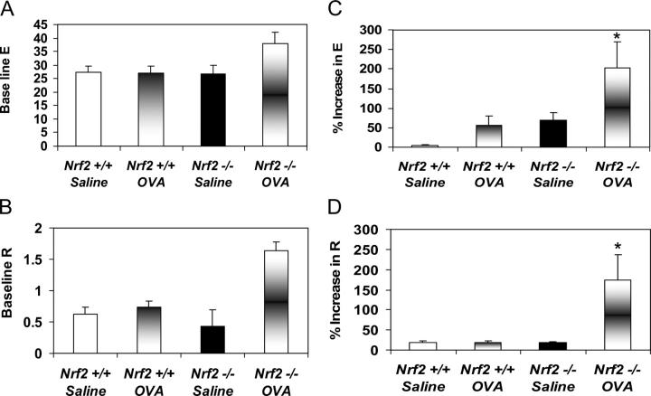
After systemic sensitization and challenges to OVA, airway responsiveness to acetylcholine aerosol was measured. In the absence of an acetylcholine challenge, no substantial differences in baseline elastance (Fig. 5 A) and resistance (Fig. 5 B) were observed in both saline- and OVA-challenged Nrf2 − / − and wild-type mice. However, 96 h after the third OVA challenge, the Nrf2 − / − mice showed a significant increase in baseline elastance (Fig. 5 C) and resistance (Fig. 5 D) to acetylcholine compared with the wild-type counterpart (P ≤ 0.05).
Figure 5.
_Nrf2_-deficient mice show increased airway responsiveness to acetylcholine challenge. OVA-challenged Nrf2 + / + and Nrf2 − / − mice (third challenge) were challenged with acetylcholine aerosol by nebulization with an Aeroneb Pro-nebulizer (n = 7). Lung resistance and compliance were measured. The percent increase in elastance (E; panel C) and resistance (R; panel D) to acetylecholine challenge were significantly higher (*, P ≤ 0.05) in the Nrf2 − / − OVA mice when compared with Nrf2 + / + OVA mice and the respective saline-challenged mice. However, no significant difference in baseline elastance (A) and resistance (B) were observed in both the saline- and OVA-challenged Nrf2 + / + and Nrf2 − / − mice in the absence of acetylcholine challenge. Data are mean ± SEM.
Cytokine levels in BAL fluid
Analysis of BAL fluid by ELISA showed a marked increase in the levels of IL-4 (76 vs. 42 pg/ml BAL fluid) and IL-13 (154 vs. 72 pg/ml BAL fluid) in the Nrf2 − / − OVA mice relative to the Nrf2 + / + OVA mice. The levels of these cytokines were very low in the BAL fluid of saline- treated control mice of both genotypes (Fig. 6, A and B).
Figure 6.
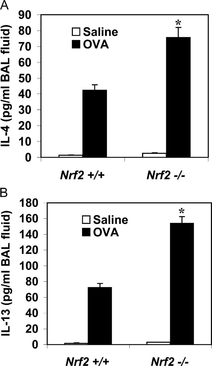
Th2 cytokine levels in the BAL fluid of Nrf2 +/+ and Nrf2 −/− mice challenged with OVA. BAL fluids collected 48 h after the second OVA challenge were used for cytokine assays using ELISA. The amounts of both IL-4 (A) and IL-13 (B) were significantly higher (*, P ≤ 0.05) in the BAL fluid of Nrf2 − / − OVA mice than in Nrf2 + / + OVA mice (n = 8). Data are mean ± SEM.
Inflammatory cytokine response of the splenocytes
To determine whether enhanced Th2 cell secretion in OVA-challenged mice was reflected at the level of systemic sensitization, we isolated splenocytes from mice 48 h after the second challenge and studied cytokine secretion in vitro after culture with OVA or antibodies directed against CD3 and CD28. Table S1 (available at http://www.jem.org/cgi/content/full/jem.20050538/DC1) shows the results from these experiments, indicating that the production of IL-4 and IL-13 was consistently higher using splenocytes from Nrf2 − / − versus wild-type mice when stimulated ex vivo. Recall production of IL-4 was generally low in these mice, in keeping with prior experience with this strain. Enhanced Th2 cytokine production in these experiments could reflect a direct repressive effect of Nrf2 on Th2 cytokine gene expression, or an indirect effect via regulation of the oxidant/antioxidant balance. To distinguish between these possibilities, we first isolated spleen CD4+ cells from unchallenged wild-type and Nrf2 − / − mice and studied cytokine production ex vivo. No significant differences (P ≥ 0.05) in IL-4 or IL-13 secretion were observed in these experiments (Table S2, available at http://www.jem.org/cgi/content/full/jem.20050538/DC1). We determined whether Nrf2 could directly regulate IL-4 or IL-13 gene expression or promoter activity in transient transfection assays. Although overexpression of Nrf2 substantially increased the expression of its known target genes glutathione cysteine ligase catalytic subunit (GCLc) and NADPH:quinone oxidoreductase (NQO1), there was no effect on IL-13 gene expression (Fig. S2, available at http://www.jem.org/cgi/content/full/jem.20050538/DC1). In parallel experiments, we found that overexpressed Nrf2 did not affect transcription driven by the IL-4 or IL-3 promoters (Fig. S3). Thus, we concluded from these data that Nrf2 deficiency indirectly enhanced Th2 cytokine production via regulation of the oxidant/antioxidant balance.
Activation of Nrf2 in the lungs of Nrf2 +/+ mice
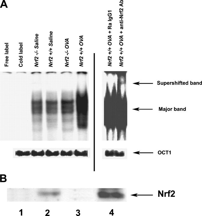
Electrophoretic mobility shift assay (EMSA) was used to determine the activation and DNA binding activity of Nrf2 in the lungs in response to allergen challenge (Fig. 7 A). EMSA analysis showed the increased binding of nuclear proteins isolated from the lungs of OVA-challenged Nrf2 + / + mice to ARE consensus sequence relative to the OVA-challenged Nrf2 − / − mice or saline-challenged control mice. Supershift analysis with anti-Nrf2 antibody also showed the binding of Nrf2 to the ARE consensus sequence, suggesting the finding that OVA challenge leads to the activation of Nrf2 in the lungs of Nrf2 + / + mice.
Figure 7.
Activation of Nrf2 in the lungs of OVA-challenged Nrf2 +/+ mice. (A) EMSA was used to determine the activation of Nrf2 in the lungs of Nrf2 + / + OVA mice. Equal amounts of nuclear extracts (10 μg) prepared from lungs were incubated with radiolabeled ARE from the NQO1 promoter and analyzed by EMSA. EMSA analysis showed the increased binding of nuclear proteins isolated from the lungs of OVA-challenged Nrf2 + / + mice to ARE sequence. The arrows indicate the supershifted band, the major band, and OCT1, respectively. (B) Level of nuclear Nrf2. Nrf2 level in nuclear extracts were determined by immunoblot analysis with anti-Nrf2 antibody. (lanes 1 and 2) Saline-challenged Nrf2 − / − and Nrf2 + / + mice, respectively; (lanes 3 and 4) OVA-challenged Nrf2 − / − and Nrf2 + / + mice, respectively. The figure is representative of three experiments.
Immunoblot analysis showed (Fig. 7 B) an increased level of Nrf2 in the lung nuclear extracts of Nrf2 + / + OVA mice compared with the saline-challenged counterpart, suggesting the accumulation of Nrf2 in the lungs of wild-type mice in response to allergen challenge. Increase of nuclear Nrf2 is needed for the activation of ARE and the transcriptional induction of various antioxidant genes.
Induction of antioxidant genes
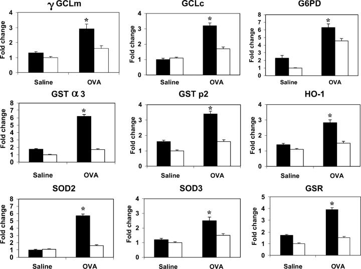
There was a substantial and coordinated elevation in transcript levels of several antioxidant genes in the lungs of Nrf2 + / + OVA mice when compared with the OVA-challenged _Nrf2-_disrupted mice. The fold changes in mRNA of various antioxidant genes as determined by real-time RT-PCR were γ GCL modifier subunit (γ GCLm; 2.9 vs. 1.6), GCLc (3.2 vs 1.7), glucose 6 phosphate dehydrogenase (G6PD; 6.3 vs. 4.6), GST α3 (6.2 vs. 1.7), GST p2 (3.4 vs. 1.6), HO-1 (2.8 vs. 1.5), SOD2 (5.7 vs 1.6), SOD3 (2.5 vs. 1.5), and glutathione S-reductase (GSR; 3.9 vs. 1.5) in the lungs of Nrf2 + / + OVA (24 h after first challenge) and Nrf2 − / − OVA mice, respectively (Fig. 8). The magnitude of the induction of panel of these antioxidant genes was considerably higher in Nrf2 wild-type than in _Nrf2_-disrupted mice, clearly showing an association with the activation of Nrf2 in response to allergen-induced lung inflammation.
Figure 8.
Real-time RT-PCR analysis of selected antioxidant genes in the lungs of OVA-challenged Nrf2 +/+ and Nrf2 −/− mice. Real-time RT-PCR analysis showed increased levels of mRNA for genes such as γ GCLm, GCLc, G6PD, GST α3, GST p2, HO-1, SOD2, SOD3, and GSR in the lungs of Nrf2 + / + OVA compared with the lungs of Nrf2 − / − OVA mice and the respective saline-challenged mice. Closed bar, Nrf2 + / + mice; open bar, Nrf2 − / − mice.
Real-time PCR analysis showed the expression of Nrf2 in CD4+ T cells and macrophages isolated from the lungs of Nrf2 + / + OVA mice (Fig. S3 A, available at http://www.jem.org/cgi/content/full/jem.20050538/DC1). Quantitative real-time RT-PCR revealed the increased expression of _Nrf2_-regulated antioxidant genes, HO-1 (CD4+ T cells, 2.5-fold; macrophages, 11.2-fold), GCLc (CD4+ T cells, 2.5-fold; macrophages, 4.6-fold), and g GCLm (CD4+ T cells, 2.5-fold; macrophages, 7.8-fold) in the CD4+ T cells (Fig. S3 B) and macrophages (Fig. S3 C) isolated from the lungs of Nrf2 + / + OVA mice when compared with the knock out counterpart.
Discussion
Our findings have indicated that Nrf2 is a critical determinant of susceptibility to allergen-induced asthma. The role of oxidative stress as a result of inflammation in the pathogenesis and disease progression of asthma has gained increased attention in recent years. However, it has been unclear whether the antioxidant status of the lung plays an important role in susceptibility to asthma. The human studies to date have by necessity been correlative and unable to address the issue of causality (i.e., did oxidative stress contribute to or result from allergic airway inflammation?). Furthermore, studies in animal models using the exogenous administration of antioxidants or antioxidant mimetics to decrease allergen-induced airway inflammation (23–25) suffer from concerns about nonspecific effects of the compounds used. Here we used mice deficient in a key regulatory transcription factor, Nrf2, to specifically dissect the role of antioxidant responses in allergic airway inflammation. Our results demonstrate that Nrf2-mediated signaling pathways serve to limit airway eosinophilia, mucus hypersecretion, and AHR in response to allergen challenge in a relevant model of asthma. We documented the occurrence of excess lung oxidative stress in the setting of Nrf2 deficiency, which was considerably attenuated by the antioxidant NAC. Collectively, these data provide compelling evidence for the pathogenic role of oxidative stress in regulating the severity of allergic airway inflammation and implicate a novel transcription factor pathway in asthma pathogenesis.
Under normal conditions, Nrf2 is anchored in the cytoplasm by binding to an inhibitory protein called Keap1. In response to oxidative stress, Nrf2 dissociates from Keap1 and then binds to AREs in the upstream sequence of target genes in association with several other proteins such as small maf proteins, c-jun, ARE-binding protein 1, and CBP/p300 and p160 family coactivators (26). In response to allergen challenge, there was an increased binding of nuclear proteins isolated from the lungs of Nrf2 + / + mice to probes containing the ARE consensus sequence. This binding is presumably caused by the interaction of nuclear proteins with the ARE, particularly of Nrf2 in the lungs of allergen-exposed Nrf2 + / + mice, as confirmed by the supershift analysis with anti-Nrf2 antibody. The slight increase in the binding of nuclear proteins from Nrf2 − / − mice lung extracts to the ARE is probably caused by the interaction of proteins other than Nrf2. Western blot analysis confirmed the nuclear accumulation of Nrf2 in the lungs of OVA-challenged Nrf2 + / + mice.
In response to allergen challenge, there was an increased induction of an array of antioxidant genes in the lungs of Nrf2 + / + mice, including the genes (γ GCLm and GCLc) involved in GSH synthesis. GSH is the major intracellular thiol antioxidant that acts directly as an ROS scavenger. The GSH redox system (GSH and GSSG) plays a critical role in determining intracellular redox balance and antioxidant function (27). There was a 2.6-fold up-regulation of GSR in the OVA-challenged wild-type mice in contrast to 1.3-fold in the _Nrf2-_disrupted mice challenged with the allergen. GSR uses NADPH for the regeneration of reduced glutathione. G6PD, the enzyme involved in the regeneration of NADPH, was also considerably induced in the wild-type mice in response to allergen. Thus, our data indicate that Nrf2 regulates several antioxidant genes that act in concert to counteract oxidative stress and inflammation.
Our recent study (28) revealed the importance of >50 _Nrf2_-dependent antioxidant and cytoprotective genes in the protection of lungs against cigarette smoke–induced emphysema in Nrf2 wild-type mice. In the present study, to determine whether the increased airway inflammation in the OVA-challenged Nrf2 − / − mice was caused by the deficient antioxidant levels in the lungs, we treated the animals with NAC 7 d before the first OVA challenge. Pretreatment of Nrf2 knockout mice with the antioxidant NAC greatly reduced eosinophil levels in the BAL fluid and inhibited the infiltration of inflammatory cells in the peribronchial and perivascular region of the lungs, providing direct evidence that the deficient antioxidant levels may increase the susceptibility to airway inflammation in response to allergen challenge. Given that a single antioxidant was able to greatly reduce eosinophilia in the OVA-challenged Nrf2 − / − mice, we believe that the attenuation of the development of airway inflammation in Nrf2 wild-type mice is mediated by the coordinated polygenic response of _Nrf2_-regulated antioxidant genes.
Genes for several other _Nrf2_-regulated antioxidant enzymes were also up-regulated to a greater extent in the mice with functional Nrf2, including GST α3, GST p2, SOD2, SOD3, and HO-1. Interestingly, polymorphisms in the pi class GST locus are associated with asthma and homozygosity for the GSTP1*Val allele has been shown to confer protection against toluene diisocyanate-induced asthma and airway hyperresponsiveness (29). HO-1 has been shown to be a major antioxidant enzyme in the lungs that protects against oxidative stress, and adenoviral-mediated overexpression of HO-1 has been shown to confer protection against hyperoxic lung injury in mice (30). The increased expression of this antioxidant gene has been shown to inhibit TNF-α (31). SOD3 is the major extracellular enzyme that counteracts ROS in lungs. SOD3 is mainly associated with the connective tissue matrix around vessels and airways in the lung, but is also found in close proximity to airway and vascular smooth muscle cells (32, 33). Intraperitoneal injection of the SOD mimetic reduced airway hyperresponsivenes in guinea pigs (34).
Disruption of the Nrf2 gene caused a greater increase in the total number of inflammatory cells, predominantly eosinophils, in BAL fluid relative to Nrf2 wild-type mice from the first to third OVA challenges. OVA-challenged _Nrf2_-deficient mice showed a 2.9-fold increase in the level of eosinophils in the BAL fluid when compared with OVA-challenged Nrf2 wild-type mice. Furthermore, the increase in asthmatic inflammation was reflected by severe extravasation of eosinophils in the perivascular, peribronchial, and parenchymal tissues of the lungs of Nrf2 − / − OVA mice. Disruption of the Nrf2 gene also caused higher numbers of neutrophils in the BAL in response to OVA challenge. A role for neutrophils in the development of airway remodeling in severe asthma has been suggested (35). In many cases of fatal and nocturnal asthma, considerable increases in lung neutrophils have been observed (4). Eosinophils and neutrophils generate reactive oxygen species that can potentially cause destruction to host lung tissue and contribute to inflammatory injury. The oxidative injury caused by eosinophils can be substantial because of their enhanced capacity to generate O2 − and H2O2 to levels higher than those produced by neutrophils (36). Thus, Nrf2 deficiency not only results in enhanced lung eosinophilia and neutrophilia, but also leads to the reduced expression of antioxidant genes that would counteract oxidative injury induced by these cells.
Oxidative stress can regulate cell recruitment to the lung at multiple levels. First, ROS can activate NF-κB, which is known to induce the expression of proinflammatory factors such as IL-8, RANTES, and the eosinophil attracting chemokine eotaxin-1 (7, 9, 22). Eotaxin-1 is a potent eosinophil-attracting CC chemokine and plays a critical role in inducing the accumulation of eosinophils in vivo (37). We provide evidence for both enhanced NF-κB activation and eotaxin secretion in the setting of Nrf2 deficiency, supporting a model in which excess oxidative stress enhances cell recruitment via NF-κB–dependent chemokine secretion. Second, oxidative stress has recently been shown to be indirectly involved in the accumulation of eosinophils by stimulating the synthesis of the eosinophil chemoattractant 5-oxo-6,8,11,14-eicosatetraenoic acid (38, 39). In future studies, it will be interesting to determine the role of this and other lipid mediators in eosinophil recruitment during allergic inflammation. We observed a considerably increased number of epithelial cells in the BAL fluid of third OVA-challenged _Nrf2_-deficient mice than the Nrf2 wild-type mice challenged with OVA. Damage to the epithelium by oxidants released from inflammatory cells is a critical factor in the pathogenesis of bronchial asthma (40), and the shedding of epithelial cells is an important histological feature observed in bronchial biopsy specimens from asthmatic patients (41). Excessive apoptosis of epithelial cells may be a likely mechanism responsible for damage to, and sloughing of, airway epithelial cells (40). Apoptosis is closely linked with oxidative stress and many agents that induce apoptosis are either oxidants or stimulators of cellular oxidative metabolism (42). The increased epithelial cells observed in the BAL fluid of _Nrf2_-deficient mice may be caused by the shedding of bronchial epithelial cells partly caused by excessive oxidative stress occurring in the lungs after OVA challenge. The mechanism by which Nrf2 protects against apoptosis in the asthma model is a subject of a future investigation.
We found that the expression of the Th2 cytokines IL-4 and IL-13 was considerably higher in the BAL fluid of OVA-challenged Nrf2 − / − mice compared with wild-type controls. This helps explain the excess production of mucus glycoproteins in Nrf2 − / − mice given the strong link between Th2 cytokines (especially IL-13) and goblet cell metaplasia (43). Enhanced Th2 cytokine production appears to be caused by allergen-driven Th2 cell sensitization in the context of oxidative stress. We did not find evidence that Nrf2 acts in a T cell–intrinsic manner to repress IL-4 or IL-13 cytokine gene expression or promoter activity. Enhanced Th2 sensitization has been reported in other models of oxidative stress, including ozone inhalation (44, 45). The precise molecular mechanism of this requires further study, but may reflect the effects of oxidative stress on cells that influence Th cell differentiation or on Th2 cell survival.
The OVA-challenged Nrf2 − / − mice showed increased lung elastance and airway resistance under baseline conditions compared with either knockout controls or OVA-challenged Nrf2 + / + mice. Interestingly, the percent increase in elastance and resistance was notably higher in Nrf2 − / − OVA compared with Nrf2 + / + OVA mice. Increased baseline resistance and elastance suggest that Nrf2 deficiency could have resulted in a chronic structural effect in the lung and it can also affect the subsequent responsiveness to inhaled bronchoconstrictor. The increased baseline elastance suggests the possibility that there may be increased fluid in the lungs that obstructs ventilation. This inflammatory edema may also explain the narrowed airways at baseline. Recent studies revealed the contribution of several factors, including the lipid mediator leukotriene and the Th2 cytokines IL-4 and IL-13, to airway hyperreactivity. IL-4 and IL-13 are required for AHR induction through the induction of allergen-specific IgG1 and IgE production, resulting in the activation of mast cells via antigen/antibody complex–Fc receptor signaling (46, 47).
Altogether, these results provide a clear link between inflammation and the severity of asthma with the defect in signaling mediated by the transcription factor Nrf2. Nrf2 is activated in response to allergen challenge in the lungs of wild-type mice, leading to the transcriptional induction of many antioxidant genes that might provide resistance against the development of asthma. Maintenance of a proper balance between ROS production and antioxidant capability regulated by Nrf2 is essential for the integrity and function of the different cellular components of the lungs that determines the outcome of allergen-induced asthma. Conversely, a lack of responsiveness of the Nrf2 pathway confers susceptibility to severe asthma because of allergen exposure in this model. The identification of Nrf2 as a determinant of asthma severity has wide implications for lung diseases, where oxidative stress and inflammation play an important role. Realizing the importance of Nrf2 in the maintenance of antioxidant gene regulation in lungs and its link with asthma from this work, future studies on asthma susceptibility and variation in Nrf2 response should prove worthwhile.
Materials and Methods
Animals and care
_Nrf2_-deficient CD1:ICR mice (SLC Japan Inc.) were generated as previously described (48). _Nrf2_-deficient mice were generated by replacing the b-ZIP region of the _Nrf_2 gene with the SV40 nuclear localization signal and β-galactosidase gene (48). Mice were genotyped for Nrf2 status by PCR amplification of genomic DNA extracted from the blood (49). PCR amplification was performed using three different primers: 5′-TGGACGGGACTATTGAAGGCTG-3′ (sense for both genotypes), 5′-CGCCTTTTCAGTAGATGGAGG-3′ (antisense for WT Nrf2 mice), and 5′-GCGGATTGACCGTAATGGGATAGG-3′ (antisense for LacZ). Mice were fed an AIN-76A diet and water ad libitum and housed under controlled conditions (23 ± 2°C; 12-h light/dark periods). All experimental protocols conducted on the mice were performed in accordance with the standards established by the US Animal Welfare Acts, as set forth in National Institutes of Health (NIH) guidelines and the Policy and Procedures Manual of the Johns Hopkins University Animal Care and Use Committee.
Sensitization and challenge protocols
8-wk-old male mice were sensitized on day 0 by i.p. injection (100 μl/mouse) with 20 μg OVA complexed with aluminum potassium sulfate. On day 14, mice were sensitized a second time with 100 μg OVA. On days 24, 26, and 28, the mice were anesthetized by i.p. injection with 0.1 ml of a mixture of 10 mg/ml ketamine and 1 mg/ml xylazine diluted in sterile PBS and challenged with 200 μg OVA in 100 μl of sterile PBS by intratracheal instillation. The control groups received sterile PBS with aluminum potassium sulfate by the i.p. route on days 0 and 14 and 0.1 ml of sterile PBS on days 24, 26, and 28. Mice were killed at different time points after OVA challenge for BAL, RNA isolation, histopathology, and for AHR measurements.
BAL fluid and phenotyping
Mice (n = 8) were anesthetized with 0.3 ml of 65 mg/ml pentobarbital and the tracheas were cannulated. BAL fluid was collected with 1 ml followed by 2× 1 ml of sterile PBS containing 5 mM EDTA, 5 mM DTT, and 5 mM PMSF. The BAL fluid was immediately centrifuged at 1,500 g. The total cell count was measured, and cytospin preparation (Shandon Scientific Inc.) was performed. Cells were stained with Diff-Quick reagent (Baxter Dade), and a differential count of 300 cells was performed using standard morphological criteria (50).
Histochemistry
The lungs were inflated with 0.6 ml of 10% buffered formalin, fixed for 24 h at 4°C, before histochemical processing. The whole lung was embedded in paraffin, sectioned at a 5-μm thickness, and stained with H&E (n = 6) for routine histopathology. Tissue sections were also stained with PAS for the identification of stored mucosubstances within the mucus goblet cells lining the main axial airways (proximal), as previously described (51). The number of PAS-positive cells were counted on longitudinal lung sections of the proximal airways. The percentage of PAS-positive cells was determined by counting the mucus-positive cells and unstained epithelial cells in the proximal airways under the microscope with a grid at 100×. Six animals were used for each treatment. The sum of the values of five fields per slide for five slides is provided for each animal. The data are expressed as means ± SEM.
Immunohistochemical staining of eosinophils in the lungs
For detection of eosinophils in tissues, the lung sections from the saline- and OVA-challenged (72 h after third challenged) mice (n = 6) were deparafinized and dehydrated in benzene and alcohol, respectively, and the endogenous peroxidase activity was quenched with 0.6% H2O2 in 80% methanol for 20 min. Sections were then digested with pepsin for 10 min before blocking with 5% normal rabbit serum for 30 min at room temperature. Rat anti–mouse MBP-1 antibody (provided by J. Lee, Mayo Clinic, Scottsdale, AZ) was then applied for 60 min, followed by a period of incubation with rabbit anti–rat IgG–HRP conjugate for 60 min. HRP was visualized with diaminobenzidine. Nuclei were then stained by application of purified 2% methyl green for 2 min.
Intervention with NAC
Nrf2 + / + and Nrf2 − / − mice (n = 6) were sensitized with OVA according to the procedure described in the Sensitization and challenge protocols section. Sensitized animals were randomly distributed into positive control (saline plus OVA), negative control (saline), and NAC-treated (NAC plus saline or antigen) groups. NAC was dissolved in distilled water (3 mmol/kg body weight, pH 7.0) and administered orally by gavage (23) as a single daily dose for 7 d before challenge with the last dose being given 2 h before OVA challenge. 24 h after challenge, BAL fluids and lung tissues were harvested and analyzed as described in the BAL fluid and phenotyping and Histochemistry sections. The experiment was repeated twice.
Determination of lipid hydroperoxides and protein carbonyls in the lungs
To quantify lipid hydroperoxides, lung tissues were homogenized in 10 mM PBS containing 10 μM cupric sulfate and incubated for 30 min at 37°C in a shaking water bath. 5 vol of methanol were added to the lung homogenate, vortexed vigorously for 2 min, and centrifuged at 8,000 g for 5 min. 0.9 ml of Fox reagent was added to 0.1 ml methanol extract and incubated for 30 min at room temperature. The absorbance was read at 560 nM using a spectrophotometer. H2O2 was used as the standard. Data were expressed as microgram of lipid hydroperoxide per milligram of protein using the molar extinction coefficient of 43,000 for hydroperoxides (52).
To determine the protein carbonyls, the lungs were homogenized in 10 mM Hepes buffer (137 mM NaCl, 4.6 mM KCl, 1.1 mM KH2PO4, 0.6 mM MgSO4, 1.1 mM EDTA, 5 mg/liter Tween 20, 1 μM butylated hydroxytoluene, 0.5 μg/ml leupeptine, 0.7 μg/ml pepstatin, 0.5 μg/ml aprotinin, and 40 μg/ml PMSF) and centrifuged at 8,000 g for 10 min at 4°C. Supernatant fractions were divided into two equal aliquots containing from 0.7 to 1 mg of protein each, precipitated with 10% TCA, and centrifuged at 8,000 g for 5 min at room temperature. One pellet was treated with 2.5 M HCl, and the other was treated with an equal volume of 10 mM dinitrophenyl hydrazine in 2.5 M HCl at room temperature for 1 h. Samples were reprecipitated with 10% TCA and subsequently with 1:1 ethanol/ethyl acetate and again reprecipitated with 10% TCA. The pellets were dissolved in 20 mM phosphate buffer, pH 6.5 (containing 6 M guanidine hydrochloride), and left for 10 min at 37°C with general vortex mixing. Samples were centrifuged at 6,000 g for 5 min and the clear supernatants were collected. The difference in absorbance between DNPH-treated and the HCl control was determined at 370 nM. Data were expressed as nanomole of carbonyl groups/milligram of protein using the molar extinction coefficient of 21,000 for NADPH derivatives (53).
Measurement of airway responsiveness
On day 31 (96 h after the third OVA challenge), mice (n = 7) were anesthetized with sodium pentobarbital, and their tracheas were cannulated via tracheostomy. The animals were ventilated as previously described (54) with a tidal volume of 0.2 ml at 2 Hz. 0.5 mg/kg body weight of succinylcholine was given i.p. to eliminate all respiratory efforts. Aerosol acetyl choline challenges were administered by nebulization with a nebulizer (Aeroneb Pro; Aerogen, Inc.) modified to decrease the dead space to 1 ml. Data were plotted as lung resistance and compliance at baseline and in response to a 10-s challenge of 0.3 mg/ml acetylcholine.
Assay of T lymphocyte activation
To determine whether Nrf2 played a T cell–intrinsic role in regulating Th2 cytokine gene expression, we isolated CD4+ T cells and splenocytes from the spleen of saline- and OVA-challenged Nrf2 + / + and Nrf2 − / − mice, respectively and stimulated them for 24 h in the absence or presence of anti-CD3 plus anti-CD28 antibodies or the calcium ionophore A23187 plus the phorbol ester PMA, followed by analysis of cytokine secretion by ELISA (Supplemental materials and methods, available at http://www.jem.org/cgi/content/full/jem.20050538/DC1).
Luciferase promoter assay and Nrf2 overexpression
Reporter constructs containing the human IL-4 and IL-13 promoter regions linked to the firefly luciferase gene were synthesized using standard techniques (pGL3 Basic; Promega). Promoter reporter constructs were cotransfected with an Nrf2 expression vector into Jurkat T cells followed by analysis of reporter gene expression using luminometry or endogenous gene expression by real-time RT-PCR and ELISA (Supplemental materials and methods).
ELISA measurements of IL-4, IL-13, and eotaxin
To measure cytokine levels, BAL fluid was collected from the lungs of each mouse (n = 8) with 0.7 ml PBS containing a cocktail of protease inhibitors and immediately centrifuged at 4°C for 5 min at 1,500 g. The supernatant was collected, aliquoted, and frozen in liquid nitrogen. The levels of IL-4 and IL-3 in BAL fluid, as well as in the supernatants from the splenocyte culture, were determined by ELISA using IL-4 and IL-13 quantikine ELISA kits. Eotaxin level in BAL fluid was analyzed using a mouse eotaxin ELISA kit.
EMSA
EMSA was performed according to the previously published procedure (55).
Quantification of GSH and GSSG in lung tissue
The concentrations of reduced and oxidized glutathiones in the lung tissues were measured using BIOXYTECH GSH/GSSG-412 kit (Supplemental materials and methods).
Western blot analysis
Western blot analysis was performed according to previously published procedures (28). In brief, 50 μg of the nuclear proteins isolated from the lungs of saline- and OVA-challenged (first) Nrf2 + / + and Nrf2 − / − mice were separated by 10% SDS-PAGE (SDS-PAGE) and transferred to PVDF membrane. The PVDF membrane was incubated with polyclonal rabbit anti-Nrf2 antibody followed by incubation with HRP-conjugated secondary antibody and developed using an ECL chemiluminescence detection kit.
To determine the activation of NF-κB, 15-μg nuclear extracts isolated from the lungs of saline- or OVA-challenged (first challenge) Nrf2 + / + and Nrf2 –/– mice were subjected to SDS-PAGE, as described above. NF-κB was detected by incubating the blots with anti–NF-κB p65 and anti–NF-κB p50 rabbit polyclonal antibodies. The blots were stripped and reprobed with anti–lamin B1 antibody. Western blot was performed with protein extracts from three different saline- or OVA-challenged Nrf2 + / + and Nrf2 –/– mice, and band intensities of p65 and p50 subunits of NF-κB of the three blots were determined using the NIH Image-Pro Plus software. Values are represented as means ± SEM.
P65/Rel A DNA binding activity
DNA binding activity of the p65/Rel A subunit of NF-κB was determined using Mercury TransFactor Kit (BD Biosciences). An equal amount of nuclear extracts isolated from the lungs were added to incubation wells precoated with the DNA-binding consensus sequence. The presence of translocated p65/Rel A subunit was then assessed by using the Mercury TransFactor kit according to manufacturer instructions. Plates were read at 655 nm, and results were expressed as OD.
Quantitative real-time RT-PCR
Total RNA was extracted from the lung tissues (n = 3) with TRIzol reagent, and then used for first-strand cDNA synthesis. Reverse transcription was performed with random hexamer primers and SuperScribe II reverse transcriptase. Using 100 ng cDNA as a template, quantification was performed by a sequence detector (ABI Prism 7000; Applied Biosystems) using the TaqMan 5′ nuclease activity from the TaqMan Universal PCR Master Mix, fluorogenic probes, and oligonucleotide primers. The copy numbers of cDNA targets were quantified by the point during cycling when the PCR product was first detected. The PCR primers and probes detecting GST α3 (available from GenBank/EMBL/DDBJ under accession no. X65021) were designed based on the sequences reported in GenBank with the Primer Express software, version 2.0 (Applied Biosystems), as follows: GST α3 forward primer, 5′-CCTGGCAAGGTTACGAAGTGA-3′; GST α3 reverse primer 5′-CAGTTTCATCCC GTCGATCTC-3′; GST α3 probe FAM 5′-CTGATGTTCCAGCAAGTGCCC-3′ TAMRA. For the rest of the genes, including GAPDH control, the (Applied Biosystems) assay on-demand kits containing the respective primers were used. TaqMan assays were repeated in triplicate samples for each of nine selected antioxidant genes (γ GCLm, GCLc, GSR, GST α3, GST p2, G6PD, SOD2, SOD3, and HO-1) in each lung sample. The mRNA expression levels for all samples were normalized to the level of the housekeeping gene GAPDH.
Statistical analysis
Results are shown as means ± SEM. Differences between groups were determined by Student's t test using the InStat program (Graph Pad Software, Inc.) and were considered statistically significant for P ≤ 0.05.
Online supplemental material
Procedures for the analysis of GSH and GSSG, isolation of lung CD4+ T cells and macrophages, T lymphocyte activation assay, construction of Nrf2 expression vector and IL-4 and IL-13 promoter constructs, RT-PCR to analyze the expression of Nrf2 gene, and antibodies and reagents used are available in Supplemental materials and methods. Fig. S1 shows the percentage of GSH increase and GSH/GSSG ratios, respectively, in the lungs of saline- and OVA-challenged Nrf2 + / + and Nrf2 − / − mice. Fig. S2 shows the Nrf2 overexpression in mouse Hepa cells (A), overexpression of Nrf2 and Nrf2-dependent antioxidant genes in the Jurkat cell line (B), the effect of Nrf2 overexpression on IL-13 promoter activity (C), and IL-13 protein level (D) in the Jurkat cell line. Fig. S3 shows the expression of Nrf2 and _Nrf2_-dependent antioxidant genes (HO-1, GCLc, and γ GCLm) in the lung CD4+ T cells and macrophages isolated from the OVA-challenged Nrf2 + / + and Nrf2 − / − mice. Table S1 shows the Th2 cytokine response of the spleenocytes of OVA-challenged Nrf2 + / + and Nrf2 − / − mice. Table S2 shows the Th2 cytokine response of the CD4+ T cells isolated from the spleen of saline-challenged Nrf2 + / + and Nrf2 − / − mice. Online supplemental material is available at http://www.jem.org/cgi/content/full/jem.20050538/DC1.
Acknowledgments
We thank Brian Schofield, Sorachai Srisuma, Rajesh Thimmulappa, and Vikas Misra for their assistance and James J. Lee for providing the anti-MBP antibody.
This work was supported by National Institutes of Health grants P50 CA058184 (to S. Biswal), RO1 HL073952 (to S.N. Georas), and R01 66554 (to R.M. Tuder); National Institutes of Environmental Health Sciences center grant P30 ES 038819; Maryland Cigarette Restitution Fund grant 8562140 (to S. Biswal); Flight Attendant Research Institute grant CH964CRF (to S. Biswal); and a grant from the Thomas and Carol McCann Innovative Research Fund for Asthma and Respiratory Disease (to S. Biswal).
The authors have no conflicting financial interest.
Abbreviations used: AHR, airway hyperreactivity; ARE, antioxidant response element; BAL, bronchoalveolar lavage; EMSA, electrophoretic mobility shift assay; G6PD, glucose 6 phosphate dehydrogenase; γ GCLm, γ GCL modifier subunit; GCLc, GSH cysteine ligase catalytic subunit; GSH, glutathione; GSR, GSH S-reductase; GSSG, GSH disulfide; GST, glutathione _S_-transferase; H&E, hematoxylin and eosin; HO-1, heme oxygenase 1; MBP, major basophilic protein; NAC, _N_-acetyl l-cysteine; Nrf2, nuclear erythroid 2 p45-related factor 2; PAS, periodic acid–Schiff; ROS, reactive oxygen species; SOD, superoxide dismutase.
References
- 1.Elias, J.A., C.G. Lee, T. Zheng, B. Ma, R.J. Homer, and Z. Zhu. 2003. New insights into the pathogenesis of asthma. J. Clin. Invest. 111:291–297. [DOI] [PMC free article] [PubMed] [Google Scholar]
- 2.Renauld, J.C. 2001. New insights into the role of cytokines in asthma. J. Clin. Pathol. 54:577–589. [DOI] [PMC free article] [PubMed] [Google Scholar]
- 3.Wills-Karp, M. 1999. Immunologic basis of antigen-induced airway hyperresponsiveness. Annu. Rev. Immunol. 17:255–281. [DOI] [PubMed] [Google Scholar]
- 4.Andreadis, A.A., S.L. Hazen, S.A. Comhair, and S.C. Erzurum. 2003. Oxidative and nitrosative events in asthma. Free Radic. Biol. Med. 35:213–225. [DOI] [PubMed] [Google Scholar]
- 5.Henricks, P.A., and F.P. Nijkamp. 2001. Reactive oxygen species as mediators in asthma. Pulm. Pharmacol. Ther. 14:409–420. [DOI] [PubMed] [Google Scholar]
- 6.Sanders, S.P., J.L. Zweier, S.J. Harrison, M.A. Trush, S.J. Rembish, and M.C. Liu. 1995. Spontaneous oxygen radical production at sites of antigen challenge in allergic subjects. Am. J. Respir. Crit. Care Med. 151:1725–1733. [DOI] [PubMed] [Google Scholar]
- 7.Drost, E.M., and W. MacNee. 2002. Potential role of IL-8, platelet-activating factor and TNF-alpha in the sequestration of neutrophils in the lung: effects on neutrophil deformability, adhesion receptor expression, and chemotaxis. Eur. J. Immunol. 32:393–403. [DOI] [PubMed] [Google Scholar]
- 8.Thomas, P.S. 2001. Tumour necrosis factor-alpha: the role of this multifunctional cytokine in asthma. Immunol. Cell Biol. 79:132–140. [DOI] [PubMed] [Google Scholar]
- 9.Rojas-Ramos, E., A.F. Avalos, L. Perez-Fernandez, F. Cuevas-Schacht, E. Valencia-Maqueda, and L.M. Teran. 2003. Role of the chemokines RANTES, monocyte chemotactic proteins-3 and -4, and eotaxins-1 and -2 in childhood asthma. Eur. Respir. J. 22:310–316. [DOI] [PubMed] [Google Scholar]
- 10.Nijkamp, F.P., and P.A. Henricks. 1990. Receptors in airway disease. Beta-adrenoceptors in lung inflammation. Am. Rev. Respir. Dis. 141:S145–S150. [DOI] [PubMed] [Google Scholar]
- 11.Katsumata, U., M. Miura, M. Ichinose, K. Kimura, T. Takahashi, H. Inoue, and T. Takishima. 1990. Oxygen radicals produce airway constriction and hyperresponsiveness in anesthetized cats. Am. Rev. Respir. Dis. 141:1158–1161. [DOI] [PubMed] [Google Scholar]
- 12.Ashutosh, K. 2000. Nitric oxide and asthma: a review. Curr. Opin. Pulm. Med. 6:21–25. [DOI] [PubMed] [Google Scholar]
- 13.Adam, L., M. Bouvier, and T.L. Jones. 1999. Nitric oxide modulates beta(2)-adrenergic receptor palmitoylation and signaling. J. Biol. Chem. 274:26337–26343. [DOI] [PubMed] [Google Scholar]
- 14.Sadeghi-Hashjin, G., G. Folkerts, P.A. Henricks, A.K. Verheyen, H.J. van der Linde, I. van Ark, A. Coene, and F.P. Nijkamp. 1996. Peroxynitrite induces airway hyperresponsiveness in guinea pigs in vitro and in vivo. Am. J. Respir. Crit. Care Med. 153:1697–1701. [DOI] [PubMed] [Google Scholar]
- 15.Potoka, D.A., J.S. Upperman, X.R. Zhang, J.R. Kaplan, S.J. Corey, A. Grishin, R. Zamora, and H.R. Ford. 2003. Peroxynitrite inhibits enterocyte proliferation and modulates Src kinase activity in vitro. Am. J. Physiol. Gastrointest. Liver Physiol. 285:G861–G869. [DOI] [PubMed] [Google Scholar]
- 16.Zhang, P., Y.Z. Wang, E. Kagan, and J.C. Bonner. 2000. Peroxynitrite targets the epidermal growth factor receptor, Raf-1, and MEK independently to activate MAPK. J. Biol. Chem. 275:22479–22486. [DOI] [PubMed] [Google Scholar]
- 17.Crapo, J.D. 2003. Redox active agents in inflammatory lung injury. Am. J. Respir. Crit. Care Med. 168:1027–1028. [DOI] [PubMed] [Google Scholar]
- 18.Bowler, R.P., and J.D. Crapo. 2002. Oxidative stress in allergic respiratory diseases. J. Allergy Clin. Immunol. 110:349–356. [DOI] [PubMed] [Google Scholar]
- 19.Kelly, F.J., I. Mudway, A. Blomberg, A. Frew, and T. Sandstrom. 1999. Altered lung antioxidant status in patients with mild asthma. Lancet. 354:482–483. [DOI] [PubMed] [Google Scholar]
- 20.Rubin, R.N., L. Navon, and P.A. Cassano. 2004. Relationship of serum antioxidants to asthma prevalence in youth. Am. J. Respir. Crit. Care Med. 169:393–398. [DOI] [PubMed] [Google Scholar]
- 21.Nguyen, T., P.J. Sherratt, and C.B. Pickett. 2003. Regulatory mechanisms controlling gene expression mediated by the antioxidant response element. Annu. Rev. Pharmacol. Toxicol. 43:233-260. [DOI] [PubMed] [Google Scholar]
- 22.Huber, M.A., A. Denk, R.U. Peter, L. Weber, N. Kraut, and T. Wirth. 2002. The IKK-2/Ikappa Balpha/NF-kappa B pathway plays a key role in the regulation of CCR3 and eotaxin-1 in fibroblasts. A critical link to dermatitis in Ikappa Balpha-deficient mice. J. Biol. Chem. 277:1268–1275. [DOI] [PubMed] [Google Scholar]
- 23.Blesa, S., J. Cortijo, M. Mata, A. Serrano, D. Closa, F. Santangelo, J.M. Estrela, J. Suchankova, and E.J. Morcillo. 2003. Oral N-acetylcysteine attenuates the rat pulmonary inflammatory response to antigen. Eur. Respir. J. 21:394–400. [DOI] [PubMed] [Google Scholar]
- 24.Chang, L.Y., M. Subramaniam, B.A. Yoder, B.J. Day, M.C. Ellison, M.E. Sunday, and J.D. Crapo. 2003. A catalytic antioxidant attenuates alveolar structural remodeling in bronchopulmonary dysplasia. Am. J. Respir. Crit. Care Med. 167:57–64. [DOI] [PubMed] [Google Scholar]
- 25.Kloek, J., I. Van Ark, F. De Clerck, N. Bloksma, F.P. Nijkamp, and G. Folkerts. 2003. Modulation of airway hyperresponsiveness by thiols in a murine in vivo model of allergic asthma. Inflamm. Res. 52:126–131. [DOI] [PubMed] [Google Scholar]
- 26.Zhu, M., and W.E. Fahl. 2001. Functional characterization of transcription regulators that interact with the electrophile response element. Biochem. Biophys. Res. Commun. 289:212–219. [DOI] [PubMed] [Google Scholar]
- 27.Meister, A. 1991. Glutathione deficiency produced by inhibition of its synthesis, and its reversal: applications in research and therapy. Pharmacol. Ther. 51:155–194. [DOI] [PubMed] [Google Scholar]
- 28.Rangasamy, T., C.Y. Cho, R.K. Thimmulappa, L. Zhen, S.S. Srisuma, T.W. Kensler, M. Yamamoto, I. Petrache, R.M. Tuder, and S. Biswal. 2004. Genetic ablation of Nrf2 enhances susceptibility to cigarette smoke-induced emphysema in mice. J. Clin. Invest. 114:1248–1259. [DOI] [PMC free article] [PubMed] [Google Scholar]
- 29.Mapp, C.E., A.A. Fryer, N. De Marzo, V. Pozzato, M. Padoan, P. Boschetto, R.C. Strange, A. Hemmingsen, and M.A. Spiteri. 2002. Glutathione S-transferase GSTP1 is a susceptibility gene for occupational asthma induced by isocyanates. J. Allergy Clin. Immunol. 109:867–872. [DOI] [PubMed] [Google Scholar]
- 30.Otterbein, L.E., J.K. Kolls, L.L. Mantell, J.L. Cook, J. Alam, and A.M. Choi. 1999. Exogenous administration of heme oxygenase-1 by gene transfer provides protection against hyperoxia-induced lung injury. J. Clin. Invest. 103:1047–1054. [DOI] [PMC free article] [PubMed] [Google Scholar]
- 31.Otterbein, L.E., F.H. Bach, J. Alam, M. Soares, H. Tao Lu, M. Wysk, R.J. Davis, R.A. Flavell, and A.M. Choi. 2000. Carbon monoxide has anti-inflammatory effects involving the mitogen-activated protein kinase pathway. Nat. Med. 6:422–428. [DOI] [PubMed] [Google Scholar]
- 32.Oury, T.D., L.Y. Chang, S.L. Marklund, B.J. Day, and J.D. Crapo. 1994. Immunocytochemical localization of extracellular superoxide dismutase in human lung. Lab. Invest. 70:889–898. [PubMed] [Google Scholar]
- 33.Oury, T.D., B.J. Day, and J.D. Crapo. 1996. Extracellular superoxide dismutase in vessels and airways of humans and baboons. Free Radic. Biol. Med. 20:957–965. [DOI] [PubMed] [Google Scholar]
- 34.Ikuta, N., S. Sugiyama, K. Takagi, T. Satake, and T. Ozawa. 1992. Implication of oxygen radicals on airway hyperresponsiveness after ovalbumin challenge in guinea pigs. Am. Rev. Respir. Dis. 145:561–565. [DOI] [PubMed] [Google Scholar]
- 35.Fahy, J.V., J. Liu, H. Wong, and H.A. Boushey. 1993. Cellular and biochemical analysis of induced sputum from asthmatic and from healthy subjects. Am. Rev. Respir. Dis. 147:1126–1131. [DOI] [PubMed] [Google Scholar]
- 36.Slungaard, A., G.M. Vercellotti, G. Walker, R.D. Nelson, and H.S. Jacob. 1990. Tumor necrosis factor–α/cachectin stimulates eosinophil oxidant production and toxicity towards human endothelium. J. Exp. Med. 171:2025–2041. [DOI] [PMC free article] [PubMed] [Google Scholar]
- 37.Griffiths-Johnson, D.A., P.D. Collins, A.G. Rossi, P.J. Jose, and T.J. Williams. 1993. The chemokine, eotaxin, activates guinea-pig eosinophils in vitro and causes their accumulation into the lung in vivo. Biochem. Biophys. Res. Commun. 197:1167–1172. [DOI] [PubMed] [Google Scholar]
- 38.Erlemann, K.R., J. Rokach, and W.S. Powell. 2004. Oxidative stress stimulates the synthesis of the eosinophil chemoattractant 5-oxo-6,8,11,14-eicosatetraenoic acid by inflammatory cells. J. Biol. Chem. 279:40376–40384. [DOI] [PubMed] [Google Scholar]
- 39.Stamatiou, P., Q. Hamid, R. Taha, W. Yu, T.B. Issekutz, J. Rokach, S.P. Khanapure, and W.S. Powell. 1998. 5-oxo-ETE induces pulmonary eosinophilia in an integrin-dependent manner in Brown Norway rats. J. Clin. Invest. 102:2165–2172. [DOI] [PMC free article] [PubMed] [Google Scholar]
- 40.Truong-Tran, A.Q., D. Grosser, R.E. Ruffin, C. Murgia, and P.D. Zalewski. 2003. Apoptosis in the normal and inflamed airway epithelium: role of zinc in epithelial protection and procaspase-3 regulation. Biochem. Pharmacol. 66:1459–1468. [DOI] [PubMed] [Google Scholar]
- 41.Laitinen, L.A., M. Heino, A. Laitinen, T. Kava, and T. Haahtela. 1985. Damage of the airway epithelium and bronchial reactivity in patients with asthma. Am. Rev. Respir. Dis. 131:599–606. [DOI] [PubMed] [Google Scholar]
- 42.Frade, J.M., and T.M. Michaelidis. 1997. Origin of eukaryotic programmed cell death: a consequence of aerobic metabolism? Bioessays. 19:827–832. [DOI] [PubMed] [Google Scholar]
- 43.Shim, J.J., K. Dabbagh, I.F. Ueki, T. Dao-Pick, P.R. Burgel, K. Takeyama, D.C. Tam, and J.A. Nadel. 2001. IL-13 induces mucin production by stimulating epidermal growth factor receptors and by activating neutrophils. Am. J. Physiol. Lung Cell. Mol. Physiol. 280:L134–L140. [DOI] [PubMed] [Google Scholar]
- 44.Osebold, J.W., Y.C. Zee, and L.J. Gershwin. 1988. Enhancement of allergic lung sensitization in mice by ozone inhalation. Proc. Soc. Exp. Biol. Med. 188:259–264. [DOI] [PubMed] [Google Scholar]
- 45.Neuhaus-Steinmetz, U., F. Uffhausen, U. Herz, and H. Renz. 2000. Priming of allergic immune responses by repeated ozone exposure in mice. Am. J. Respir. Cell Mol. Biol. 23:228–233. [DOI] [PubMed] [Google Scholar]
- 46.Wedemeyer, J., M. Tsai, and S.J. Galli. 2000. Roles of mast cells and basophils in innate and acquired immunity. Curr. Opin. Immunol. 12:624–631. [DOI] [PubMed] [Google Scholar]
- 47.Walter, D.M., J.J. McIntire, G. Berry, A.N. McKenzie, D.D. Donaldson, R.H. DeKruyff, and D.T. Umetsu. 2001. Critical role for IL-13 in the development of allergen-induced airway hyperreactivity. J. Immunol. 167:4668–4675. [DOI] [PubMed] [Google Scholar]
- 48.Itoh, K., T. Chiba, S. Takahashi, T. Ishii, K. Igarashi, Y. Katoh, T. Oyake, N. Hayashi, K. Satoh, I. Hatayama, et al. 1997. An Nrf2/small Maf heterodimer mediates the induction of phase II detoxifying enzyme genes through antioxidant response elements. Biochem. Biophys. Res. Commun. 236:313–322. [DOI] [PubMed] [Google Scholar]
- 49.Ramos-Gomez, M., M.K. Kwak, P.M. Dolan, K. Itoh, M. Yamamoto, P. Talalay, and T.W. Kensler. 2001. Sensitivity to carcinogenesis is increased and chemoprotective efficacy of enzyme inducers is lost in Nrf2 transcription factor-deficient mice. Proc. Natl. Acad. Sci. USA. 98:3410–3415. [DOI] [PMC free article] [PubMed] [Google Scholar]
- 50.Saltini, C., A.J. Hance, V.J. Ferrans, F. Basset, P.B. Bitterman, and R.G. Crystal. 1984. Accurate quantification of cells recovered by bronchoalveolar lavage. Am. Rev. Respir. Dis. 130:650–658. [DOI] [PubMed] [Google Scholar]
- 51.Steiger, D., J. Hotchkiss, L. Bajaj, J. Harkema, and C. Basbaum. 1995. Concurrent increases in the storage and release of mucin-like molecules by rat airway epithelial cells in response to bacterial endotoxin. Am. J. Respir. Cell Mol. Biol. 12:307–314. [DOI] [PubMed] [Google Scholar]
- 52.Jiang, Z.Y., J.V. Hunt, and S.P. Wolff. 1992. Ferrous ion oxidation in the presence of xylenol orange for detection of lipid hydroperoxide in low density lipoprotein. Anal. Biochem. 202:384–389. [DOI] [PubMed] [Google Scholar]
- 53.Oliver, C.N., B.W. Ahn, E.J. Moerman, S. Goldstein, and E.R. Stadtman. 1987. Age-related changes in oxidized proteins. J. Biol. Chem. 262:5488–5491. [PubMed] [Google Scholar]
- 54.Ewart, S., R. Levitt, and W. Mitzner. 1995. Respiratory system mechanics in mice measured by end-inflation occlusion. J. Appl. Physiol. 79:560–566. [DOI] [PubMed] [Google Scholar]
- 55.Tirumalai, R., T. Rajesh Kumar, K.H. Mai, and S. Biswal. 2002. Acrolein causes transcriptional induction of phase II genes by activation of Nrf2 in human lung type II epithelial (A549) cells. Toxicol. Lett. 132:27–36. [DOI] [PubMed] [Google Scholar]
