The timing of TCRα expression critically influences T cell development and selection (original) (raw)
Abstract
Sequential rearrangement of the T cell receptor for antigen (TCR) β and α chains is a hallmark of thymocyte development. This temporal control is lost in TCR transgenics because the α chain is expressed prematurely at the CD4−CD8− double negative (DN) stage. To test the importance of this, we expressed the HYα chain at the physiological CD4**+CD8+** double positive (DP) stage. The reduced DP and increased DN cellularity typically seen in TCR transgenics was not observed when the α chain was expressed at the appropriate stage. Surprisingly, antigen-driven selection events were also altered. In male mice, thymocyte deletion now occurred at the single positive or medullary stage. In addition, no expansion of CD8αα intestinal intraepithelial lymphocytes (IELs) was observed, despite the fact that HY transgenics have been used to model IEL development. Collectively, these data establish the importance of proper timing of TCR expression in thymic development and selection and emphasize the need to use models that most accurately reflect the physiologic process.
During T cell development, progenitors seed the thymus from the blood and begin a sequential program of maturation marked by changes in cell surface phenotype (for review see reference 1). The earliest progenitors lack expression of the CD4 and CD8 coreceptors and are therefore termed double negative (DN). DN thymocytes can be further subdivided based on the expression of CD44 and CD25 into DN1–DN4 stages. The DN3 stage of development is where thymocytes must pass their first test of fitness, β-selection. If TCRβ gene rearrangement is successful, the polypeptide chain pairs with an invariant pre-TCRα and signals the thymocyte to undergo further differentiation. The events of β-selection include survival, proliferation, differentiation, and allelic exclusion at the TCRβ locus. At this point, the progenitor also up-regulates CD4 and CD8 to become double positive (DP) and initiates rearrangement at the TCRα gene locus. If a productive TCRα gene rearrangement occurs, the α chain can pair with the already expressed TCRβ chain and be expressed on the surface. All subsequent selection events are based on the antigen binding site formed by this heterodimer. It is currently held that thymocytes bearing a TCR with high affinity for self-MHC–peptide complexes are deleted from the repertoire, whereas those with a low affinity are positively selected. If the TCR has negligible affinity for self-MHC, the thymocyte undergoes death by neglect. A key feature of these selective events and a hallmark of T cell development is the ordered and sequential rearrangement and expression of the TCRβ and TCRα chains, respectively. This highly regulated process ensures the production of a clonally expressed repertoire with a minimum of energy expenditure.
Despite the normally sequential expression of TCRβ and -α in normal mice in most TCR transgenic model systems both TCRα and -β chains are expressed early in development. This early expression of TCRα has been suggested to affect β-selection even in the presence of the pre-TCR because TCRβ has a higher affinity for TCRα than it does for pre-Tα (2). Although the αβTCR heterodimer can mediate β-selection if expressed at the DN stage, it is highly inefficient (3). In addition, early expression may affect αβ/γδ lineage commitment, resulting in a large population of mature DN TCR+ cells both in the thymus and the periphery (4–7). In the thymus, these cells are thought to represent a terminally differentiated population without the ability to seed the DP compartment (6). In the periphery, DN TCR+ cells display properties consistent with a γδ-lineage cell (7). These lineage-misdirected cells are not observed in wild-type mice or mice that express a transgenic TCRβ chain. Therefore, it has been suggested that early TCRα expression results in the previously mentioned abnormalities.
To directly test this, we sought to create a model in which TCRα expression would be delayed until the DP stage (as is the case in normal animals). Using a Cre/lox-based conditional strategy, we expressed the HY TCRα at the DP stage of development (HYcd4 mice). In this model, the defects in β-selection and lineage commitment observed in conventional HY transgenics were corrected. Other developmental characteristics, including positive selection and lymphopenia-induced proliferation, were unchanged. Interestingly, in HYcd4 male mice, clonal deletion did not occur until the single positive (SP) stage, despite antigen encounter at the DP stage. In addition, the prominent expansion of CD8αα+ intraepithelial lymphocytes (IELs) observed in conventional HY male mice was not apparent in HYcd4 mice. These observations suggest that certain properties of conventional TCR transgenics are nonphysiologic and demonstrate that T cell selection is critically influenced by the appropriate timing of TCRα expression.
Results
Conditional expression of HY TCR
To conditionally express the HY TCRα chain at the DP stage, we used the CD4 promoter-enhancer. However, because this promoter is not active in mature CD8 T cells, we combined it with a Cre/lox-based strategy. The HY TCRα was cloned immediately downstream of a transcriptional and translation “STOP” cassette flanked by loxP sites. After removal of the STOP cassette by Cre-mediated recombination, a constitutively active promoter, pCAGGS, drives transcription of the HY TCRα (Fig. 1 A). By this strategy, expression of HY TCRα should be completely dependent on Cre expression from the CD4 promoter but not extinguished in CD8 T cells. Transient cotransfection of the conditional HY TCRα construct and a Cre expression vector into the TCR− BW5147 58−/− hybridoma cell line indicated that expression of HY TCRα was dependent on the presence of Cre (unpublished data). Previous data indicated that in CD4–Cre mice, Cre-mediated recombination was initiated at the late DN3 stage and completed at the DN4 stage (8, 9). Therefore, by using CD4-driven Cre, we predicted that HY TCRα would not be expressed until after β-selection, which fairly accurately mimics when endogenous TCRα rearrangement and expression occur. Conditional HY TCRα mice were created, crossed to CD4–Cre mice, and bred to HY TCRβ transgenic mice. Mice bearing all three transgenes are referred to as HYcd4 mice. Although the construct encodes a bicistronic message containing GFP, no GFP expression was observed (unpublished data), similar to a conditional construct reported previously (10). HYcd4 mice showed no gross abnormalities in the CD4/CD8 thymic profile (Fig. 1 B, top). The CD44/CD25 profile of DN thymocytes from HYcd4 mice was similar to HYβ-only mice, which display a slight acceleration through the DN subsets (Fig. 1 B, bottom). To determine when the HY αβTCR is expressed at the cell surface of thymocytes, thymic subpopulations were electronically gated and examined for HY TCRα expression using the T3.70 antibody (11). T3.70 staining was observed in the DP and CD8SP subsets and to a lesser extent in CD4SP cells (Fig. 1 C). As predicted, no T3.70 expression was observed in the bulk DN compartment (Fig. 1 C) or in any DN subcompartment (Fig. 1 D). This is in contrast to conventional HY TCR transgenic mice that express the TCRα chain as early as the DN2 stage (Fig. 1 D). Although there was expression of conventional HY TCR in DN1 phenotype cells, this subset is heterogenous (12), and it is unclear if canonical DN1 progenitors express it. We also observed a slightly reduced level of surface TCR in all subsets of HYcd4 mice when compared with conventional HY mice (Fig. 1 C), which could be caused by the strength of the ubiquitous promoter chosen. When comparing with wild-type thymocytes, the HYcd4 DP TCR levels were closer: HY DPs express 4–5-fold more receptor, whereas HYcd4 DPs express only 2.5–3-fold more receptor (Fig. S1, available at http://www.jem.org/cgi/content/full/jem.20050359/DC1). In the CD8SP compartment, HYcd4 thymocytes expressed approximately half the level of TCR as the wild type, whereas HY thymocytes expresses similar levels (Fig. S1). Overall, as predicted, the HY TCR complex was expressed beginning at the DP stage and continued through the CD8SP stage in HYcd4 mice.
Figure 1.
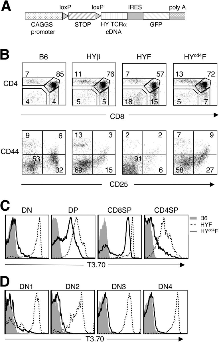
Conditional expression of the HY TCR beginning at the DP stage in HYcd4 mice. (A) Schematic representation of the conditional HY TCRα transgene. (B) Thymocytes from the indicated mice were stained with either anti-CD4 and anti-CD8 (top) or with anti-CD4, -CD8, -B220, and –NK1.1 to exclude lineage-positive cells and with anti-CD44 and anti-CD25 (bottom). Cells were analyzed by flow cytometry. Numbers represent the percentage of cells in each gate. (C) Thymocytes from B6 (shaded region), HY female (dotted line), and HYcd4 female (continuous line) were stained with anti-CD4, anti-CD8, and T3.70 and analyzed by flow cytometry. Individual subpopulations were gated, and T3.70 expression is depicted. (D) Thymocytes from B6 (shaded region), HY female (dotted line), and HYcd4 female (continuous line) were stained with anti-CD4, -CD8, -B220, and -NK1.1 to exclude lineage-positive cells and with anti-CD44, anti-CD25, and T3.70 to allow electronic gating of the DN subpopulations. T3.70 expression is depicted for the individual DN subpopulations. The y axes in C and D represent the percentage of maximum expression.
Corrected β-selection and lineage commitment in HYcd4 mice
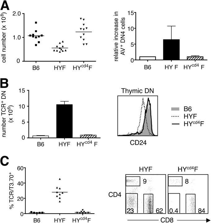
Early expression of a mature TCR was proposed to result in impaired β-selection and altered lineage commitment in HY and other TCR transgenics (5, 6, 13). Therefore, we wished to determine whether delaying TCRα expression until the DP stage corrected these events. Because little or no cell division occurs in the DP and SP compartment, the total number of thymocytes present in the thymus largely reflects the proliferative burst that accompanies β-selection. The thymic cellularity of HYcd4 female mice was equivalent to that of B6 mice, whereas conventional HY female mice showed a two- to threefold reduction in total thymocyte numbers (Fig. 2 A, left). Additionally, β-selection mediated by an αβTCR, as opposed to a pre-TCR, resulted in an increase in annexin V+ cells in the DN4 compartment (3). By expressing HY TCRα at the DP stage, we found a similar percentage of annexin V+ DN4 cells as in wild-type mice, whereas the early TCRα expression observed in conventional HY mice resulted in a 5–10-fold increase in annexin V+ DN4 cells (Fig. 2 A, right). These results provide further evidence that the early expression of an αβ heterodimer impairs β-selection.
Figure 2.
β-selection and lineage misdirection is corrected in HYcd4 mice. (A) Total thymocyte numbers from B6 (107 ± 25 × 106), HY female (56 ± 14 × 106), and HYcd4 female (123 ± 38 × 106) are shown (left). Thymocytes were stained with anti-CD4, -CD8, -B220, and -NK1.1 to exclude lineage-positive cells. The horizontal line represents the mean. Anti-CD44, anti-CD25, and annexin V staining were performed to identify apoptotic cells in the DN4 compartment (right). Data are expressed as a ratio of the percentage of annexin V+ DN4 cells compared with B6. Error bar represents SD. (B) DN thymocytes were analyzed for TCRβ or CD24 expression. The number of DN TCR+ cells from B6 (6.7 ± 1.8 × 10⁵), HY female (11 ± 1.7 × 10⁶), and HYcd4 female (9.0 ± 1.8 × 10⁵) (left; error bar represents SD) or the expression level of CD24 on B6 (shaded region), HY female (dotted line), and HYcd4 female (continuous line) (right; y axis represents the percentage of maximum expression) was determined. (C) LN cells from the indicated mice were stained with anti-CD4, anti-CD8, H57-597 (anti-TCRβ), and T3.70. The percentage of CD4−CD8− cells expressing T3.70 (HY female and HYcd4 female) or TCRβ (B6) was assessed (left). Additionally, the CD4/CD8 profile of T3.70+ LN cells is shown for HY female and HYcd4 female mice (right). The numbers within the FACS plots represent the percentage of cells within that gate.
It has additionally been suggested that αβ/γδ lineage commitment is disrupted by early expression of TCRα, resulting in mature αβTCR+ DN cells in both the thymus and periphery (5, 7, 13). In examining the thymic DN compartment, conventional HY female mice had a large number of resident αβTCR+ cells, whereas this population was substantially reduced in wild-type and HYcd4 mice (Fig. 2 B, left). As cells mature, the expression of CD24 (heat-stable antigen) decreases with the most mature thymic cells being CD24lo. No difference in CD24 levels between wild-type and HYcd4 mice was observed (Fig. 2 B, right). However, the vast majority of conventional HY mice had lower levels of CD24, suggesting that they are more mature and may comprise a population of lineage-misdirected cells (so-called “γδ-wannabes”; Fig. 2 B, right). Mature αβTCR+ DN cells are also prominent in the periphery of conventional HY mice and, again, were absent in HYcd4 mice (Fig. 2 C). Collectively, these data suggest that delaying TCRα expression in a transgenic mouse allows β-selection and early lineage commitment to occur normally.
Timing of TCRα does not appear to affect positive selection or homeostatic proliferation (HP)
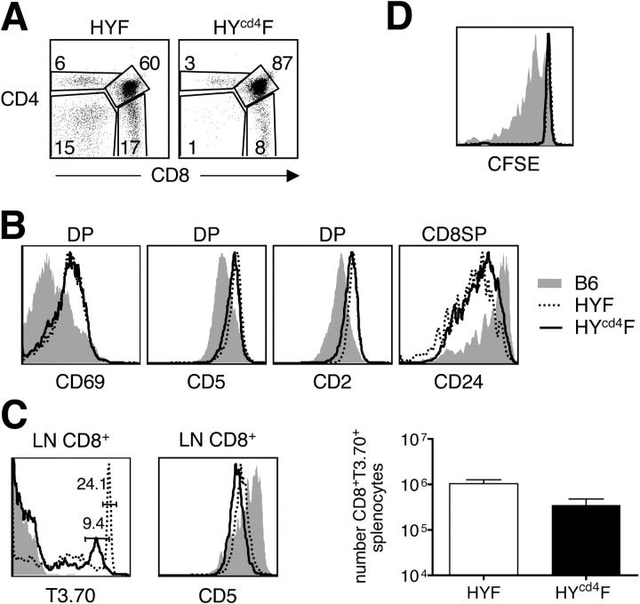
It was unclear at this point whether or not early expression of TCRα would affect positive selection events. By first gating on T3.70+ cells and then examining the CD4/CD8 distribution, we observed a prominent CD8SP population, indicating positive selection in HYcd4 female mice, as in the conventional model (Fig. 3 A). The population of T3.70+ CD8SPs was somewhat lower in both percentage and number in HYcd4 mice compared with conventional HY mice, suggesting a reduced efficiency. The reason for this difference is currently unclear; however, it could be caused by endogenous TCR gene rearrangements, which are likely to be more prevalent when the transgene comes on later. Indeed, the phenotype of HYcd4TCRα−/− mice (see Fig. 5 C) supports this. Positive selection is known to induce changes in gene expression that can be monitored by changes in the cell surface phenotype of DP thymocytes, including up-regulation of CD69, CD5, and CD2, and down-regulation of heat-stable antigen. Compared with B6 DP thymocytes, both conventional HY and HYcd4 DP thymocytes up-regulated CD69, CD5, CD2 (Fig. 3 B), and CD53 (not depicted) to a similar extent. In the CD8SP compartment, both showed a reduction in CD24, similar to wild-type controls (Fig. 3 B). Examination of the LNs from conventional HY and HYcd4 mice indicated the presence of CD8+ T3.70+ cells (Fig. 3 C). Although there was a slight difference in the percentage of CD8+ T3.70+ cells in the LNs and in the number of T3.70+ CD8+ cells in the spleens of conventional HY and HYcd4 mice (Fig. 3 C), this difference is likely attributable to a reduced number of T3.70+ CD8s exiting the thymus in HYcd4 mice because not all progenitors express the HY TCRα chain (Fig. 1 C). Additionally, the level of CD5 expressed by T3.70+ LN CD8 cells was equivalent in HY and HYcd4 mice, suggesting that the “tuning” of the TCR signal is similar in both strains of mice (Fig. 3 C).
Figure 3.
Positive selection and HP are not affected by early TCRα expression. (A) Thymocytes from HY female and HYcd4 female mice were stained with anti-CD4, anti-CD8, and T3.70 and analyzed by flow cytometry. The CD4/CD8 profile of T3.70+ cells is shown. The numbers within the FACS plots represent the percentage of cells within that gate. (B) T3.70+ DP or CD8SP thymocytes from B6 (shaded region), HY female (dotted line), and HYcd4 female (continuous line) were assessed for levels of CD69, CD5, CD2, and CD24. (C) LN cells from B6 (shaded region), HY female (dotted line), and HYcd4 female (continuous line) mice were stained with anti-CD4, anti-CD8, T3.70, and CD5. The level of T3.70 on gated CD8 cells (left) and CD5 on gated CD8+ T3.70+ cells (middle) is shown. The numbers in the left panel indicate the percentage of T3.70hi CD8 cells. The right panel shows the absolute number of CD8+ T3.70hi splenocytes from HY female (1.0 ± 0.6 × 10⁶) and HYcd4 female (3.5 ± 3 × 10⁵) mice. Error bars represent SD. (D) Bulk thymocytes from HYcd4 female (continuous line) or a mixture of B6 (shaded region) and HY female (dotted line) thymocytes were CFSE labeled and adoptively transferred into sublethally irradiated B6.SJL mice. Recipient mice were harvested 9 d after transfer and CD45.2+ CD8+ T3.70+ cells were analyzed for CFSE dilution. CD45.2+ CD8+ T3.70− cells were used as an internal control. Representative data from one recipient mouse out of three receiving either HYcd4 female or B6/HY female cells are shown. The y axes in B–D represent the percentage of maximum expression.
Figure 5.
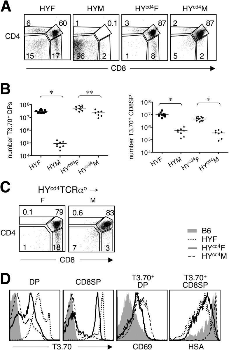
Deletion occurs late in HYcd4 male mice. (A) Thymocytes from the indicated mice were stained with anti-CD4, anti-CD8, and T3.70 and analyzed by flow cytometry. The T3.70+ thymocytes were gated and the CD4/CD8 profile is indicated. The numbers within the FACS plots represent the percentage of cells falling within that gate. (B) Comparisons of the absolute number of DP (HY female, 30 ± 7.6 × 10⁶; HY male, 9.0 ± 5 × 10⁴; HYcd4 female, 53 ± 18 × 106; HYcd4 male, 23 ± 12 × 106), and CD8SP (HY female, 10 ± 4 × 10⁶; HY male, 5.3 ± 3.3 × 10⁵; HYcd4 female, 4.4 ± 1.4 × 10⁶; HYcd4 male, 3.4 ± 1.9 × 10⁵) T3.70+ thymocytes from the different mouse strains. (*, P < 0.0001; **, P < 0.002). The horizontal lines represent the means. (C) Bone marrow from HYcd4 TCRαo female mice was mixed with either female or male B6.PL bone marrow, and 7 × 10⁶ cells were injected i.v. into lethally irradiated female or male B6.PL recipients, respectively. The female (left) and male (right) recipients were harvested 5–8 wk after transfer. CD4/CD8 profile of Thy1.2+ cells is indicated. (D) DP and CD8SP thymocytes from B6 (shaded region), HY female (dotted line), HYcd4 female (continuous line), and HYcd4 male (dashed line) mice were analyzed by flow cytometry for T3.70 expression (far left and left). T3.70+ DP (right) and T3.70+ CD8SP (far right) were assessed for CD69 up-regulation and CD24 down-regulation, respectively. The y axis represents the percentage of maximum expression.
To survive in the periphery, CD8 cells must receive a tonic signal through the TCR (14). The ability to undergo HP has an effect on the number of cells present in the periphery and has been suggested to correlate with TCR affinity and, thus, the level of CD5 expressed on peripheral CD8 T cells (15). HY transgenic CD8 T cells have a notable inefficiency in this homeostasis, particularly in lymphopenic recipients (16), that could either be caused by an inherently low affinity for self-antigen or by some other nonphysiologic aspect of the conventional HY transgenic model, such as early expression of the TCRα chain in development. Thus, we evaluated the capacity of CD8SP thymocytes to undergo HP in lymphopenic recipients. CD8SP thymocytes were used in this assay because this cell population is phenotypically equivalent between the two mouse strains, and the cells had not yet undergone any competition with other peripheral T cells. Carboxyfluorescein diacetate succinimidyl ester (CFSE)–labeled bulk thymocytes from HYcd4 or conventional HY thymocytes mixed with B6 thymocytes were injected into sublethally irradiated congenic recipients and parked for 9 d. B6 thymocytes were mixed with conventional HY thymocytes to provide a reference population for determining the extent of HP because the CD8SP pool of thymocytes from HYcd4 mice contain T3.70− cells with a polyclonal repertoire. Neither the T3.70+ CD8 cells from conventional HY mice nor those from HYcd4 mice underwent HP (Fig. 3 D), as was previously reported (17). Control polyclonal cells did undergo division as measured by CFSE dilution. Overall, these data suggest that positive selection and HP are not affected by early expression of TCRα.
Antigen sensitivity of DP and CD8SP thymocytes are identical in conventional HY and HYcd4 mice
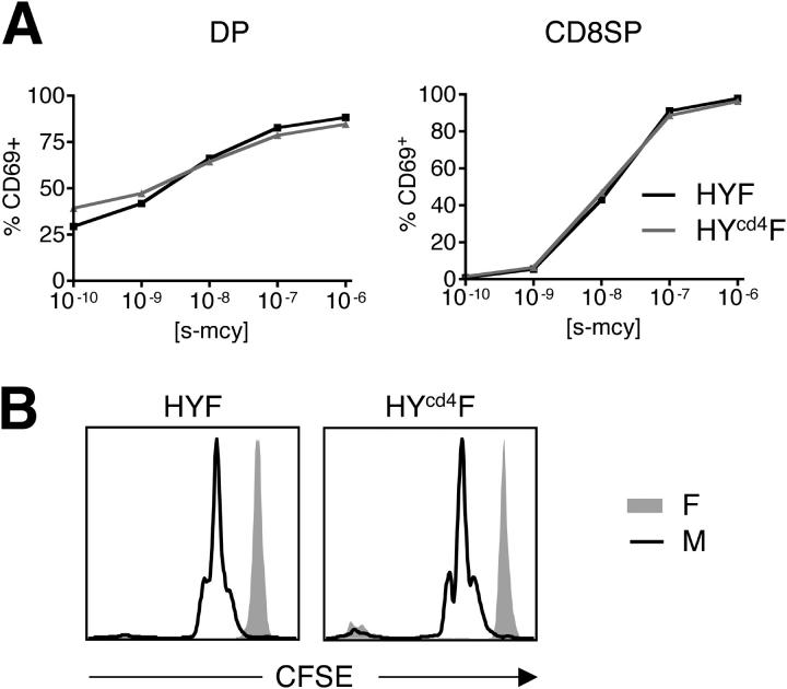
Because of the reduction in TCR levels, we wanted to examine the antigen sensitivity of T3.70+ CD8SP cells from HYcd4 mice. Total thymocytes were cultured at a 1:1 ratio with B6 splenocytes and increasing doses of agonist peptide, smcy, in vitro for 20 h, and the response of T3.70+ CD8SP cells was evaluated by CD69 induction. Both T3.70+ DP and CD8SP thymocytes from conventional HY and HYcd4 mice responded equivalently to agonist peptide at all concentrations (Fig. 4 A). Additionally, the T3.70+ CD8SP divided similarly in response to 100 nM smcy as measured by CFSE dilution (unpublished data). To compare the functional potential of HY and HYcd4 cells in vivo, bulk thymocytes were CFSE labeled and injected i.v. into intact male or female recipients. 2 d after transfer, the LNs and spleens of the recipient mice were harvested and the CFSE dilution of the CD8SP T3.70+ cells was determined. Both HY and HYcd4 cells diluted CFSE equivalently in male recipients (Fig. 4 B). Therefore, despite a twofold difference in surface TCR levels, the antigen responsiveness of cells from HY and HYcd4 mice appears equivalent.
Figure 4.
In vitro and in vivo responsiveness of HY and HYcd4 CD8 thymocytes is equivalent. (A) Bulk thymocytes from HY female (black line) or HYcd4 female (gray line) mice were mixed at a 1:1 ratio with female B6 splenocytes and increasing concentrations of agonist smcy peptide were added to the culture and incubated for 20 h. Cells were harvested and stained with anti-CD4, anti-CD8, T3.70, and anti-CD69 and analyzed by flow cytometry. CD4+CD8+T3.70hi (left) and CD4−CD8+T3.70+ (right) cells were gated, and the induction of CD69 was measured. Data are expressed as a percentage of cells that maximally up-regulated CD69. (B) HY female (left) or HYcd4 female (right) bulk thymocytes were CFSE labeled and injected i.v. into intact female (shaded region) or male (continuous line) recipients. 48 h after injection, spleens were harvested, and the CFSE dilution of CD8+ T3.70+ cells was measured. The y axis represents the percentage of maximum expression.
Negative selection occurs late in development in HYcd4 mice
Some discrepancy exists in the literature regarding the timing of negative selection during development. In wild-type mice, superantigen-mediated deletion generally occurs late in development, as DP mature into SP cells, whereas in other models, particularly some transgenic models (including the conventional HY model), deletion occurs early, either right at or preceding DP generation. Examination of the CD4/CD8 profile of T3.70+ cells in the thymus revealed striking differences between conventional HY and HYcd4 male mice. The overwhelming majority of T3.70+ cells in conventional HY male mice were of the DN phenotype with virtually no DP, CD4SP, or CD8SP populations (Fig. 5 A). In contrast, in HYcd4 male mice, T3.70+ cells were predominantly DP thymocytes with very few CD8SP cells (Fig. 5 A). Comparing HYcd4 female to HYcd4 male T3.70+ thymocytes, there was no difference in the percentage of DP cells, whereas there was a dramatic loss of CD8SP cells in HYcd4 male mice (Fig. 5 A). To confirm that this phenotype was dependent on the timing of HY TCR expression and not some other variable of the conditional expression strategy, we also examined HYlck animals. In this strain, Cre is driven by the lck promoter and results in HY TCR expression in the early DN stage, which is similar to conventional HY mice (unpublished data). Indeed, the thymus of the HYlck male animal resembled the conventional HY male thymus, where the majority of T3.70+ cells were DN (Fig. S2 A, available at http://www.jem.org/cgi/content/full/jem.20050359/DC1). The difference between the conventional HY and HYcd4 strains was further highlighted when comparing the number of T3.70+ cells in the DP and CD8SP populations from the various mice. There was an ∼500–1,000-fold reduction in DP thymocytes when comparing conventional HY male and female mice, whereas only a 2-fold reduction in the numbers of DP thymocytes is observed between HYcd4 male and female mice (Fig. 5 B). Again, very few T3.70+ DP thymocytes were present in HYlck males (Fig. S2 B), suggesting that this difference is caused by the timing of TCRα gene expression. The reduction in HYcd4 mice was predominantly seen in the CD8SP compartment, which is 15-fold smaller in HYcd4 male compared with female mice (Fig. 5 B). Thus, although male antigen-mediated deletion occurs at the DN–DP transition in the conventional HY model, it occurred at the DP–SP transition in the HYcd4 model, and this difference was caused by the timing of TCRα expression.
To rule out the possible contribution of endogenous TCRα chains to our observations, we constructed bone marrow chimeras with HYcd4 TCRαo female bone marrow. We created mixed chimeras with either B6.PL male or female bone marrow and injected them into male or female B6.PL recipients, respectively, to provide male antigen-expressing, bone marrow–derived, antigen-presenting cells. The only difference in the CD4/CD8 profile of T3.70+ cells from the thymus of HYcd4 mice on a TCRα-sufficient or -deficient background was a slightly enhanced CD8 percentage in the females (Fig. 5 C). A large population of T3.70+ DP, with an absence of CD8SP, was still observed in the male. Additionally, the relative chimerism of the DP and CD8SP compartments in male and female recipients supported late deletion (unpublished data).
No difference in the level of T3.70 expression was observed between HYcd4 male and female DP cells (Fig. 5 D). Thus, it is possible that T3.70+ DP cells are present in HYcd4 male mice because they have yet to encounter antigen (e.g., because of a lack of presentation by cortical epithelial cells). However, this does not appear to be the case because CD69 was expressed at a high level in HYcd4 T3.70+ male DP cells, indicating that those cells had in fact responded to antigen but were not yet deleted (Fig. 5 D). Additionally, an examination of the few remaining T3.70+ CD8SP cells in HYcd4 male mice revealed a high expression of CD24, indicating an immature state (Fig. 5 D). Collectively, these data indicate that early expression of HY TCRα in conventional HY male mice leads to immediate and early deletion, whereas HY TCRα expression at the DP stage leads to a later and delayed deletion, which occurs as cells transition from DP to SP.
CD8αα IEL expansion occurs less in HYcd4 male mice
There is a unique population of lymphocytes in the gut that express αβTCR and CD8αα homodimers. A great deal of debate has surrounded the issue of how this population develops. Although it was thought that CD8αα IELs develop extrathymically, this might only be the case in lymphopenic conditions (18); other evidence suggests a thymic origin (19). Additionally, this population is thought to be self-reactive and to serve a regulatory function in the gut (20). Indeed, consistent with this idea, conventional HY male mice display at least a 100-fold expansion in CD8αα IELs (21, 22). Nevertheless, it remains controversial as to whether CD8αα IELs develop from DN or DP progenitors. We felt that our model would be a good means to evaluate this issue.
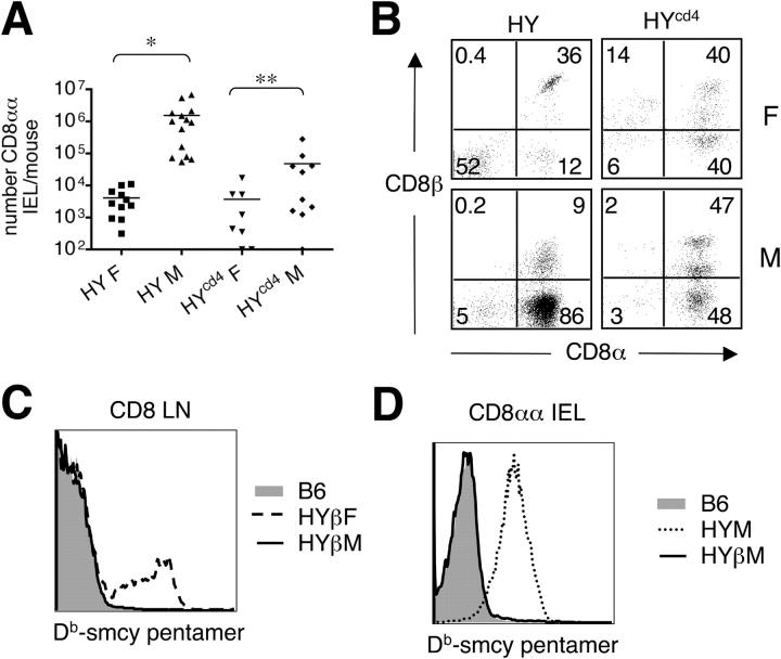
To this end, we purified IELs according to standard protocols and evaluated the number and percentage of T3.70+ CD8αα IELs in the different transgenic strains. As previously reported, the number of T3.70+ CD8αα IELs was ∼500-fold higher in conventional HY male mice compared with females (Fig. 6 A; references 21 and 22). In contrast, there was only a fivefold change in the number of these cells between male and female HYcd4mice, which was not significant with the number of animals analyzed (P > 0.05; Fig. 6 A). Examination of HYlck mice showed a 200-fold increase in T3.70+ CD8αα IELs in males over females, similar to conventional HY transgenics, further suggesting that the timing of TCRα expression is critical for the expansion of these cells (Fig. S2 D). Additionally, the T3.70+ IELs from HYcd4 mice showed a similar percentage of CD8αβ- and CD8αα-expressing cells in both males and females, whereas there was a dramatic increase in CD8αα-expressing cells in conventional HY and HYlck male mice (Fig. 6 B and Fig. S2 D). Curiously, the IELs from HYcd4 male mice display a small CD8αβhi population that is not present in HY or HYlck males, of which we do not understand the importance at this time.
Figure 6.
Early TCRα expression is required for expansion of gut CD8αα IELs. (A) IELs were isolated and stained with anti-CD8β, -CD8α, -CD3, and T3.70 and analyzed by flow cytometry. CD3+ T3.70+ cells were electronically gated and the absolute number of CD8αα+ cells was quantified by comparison to the acquisition of a known number of latex beads included in the sample (HY female, 4.1 ± 3.6 × 10³; HY male, 1.5 ± 2.1 × 10⁶; HYcd4 female, 3.7 ± 5.8 × 10³; HYcd4 male, 4.8 ± 8.6 × 10⁴). The horizontal lines represent the means. *, P < 0.05; **, P > 0.05. (B) CD8β/CD8α profile of the CD3+ T3.70+ IELs is shown for the indicated mice. The numbers within the FACS plots represent the percentage of cells falling within that gate. (C) LN cells from B6 (shaded region), HYβ female (dashed line), and HYβ male (continuous line) mice were stained with anti-CD8α, -CD8β, -CD3, and Db–smcy pentamer. Db–smcy pentamer staining for CD3+ CD8αβ+ cells is indicated. (D) IELs were harvested from B6 (shaded region), HYβ male (continuous line), and HY male (dotted line) mice and stained with anti-CD8α, -CD8β, -CD3, and Db–smcy pentamers. Db–smcy pentamer staining from CD3+ CD8αα+ cells is indicated. The y axes in C and D represent the percentage of maximum expression.
To determine whether early versus late TCR expression has an impact on the phenotype of T3.70+ CD8αα IELs, we examined the expression pattern of several molecules reported to be differentially expressed on CD8αβ versus CD8αα IELs by the gene profiling (23). CD8αα and CD8αβ IELs from B6 display distinct patterns of CD5, CD122, and CD11a (Fig. S3, available at http://www.jem.org/cgi/content/full/jem.20050359/DC1). Interestingly, the level of expression of these markers on T3.70+ CD8αα IELs from both conventional HY and HYcd4 males was not similar to either normal population (Fig. S3). In fact, for all three markers, the level of expression was intermediate between CD8αα and CD8αβ IELs from B6 (Fig. S3). The reason for this difference is also currently unclear. Together, these data suggest that the expansion of T3.70+ CD8αα IELs in response to high affinity ligand is dependent on TCR expression early in development. Furthermore, CD8αα IELs that do develop in conventional HY or HYcd4 male mice have an unusual phenotype when compared with nontransgenic CD8αα IELs.
If CD8αα IEL expansion requires TCR gene rearrangement and expression at the DN stage, then one would predict that such cells would not be prominent in HYβ transgenic mice. HYβ transgenic mice express only the TCRβ chain early in development. The TCRα chain is derived from the endogenous repertoire, which does not undergo gene rearrangement and expression until the DP stage. Thus, potential male reactive receptors are not generated until the DP stage. Female HYβ transgenic mice displayed a prominent population of male reactive CD8+ cells in the LNs as judged by their ability to bind Db/male antigen tetramers (Fig. 6 C; reference 24), which are presumably derived by positive selection at the DP stage. If self-reactivity were to result in the positive selection of CD8αα IELs in HYβ mice, then CD8αα IELs from male mice should display an increase in male reactivity. This was not the case, as CD8αα IELs from male HYβ mice do not show detectable Db–smcy pentamer binding (Fig. 6 D). This does not reflect a technical inability of CD8αα cells to bind pentamers because the CD8αα IELs from HY conventional mice do bind the pentamer (Fig. 6 D). This result confirms the findings from the HYcd4 mouse and suggests that development of CD8αα IELs in HY mice depends on early rearrangement and expression of the TCR.
Discussion
Our data indicate that early expression of the TCR affects several properties in TCR transgenic mice. This is important because TCR transgenics represent the predominant tool used to study lymphocyte development, particularly as it relates to the specificity and affinity of the rearranged antigen receptor. When the male-specific HY TCR was expressed at the physiological DP stage of development, the β-selection defect and lineage misdirection seen in conventional TCR transgenics was corrected. Furthermore, deletion occurred late in development and expansion of the CD8αα+ gut IELs was lost. Positive selection and HP were unaffected by early TCR expression. In general, we feel this model represents a more faithful recapitulation of T cell development processes.
β-selection and lineage misdirection
β-selection is a process initiated by the pre-TCR at the DN3 stage. It signals the successful completion of TCRβ chain gene rearrangement and results in cellular expansion, CD4 and CD8 expression, termination of further TCRβ chain gene rearrangement, initiation of TCRα chain gene rearrangement, migration into the cortex, and development of positive and negative selection “competence” factors (25, 26). Although a mature αβTCR is capable of inducing β-selection at the DN stage, it is much less efficient than that mediated by the pre-TCR (3). In HYcd4 mice, the β-selection impairment seen in conventional HY mice did not occur. This finding is consistent with a pre-Tα competition model, where a higher affinity of TCRβ for TCRα, compared with pre-Tα, results in less efficient β-selection. In support of this hypothesis, studies by Lacorazza et al. have indicated that premature TCR expression impairs the proliferative burst of DN thymocytes and increases the number of terminally differentiated DNs (6).
In the periphery of conventional HY mice, there is a large population of DN T3.70+ cells. These cells have been shown to represent lineage-misdirected cells, arising because of signals from the αβTCR at the DN stage as would be the case if a γδTCR were expressed (4, 13). When the HY TCR was not expressed until the DP stage, this population of cells was absent. Importantly, some studies have suggested that these DN TCR+ cells have regulatory potential (27, 28). Because the DN cells have the same specificity as the CD8 or CD4 cells in the periphery, they have the potential to influence the immune response in a nonphysiologic manner if intact TCR transgenics are used to evaluate an antigen-specific response. This, however, is not a concern when the adoptive transfer of purified CD8 or CD4 transgenic cells is used to evaluate an antigen-specific response.
Positive selection and homeostasis
Early TCR expression does not seem to affect positive selection or homeostasis. We found no differences in the phenotypes of T3.70+ DP or CD8SP cells in HYcd4 mice compared with conventional HY mice. In both cases, markers shown to be up-regulated in response to TCR stimulation, including CD69, CD2, CD5, and CD53, were all induced to the same extent. Additionally, T3.70+ CD8SP cells responded equivalently in vitro and in vivo to agonist ligand. Evaluation of the HP potential of T3.70+ CD8SP thymocytes revealed that cells from neither model were able to divide in response to lymphopenic conditions, which has been previously demonstrated for conventional HY cells (17). The reason for this lack of HP is currently unclear; however, at least two different explanations can be offered. First, it is possible that the selecting ligand in the thymus is either absent or reduced in quantity in the periphery. Work by Santori et al. identified, through homology-based searches, a peptide that could select HY TCR thymocytes in fetal thymic organ culture, but it is unclear if, where, and in what quantities this peptide is presented in the periphery (29). Alternatively, it has been suggested that the failure of HY T cells to undergo HP is caused by the low affinity of the HY TCR for the selecting ligand (15). This reasoning stems from the observation that both peripheral CD8s and CD8SP thymocytes express lower levels of CD5 than do bulk B6 thymocytes or other TCR transgenics that do undergo HP. We find this possibility somewhat unlikely since positive selection of HY thymocytes does occur and appears to be quite efficient. We favor a combination of these two hypotheses in which the affinity of the HY TCR for the selecting ligand is somewhat lower than for other TCR transgenics, as illustrated by reduced CD5 levels, but still high enough to mediate positive selection, and the expression of this ligand is limited in the periphery. Further experiments are necessary to understand this issue fully.
Clonal deletion
Perhaps some of the most intriguing results were obtained when examining T cell development in male HYcd4 mice. We observed that negative selection in HYcd4 male mice occurs late in development, at the DP to CD8SP transition. Discrepancies exist in the literature regarding the timing of negative selection. Some models, including the conventional HY and other TCR transgenic models, find that negative selection occurs immediately at or preceding DP generation. However, deletion in other transgenic models (30–32), as well as endogenous superantigen-mediated deletion (33, 34), occurs at the DP–SP transition, as is found in the HYcd4 model. In addition, analysis of the anatomy of apoptosis in MHC-sufficient versus -deficient mice suggests that deletion is primarily cortico-medullary (35), corresponding to a late deletion at the DP or SP stage. Based on the findings here, we hypothesize that the early deletion seen in some TCR transgenic models of negative selection is caused by the nonphysiologic early TCR expression. Clearly, the anatomical location of self-antigen can be a factor in when negative selection occurs. However, because the male self-antigen is ubiquitously expressed in the thymus, and its presentation does not vary between conventional HY, HYlck, and HYcd4 models, the timing of TCR expression appears to be a more critical factor in when negative selection occurs.
One surprising feature of the response to male antigen in the HYcd4 model is the presence of a large population of activated DP progenitors. In conventional HY male mice, it is difficult to find progenitors (DN or DP) with an activated phenotype, presumably because they had undergone immediate apoptosis. This implies that the death induced in the more physiologic HYcd4 model is somehow delayed. Previous studies have suggested that the medulla and cortico-medullary junction is the primary site for clonal deletion to superantigen (35) or to the complement component C5 in A18 TCR transgenic animals (36), and genetic studies suggest that costimulatory molecules that are expressed in the medulla, but not the cortex, are important for clonal deletion (37–40). Therefore, an interesting possibility is that self-antigen–specific progenitors normally do not undergo clonal deletion until they migrate to the cortico-medullary junction, a possibility that could be evaluated by determining the site of deletion in the HYcd4 model.
In addition to there being discrepancies about the stage of deletion, there is also controversy about the molecular signaling pathways leading to deletion. For example, the role of CD40–CD40L interaction has been demonstrated to be necessary in some models of deletion, yet appears to be dispensable in others (including the conventional HY model; reference 41). Other molecules, including TNFR, CD28, and Fas, also have controversial roles in negative selection (42). Because deletion occurs late in the HYcd4 model it may be interesting to reevaluate the role of these molecules in central tolerance to male antigens.
CD8αα IELs
Although clonal deletion is an important central tolerance mechanism, recent evidence suggests that progenitors can also be positively selected into a regulatory T cell lineage upon encountering high affinity ligand in the thymus (43, 44). This process has been referred to as “agonist selection” and applies to three different T cell populations: CD4+CD25+ regulatory T cells, NKT cells, and CD8αα IELs (45). Many questions remain about the factors that lead to agonist selection. Early work in the field of CD8αα IEL development suggested that this cell type arose extrathymically. However, recently, it was demonstrated that the extrathymic pathway might only operate under conditions of lymphopenia, and that the thymus is the primary developmental site for CD8αα IELs (18, 19). In addition to where these cells develop, the progenitor cell giving rise to CD8αα IELs has been controversial. Using TCR transgenic models for CD8αα IEL development, it was suggested that a DN progenitor gives rise to CD8αα IELs, and that in cases where there are few DP cells (such as in conventional HY male mice) there are many CD8αα IELs (21, 22). Gene profiling studies also showed striking commonalities between αβTCR CD8αα IELs and γδTCR DN IELs (23, 46). Because γδTCR-expressing cells arise from DN progenitors and do not develop through a DP intermediate, it was suggested that DN thymocytes are the immediate precursors of CD8αα IELs. Indeed, we found that although CD8αα IELs were present in HYcd4 male mice, there was little agonist ligand–dependent expansion, suggesting that the large CD8αα IEL population in conventional HY male mice is an unnatural consequence of the early expression of the receptor at the DN stage. This result was further confirmed using the HYβ transgenic model in which there was no male reactivity in the CD8αα IEL population of male mice. In an elegant fate mapping experiment, Eberl et al. demonstrated that CD8αα IELs arose from at least a DN4 stage thymocyte and likely a DP thymocyte (47). Additionally, Yamagata et al. were able to generate CD8αα+ cells from in vitro culture of nonselected HY DP with agonist-expressing stroma in a reaggregate culture (48). Our experiments do not preclude DP thymocytes as intermediates in the generation of CD8αα IELs; however, they do suggest that early TCR expression (within the DN compartment) is required. It is unclear why early TCR expression is necessary for the generation of CD8αα IELs, but one could postulate that signals received by a DN thymocyte from a mature αβTCR, as opposed to a pre-TCR, could in some manner imprint the cell to develop along this pathway when faced with agonist ligand.
In conclusion, the HYcd4 model has several advantages over other TCR transgenic and nontransgenic models of development, but it also has a few drawbacks. First, it contains a high frequency of monoclonal cells, which facilitates analysis, but may lead to unnatural effects (as in other TCR transgenics). Second, in order to create the HYcd4 mouse, three transgenes are required. This makes it quite laborious and time consuming when wanting to introduce other transgenes or breed onto a gene-deficient strain. Finally, the level of TCR expression on mature T cells in these animals is lower than what is observed in wild-type mice. Although this issue did not appear to affect the sensitivity of the cells to ligand, there could be cases in which this would be a problem. It would be of substantial benefit to engineer a system where the TCR was expressed at the appropriate time developmentally, expressed similar amounts of surface TCR to wild-type mice, and is facile enough to allow the study of multiple genetic factors.
Overall, the HYcd4 model allowed us to define those properties of TCR transgenic lymphocyte development that are nonphysiological and caused by the early expression of the TCR. Our findings suggest that late (DP to SP) models of negative selection may be the most appropriate models to evaluate molecular pathways of clonal deletion, and that improved models are needed to study the development of CD8αα IELs.
Materials and Methods
DNA constructs.
pCAGGS STOP HY TCRα was generated using standard molecular biology techniques. In brief, the pEF321 ShcFFF plasmid containing a floxed transcriptional and translational STOP cassette was obtained from K. Ravichandran (University of Virginia, Charlottesville, VA; reference 10). The internal ribosomal entry site (IRES)–GFP portion of pEF321 ShcFFF was replaced with an IRES–GFP cassette provided by S. Casola (Harvard University, Boston, MA). The HY TCRα cDNA was amplified from a cDNA library generated from bulk HY thymocytes and replaced the ShcFFF cDNA by directional cloning. The floxed STOP HY TCRα IRES–GFP fragment was inserted into the pCAGGS vector, generating pCAGGS STOP HY TCRα.
Animals.
C57BL/6 (B6), Thy 1 congenic C57BL/6.PL (B6.PL), CD45 congenic C57BL/6.SJL (B6.SJL), and TCRα−/− mice were purchased from the Jackson Laboratory. CD4–Cre and lck–Cre mice were purchased from Taconic. The conditional HY TCRα–expressing mice were generated by microinjection of the Sal–Not fragment from pCAGGS STOP HY TCRα into preimplantation C57BL/6 embryos by the Mouse Genetics Laboratory at the University of Minnesota. Founders were identified by PCR using primers directed against the 3′ region of the STOP cassette. Nine founders were identified, of which only two appeared to express HY TCRα. Data from founder 8820 are presented. Conventional HY and HYβ mice were a gift of H. von Boehmer (Harvard University, Boston, MA). Mice containing the HYβ, CD4–Cre and conditional HY TCRα (HYcd4) or HYβ, lck–Cre, and conditional HY TCRα (HYlck) transgenes were obtained by breeding. All animals were maintained and treated in accordance with federal guidelines approved by the University of Minnesota Institutional Animal Care Committee.
Antibodies and flow cytometry.
All fluorochrome and biotinylated antibodies were purchased from BD Biosciences, eBioscience, or Biolegend. Cells were stained with antibody for 30 min on ice in FACS buffer (PBS, 1% FCS, and 0.02% azide, pH 7.2) and washed two times in FACS buffer after each antibody incubation. Cell events were collected using a cytometer (FACSCalibur or LSRII; BD Biosciences) and analyzed with FlowJo software (Tree Star Inc.). Db–smcy pentamers were purchased from ProImmune. Pentamer staining was performed in FACS buffer at room temperature for 30 min before staining with other antibodies.
Purification of IELs.
Lymphocytes from the small intestine were purified using a protocol described by Podd et al. (49). In brief, the small intestine was isolated, cut longitudinally, and the contents were rinsed out with ice-cold HBSS. The intestine was then cut into 0.5-cm pieces and incubated three times at 37°C for 20 min in HBSS and 1 mM dithiothreitol with shaking. After each round of shaking, the supernatant was strained with a 70-μM nylon filter. Cells were pelleted and lymphocytes were enriched by centrifugation over a 70×/40× Percoll gradient, and the band at the 70×/40× interface was collected. Cells were washed in RP10 before staining.
In vitro and in vivo stimulation.
Responder HY or HYcd4 female thymocytes or bulk LN cells were mixed at a 1:1 ratio with female B6 splenocytes and the indicated concentration of agonist smcy peptide in vitro (50). Cultures were incubated for 20 h at 37°C and stained with anti-CD4, anti-CD8, T3.70, and anti-CD69. T3.70+ DP or CD8SP cells were electronically gated, and the expression of CD69 was measured. Data are represented as the percentages of indicated population that maximally up-regulated CD69. Bulk thymocytes from HY and HYcd4 female mice were CFSE labeled (Molecular Probes) and injected i.v. into unmanipulated male or female recipients. LNs and spleens were harvested 48 h after injection, stained with anti-CD4, CD8, CD69, and T3.70, and analyzed on a cytometer (LSRII; BD Biosciences). Data are presented as the CFSE profile for CD8+ T3.70+ cells from the spleen from male or female recipients.
HP.
Bulk thymocytes from HY, HYcd4, and B6 female mice were isolated. HY thymocytes were spiked with B6 thymocytes to provide a reference population for measurement of HP (HYcd4 mice have a population of CD8SP that do not express the HY receptor and thus provide a preexisting internal control). Thymocytes were then CFSE labeled and injected i.v. into sublethally irradiated congenic B6.SJL mice. LNs and spleens were harvested 9 d after injection, and CFSE dilution was measured in the B6, HY, and HYcd4 CD8 populations.
Online supplemental material.
Fig. S1 depicts a quantitative comparison of TCR levels in CD69+ (positively selected) DP thymocytes from B6, HY, and HYcd4 mice. Fig. S2 shows a phenotypic and quantitative analysis of thymocytes (A and B) and IELs (C and D) from HYlck female and male mice. Fig. S3 depicts a phenotypic analysis of IEL populations (CD8αα and CD8αβ) from various mice. Online supplemental material is available at http://www.jem.org/cgi/content/full/jem.20050359/DC1.
Acknowledgments
The authors wish to thank Dr. Lisa McNeil for her critical review of the manuscript, Dr. Kodi Ravichandran and Dr. Stefano Casola for DNA constructs, and Dr. Harald von Boehmer for mice. We would also like to thank Dr. Victoria Camerini for her help in IEL isolation.
This work was supported by National Institutes of Health grants AI39560, AI50105 (to K.A. Hogquist), and AI38903 (to S.C. Jameson). T.A. Baldwin is supported by fellowships from the Alberta Heritage Foundation for Medical Research and the Canadian Institutes of Health Research.
The authors have no conflicting financial interests.
Abbreviations used: CFSE, carboxyfluorescein diacetate succinimidyl ester; DN, double negative; DP, double positive; HP, homeostatic proliferation; IEL, intraepithelial lymphocyte; SP, single positive.
References
- 1.Starr, T.K., S.C. Jameson, and K.A. Hogquist. 2003. Positive and negative selection of T cells. Annu. Rev. Immunol. 21:139–176. [DOI] [PubMed] [Google Scholar]
- 2.Trop, S., M. Rhodes, D.L. Wiest, P. Hugo, and J.C. Zuniga-Pflucker. 2000. Competitive displacement of pT alpha by TCR-alpha during TCR assembly prevents surface coexpression of pre-TCR and alpha beta TCR. J. Immunol. 165:5566–5572. [DOI] [PubMed] [Google Scholar]
- 3.Borowski, C., X. Li, I. Aifantis, F. Gounari, and H. von Boehmer. 2004. Pre-TCRα and TCRα are not interchangeable partners of TCRβ during T lymphocyte development. J. Exp. Med. 199:607–615. [DOI] [PMC free article] [PubMed] [Google Scholar]
- 4.Bruno, L., H.J. Fehling, and H. von Boehmer. 1996. The alpha beta T cell receptor can replace the gamma delta receptor in the development of gamma delta lineage cells. Immunity. 5:343–352. [DOI] [PubMed] [Google Scholar]
- 5.Fritsch, M., A. Andersson, K. Petersson, and F. Ivars. 1998. A TCR alpha chain transgene induces maturation of CD4- CD8- alpha beta+ T cells from gamma delta T cell precursors. Eur. J. Immunol. 28:828–837. [DOI] [PubMed] [Google Scholar]
- 6.Lacorazza, H.D., C. Tucek-Szabo, L.V. Vasovic, K. Remus, and J. Nikolich-Zugich. 2001. Premature TCR alpha beta expression and signaling in early thymocytes impair thymocyte expansion and partially block their development. J. Immunol. 166:3184–3193. [DOI] [PubMed] [Google Scholar]
- 7.Terrence, K., C.P. Pavlovich, E.O. Matechak, and B.J. Fowlkes. 2000. Premature expression of T cell receptor (TCR) αβ suppresses TCRγδ gene rearrangement but permits development of γδ lineage T cells. J. Exp. Med. 192:537–548. [DOI] [PMC free article] [PubMed] [Google Scholar]
- 8.Wolfer, A., T. Bakker, A. Wilson, M. Nicolas, V. Ioannidis, D.R. Littman, P.P. Lee, C.B. Wilson, W. Held, H.R. MacDonald, and F. Radtke. 2001. Inactivation of Notch 1 in immature thymocytes does not perturb CD4 or CD8 T cell development. Nat. Immunol. 2:235–241. [DOI] [PubMed] [Google Scholar]
- 9.Wolfer, A., A. Wilson, M. Nemir, H.R. MacDonald, and F. Radtke. 2002. Inactivation of Notch1 impairs VDJbeta rearrangement and allows pre-TCR-independent survival of early alpha beta lineage thymocytes. Immunity. 16:869–879. [DOI] [PubMed] [Google Scholar]
- 10.Zhang, L., V. Camerini, T.P. Bender, and K.S. Ravichandran. 2002. A nonredundant role for the adapter protein Shc in thymic T cell development. Nat. Immunol. 3:749–755. [DOI] [PubMed] [Google Scholar]
- 11.Teh, H.S., P. Kisielow, B. Scott, H. Kishi, Y. Uematsu, H. Bluthmann, and H. von Boehmer. 1988. Thymic major histocompatibility complex antigens and the alpha beta T-cell receptor determine the CD4/CD8 phenotype of T cells. Nature. 335:229–233. [DOI] [PubMed] [Google Scholar]
- 12.Porritt, H.E., L.L. Rumfelt, S. Tabrizifard, T.M. Schmitt, J.C. Zuniga-Pflucker, and H.T. Petrie. 2004. Heterogeneity among DN1 prothymocytes reveals multiple progenitors with different capacities to generate T cell and non-T cell lineages. Immunity. 20:735–745. [DOI] [PubMed] [Google Scholar]
- 13.Buer, J., I. Aifantis, J.P. DiSanto, H.J. Fehling, and H. von Boehmer. 1997. T-cell development in the absence of the pre-T-cell receptor. Immunol. Lett. 57:5–8. [DOI] [PubMed] [Google Scholar]
- 14.Jameson, S.C. 2002. Maintaining the norm: T-cell homeostasis. Nat. Rev. Immunol. 2:547–556. [DOI] [PubMed] [Google Scholar]
- 15.Kieper, W.C., J.T. Burghardt, and C.D. Surh. 2004. A role for TCR affinity in regulating naive T cell homeostasis. J. Immunol. 172:40–44. [DOI] [PubMed] [Google Scholar]
- 16.Tanchot, C., F.A. Lemonnier, B. Perarnau, A.A. Freitas, and B. Rocha. 1997. Differential requirements for survival and proliferation of CD8 naive or memory T cells. Science. 276:2057–2062. [DOI] [PubMed] [Google Scholar]
- 17.Rocha, B., and H. von Boehmer. 1991. Peripheral selection of the T cell repertoire. Science. 251:1225–1228. [DOI] [PubMed] [Google Scholar]
- 18.Guy-Grand, D., O. Azogui, S. Celli, S. Darche, M.C. Nussenzweig, P. Kourilsky, and P. Vassalli. 2003. Extrathymic T cell lymphopoiesis: ontogeny and contribution to gut intraepithelial lymphocytes in athymic and euthymic mice. J. Exp. Med. 197:333–341. [DOI] [PMC free article] [PubMed] [Google Scholar]
- 19.Leishman, A.J., L. Gapin, M. Capone, E. Palmer, H.R. MacDonald, M. Kronenberg, and H. Cheroutre. 2002. Precursors of functional MHC class I- or class II-restricted CD8alphaalpha(+) T cells are positively selected in the thymus by agonist self-peptides. Immunity. 16:355–364. [DOI] [PubMed] [Google Scholar]
- 20.Cheroutre, H. 2004. Starting at the beginning: new perspectives on the biology of mucosal T cells. Annu. Rev. Immunol. 22:217–246. [DOI] [PubMed] [Google Scholar]
- 21.Cruz, D., B.C. Sydora, K. Hetzel, G. Yakoub, M. Kronenberg, and H. Cheroutre. 1998. An opposite pattern of selection of a single T cell antigen receptor in the thymus and among intraepithelial lymphocytes. J. Exp. Med. 188:255–265. [DOI] [PMC free article] [PubMed] [Google Scholar]
- 22.Rocha, B., H. von Boehmer, and D. Guy-Grand. 1992. Selection of intraepithelial lymphocytes with CD8 alpha/alpha co-receptors by self-antigen in the murine gut. Proc. Natl. Acad. Sci. USA. 89:5336–5340. [DOI] [PMC free article] [PubMed] [Google Scholar]
- 23.Pennington, D.J., B. Silva-Santos, J. Shires, E. Theodoridis, C. Pollitt, E.L. Wise, R.E. Tigelaar, M.J. Owen, and A.C. Hayday. 2003. The inter-relatedness and interdependence of mouse T cell receptor gammadelta+ and alphabeta+ cells. Nat. Immunol. 4:991–998. [DOI] [PubMed] [Google Scholar]
- 24.Bouneaud, C., P. Kourilsky, and P. Bousso. 2000. Impact of negative selection on the T cell repertoire reactive to a self-peptide: a large fraction of T cell clones escapes clonal deletion. Immunity. 13:829–840. [DOI] [PubMed] [Google Scholar]
- 25.Michie, A.M., and J.C. Zuniga-Pflucker. 2002. Regulation of thymocyte differentiation: pre-TCR signals and beta-selection. Semin. Immunol. 14:311–323. [DOI] [PubMed] [Google Scholar]
- 26.Neilson, J.R., M.M. Winslow, E.M. Hur, and G.R. Crabtree. 2004. Calcineurin B1 is essential for positive but not negative selection during thymocyte development. Immunity. 20:255–266. [DOI] [PubMed] [Google Scholar]
- 27.Wang, R., Y. Wang-Zhu, and H. Grey. 2002. Interactions between double positive thymocytes and high affinity ligands presented by cortical epithelial cells generate double negative thymocytes with T cell regulatory activity. Proc. Natl. Acad. Sci. USA. 99:2181–2186. [DOI] [PMC free article] [PubMed] [Google Scholar]
- 28.Priatel, J.J., O. Utting, and H.S. Teh. 2001. TCR/self-antigen interactions drive double-negative T cell peripheral expansion and differentiation into suppressor cells. J. Immunol. 167:6188–6194. [DOI] [PubMed] [Google Scholar]
- 29.Santori, F.R., S.M. Brown, Y. Lu, T.A. Neubert, and S. Vukmanovic. 2001. Cutting edge: positive selection induced by a self-peptide with TCR antagonist activity. J. Immunol. 167:6092–6095. [DOI] [PubMed] [Google Scholar]
- 30.Gallegos, A.M., and M.J. Bevan. 2004. Central tolerance to tissue-specific antigens mediated by direct and indirect antigen presentation. J. Exp. Med. 200:1039–1049. [DOI] [PMC free article] [PubMed] [Google Scholar]
- 31.Vasquez, N.J., J. Kaye, and S.M. Hedrick. 1992. In vivo and in vitro clonal deletion of double-positive thymocytes. J. Exp. Med. 175:1307–1316. [DOI] [PMC free article] [PubMed] [Google Scholar]
- 32.Zhan, Y., J.F. Purton, D.I. Godfrey, T.J. Cole, W.R. Heath, and A.M. Lew. 2003. Without peripheral interference, thymic deletion is mediated in a cohort of double-positive cells without classical activation. Proc. Natl. Acad. Sci. USA. 100:1197–1202. [DOI] [PMC free article] [PubMed] [Google Scholar]
- 33.MacDonald, H.R., R. Schneider, R.K. Lees, R.C. Howe, H. Acha-Orbea, H. Festenstein, R.M. Zinkernagel, and H. Hengartner. 1988. T-cell receptor V beta use predicts reactivity and tolerance to Mlsa-encoded antigens. Nature. 332:40–45. [DOI] [PubMed] [Google Scholar]
- 34.Kappler, J.W., N. Roehm, and P. Marrack. 1987. T cell tolerance by clonal elimination in the thymus. Cell. 49:273–280. [DOI] [PubMed] [Google Scholar]
- 35.Surh, C.D., and J. Sprent. 1994. T-cell apoptosis detected in situ during positive and negative selection in the thymus. Nature. 372:100–103. [DOI] [PubMed] [Google Scholar]
- 36.Douek, D.C., K.T. Corley, T. Zal, A. Mellor, P.J. Dyson, and D.M. Altmann. 1996. Negative selection by endogenous antigen and superantigen occurs at multiple thymic sites. Int. Immunol. 8:1413–1420. [DOI] [PubMed] [Google Scholar]
- 37.Kishimoto, H., and J. Sprent. 1999. Several different cell surface molecules control negative selection of medullary thymocytes. J. Exp. Med. 190:65–73. [DOI] [PMC free article] [PubMed] [Google Scholar]
- 38.Buhlmann, J.E., S.K. Elkin, and A.H. Sharpe. 2003. A role for the B7-1/B7-2:CD28/CTLA-4 pathway during negative selection. J. Immunol. 170:5421–5428. [DOI] [PubMed] [Google Scholar]
- 39.Foy, T.M., D.M. Page, T.J. Waldschmidt, A. Schoneveld, J.D. Laman, S.R. Masters, L. Tygrett, J.A. Ledbetter, A. Aruffo, E. Claassen, et al. 1995. An essential role for gp39, the ligand for CD40, in thymic selection. J. Exp. Med. 182:1377–1388. [DOI] [PMC free article] [PubMed] [Google Scholar]
- 40.Li, R., and D.M. Page. 2001. Requirement for a complex array of costimulators in the negative selection of autoreactive thymocytes in vivo. J. Immunol. 166:6050–6056. [DOI] [PubMed] [Google Scholar]
- 41.Page, D.M., E.M. Roberts, J.J. Peschon, and S.M. Hedrick. 1998. TNF receptor-deficient mice reveal striking differences between several models of thymocyte negative selection. J. Immunol. 160:120–133. [PubMed] [Google Scholar]
- 42.Palmer, E. 2003. Negative selection–clearing out the bad apples from the T-cell repertoire. Nat. Rev. Immunol. 3:383–391. [DOI] [PubMed] [Google Scholar]
- 43.Jordan, M.S., A. Boesteanu, A.J. Reed, A.L. Petrone, A.E. Holenbeck, M.A. Lerman, A. Naji, and A.J. Caton. 2001. Thymic selection of CD4+CD25+ regulatory T cells induced by an agonist self-peptide. Nat. Immunol. 2:301–306. [DOI] [PubMed] [Google Scholar]
- 44.Apostolou, I., A. Sarukhan, L. Klein, and H. von Boehmer. 2002. Origin of regulatory T cells with known specificity for antigen. Nat. Immunol. 3:756–763. [DOI] [PubMed] [Google Scholar]
- 45.Baldwin, T.A., K.A. Hogquist, and S.C. Jameson. 2004. The fourth way? Harnessing aggressive tendencies in the thymus. J. Immunol. 173:6515–6520. [DOI] [PubMed] [Google Scholar]
- 46.Shires, J., E. Theodoridis, and A.C. Hayday. 2001. Biological insights into TCRgammadelta+ and TCRalphabeta+ intraepithelial lymphocytes provided by serial analysis of gene expression (SAGE). Immunity. 15:419–434. [DOI] [PubMed] [Google Scholar]
- 47.Eberl, G., and D.R. Littman. 2004. Thymic origin of intestinal alphabeta T cells revealed by fate mapping of RORgammat+ cells. Science. 305:248–251. [DOI] [PubMed] [Google Scholar]
- 48.Yamagata, T., D. Mathis, and C. Benoist. 2004. Self-reactivity in thymic double-positive cells commits cells to a CD8 alpha alpha lineage with characteristics of innate immune cells. Nat. Immunol. 5:597–605. [DOI] [PubMed] [Google Scholar]
- 49.Podd, B.S., C. Aberg, K.L. Kudla, L. Keene, E. Tobias, and V. Camerini. 2001. MHC class I allele dosage alters CD8 expression by intestinal intraepithelial lymphocytes. J. Immunol. 167:2561–2568. [DOI] [PubMed] [Google Scholar]
- 50.Markiewicz, M.A., C. Girao, J.T. Opferman, J. Sun, Q. Hu, A.A. Agulnik, C.E. Bishop, C.B. Thompson, and P.G. Ashton-Rickardt. 1998. Long-term T cell memory requires the surface expression of self-peptide/major histocompatibility complex molecules. Proc. Natl. Acad. Sci. USA. 95:3065–3070. [DOI] [PMC free article] [PubMed] [Google Scholar]
