Elevated globotriaosylsphingosine is a hallmark of Fabry disease (original) (raw)
Abstract
Fabry disease is an X-linked lysosomal storage disease caused by deficiency of α-galactosidase A that affects males and shows disease expression in heterozygotes. The characteristic progressive renal insufficiency, cardiac involvement, and neuropathology usually are ascribed to globotriaosylceramide accumulation in the endothelium. However, no direct correlation exists between lipid storage and clinical manifestations, and treatment of patients with recombinant enzymes does not reverse several key signs despite clearance of lipid from the endothelium. We therefore investigated the possibility that globotriaosylceramide metabolites are a missing link in the pathogenesis. We report that deacylated globotriaosylceramide, globotriaosylsphingosine, and a minor additional metabolite are dramatically increased in plasma of classically affected male Fabry patients and plasma and tissues of Fabry mice. Plasma globotriaosylceramide levels are reduced by therapy. We show that globotriaosylsphingosine is an inhibitor of α-galactosidase A activity. Furthermore, exposure of smooth muscle cells, but not fibroblasts, to globotriaosylsphingosine at concentrations observed in plasma of patients promotes proliferation. The increased intima-media thickness in Fabry patients therefore may be related to the presence of this metabolite. Our findings suggest that measurement of circulating globotriaosylsphingosine will be useful to monitor Fabry disease and may contribute to a better understanding of the disorder.
Keywords: α-galactosidase A, globotriaosylceramide, smooth muscle cell, lysoglycolipids
Fabry disease is an X-linked lysosomal storage disorder resulting from deficient activity of α-galactosidase A (1, 2). Lysosomal accumulation of its substrate globotriaosylceramide (Gb3) that is prominent in endothelial cells is thought to cause the progressive renal insufficiency, cardiac involvement, and CNS pathology in Fabry patients (3). Two different recombinant α-galactosidase A preparations are in use for the treatment of Fabry disease (4–6). Although the enzyme therapies were found to result in the desired clearance of Gb3 from the endothelium, several of the clinical effects are not as robust as predicted. In some patients, stabilization of renal function and improvement in cardiac hypertrophy occurs upon therapy, but many experience progressive complications (7). These findings suggest that Gb3 elevation and clinical manifestation of Fabry disease do not necessarily correlate. Other observations corroborate this view. A large proportion of female carriers of Fabry disease develop symptoms similar to hemizygotes despite considerable amounts of circulating residual enzyme (8–12). This finding sharply contrasts with the general lack of symptoms among heterozygote carriers of another X-linked lysosomal hydrolase deficiency, Hunter disease. Furthermore, prominent Gb3 accumulation occurs in hemizygotes at or even before birth, long before any clinical symptoms develop (13). The discrepancy between early storage of Gb3 and clinical symptoms also is noted in Fabry mice generated by disruption of the α-galactosidase A gene (14). The absence of infantile manifestations in Fabry patients completely lacking α-galactosidase A activity indicates that Gb3 accumulation does not cause immediate, and perhaps does not even directly cause, signs of disease. Consistent with this notion, plasma or urinary levels of Gb3 in neither hemizygotes nor heterozygotes correlate with the severity of disease manifestations (12, 15, 16). Plasma Gb3 concentrations in some presymptomatic boys may exceed those in symptomatic adult hemizygotes. A recent study has provided evidence for the presence of an unidentified substance in plasma of symptomatic Fabry disease patients that stimulates proliferation of vascular smooth muscle cells and cardiomyocytes in vitro (17). It is conceivable that this substance is a causative factor in the development of left ventricular hypertrophy and increased intima-media thickness in Fabry patients. Although Gb3 accumulation is clearly a prerequisite for manifestation of Fabry disease, these observations point to the existence of another factor in addition to Gb3 that is involved in the pathogenesis of the disorder. Here, we report that plasma of Fabry patients contains markedly increased concentrations of deacylated Gb3, globotriaosylsphingosine (lyso-Gb3). The relative increase in the plasma concentrations of this cationic amphiphilic glycolipid exceeds that of Gb3 by more than an order of magnitude. At concentrations occurring in plasma of symptomatic Fabry patients, lyso-Gb3 promotes Gb3 storage and induces proliferation of smooth muscle cells in vitro suggestive of a causative role of lyso-Gb3 in the pathogenesis of Fabry disease.
Results
Identification of lyso-Gb3 in Fabry Plasma.
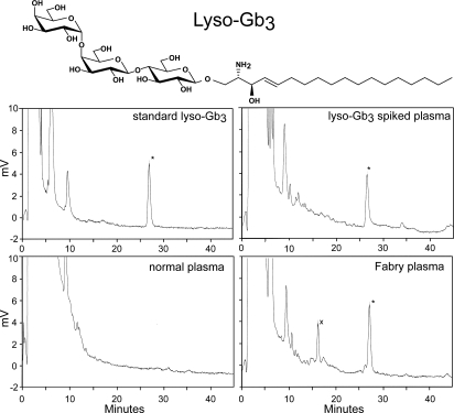
The consideration that a pathogenic, potentially vasoactive metabolite might be present in plasma from Fabry patients prompted us to investigate the presence of lyso-Gb3 (deacylated Gb3). Hitherto the existence of lyso-Gb3 has not been documented. However, various deacylated glycosphingolipids have been identified in lysosomal glycolipid storage diseases (see discussion in ref. 18 for an overview). For example, in Gaucher disease, glucosylsphingosine, the deacylated form of the accumulating glucosylceramide, is known to be increased as well (18, 19). The same holds for Krabbe disease in which galactosylsphingosine or psychosine (deacylated galactosylceramide) is elevated (20–23). It has been proposed that such lysosphingolipids play a role in the pathogenesis of these disorders (18–24). lyso-Gb3 is a cationic amphiphile with a large polar sugar moiety, rendering it relatively hydrophilic and water-soluble (see Fig. 1 Inset). Its presence therefore may have escaped attention during lipid analyses focusing on constituents of the organic phase of extractions. By using normal plasma spiked with a standard of pure lyso-Gb3, an appropriate extraction procedure for lyso-Gb3 was developed. lyso-Gb3 was recovered quantitatively from the upper phase of the extraction according to the Bligh and Dyer procedure (25), and it was purified by repeated extraction of the upper phase with 1-butanol (see Materials and Methods). The free amine moiety of lyso-Gb3 was fluorescently labeled with ortho-phthaldialdehyde according to a procedure used earlier for labeling of other lysosphingolipids (26). Labeled lysosphingolipids were separated by high-performance liquid chromatography and quantitatively analyzed (see Materials and Methods). Plasma samples from symptomatic Fabry patients were found to contain large quantities of a compound with a retention time identical to that of authentic lyso-Gb3 in the spiked plasma samples (Fig. 1). This compound could not be detected in plasma from control individuals. The identity of the abnormal structure as lyso-Gb3 was further verified. Incubation of the plasma sample with recombinant α-galactosidase A resulted in a shift to a value coinciding with that shown by lactosylsphingosine. Tandem mass spectrometry analysis confirmed the identity of the abnormal structure as lyso-Gb3 [see supporting information (SI) _Text_]. Examination of chromatograms of plasma from symptomatic patients pointed to the presence of another, more polar, abnormal compound X with a retention time of 16.2 min (see Fig. 1, Fabry plasma). Its concentration is ≈5-fold lower than that of lyso-Gb3. The shift in its peak retention by 1 min after incubation with α-galactosidase A indicates that the compound also contains a terminal α-galactoside moiety, which suggests that this compound is presumably also a lysoglycolipid and genuine storage product in Fabry disease. Based on its chromatographic behavior, it seems unlikely that the compound is sphingosinedigalactoside, the deacylated form of ceramidedigalactoside. The latter lipid has been found to be increased in kidney of patients with Fabry disease (27). The low abundance of the compound did not yet allow identification of its structure by tandem mass spectrometry. The ratio of lyso-Gb3 to the other abnormal structure in Fabry plasma specimens was remarkably constant in one individual but varied slightly among different patients.
Fig. 1.
Detection of lyso-Gb3 in plasma of Fabry patient. (Top) Structure of lyso-Gb3. (Middle and Bottom) Chromatograms obtained for pure lyso-Gb3 (Middle Left), normal plasma (Bottom Left), normal plasma spiked with lyso-Gb3 (Middle Right), and Fabry hemizygote plasma (Bottom Right).
Elevated lyso-Gb3 Levels in Plasma of Members of a Large Fabry Pedigree.
Plasma levels of lyso-Gb3, Gb3, lactosylceramide, glucosylceramide, and ceramide were determined in members of a large Fabry pedigree (see Materials and Methods). The status of all individuals was confirmed by enzyme analysis and genotyping (mutation encoding amino acid substitution D136Y). Table 1 shows that plasma specimens of the unaffected males (father and two normal sons) showed lyso-Gb3 levels below the detection level of 10 nM and normal levels of Gb3 (1.3–1.8 μM). In sharp contrast, plasma from all five affected sons showed markedly elevated lyso-Gb3 (195–407 nM) and elevated Gb3 (4.9–10.0 μM) levels. In female members of the family, no clear increases in plasma Gb3 were noted, independent of the genetic status of individuals. However, lyso-Gb3 was clearly increased in the symptomatic aunt, mother, and daughter (22–76 nM), and it was below the detection limit in the asymptomatic daughter and the unaffected siblings.
Table 1.
Plasma lyso-Gb3 and neutral glycosphingolipids in members of Fabry pedigree
| Status | Age at sampling, years | MSSI | lysoGb3, nM | Gb3, μM | LacCer, μM | GlcCer, μM | Cer, μM |
|---|---|---|---|---|---|---|---|
| Normal, daughter | 4 | 0 | ND | 1.6 | 4.2 | 4.3 | 10.3 |
| Normal, daughter | 8 | 0 | ND | 1.8 | 4.8 | 5.1 | 7.5 |
| Normal, daughter | 17 | 0 | ND | 1.1 | 3.3 | 3.9 | 7.9 |
| Heterozygous, daughter | 6 | 0 | ND | 1.1 | 3.2 | 2.9 | 8.1 |
| Heterozygous, daughter | 14 | 10 | 22 | 1.6 | 3.6 | 4.1 | 6.2 |
| Heterozygous, mother | 41 | 22 | 36 | 1.7 | 4.9 | 5.0 | 6.9 |
| Heterozygous, aunt | 45 | 32 | 76 | 2.5 | 5.6 | 5.1 | 11.7 |
| Normal, son | 9 | 0 | ND | 1.3 | 4.0 | 4.9 | 7.7 |
| Normal, son | 24 | 0 | ND | NA | NA | NA | NA |
| Normal, father | 50 | 0 | ND | 1.8 | 3.7 | 5.4 | 10.7 |
| Hemizygous, son | 11 | 10 | 407 | 10.0 | 6.1 | 8.1 | 8.8 |
| Hemizygous, son | 16 | 20 | 320 | 6.7 | 3.8 | 4.1 | 6.0 |
| Hemizygous, son | 17 | 12 | 217 | 7.0 | 6.0 | 7.3 | 5.8 |
| Hemizygous, son | 18 | 19 | 222 | 6.0 | 6.5 | 6.2 | 8.6 |
| Hemizygous, son | 20 | 12 | 196 | 6.9 | 6.5 | 6.5 | 10.0 |
Plasma lyso-Gb3 and Manifestations of Disease.
Plotting the plasma concentration of lyso-Gb3 against that of Gb3 for individual members of a Dutch Fabry cohort (see SI Fig. 4) indicated that the elevation of plasma lyso-Gb3, like that of Gb3, is most prominent in hemizygotes. There is a strong correlation between the concentration of plasma lyso-Gb3 and Gb3 in hemizygous patients (n = 22, ρ = 0.75, P < 0.001). In sharp contrast to the patients with classic Fabry disease manifestations, individuals carrying the mutation encoding R112H α-galactosidase A, which is known to be associated with an atypical manifestation of symptoms (28), showed normal Gb3 levels and very low lyso-Gb3 levels.
The relationship of individual plasma lyso-Gb3 concentrations with age and the commonly used Mainz Severity Score Index (MSSI) of disease severity (29) was determined (see also SI Fig. 5). In the case of hemizygotes, no correlation was noted between plasma lyso-Gb3 with regard to age or MSSI. Very high lyso-Gb3 concentrations were detected in plasma specimens of all classically affected Fabry hemizygotes. Of note is that specifically young boys with little disease manifestations already showed high plasma lyso-Gb3. Also in the case of a newborn hemizygote, a prominently increased lyso-Gb3 (81 nM) was demonstrable. No significant correlation of plasma lyso-Gb3 in hemizygotes with left ventricular mass or other disease signs was observed.
A clear exception among the hemizygotes were individuals carrying the R112H mutation. Two atypically affected adult male patients with the R112H mutation (one with polyneuropathy as the only symptom and the other with isolated renal disease, possibly also related to hypertension) showed relatively low lyso-Gb3 concentrations. Their MSSI scores were 22 and 38, respectively. Three very mildly affected, middle-aged hemizygotes for the R112H mutation (MSSI of 2, 2, and 4) also showed very low plasma lyso-Gb3 levels.
Examination of female heterozygotes revealed that, with increasing age, lyso-Gb3 tends to be higher, although not significantly (P = 0.08). In females, lyso-Gb3 also tends to be more elevated with increasing MSSI score (Spearman's ρ = 0.49, P = 0.05). A clear exception in this respect was a 13-year-old heterozygote showing relatively high plasma lyso-Gb3 in combination with a low MSSI of 6. The patient, now receiving enzyme-replacement therapy (ERT), suffered from acroparesthesias on exercise and showed a white matter lesion on the MRI of the brain. The latter is relatively rare in young hemizygotes and suggests a rapidly progressive disease manifestation. Analysis of neuropathology and renal and cardiac involvement revealed that only left ventricular mass in Fabry heterozygotes correlated with plasma lyso-Gb3 concentration (Spearman's ρ = 0.68, P < 0.05). In conclusion, in Fabry hemizygotes marked increases in lyso-Gb3 are noted already at young age, with the exception of atypical carriers of the R112H mutation. In the case of female heterozygotes, a larger series of patients has to be analyzed to establish the relation of plasma lyso-Gb3 with age and disease manifestations.
lyso-Gb3 in Fabry Mice.
The plasma concentrations of lyso-Gb3 and Gb3 were determined in mice. Table 2 shows that, at 20 weeks of age, plasma lyso-Gb3 concentrations are clearly less elevated in female heterozygous mice compared with female mice homozygous for the α-galactosidase A gene disruption and male hemizygous mice. Plasma lyso-Gb3 was found to be prominently increased at very young age in hemizygous mice, being 476, 510, and 614 nM at 70, 138, and 243 days, respectively.
Table 2.
Plasma lyso-Gb3 and neutral glycosphingolipids in Fabry mice
| Sex | Status | X, nmol/ml | lysoGb3, nmol/ml | Gb3, nmol/ml | LacCer, nmol/ml | GlyCer, nmol/ml | Cer, nmol/ml |
|---|---|---|---|---|---|---|---|
| Female | Heterozygote | 0.004 | 0.035 | 0.2 | 0.1 | 6.4 | 3.0 |
| Female | Homozygote | 0.143 | 0.737 | 1.9 | 0.1 | 7.1 | 2.2 |
| Female | Wild-type | <0.010 | <0.010 | 0.1 | 0.2 | 3.2 | 4.5 |
| Male | Hemizygote | 0.107 | 0.510 | 2.6 | 0.2 | 4.9 | 3.4 |
| Male | Wild-type | <0.010 | <0.010 | 0.1 | 0.1 | 3.9 | 3.2 |
The presence of lyso-Gb3 in tissues was studied in 20-week-old hemizygous mice. In parallel, concentrations of Gb3 and some other glycosphingolipids were determined. Dramatic increases in Gb3 and lyso-Gb3 concentrations were noted in most tissues of hemizygous mice (Table 3). Very high concentrations of lyso-Gb3 were observed in liver and intestine, clearly exceeding that in plasma. This finding suggests that lyso-Gb3 is either actively formed or preferentially trapped in these organs.
Table 3.
Tissue content on lyso-Gb3 in hemizygous Fabry mice
| Status | Tissue | X, nmol/g | lysoGb3, nmol/g | Gb3, nmol/g | LacCer, nmol/g | GlyCer, nmol/g | Cer, nmol/g |
|---|---|---|---|---|---|---|---|
| Hemizygote | Aorta | 0.22 | 0.80 | 234 | 20.1 | 123 | 84.6 |
| Duodenum | 1.33 | 4.10 | 1,083 | 52.3 | 140 | 264 | |
| Brain | 0.15 | 0.53 | 35.7 | 21.3 | 11,400 | 542 | |
| Heart | 0.18 | 0.44 | 242 | 32.0 | 28.6 | 113 | |
| Kidney | 0.12 | 0.51 | 707 | 38.4 | 75.3 | 267 | |
| Liver | 2.30 | 10.9 | 713 | 28.9 | 47.5 | 292 | |
| Control | Aorta | <0.010 | <0.010 | 4.4 | 5.0 | 137 | 63.8 |
| Duodenum | <0.010 | <0.010 | 5.1 | 14.1 | 111 | 221 | |
| Brain | <0.010 | <0.010 | 4.6 | 23.5 | 10,700 | 628 | |
| Heart | <0.010 | <0.010 | 5.2 | 19.5 | 23.3 | 116 | |
| Kidney | <0.010 | <0.010 | 196 | 15.2 | 72.6 | 202 | |
| Liver | <0.010 | <0.010 | 2.9 | 12.6 | 51.6 | 252 |
Hydrophilic Nature of lyso-Gb3.
The structure of lyso-Gb3 predicts a high degree of solubility in water. Consistent with this notion, lyso-Gb3 is largely recovered in the methanol/water phase upon Bligh and Dyer extraction (see also Materials and Methods). Given the high concentration of lyso-Gb3 in plasma of symptomatic Fabry patients, we investigated its distribution among lipoproteins. In contrast to Gb3, which is completely lipoprotein-bound, only a very small percentage of lyso-Gb3 was found to be associated with lipoproteins. The vast majority of lyso-Gb3 (>95%), was recovered in the lipoprotein-free fraction, presumably associated to albumin, as is the case with other hydrophilic lipids such as fatty acids. Unfortunately, the possibility that lyso-Gb3 is secreted into urine can presently not be confirmed nor excluded with certainty. The detection of lyso-Gb3 in urine samples with the present procedure is hampered by the presence of interfering substances in this fluid.
Inhibitory Properties of lyso-Gb3 in Vitro and in Vivo.
Given the structural similarity of lyso-Gb3 to Gb3, its capacity to inhibit α-galactosidase A was investigated. First, the effect of lyso-Gb3 on activity of recombinant α-galactosidase A toward 4-methylumbeliferyl-α-galactoside was determined. Potent competitive inhibition (_K_i = 34 μM) was observed to be identical for the two tested recombinant α-galactosidase A preparations. The activity of recombinant α-galactosidase A preparations toward Gb3 as substrate also was pronouncedly inhibited by the presence of lyso-Gb3. IC50 values of ≈20 μM were observed.
Cultured fibroblasts from normal subjects accumulate Gb3 when exposed to lyso-Gb3. An illustrative example of this is depicted in SI Fig. 6. lyso-Gb3 at concentrations ≈100 nM, similar to those encountered in plasma of symptomatic Fabry patients, is sufficient to increase cellular Gb3. Incubation with 500 nM lyso-Gb3 resulted in an increase of cellular Gb3 from 2.03 nmol/mg protein to 3.16 nmol/mg protein within 9 days. This effect is presumed to be caused by inhibition of α-galactosidase A activity, but it cannot be excluded that, after uptake, some lyso-Gb3 is converted to Gb3. An ≈200-fold more potent inhibitory capacity of lyso-Gb3 in vivo compared with in vitro could be explained by trapping of the membrane-permeable lyso-Gb3 in the acidic lysosome. Its pKa predicts accumulation of lyso-Gb3 in lysosomes by a factor of ≈200, coinciding with the noted difference in apparent IC50 between in vitro and in vivo experiments.
Biological Effect of lyso-Gb3.
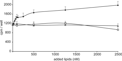
A characteristic feature of Fabry disease is vascular remodeling. This phenomenon is reflected in increased carotid intima-media thickness as the result of proliferation of smooth muscle cells (29, 30). To establish whether lyso-Gb3 exerts itself deleterious effects on the vasculature, the influence of the compound on smooth muscle cell proliferation was studied. Fig. 2 shows that low (50–100 nM) concentrations of lyso-Gb3, similar to those observed in plasma of symptomatic Fabry patients, markedly promote proliferation of smooth muscle cells in culture. In sharp contrast, the structurally related compounds like Gb3 and lactosylsphingosine have no stimulating effect on smooth muscle proliferation.
Fig. 2.
Increased proliferation of smooth muscle cells by exposure to lyso-Gb3. The proliferation rate of smooth muscle cells was determined as described in Materials and Methods. Cells were exposed for 24 h to indicated concentrations of lyso-Gb3 (♦), lactosylsphingosine (□), or Gb3 (△). The incorporation of radioactive thymidine per well is depicted.
We did not observe any stimulation by lyso-Gb3 of proliferation of fibroblasts (data not shown). These findings suggests that the elevation of lyso-Gb3 in plasma of Fabry patients may underlie the observed increase in intima-media thickness.
Degradation of lyso-Gb3 by α-Galactosidase A and Effect of ERT.
lyso-Gb3 can act as substrate for α-galactosidase A, resulting in its conversion to lactosylsphingosine. Compared with Gb3, the degradation of pure lyso-Gb3 in the test tube by recombinant α-galactosidase A preparations was found to be at least 50-fold less efficient (data not shown). At neutral pH, recombinant α-galactosidases A hardly hydrolyze lyso-Gb3 because of their acid pH optimum (see SI Text).
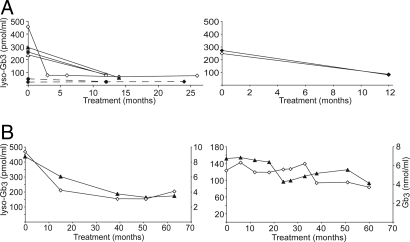
The effect of prolonged ERT on plasma lyso-Gb3 was studied for a selected subset of patients. Fig. 3 shows representative examples of the observed responses in plasma lyso-Gb3 to ERT with commercially available enzyme preparations agalsidase alfa (Shire) and agalsidase beta (Genzyme). Patients receiving either agalsidase alfa (Fig. 3 Left) or agalsidase beta (Fig. 3 Right), without developing antibodies, showed a reduction of lyso-Gb3 within 1 year, although a complete normalization was not observed. Some of the male hemizygotes lacking α-galactosidase A develop neutralizing antibodies upon ERT (31–33). Fig. 3B show the responses in plasma lyso-Gb3 and Gb3 to ERT for patients that had developed antibodies after receiving agalsidase alfa (Left) and agalsidase beta (Right). Reduction of plasma lyso-Gb3 in these individuals was relatively poor. At present, no statements can be made about the relative efficacy of the two commercial recombinant α-galactosidase A preparations or the influence of enzyme dose and antibody formation. A detailed investigation with carefully matched patients will be required to answer these important questions.
Fig. 3.
Effect of ERT on plasma lyso-Gb3. Illustrative examples of responses in plasma lyso-Gb3 in Fabry patients receiving ERT. (A) Responses in plasma lyso-Gb3 to agalsidase alfa treatment (0.2 mg/kg body weight, 2 weeks) (Left) or agalsidase beta treatment (0.2 mg/kg body weight, 2 weeks) (Right) in patients developing no antibodies. All patients were hemizygotes, except two indicated by the dotted line. (B) Response in plasma lyso-Gb3 (▲) and Gb3 (◇) to agalsidase alfa treatment (0.2 mg/kg body weight, 2 weeks) (Left) or agalsidase beta treatment (0.2 mg/kg body weight, 2 weeks) (Right) in patients developing antibodies. The dose of agalsidase beta was adjusted to (1.0 mg/kg body weight, 2 weeks) at 36 months of treatment.
Discussion
The striking abnormality in lyso-Gb3 in plasma of Fabry patients noticed in this study has been overlooked for many decades, likely because the compound is very water-soluble and does not distribute into the organic phase during organic solvent extractions. Our discovery of markedly elevated concentrations lyso-Gb3 in plasma of symptomatic Fabry patients and the potential of the compound to inhibit α-galactosidase A activity and to induce smooth muscle cell proliferation is of considerable interest. Vascular involvement in Fabry patients is known to include an accelerated hypertrophy of the walls of the radial artery and a thickening of the intima media of the common carotid artery because of proliferation of smooth muscle cells (29, 30). In the near future, a large number of patients, in particular heterozygotes, should be studied to determine the relationship between intima-media thickness and plasma lyso-Gb3 levels. Left ventricular hypertrophy also is common in Fabry disease. Of note, a correlation was observed between left ventricular hypertrophy and plasma lyso-Gb3 concentration in heterozygotes.
The elevated lyso-Gb3 in plasma of symptomatic patients might partially explain the finding by Barbey et al. (17) of an unidentified substance in plasma of symptomatic Fabry disease patients that stimulates proliferation of vascular smooth muscle cells and cardiomyocytes in vitro. Elevated plasma lyso-Gb3 also may provide plausible explanations for other poorly understood phenomena in Fabry disease. First, the inhibitory properties of lyso-Gb3 may help to explain the repeated finding of Gb3 accumulation in renal transplants; prior observations already pointed to a circulating inhibitor of α-galactosidase A in Fabry patients (34, 35). Second, manifestation of the disease in female heterozygotes, despite considerable residual circulating α-galactosidase A and a significant proportion of enzyme-competent cells, becomes more understandable. It can be envisioned that formation of lyso-Gb3 in enzyme-deficient cells, followed by its release into the circulation and subsequent uptake in other cells, ultimately also will inhibit α-galactosidase A activity in enzyme-competent cells. Moreover, the circulating lyso-Gb3 may stimulate vascular remodeling. The disease thus may eventually manifest itself in female heterozygotes as devastatingly as in male hemizygotes. An additional consequence of high lyso-Gb3 concentrations in plasma also should be considered. It is conceivable that the lyso-Gb3 after its uptake by cells subsequently may be acylated to Gb3, which could lead to increased concentrations of extralysosomal Gb3. This pool of Gb3 would not be accessible to endogenous α-galactosidase A or enzyme administered during ERT. Extralysosomal accumulation of Gb3 recently has been documented in tissues from Fabry patients (36).
The clear increase in plasma lyso-Gb3 in young hemizygotes, and similar findings in hemizygous mice, is of particular interest. Markedly elevated plasma lyso-Gb3 in a newborn is a particularly striking finding on the basis of which it seems that, in some hemizygotes, vascular remodeling might already be initiated at or even before birth. This finding would imply that there exists a considerable window between increase of plasma lyso-Gb3 and detectable disease manifestations. In female heterozygotes, the build-up of lyso-Gb3 clearly proceeds more slowly than in the hemizygotes. Monitoring plasma lyso-Gb3 in clinical studies aimed on prevention of signs and symptoms seems of interest. It is of interest to note that Fabry mice, despite relatively high lyso-Gb3 and Gb3 contents of plasma and kidney, do not show overt kidney disease, which suggests that microvascular complications in the kidney are not swiftly induced by the lipid abnormalities.
The origin of lyso-Gb3 is elusive. It might be synthesized by sequential glycosylation of sphingoid bases and accumulate because of the deficiency in α-galactosidase A. Based on the analysis of the sphingoid base composition, Kobayashi et al. (37) earlier suggested that in brain from patients with GM1 gangliosidosis and GM2 gangliosidosis elevated levels of corresponding lysosphingolipids might originate from biosynthesis and not from deacylation of sphingolipids. Biosynthesis of galactosylsphingosine (psychosine) from sphingosine also has been reported (20, 38). Alternatively, it might still be that lyso-Gb3 is formed by deacylation of the stored Gb3 in Fabry patients. The strikingly constant ratio between lyso-Gb3 and Gb3 in individual Fabry patients and Fabry mice is consistent with this but does not discriminate between the two possibilities. It has to be established whether lyso-Gb3 can originate spontaneously from stored Gb3 or even is actively formed by the action of a specific enzyme. Candidates in this connection are the lysosomal ceramidase, which might be promiscuous toward Gb3, or a hitherto unknown specific glycosphingolipid deacylase. Intraindividual differences in these tentative lyso-Gb3-generating enzyme activities might conceivably impact on the age of onset and progression of Fabry disease.
Our observation that lyso-Gb3 induces smooth muscle cell proliferation warrants additional investigations on the cellular mechanisms involved. It already has been demonstrated that lysosphingolipids can affect cellular proliferation via interference in signal transduction. For example, sphingosine-1-phosphate is known to stimulate proliferation of smooth muscle cells by a variety of signal transduction pathways including ERK and MAPK (39, 40).
Our findings suggest that Fabry disease might no longer be viewed as a prototypic lysosomal storage disorder. Although lysosomal storage of Gb3 is clearly a prerequisite for manifestations of Fabry disease, the formation of pathogenic lyso-Gb3 may be instrumental with regard to the onset and progression of some manifestations. In this sense, Fabry disease would resemble more closely Krabbe disease in which the toxic metabolite galactosylphingosine (psychosine) rather than galactosylceramide is considered to be a crucial factor in the pathogenesis (23). In this respect, it will be valuable to establish the identity of lipid X (presumably also a lyso-glycolipid) that also is present in Fabry plasma.
The role of lyso-Gb3 in the onset of various clinical signs of Fabry disease should be firmly established. Provided that it is unequivocally demonstrated that lyso-Gb3 contributes to disease manifestations, measurement of its concentration in plasma may become a valuable guide for clinical management. Monitoring of lyso-Gb3 possibly could assist in decision-making when therapeutic intervention has to be initiated and which treatment regimen results in individual patients in optimal prevention and/or correction. Our investigation has indicated that ERT with recombinant α-galactosidases can reduce, but not easily normalize, plasma lyso-Gb3. However, it is reassuring that the abnormality in lyso-Gb3 in Fabry patients is at least partly responsive to ERT. The observed reductions in plasma lyso-Gb3 after ERT could be either caused by direct intralysosomal degradation of lyso-Gb3 or result from intralysosomal clearance of stored Gb3, limiting the source for production of lyso-Gb3. In addition to existing ERTs, novel therapeutic avenues that specifically target prevention of lyso-Gb3 formation or promote its removal also may become of interest in the future.
In summary, markedly elevated lyso-Gb3 constitutes a hallmark of Fabry disease, and the potential role of this metabolite in the manifestations of the disease warrants further investigation.
Materials and Methods
Fabry Patients, Plasma Specimens, and Cultured Fibroblasts.
Selection from the cohort of patients with Fabry disease at the Academic Medical Center in Amsterdam was carried out for analysis of (glyco)sphingolipids and disease manifestations before the initiation of ERT. This selection included 17 classically affected hemizygotes (age range 6–45 years), 17 heterozygotes (age range 11–52 years), two newborns (1 hemizygote and 1 heterozygote), and 5 atypical hemizygotes with the R112H mutation. In addition, 10 control subjects (five males and five females) were included. A large Fabry family was part of this study, including an affected mother and her affected sister, a healthy father, five hemizygous sons, three heterozygous individuals, and five healthy siblings. Ten patients (eight hemizygotes and two heterozygotes) received either 0.2 mg/kg agalsidase alfa (Replagal, Shire) or 0.2 mg/kg or 1.0 mg/kg agalsidase beta (Fabrazyme, Genzyme). The diagnosis of Fabry disease was confirmed by demonstration of deficient activity of α-galactosidase A in leukocytes and genotyping. Blood samples were obtained with informed consent for investigations. Skin fibroblasts from Fabry patients (n = 2) and control individuals (n = 2) were cultured as described in ref. 41.
Clinical Assessments.
A complete medical history was obtained, and physical examination was performed on all patients. Clinical assessments are described in detail in SI Text. Briefly, as described in ref. 7, renal function was analyzed and brain magnetic resonance imaging, electrocardiograms, cardiac ultrasound investigations, hearing tests, and assessments of pain were carried out. Overall severity of disease was assessed by using the MSSI (9).
Statistical Analysis.
Correlations were calculated for hemizygotes and heterozygotes separately, with the exclusion of the atypical cases carrying the R112H mutation and the two newborns. Correlations between variables were described with the use of Spearman rank correlation coefficients. P values of <0.05 were considered significant.
Fabry Mice.
C3HeB/FeJ and B6;129-Gla tm1Kul (stock number 003535) strains were obtained from The Jackson Laboratory. Animals were fed a commercially available laboratory diet (RMH-B; Hope Farms, Woerden, The Netherlands).
Culture and Measurement of Proliferation of Smooth Muscle Cells.
Smooth muscle cells were obtained from Carlie de Vries (Amsterdam). Smooth muscle cells were seeded in 24-well plates at 1–4 × 10⁴ cells per well and reached 60–70% confluence after 24 h. Smooth muscle cells were made quiescent by incubation for 24 h in FBS-free medium and then were stimulated for 24 h with 5% (vol/vol) FBS. Subsequently, cells were labeled for 4 h with 0.25 μCi per well [methyl-3H]thymidine (Amersham Biosciences). Incorporated [3H]thymidine was precipitated for 30 min at 4°C with 10% (wt/vol) trichloroacetic acid, washed twice with 5% (wt/vol) trichloroacetic acid, and dissolved in 0.5 M NaOH (0.5 ml per well), and radioactivity was measured by liquid-scintillation counting.
Recombinant α-Galactosidases.
Agalsidase alfa (Replagal, Shire) and agalsidase beta (Fabrazyme, Genzyme) were purchased from the local hospital pharmacy.
Enzyme Activity Assays.
The enzymatic activity of α-galactosidase A was determined with fluorogenic 4-methylumbelliferyl-α-galactoside as substrate (41). Enzyme preparations were incubated at 37°C with 3.5 mM substrate in 100 mM citrate/200 mM phosphate buffer (pH 4.6) at 37°C. Alternatively, enzymatic activity was measured toward Gb3 in the presence of 0.5% (wt/vol) sodium taurocholate in 50 mM sodium acetate buffer (pH 4.2). The formation lactosylceramide was determined by high-performance thin layer chromatography (HPTLC) analysis (12).
Plasma Fractionation.
Fasted EDTA plasma was obtained from a patient with Fabry disease and an age- and gender-matched control subject. Lipoproteins were separated by gradient ultracentrifugation according to the procedure of Redgrave et al. (42).
Measurement of Neutral (Glyco)sphingolipids.
Gb3, lactosylceramide, glucosylceramide, and ceramide were measured by using high-performance liquid chromatography as described in ref. 26. The mean Gb3 concentration in plasma from controls was 1.7 (0.5) μmol/liter (n = 40), mean concentration of glucosylceramide in plasma from controls was 6.3 (1.9) μmol/liter (n = 40), and mean concentration of ceramide in plasma from controls was 9.0 (2.3) μmol/liter (n = 40) (26).
Measurement of lyso-Gb3 in Fabry Plasma and Tissues.
To 100 μl of plasma or tissue homogenates, 50 μl of water was added, and the lipids were extracted with 900 μl of chloroform/methanol 1/2 (vol/vol). The extract was centrifuged for 10 min at 14,000 × g, and the supernatant was removed. To the supernatant, 0.3 ml of chloroform and 0.45 ml of water were added, and after mixing, the phases were separated by brief centrifugation. The upper phase was removed, and the lower chloroform phase was extracted once more with 1.2 ml of methanol/water 1/1 (vol/vol). The combined upper phases were dried and taken up in 1 ml of water, and the water phase was extracted twice with 1 ml of water-saturated 1-butanol. lyso-Gb3 was recovered from the butanol phase with an overall recovery of >90%. The butanol phase was dried and dissolved in 120 μl of methanol. Of this solution, 50 μl was taken to dryness, and the lyso-Gb3 was derivatized with 25 μl of _o_-phtaldialdehyde (OPA) reagent (5 mg of OPA, 0.1 ml of ethanol, 5 μl of 2-mercaptoethanol, and 10 ml of 3% boric acid, pH 10.7). The OPA-derivatized lyso-Gb3 was separated by HPLC and identified by fluorescence detection as described in ref. 26. Quantification was by external standardization after standard addition of authentic lyso-Gb3 (Sigma-Aldrich) to normal plasma so that increasing concentrations from 0 to 1,000 nmol/liter plasma were obtained. The response was strictly linear with the lyso-Gb3 concentration, and the limit of quantification was 10 nmol/liter. Peak identification was by comparison of the retention time with that of authentic lyso-Gb3.
Supplementary Material
Supporting Information
ACKNOWLEDGMENTS.
We thank the members of the Dutch Fabry patient society (FSIGN) for their collaboration. Els Ormel is acknowledged for assistance in the clinic, Carlie de Vries and Thijs Pols for advice regarding experiments with smooth muscle cells, and Anton Bussink and Albert Groen for stimulating discussions. This research was funded by the Academic Medical Center in Amsterdam.
Footnotes
The authors declare no conflict of interest.
References
- 1.Brady RO, et al. Enzymatic defect in Fabry's disease: Ceramidetrihexosidase deficiency. N Engl J Med. 1967;276:1163–1167. doi: 10.1056/NEJM196705252762101. [DOI] [PubMed] [Google Scholar]
- 2.Kint JA. Fabry's disease: α-Galactosidase deficiency. Science. 1970;167:1268–1269. doi: 10.1126/science.167.3922.1268. [DOI] [PubMed] [Google Scholar]
- 3.Desnick RJ, Ioannou YA. α-Galactosidase A deficiency: Fabry disease. In: Scriver R, et al., editors. The Metabolic and Molecular Bases of Inherited Disease. New York: McGraw-Hill; 2001. pp. 3733–3774. [Google Scholar]
- 4.Brady RO, et al. Replacement therapy for inherited enzyme deficiency: Use of purified ceramidetrihexosidase in Fabry's disease. N Engl J Med. 1973;289:9–14. doi: 10.1056/NEJM197307052890103. [DOI] [PubMed] [Google Scholar]
- 5.Schiffmann R, et al. Enzyme-replacement therapy in Fabry disease: A randomized controlled trial. J Am Med Assoc. 2001;285:2743–2749. doi: 10.1001/jama.285.21.2743. [DOI] [PubMed] [Google Scholar]
- 6.Eng CM, et al. Safety and efficacy of recombinant human α-galactosidase A—Replacement therapy in Fabry's disease. N Engl J Med. 2001;345:9–16. doi: 10.1056/NEJM200107053450102. [DOI] [PubMed] [Google Scholar]
- 7.Vedder AC, et al. Treatment of Fabry disease: Outcome of a comparative trial with agalsidase alfa or β at a dose of 0.2 mg/kg. PLoS ONE. 2007;2:e598. doi: 10.1371/journal.pone.0000598. [DOI] [PMC free article] [PubMed] [Google Scholar]
- 8.MacDermot KD, Holmes A, Miners AH. Natural history of Fabry disease in affected males and obligate carrier females. J Inherit Metab Dis. 2001;24(Suppl 2):13–14. doi: 10.1023/a:1012447102358. [DOI] [PubMed] [Google Scholar]
- 9.Whybra C, et al. Anderson-Fabry disease: Clinical manifestations of disease in female heterozygotes. J Inherit Metab Dis. 2001;24:715–724. doi: 10.1023/a:1012993305223. [DOI] [PubMed] [Google Scholar]
- 10.Deegan PB, et al. Natural history of Fabry disease in females in the Fabry Outcome Survey. J Med Genet. 2006;43:347–352. doi: 10.1136/jmg.2005.036327. [DOI] [PMC free article] [PubMed] [Google Scholar]
- 11.Gupta S, Ries M, Kotsopoulos S, Schiffmann R. The relationship of vascular glycolipid storage to clinical manifestations of Fabry disease: A cross-sectional study of a large cohort of clinically affected heterozygous women. Medicine. 2005;84:261–268. doi: 10.1097/01.md.0000178976.62537.6b. [DOI] [PubMed] [Google Scholar]
- 12.Vedder AC, et al. The Dutch Fabry cohort: Diversity of clinical manifestations and Gb3 levels. J Inherit Metab Dis. 2007;30:68–78. doi: 10.1007/s10545-006-0484-8. [DOI] [PubMed] [Google Scholar]
- 13.Vedder AC, et al. Manifestations of Fabry disease in placental tissue. J Inherit Metab Dis. 2006;29:106–111. doi: 10.1007/s10545-006-0196-0. [DOI] [PubMed] [Google Scholar]
- 14.Ohshima T, et al. α-Galactosidase A deficient mice: A model of Fabry disease. Proc Natl Acad Sci USA. 1997;94:2540–2544. doi: 10.1073/pnas.94.6.2540. [DOI] [PMC free article] [PubMed] [Google Scholar]
- 15.Young E, et al. Is globotriaosylceramide a useful biomarker in Fabry disease? Acta Paediatr Suppl. 2005;94:51–54. doi: 10.1111/j.1651-2227.2005.tb02112.x. [DOI] [PubMed] [Google Scholar]
- 16.Bekri S, Lidove O, Jaussaud R, Knebelmann B, Barbey F. The role of ceramide trihexoside (globotriaosylceramide) in the diagnosis and follow-up of the efficacy of treatment of Fabry disease: A review of the literature. Cardiovasc Hematol Agents Med Chem. 2006;4:289–297. doi: 10.2174/187152506778520718. [DOI] [PubMed] [Google Scholar]
- 17.Barbey F, et al. Cardiac and vascular hypertrophy in Fabry disease: Evidence for a new mechanism independent of blood pressure and glycosphingolipid deposition. Arterioscler Thromb Vasc Biol. 2006;26:839–844. doi: 10.1161/01.ATV.0000209649.60409.38. [DOI] [PubMed] [Google Scholar]
- 18.Orvisky E, et al. Glucosylsphingosine accumulation in tissues from patients with Gaucher disease: Correlation with phenotype and genotype. Mol Genet Metab. 2002;76:262–270. doi: 10.1016/s1096-7192(02)00117-8. [DOI] [PubMed] [Google Scholar]
- 19.Nilsson O, Svennerholm L. Accumulation of glucosylceramide and glucosylsphingosine (psychosine) in cerebrum and cerebellum in infantile and juvenile Gaucher disease. J Neurochem. 1982;39:709–718. doi: 10.1111/j.1471-4159.1982.tb07950.x. [DOI] [PubMed] [Google Scholar]
- 20.Vanier M, Svennerholm L. Chemical pathology of Krabbe disease: The occurrence of psychosine and other neutral sphingoglycolipids. Adv Exp Med Biol. 1976;68:115–126. doi: 10.1007/978-1-4684-7735-1_8. [DOI] [PubMed] [Google Scholar]
- 21.Svennerholm L, Vanier MT, Månsson JE. Krabbe disease: A galactosylsphingosine (psychosine) lipidosis. J Lipid Res. 1980;21:53–64. [PubMed] [Google Scholar]
- 22.Igisu H, Suzuki K. Progressive accumulation of toxic metabolite in a genetic leukodystrophy. Science. 1984;224:753–755. doi: 10.1126/science.6719111. [DOI] [PubMed] [Google Scholar]
- 23.Suzuki K. Twenty five years of the “psychosine hypothesis”: A personal perspective of its history and present status. Neurochem Res. 1998;23:251–259. doi: 10.1023/a:1022436928925. [DOI] [PubMed] [Google Scholar]
- 24.Schueler UH, et al. Toxicity of glucosylsphingosine (glucopsychosine) to cultured neuronal cells: A model system for assessing neuronal damage in Gaucher disease type 2 and 3. Neurobiol Dis. 2003;14:595–601. doi: 10.1016/j.nbd.2003.08.016. [DOI] [PubMed] [Google Scholar]
- 25.Bligh EG, Dyer WJ. A rapid method for total lipid extraction and purification. Can J Biochem Physiol. 1959;37:911–917. doi: 10.1139/o59-099. [DOI] [PubMed] [Google Scholar]
- 26.Groener JE, et al. HPLC for simultaneous quantification of total ceramide, glucosylceramide, and ceramide trihexoside concentrations in plasma. Clin Chem. 2007;53:742–747. doi: 10.1373/clinchem.2006.079012. [DOI] [PubMed] [Google Scholar]
- 27.Li YT, Li SC, Dawson G. Anomeric structure of ceramide digalactoside isolated from the kidney of a patient with Fabry's disease. Biochim Biophys Acta. 1972;260:88–92. [PubMed] [Google Scholar]
- 28.Eng CM, et al. Fabry disease: Twenty-three mutations including sense and antisense CpG alterations and identification of a deletional hot-spot in the α-galactosidase A gene. Mol Med. 1997;3:174–182. doi: 10.1093/hmg/3.10.1795. [DOI] [PubMed] [Google Scholar]
- 29.Kalliokoski RJ, et al. Structural and functional changes in peripheral vasculature of Fabry patients. J Inherit Metab Dis. 2006;29:660–666. doi: 10.1007/s10545-006-0340-x. [DOI] [PubMed] [Google Scholar]
- 30.Barbey F, et al. Increased carotid intima-media thickness in the absence of atherosclerotic plaques in an adult population with Fabry disease. Acta Paediatr Suppl. 2006;95:63–68. doi: 10.1111/j.1651-2227.2006.tb02392.x. [DOI] [PubMed] [Google Scholar]
- 31.Linthorst GE, Hollak CE, Donker-Koopman WE, Strijland A, Aerts JM. Enzyme therapy for Fabry disease: Neutralizing antibodies toward agalsidase alpha and beta. Kidney Int. 2004;66:1589–1595. doi: 10.1111/j.1523-1755.2004.00924.x. [DOI] [PubMed] [Google Scholar]
- 32.Whitfield PD, et al. Monitoring enzyme-replacement therapy in Fabry disease—Role of urine globotriaosylceramide. J Inherit Metab Dis. 2005;28:21–33. doi: 10.1007/s10545-005-4415-x. [DOI] [PubMed] [Google Scholar]
- 33.Ohashi T, et al. Influence of antibody formation on reduction of globotriaosylceramide (GL-3) in urine from Fabry patients during agalsidase beta therapy. Mol Genet Metab. 2007;92:271–273. doi: 10.1016/j.ymgme.2007.06.013. [DOI] [PubMed] [Google Scholar]
- 34.Van den Bergh FA, Rietra PJ, Kolk-Vegter AJ, Bosch E, Tager JM. Biochemical outcome of successful renal transplantation in a Fabry patient. Acta Med Scand. 1976;200:249–256. [PubMed] [Google Scholar]
- 35.Mosnier JF, et al. Recurrence of Fabry's disease in a renal allograft 11 years after successful renal transplantation. Transplantation. 1991;51:759–762. doi: 10.1097/00007890-199104000-00004. [DOI] [PubMed] [Google Scholar]
- 36.Askari H, et al. Cellular and tissue localization of globotriaosylceramide in Fabry disease. Virchows Arch. 2007;451:823–834. doi: 10.1007/s00428-007-0468-6. [DOI] [PubMed] [Google Scholar]
- 37.Kobayashi T, et al. Accumulation of lysosphingolipids in tissues from patients with GM1 and GM2 gangliosidoses. J Neurochem. 1992;59:1452–1458. doi: 10.1111/j.1471-4159.1992.tb08460.x. [DOI] [PubMed] [Google Scholar]
- 38.Cleland WW, Kennedy EP. The enzymatic synthesis of psychosine. J Biol Chem. 1960;235:45–51. [PubMed] [Google Scholar]
- 39.Lockman K, et al. Sphingosine 1-phosphate stimulates smooth muscle cell differentiation and proliferation by activating separate serum response factor co-factors. J Biol Chem. 2004;279:42422–42430. doi: 10.1074/jbc.M405432200. [DOI] [PubMed] [Google Scholar]
- 40.Michel MC, Mulders AC, Jongsma M, Alewijnse AE, Peters SL. Vascular effects of sphingolipids. Acta Paediatr Suppl. 2007;96:44–48. doi: 10.1111/j.1651-2227.2007.00207.x. [DOI] [PubMed] [Google Scholar]
- 41.Blom D, et al. Recombinant enzyme therapy for Fabry disease: Absence of editing of human α-galactosidase A mRNA. Am J Hum Genet. 2003;72:23–31. doi: 10.1086/345309. [DOI] [PMC free article] [PubMed] [Google Scholar]
- 42.Redgrave TG, Roberts DC, West CE. Separation of plasma lipoproteins by density-gradient ultracentrifugation. Anal Biochem. 1975;65:42–49. doi: 10.1016/0003-2697(75)90488-1. [DOI] [PubMed] [Google Scholar]
Associated Data
This section collects any data citations, data availability statements, or supplementary materials included in this article.
Supplementary Materials
Supporting Information


