A viral suppressor of gene silencing in plants (original) (raw)
Abstract
Gene silencing is an important but little understood regulatory mechanism in plants. Here we report that a viral sequence, initially identified as a mediator of synergistic viral disease, acts to suppress the establishment of both transgene-induced and virus-induced posttranscriptional gene silencing. The viral suppressor of silencing comprises the 5′-proximal region of the tobacco etch potyviral genomic RNA encoding P1, helper component-proteinase (HC-Pro) and a small part of P3, and is termed the P1/HC-Pro sequence. A reversal of silencing assay was used to assess the effect of the P1/HC-Pro sequence on transgenic tobacco plants (line T4) that are posttranscriptionally silenced for the uidA reporter gene. Silencing was lifted in offspring of T4 crosses with four independent transgenic lines expressing P1/HC-Pro, but not in offspring of control crosses. Viral vectors were used to assess the effect of P1/HC-Pro expression on virus-induced gene silencing (VIGS). The ability of a potato virus X vector expressing green fluorescent protein to induce silencing of a green fluorescent protein transgene was eliminated or greatly reduced when P1/HC-Pro was expressed from the same vector or from coinfecting potato virus X vectors. Expression of the HC-Pro coding sequence alone was sufficient to suppress virus-induced gene silencing, and the HC-Pro protein product was required for the suppression. This discovery points to the role of gene silencing as a natural antiviral defense system in plants and offers different approaches to elucidate the molecular basis of gene silencing.
Homology-dependent gene silencing appears to be a fundamental regulatory mechanism operating in diverse types of organisms (1). The gene silencing phenotype is characterized by reduced levels of the specific mRNA encoded by the suppressed gene(s), and individual cases fall into two major mechanistic classes: (i) those in which mRNA level is regulated transcriptionally and (ii) those in which it is regulated posttranscriptionally. Because both classes of silencing are associated with sequence-specific suppression of RNA accumulation and methylation of the corresponding gene (2–5), it is possible that the two major classes of silencing are linked in an as yet undetermined way. However, the mechanisms leading to the establishment of gene silencing, and the identity of cellular genes that mediate the phenomenon, are unknown.
Homology-dependent gene silencing is probably best documented in transgenic plants where it may be induced by insertion of multiple copies of homologous transgenes or by insertion of a transgene with homology to an endogenous gene (6–9). The phenomenon may also be induced by plant viruses, and this was first observed in viruses expressing sequences with homology to host transgenes or endogenous genes (5, 10, 11). More recently it has been reported that gene silencing can be induced by plant virus infections in the absence of any known homology of the viral genome to host genes and that this silencing may occur at either the transcriptional or the posttranscriptional level (12–14). These observations of virus-induced gene silencing (VIGS) have led to the proposal that gene silencing may have evolved as a defense mechanism against viral invasion. If gene silencing serves as a general defense mechanism against plant viruses, it would not be surprising if some plant viruses have evolved ways to circumvent the defense system.
Further evidence of a general antiviral defense pathway in plants comes from studies of synergistic viral disease, in which coinfection with two heterologous viruses leads to much more severe symptoms than does infection with either virus alone. Many such synergistic diseases involve a member of the potyvirus group of plant viruses. Previously, it was found that transgenic plants expressing the 5′-proximal region of the tobacco etch potyviral (TEV) genome (termed the P1/HC-Pro sequence) develop synergistic disease when infected with any of a broad range of plant viruses (15). This result suggested that expression of the P1/HC-Pro sequence might interfere with a general antiviral system in plants, thereby permitting viruses to accumulate beyond the normal host-mediated limits. Posttranscriptional gene silencing is a candidate for such a system, and the studies presented here were designed to test the hypothesis that the P1/HC-Pro sequence interferes with the establishment of gene silencing. The effect of P1/HC-Pro expression on posttranscriptional gene silencing was tested in two different silencing systems. The results support the suggestion that P1/HC-Pro acts as a suppressor of both transgene-induced and virus-induced gene silencing.
MATERIALS AND METHODS
Transgenic Tobacco Lines.
The six independent TEV P1/HC-Pro and vector transgenic tobacco lines used in these studies are all homozygous and were the kind gift of J. Carrington (Washington State University); the construction of each has been described (16–18). In brief, the TEV sequences were first cloned into the intermediate vector pRTL2 (16) and then transferred along with the cauliflower mosaic virus 35S promoter and terminator sequences to the binary vector pGA482 (19). The U-6B and vector-only control plants are transgenic lines in Nicotiana tabacum cv Havana 425. Line U-6B carries the wild-type P1/HC-Pro sequence (nucleotides 12–2,681 of the TEV genome) and expresses HC-Pro at a level equivalent to that in TEV-infected leaves (16). Lines TEV-B, TEV-C, TEV-I, and TEV-K were constructed in N. tabacum cv Xanthi and contain the P1/HC-Pro sequence as in line U-6B, except that each contains a nine nucleotide insertion introduced by site-directed mutagenesis (20). The insertion produces an _Nco_I site in the transgene DNA and results in an insertion of the amino acid triplet Thr-Met-Ala at different locations within the TEV P1/HC-Pro polyprotein. For this study, the previously reported location of the insertion in each line was verified by PCR amplification of the transgene and restriction enzyme analysis of the PCR product (data not shown). This analysis confirmed the reported locations of the _Nco_I site for lines TEV-C, TEV-I, and TEV-K (18). However, the insertion in line TEV-B, reported to be at nucleotide 616 within the P1-coding sequence (17, 18), was determined to be approximately at nucleotide 973 of the P1-coding sequence. Primary transformants and T1 progeny of selfed primary transformants of each line originally expressed HC-Pro (16–18). However, the homozygous TEV-K line no longer accumulates detectable levels of P1/HC-Pro RNA or HC-Pro protein (data not shown) and may be posttranscriptionally silenced for the P1/HC-Pro sequence.
D. Baulcombe (Sainsbury Laboratory, Norwich, U.K.) graciously provided the β-glucuronidase (GUS)-silenced transgenic line T4 and the green fluorescent protein (GFP)-expressing Nicotiana benthamiana transgenic line.
Histochemical Staining for GUS Activity.
Leaves were assayed for GUS activity as described (21) but with minor modifications. For staining of GUS at sites of PVX–GUS infection (foci), the leaves were partially abraded on the lower side by using carborundum, fixed for 20 min in 90% acetone, vacuum infiltrated with a buffer containing 50 mM sodium phosphate (pH 7.2), 0.5 mM K3Fe (CN)6, 0.5 mM K3Fe(CN)6, and 1 mM 5-bromo-4-chloro-3-indolyl β-d-glucuronide and incubated at 37°C. Leaf pieces were subsequently treated with 95% ethanol to remove chlorophyll. The GUS staining in PVX–GUS foci is visible within 15–60 min under these conditions, whereas staining in transgenic tissues is not detectable under the same conditions (see Fig. 1). To stain for GUS in transgenic tissues as in Fig. 2, leaves were carefully and uniformly abraded on the lower side with carborundum, substrate concentration was increased 4-fold to 4 mM, and leaves were incubated for 12–24 hr at 37°C.
Figure 1.
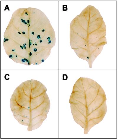
PVX–GUS infection of offspring of GUS-silenced transgenic line T4. Three-week-old seedlings were inoculated with PVX–GUS (27), and the inoculated leaves were assayed for GUS activity by histochemical staining at 5 days postinoculation. Blue foci are indicative of viral replication. (A) T4 crossed with line TEV-B, which expresses the P1/HC-Pro sequence; (B) homozygous T4; (C) T4 crossed with line TEV-K, which contains a P1/HC-Pro sequence carrying a mutation that inactivates HC-Pro; and (D) T4 crossed with nontransformed N. tabacum cv xanthi nc.
Figure 2.
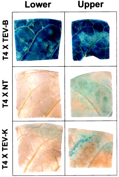
Histochemical staining of GUS activity in leaves of offspring of GUS-silenced transgenic line T4 crossed with line TEV-B (Upper), nontransformed tobacco (Middle), or line TEV-K (Bottom). Leaves of 4- to 5-week-old seedlings were assayed as described.
RNA Analysis.
RNA isolation and Northern blot analyses were carried out as described (22). The probes for the uidA mRNA and the GFP mRNA were 32P-labeled randomly primed cDNAs from PCR-amplified fragments representing the entire GUS or GFP coding regions, respectively. In all cases, ethidium bromide staining of 18S ribosomal RNA was used to confirm equal loading of total RNA per lane.
Construction of PVX Vectors.
The construction and characterization of PVX–TEV, PVX–HC, and PVX–noHC vectors have been described (15) and the PVX–GFP (with wild-type GFP) and PVX–GUS vectors were the kind gift of D. Baulcombe (Sainsbury Laboratory, Norwich, U.K.). To construct PVX–GFPt, which contains the S65T variant of GFP (23), the infectious PVX cDNA for PVX–5′TEV (15) was cut with the restriction enzymes _Eco_RV and _Asc_I to remove the 5′TEV sequence, and a DNA fragment containing the entire coding region of GFP was inserted in the same position. The GFPt fragment was obtained by PCR amplification of a cDNA encoding the S65T variant of the protein (23) by using primers carrying _Eco_RV and _Asc_I sites at the appropriate ends. PVX–P1/HC-Pro/GFPt was constructed from the PVX–5′TEV cDNA as above except that the TEV 5′ sequence was replaced with a truncated version encoding only P1 and HC-Pro and ending with the proteolytic cleavage site for HC-Pro followed by a _Not_I and an _Asc_I restriction site, respectively. The GFPt fragment, this time amplified with _Not_I and _Asc_I sites at the appropriate ends, was ligated into the _Not_I–_Asc_I sites just downstream of the HC-Pro proteolytic cleavage site. The resulting PVX vector produced a subgenomic RNA encoding a polyprotein consisting of the TEV proteins P1 and HC-Pro followed by GFPt and arranged so that the mature proteins were produced by autoproteolytic activities of both P1 and HC-Pro.
Modified PVX was prepared by infecting tobacco protoplasts with in vitro transcripts of the corresponding infectious cDNAs. Extracts of infected protoplasts were either used directly as virus inoculum for the experiments described or first passaged through Nicotiana clevelandii (by using extracts of inoculated leaves only).
GFP Photography.
GFP transgenic plants were photographed under long wavelength UV illumination (lamp model B-100AP, Fisher Scientific) by using a 35-mm camera with a Wratten (Eastman Kodak) filter (no. 8, yellow), Fujichrome Sensia II 400 color slide film, and a 15-sec exposure at F-stop 5.6.
RESULTS
Suppression of Transgene-Induced Gene Silencing.
The effect of P1/HC-Pro expression on transgene-induced gene silencing was examined by using a reversal of gene silencing assay. In this assay, a transgenic N. tabacum line (T4), which is posttranscriptionally silenced for expression of the uidA gene encoding the reporter enzyme GUS (5, 24), was crossed with four independent transgenic plants expressing either the wild-type TEV P1/HC-Pro sequence (transgenic line U-6B; ref. 16) or mutant P1/HC-Pro sequences that retained the ability to mediate synergistic disease in plants infected with PVX (lines TEV-B, TEV-C and TEV-I; ref. 18). In parallel control experiments, the T4 line was selfed or crossed with nontransformed or vector-only transformed plants of equivalent genetic background (N. tabacum cv xanthi nc or cv Havana 425) or with P1/HC-Pro transgenic line TEV-K (18), which does not mediate synergistic disease. Plants from the F1 generation of these crosses were then assayed for expression of HC-Pro and of the previously silenced uidA gene. Progeny of all crosses with P1/HC-Pro transgenic lines except TEV-K accumulated P1/HC-Pro mRNA and HC-Pro protein (data not shown).
Preliminary characterization of offspring from the crosses described above exploited the phenomenon that plants which posttranscriptionally silence a gene are resistant to a virus carrying that gene (RNA-mediated virus resistance; refs. 25 and 26). The T4 line is silenced for the GUS transgene and is resistant to infection with PVX when it carries the GUS sequence, but is susceptible to PVX carrying the GFP gene (5). Homozygous T4 plants and offspring of the six outcrosses were inoculated with PVX–GUS (27) and examined for expression of the GUS reporter gene in infection foci. Homozygous T4 plants (Fig. 1B) as well as the offspring of the crosses of T4 with nontransformed tobacco (Fig. 1D, Table 1), with TEV-K (Fig. 1C, Table 1) or with vector-only transformed tobacco (Table 1) were resistant to PVX–GUS, but susceptible to PVX–GFP (data not shown). In contrast, offspring of the T4 cross with TEV-B were susceptible to PVX–GUS as indicated by GUS positive infection foci on the inoculated leaf (Fig. 1A, Table 1). The offspring of T4 crosses with lines U-6B, TEV-C and TEV-I were similarly susceptible to PVX–GUS and developed large numbers of GUS positive infection foci on inoculated leaves (Table 1). Thus, expression of P1/HC-Pro relieves the RNA-mediated virus resistance of line T4, suggesting that the posttranscriptional silencing of GUS in this line has been reduced or eliminated by expression of the P1/HC-Pro sequence.
Table 1.
Number of infection foci on leaves of T4 offspring inoculated with PVX–GUS
| Outcross parent | No. of foci (no. of leaves tested) |
|---|---|
| U-6B | 40 (6) |
| TEV-B | 51 (5) |
| TEV-C | 21 (4) |
| TEV-I | 36 (8) |
| TEV-K | 4 (6) |
| Xanthi (nontransformed) | 4 (5) |
| Hav-425 (vector transformed) | 2 (5) |
Although the viral infection assay suggests that P1/HC-Pro interferes with gene silencing, it could be argued that the effect of P1/HC-Pro is simply to enhance replication of the PVX vector, thereby allowing it to overcome the silencing-induced resistance. To determine if the P1/HC-Pro sequence affects expression of the GUS transgene and therefore has a direct effect on gene silencing in this system, we measured GUS activity and GUS mRNA levels in the offspring of each of the outcrosses of T4 with lines TEV-B and TEV-K and with nontransformed tobacco. Leaves of the F1 offspring were assayed for GUS activity by histochemical staining. These experiments were carried out on five or six leaves of three individual offspring at both 4 and 5 weeks postgermination. GUS activity was present in all of the tested leaves of the offspring of the cross of T4 with TEV-B (Fig. 2 Top). In contrast, only weak patches of blue were observed in some of the upper leaves taken from the offspring of the T4 crosses with nontransformed tobacco and with TEV-K (Fig. 2 Middle and Bottom, respectively). A similar result was obtained with homozygous T4 plants (data not shown).
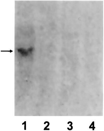
The presence of GUS activity was correlated with the level of GUS mRNA as determined by Northern blot analysis. Three offspring of each cross were assayed separately. Homozygous T4 plants and F1 offspring of the crosses with nontransformed tobacco and with TEV-K accumulated low levels of GUS mRNA (Fig. 3, lanes 2–4, respectively). The level of GUS mRNA was dramatically higher in the offspring of the cross of T4 with TEV-B (Fig. 3, lane 1). GUS mRNA levels were consistently 5- to 8-fold higher in offspring from the crosses with TEV-B as compared with those from crosses with either TEV-K or nontransformed tobacco as determined by PhosphorImager (Molecular Dynamics) analysis. These results indicate that expression of P1/HC-Pro interferes with transgene-induced posttranscriptional gene silencing. Because silencing is not suppressed in T4 × TEV-K offspring, which carry but do not express the P1/HC-Pro transgene, the suppression of silencing cannot simply be due to the presence of the DNA sequences of the transgenes independent of the P1/HC-Pro functionality.
Figure 3.
Northern blot analysis of GUS mRNA levels in offspring of line T4 × TEV-B (lane 1), T4 × nontransformed tobacco (lane 3), or T4 × TEV-K (lane 4), or in homozygous T4 plants (lane 2). For each sample, total RNA was extracted from an entire 3-week-old seedling (excluding roots), quantitated, separated by denaturing gel electrophoresis (5 μg/lane), blotted to membrane, and probed with randomly primed cDNA specific for the GUS transgene. The arrow indicates the position of GUS mRNA.
Suppression of VIGS.
A second set of experiments assessed the role of P1/HC-Pro in gene silencing induced by infection with a virus (VIGS). In VIGS, expression of a plant transgene or an endogenous gene is silenced by infection with a virus carrying the same sequence. The viral vector may also be silenced and eventually eliminated by VIGS, but the extent of silencing of the virus is variable and depends on the particular plant gene carried by the virus (28). For the VIGS experiments we used the system characterized by Ruiz et al. (28): transgenic N. benthamiana plants expressing high levels of GFP, in combination with a PVX vector that expresses GFP. In this system, the PVX–GFP-induced silencing exhibits three phases. In early stages of infection, GFP is expressed from both the virus and the transgene. In the second phase, the GFP transgene becomes completely and systemically silenced, whereas the virus becomes silenced to a lesser extent. In the third phase, the virus is completely silenced and eliminated from the plant, whereas the transgene continues to be silenced in old tissue but not in newly emerging tissues.
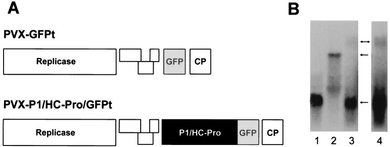
To test the effect of P1/HC-Pro expression on VIGS, we used PVX as a vector to express GFP by itself or together with P1/HC-Pro and followed the progress of VIGS in GFP transgenic plants infected with each of the two vectors. The PVX vector constructs used for these experiments are shown in Fig. 4A. In the PVX–GFPt vector, GFP was expressed from a subgenomic RNA under the control of the coat protein subgenomic promoter. In the PVX–P1/HC-Pro/GFPt vector, P1/HC-Pro/GFPt was expressed as a polyprotein from the coat protein subgenomic promoter and was subsequently processed to produce the individual proteins via the autoproteolytic activities of P1 and HC-Pro. GFPt is the S65T version of GFP which has a red-shifted excitation wavelength that does not fluoresce when exposed to the long wavelength UV light used to observe wild-type GFP.
Figure 4.
VIGS of transgene encoded GFP in the absence or presence of P1/HC-Pro expression. (A) Schematic diagrams of the PVX vector constructs carrying the GFPt coding sequence (PVX–GFPt) or the TEV P1/HC-Pro sequence and GFPt. GFPt is the S65T version of GFP. (B) Northern blot analysis of GFP transgene mRNA levels in mock-inoculated GFP transgenic plants (lane 1) or plants infected with either PVX–GFPt (lane 2) or PVX–P1/HC-Pro/GFPt (lanes 3 and 4). Lane 4 shows a 3-fold longer exposure of lane 3. Total RNA was extracted from upper leaves of the plants at 20 days after inoculation and analyzed as described in the legend for Fig. 3, except that a GFP-specific probe was used. Arrows indicate the positions of PVX–P1/HC-Pro/GFPt genomic RNA, PVX–GFPt genomic RNA, and GFP transgene mRNA (from top to bottom, respectively).
Expression of the wild-type GFP transgene in the infected plants was monitored by visual inspection of the plants under UV illumination and by Northern blot analysis of GFP transgene mRNA. Plant tissues expressing GFP exhibit green fluorescence upon exposure to UV light, whereas those that have silenced the GFP transgene exhibit a red color caused by the fluorescence of chlorophyll. Mock-inoculated GFP transgenic plants exhibited the green fluorescent phenotype throughout the experiment (data not shown), and high levels of GFP transgene mRNA were present in the upper parts of the plant several weeks after mock inoculation (Fig. 4B, lane 1). The virus-inoculated plants became systemically infected as evidenced by mild mottling on leaves in the upper part of the plant (data not shown) and by the presence of viral genomic RNA (Fig. 4B, lanes 2 and 4).
VIGS of GFP was initially detected on plants infected by PVX–GFPt by the appearance of red spots and red borders around veins on the inoculated leaf when viewed under UV illumination. The upper parts of the plants became completely silenced for the GFP transgene within 20 days postinoculation. Upper leaves were entirely red under UV illumination (data not shown) and no GFP transgene mRNA could be detected (Fig. 4B, lane 2). In contrast, the PVX–P1/HC-Pro/GFPt-infected plants did not develop signs of silencing on the inoculated leaf but instead displayed chlorotic spots in visible light that exhibited green fluorescence slightly greater than that in the surrounding tissue under UV light. By 20 days postinoculation, when the PVX–GFPt-inoculated plants were completely silenced for the GFP transgene, the PVX–P1/HC-Pro/GFPt-infected plants either continued to exhibit green fluorescence at the level seen in mock-inoculated plants or displayed only partial silencing characterized by small regions of red within an overall green background (data not shown). The level of GFP transgene mRNA in the PVX–P1/HC-Pro/GFPt-infected plants remained at the level observed in mock-infected plants (Fig. 4B, compare lanes 1 and 3), although viral genomic RNA carrying the P1/HC-Pro/GFPt sequence was detectable in the same tissue (Fig. 4B, lane 4). These results suggest that expression of P1/HC-Pro interferes with the establishment of VIGS.
Mixed Virus Infections and VIGS.
Mixed virus infections were used as a complementary strategy to determine if expression of P1/HC-Pro could suppress VIGS and to identify the regions of P1/HC-Pro required for suppression. GFP transgenic plants were infected with PVX–GFP (expressing wild-type GFP; ref. 29) alone or with a mixture of PVX-GFP and a PVX vector expressing either the wild-type TEV P1/HC-Pro sequence (PVX–5′TEV) or a subset of that sequence (PVX–HC or PVX–noHC). The organization of these vectors is similar to that of the PVX vectors shown in Fig. 4A, with expression of the TEV sequence under the control of the PVX coat protein subgenomic promoter. The vector PVX–5′TEV carries the coding region of the P1/HC-Pro sequence of TEV (nucleotides 146–2,674). PVX–HC carries only the region encoding HC-Pro (nucleotides 1,057–2,433). PVX–noHC carries the same TEV sequences as PVX–HC, except that the translation start site was mutated from AUG to ACG. N. benthamiana plants infected with PVX–5′TEV or PVX–HC accumulate both viral RNA carrying the HC-Pro nucleotide sequence and the HC-Pro gene product, whereas plants infected with PVX–noHC accumulate viral RNA but no detectable HC-Pro protein (15).
VIGS of GFP was monitored in the singly and doubly infected GFP transgenic plants by exposure to long wavelength UV light. PVX–GFP-infected plants exhibited bright green infection foci on the inoculated leaf by about one week postinoculation, followed by complete silencing of GFP in the upper leaves by 20 days postinoculation (data not shown). Plants doubly infected with PVX–GFP and PVX–noHC behaved exactly like the singly infected controls, exhibiting a similar number of infection foci on inoculated leaves and complete silencing of GFP in the upper parts of the plant in approximately 3 weeks (Fig. 5D). In contrast, plants doubly infected with PVX–GFP and PVX–5′TEV retained the green fluorescent phenotype throughout the course of the experiment (Fig. 5A), although they had about the same number of PVX–GFP infection foci as singly infected plants. The plants doubly infected with PVX–GFP and PVX–HC exhibited a range of VIGS phenotypes, some plants showing an almost complete suppression of VIGS (Fig. 5B) and others showing partial silencing of GFP, usually beginning within veins and the immediately surrounding regions (Fig. 5C). These results suggest that P1/HC-Pro suppresses VIGS and that the HC-Pro sequence alone is sufficient for much of the suppression. The failure of PVX–noHC to interfere with VIGS indicates that the suppression of silencing is a function of the HC-Pro gene product.
Figure 5.
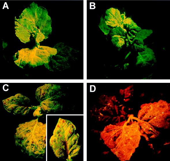
VIGS of transgene encoded GFP in mixed virus infections. Transgenic GFP N. benthamiana plants coinoculated with (A) PVX–GFP and PVX–5′TEV showing complete suppression of VIGS of GFP, (B) PVX–GFP and PVX–HC showing an almost complete suppression of VIGS of GFP, (C) PVX–GFP and PVX–HC showing partial suppression of VIGS of GFP, and (D) PVX–GFP and PVX–noHC showing complete VIGS of GFP. The Inset in C is a closer view of a partially silenced leaf showing silencing in the vicinity of veins.
DISCUSSION
The results presented here show that P1/HC-Pro acts as a suppressor of posttranscriptional gene silencing. P1/HC-Pro has been shown to transactivate the replication and enhance the pathogenicity of a broad range of heterologous plant viruses (15). These results are consistent with the hypothesis that posttranscriptional gene silencing is a defense mechanism that limits the accumulation of viral RNAs in the plant (12–14). From the data presented here it is reasonable to conclude that both poty- and potexviruses are normally constrained by gene silencing in the infected plant. As these are not closely related viruses, it is likely that many other viruses also are constrained in the same way, raising the possibility that the pathogenicity factors of many types of viruses may act to suppress gene silencing.
The ability of the TEV-P1/HC-Pro sequence to suppress gene silencing may explain the mechanism of synergistic plant diseases involving a potyvirus. Production of P1/HC-Pro by the potyvirus may suppress this silencing-based defense pathway, allowing the other virus of the synergistic pair to accumulate past the normal silencing-imposed limits and induce a more severe disease. The large number of unrelated viruses that interact with a member of the potyvirus group to cause a synergistic disease is additional support for the suggestion that gene silencing is used by plants to control the replication of many different viruses.
The failure of PVX–noHC to suppress VIGS in mixed infection experiments points to the importance of the HC-Pro gene product in suppression of silencing. HC-Pro, like many viral proteins, is multifunctional (reviewed in ref. 30). The central region of the protein is of particular interest to this study because it regulates both pathogenicity and RNA replication in potyviruses (31–35) and is required to mediate the synergistic interaction that occurs between PVX and a variety of potyviruses (15, 18, 36). There is some evidence that the central domain may also be involved in suppression of silencing. The TEV-K transgenic tobacco line carries the TEV P1/HC-Pro sequence with a mutation within the central domain. Plants of the T1 generation of the TEV-K line expressed P1/HC-Pro mRNA and detectable levels of HC-Pro gene product, but failed to develop synergistic disease when infected with PVX (18). However, during the process of producing the homozygous TEV-K line used in the crosses presented here, the TEV-K line developed two new characteristics. It fails to accumulate either P1/HC-Pro mRNA or HC-Pro gene product and is resistant to infection by TEV (unpublished results). These results suggest that the TEV-K line has become posttranscriptionally silenced for P1/HC-Pro, consistent with the idea that the mutation in the central domain of HC-Pro renders it incapable of suppressing silencing.
If the P1/HC-Pro sequence acts to inhibit posttranscriptional gene silencing, how is it possible that potyviruses, which express P1/HC-Pro as part of their natural life cycle, are subject to this type of silencing (10)? In the case of transgenic plants that are silenced for the TEV coat protein (CP) gene and therefore resistant to TEV, the virus may be hampered by the fact that P1/HC-Pro must be expressed from the viral genomic RNA which also contains the already targeted CP sequence. However, this cannot explain the failure of P1/HC-Pro to suppress silencing in CP-expressing transgenic lines that exhibit the recovery phenomenon (3). These transgenic plants are initially susceptible to the virus, but later the upper part of the plant recovers due to posttranscriptional gene silencing. The recovery is actually an example of VIGS where both the transgene and the virus become silenced, but in this case, unlike the VIGS presented here, expression of P1/HC-Pro fails to suppress silencing. Probably the simplest explanation for these differences is that PVX replicates faster than TEV and produces P1/HC-Pro at a higher level early in the infection cycle. The difference may also reflect a difference in the transgenic plants in the two cases. Induction of silencing in these plants may occur in a series of steps with some plants having already progressed past a critical point where P1/HC-Pro can exert its action. Another alternative is that the P1/HC-Pro suppression of silencing is not complete and the plant sometimes overcomes it.
Gene silencing is an important genetic mechanism with both theoretical and practical implications. The discovery of a viral sequence that suppresses gene silencing opens a new and promising avenue to understanding the mechanisms that regulate this pathway in plants. P1/HC-Pro can be used to help establish whether different classes of gene silencing employ similar mechanisms or pathways. The practical implication of this finding centers on potential improvements in genetic engineering in plants, where it is desirable to manipulate silencing. Technologies that use plants to express foreign gene products or to overexpress endogenous genes are often hampered by gene silencing, which prevents long-term high level expression of the transgene. It may be possible to use the P1/HC-Pro silencing suppressor to directly counter silencing in these situations. Elucidation of the mechanism by which P1/HC-Pro suppresses silencing may also lead to the development of strategies to enhance silencing in plants, thereby improving technologies that exploit gene silencing to eliminate expression of undesirable genes.
Acknowledgments
We thank D. Baulcombe for providing PVX–GUS and PVX–GFP viruses, and D. Baulcombe, L. Bowman, B. Krizek, and L. Marton for helpful discussions. This research was supported by Grant 9702709 from the U.S. Department of Agriculture National Research Initiative Competitive Grants Program, Plant Pathology panel.
ABBREVIATIONS
VIGS
virus-induced gene silencing
GFP
green fluorescent protein
GUS
β-glucuronidase
HC-Pro
helper component-proteinase
P1/HC-Pro
the 5′-proximal region of the tobacco etch genomic RNA encoding P1, HC-Pro, and a small part of the P3 proteins
PVX
potato virus X
TEV
tobacco etch virus
References
- Bingham P M. Cell. 1997;90:385–387. doi: 10.1016/s0092-8674(00)80496-1. [DOI] [PubMed] [Google Scholar]
- 2.Ingelbrecht I, Van Houdt H, Van Montagu M, Depicker A. Proc Natl Acad Sci USA. 1994;91:10502–1453. doi: 10.1073/pnas.91.22.10502. [DOI] [PMC free article] [PubMed] [Google Scholar]
- 3.Smith H A, Swaney S L, Parks T D, Wernsman E A, Dougherty W G. Plant Cell. 1994;6:1441–1443. doi: 10.1105/tpc.6.10.1441. [DOI] [PMC free article] [PubMed] [Google Scholar]
- 4.Park Y-D, Papp I, Moscone E A, Iglesias V A, Vaucheret H, Matzke M A, Matzke A J M. Plant J. 1996;9:183–194. doi: 10.1046/j.1365-313x.1996.09020183.x. [DOI] [PubMed] [Google Scholar]
- 5.English J J, Mueller E, Baulcombe D C. Plant Cell. 1996;8:179–188. doi: 10.1105/tpc.8.2.179. [DOI] [PMC free article] [PubMed] [Google Scholar]
- 6.Finnegan J, McElroy D. Bio/Technology. 1994;12:883–888. [Google Scholar]
- 7.Matzke M A, Matzke A J M. Trends Genet. 1995;11:1–3. doi: 10.1016/s0168-9525(00)88973-8. [DOI] [PubMed] [Google Scholar]
- 8.Meyer P. Annu Rev Plant Physiol Plant Mol Biol. 1996;47:23–48. doi: 10.1146/annurev.arplant.47.1.23. [DOI] [PubMed] [Google Scholar]
- 9.Crispin B T. Plant Cell. 1997;9:1245–1249. [Google Scholar]
- 10.Lindbo J A, Silva-Rosales L, Proebsting W M, Dougherty W G. Plant Cell. 1993;5:1749–1759. doi: 10.1105/tpc.5.12.1749. [DOI] [PMC free article] [PubMed] [Google Scholar]
- 11.Kumagai M H, Donson J, Della-Cioppa G, Harvey D, Hanley K, Grill L K. Proc Natl Acad Sci USA. 1995;92:1679–1683. doi: 10.1073/pnas.92.5.1679. [DOI] [PMC free article] [PubMed] [Google Scholar]
- 12.Ratcliff F, Harrison B, Baulcombe D. Science. 1997;276:1558–1560. doi: 10.1126/science.276.5318.1558. [DOI] [PubMed] [Google Scholar]
- 13.Covey S N, Al-Kaff N S, Langara A, Turner D S. Nature (London) 1997;385:781–782. [Google Scholar]
- 14.Al-Kaff N S, Covey S N, Kreike M M, Page A M, Pinder R, Dale P J. Science. 1998;279:2113–2115. doi: 10.1126/science.279.5359.2113. [DOI] [PubMed] [Google Scholar]
- 15.Pruss G, Ge X, Shi X M, Carrington J C, Vance V B. Plant Cell. 1997;9:859–868. doi: 10.1105/tpc.9.6.859. [DOI] [PMC free article] [PubMed] [Google Scholar]
- 16.Carrington J C, Freed D D, Oh C-S. EMBO J. 1990;9:1347–1353. doi: 10.1002/j.1460-2075.1990.tb08249.x. [DOI] [PMC free article] [PubMed] [Google Scholar]
- 17.Verchot J, Carrington J C. J Virol. 1995;69:3668–3674. doi: 10.1128/jvi.69.6.3668-3674.1995. [DOI] [PMC free article] [PubMed] [Google Scholar]
- 18.Shi X M, Miller H, Verchot J, Carrington J C, Vance V B. Virology. 1997;231:35. doi: 10.1006/viro.1997.8488. 42. [DOI] [PubMed] [Google Scholar]
- 19.An G. Plant Physiol. 1985;79:568–570. doi: 10.1104/pp.79.2.568. [DOI] [PMC free article] [PubMed] [Google Scholar]
- 20.Verchot J, Koonin E V, Carrington J C. Virology. 1991;185:527–535. doi: 10.1016/0042-6822(91)90522-d. [DOI] [PubMed] [Google Scholar]
- 21.Jefferson R A, Kavanagh T A, Bevan M W. EMBO J. 1987;6:3901–3907. doi: 10.1002/j.1460-2075.1987.tb02730.x. [DOI] [PMC free article] [PubMed] [Google Scholar]
- 22.Vance V B. Virology. 1991;182:486–494. doi: 10.1016/0042-6822(91)90589-4. [DOI] [PubMed] [Google Scholar]
- 23.Heim R, Cubitt A B, Tsien R Y. Nature (London) 1995;373:663–664. doi: 10.1038/373663b0. [DOI] [PubMed] [Google Scholar]
- 24.Hobbs S L A, Kpodar P, DeLong C M O. Plant Mol Biol. 1990;15:851–864. doi: 10.1007/BF00039425. [DOI] [PubMed] [Google Scholar]
- 25.Mueller E, Gilbert J E, Davenport G, Brigneti G, Baulcombe D C. Plant J. 1995;7:1001–1013. [Google Scholar]
- 26.Baulcombe D C. Plant Cell. 1996;8:1833–1844. doi: 10.1105/tpc.8.10.1833. [DOI] [PMC free article] [PubMed] [Google Scholar]
- 27.Chapman S, Kavanagh T, Baulcombe D. Plant J. 1992;2:549–557. doi: 10.1046/j.1365-313x.1992.t01-24-00999.x. [DOI] [PubMed] [Google Scholar]
- 28.Ruiz M T, Voinnet O, Baulcombe D C. Plant Cell. 1998;10:937–946. doi: 10.1105/tpc.10.6.937. [DOI] [PMC free article] [PubMed] [Google Scholar]
- 29.Baulcombe D C, Chapman S, Cruz S S. Plant J. 1995;7:1045–1053. doi: 10.1046/j.1365-313x.1995.07061045.x. [DOI] [PubMed] [Google Scholar]
- 30.Maia I G, Haenni A-L, Bernardi F. J Gen Virol. 1996;77:1335–1341. doi: 10.1099/0022-1317-77-7-1335. [DOI] [PubMed] [Google Scholar]
- 31.Atreya C D, Pirone T P. Proc Natl Acad Sci USA. 1993;90:11919–11923. doi: 10.1073/pnas.90.24.11919. [DOI] [PMC free article] [PubMed] [Google Scholar]
- 32.Atreya C D, Atreya P L, Thornbury D W, Pirone T P. Virology. 1992;191:106–111. doi: 10.1016/0042-6822(92)90171-k. [DOI] [PubMed] [Google Scholar]
- 33.Cronin S, Verchot J, Haldeman-Cahill M C, Schaad M C, Carrington J C. Plant Cell. 1995;7:549–559. doi: 10.1105/tpc.7.5.549. [DOI] [PMC free article] [PubMed] [Google Scholar]
- 34.Kassachau K D, Cronin S, Carrington J C. Virology. 1997;228:251–262. doi: 10.1006/viro.1996.8368. [DOI] [PubMed] [Google Scholar]
- 35.Klein P G, Klein R R, Rodrigues-Cerezo E, Hunt A G, Shaw J G. Virology. 1994;204:759–769. doi: 10.1006/viro.1994.1591. [DOI] [PubMed] [Google Scholar]
- 36.Vance V B, Berger P H, Carrington J C, Hunt A G, Shi X M. Virology. 1995;206:583–590. doi: 10.1016/s0042-6822(95)80075-1. [DOI] [PubMed] [Google Scholar]
