Chemotherapeutic Drugs Induce PPAR-γ Expression and Show Sequence-Specific Synergy with PPAR-γ Ligands in Inhibition of Non-Small Cell Lung Cancer (original) (raw)
Abstract
Preclinical studies have shown that peroxisome proliferator-activated receptor γ (PPAR-γ) ligands can exert antitumor effects against non-small cell lung cancer (NSCLC) and a variety of other cancers. In this study, we investigate the potential use of a PPAR-γ ligand, troglitazone (Tro), in combination with either of two chemotherapeutic agents, cisplatin (Cis) or paclitaxel (Pac), for the treatment of NSCLC. In vitro, treatment of NSCLC cell lines with Tro potentiated Cis- or Pac-induced growth inhibition. The potentiation of growth inhibition was observed only when Cis or Pac treatment was followed by Tro and not vice versa, demonstrating a sequence-specific effect. Median effect analysis revealed a synergistic interaction between Tro and Cis in the inhibition of NSCLC cell growth and confirmed the sequence-specific effect. We also found that Cis or Pac up-regulated the expression of PPAR-γ protein, accounting for the observed sequence-specific synergy. Similarly, experiments performed using a NSCLC xenograft model demonstrated enhanced effectiveness of combined treatment with Cis and PPAR-γ ligands, Tro or pioglitazone. Tumors from Cis-treated mice also demonstrated enhanced PPAR-γ expression. Together, our data demonstrate a novel sequence-specific synergy between PPAR-γ ligands and chemotherapeutic agents for lung cancer treatment.
Introduction
Treatment for lung cancer remains disappointing, with a 5-year survival rate of only 15% [1]. Induction and maintenance of the differentiated state has been an important strategy in the search for new cancer therapeutics. Peroxisome proliferator-activated receptor γ (PPAR-γ), a ligand-activated nuclear transcription factor, has been shown to exhibit antiproliferative effects through its ability to promote differentiation in human breast, prostate, colon, pituitary, and lung tumors [2–6], as well as through apoptosis [7–12] and cell cycle arrest [10,11,13,14]. Similarly, we have shown that PPAR-γ ligands promote differentiation and inhibit tumor growth, metastasis, and angiogenesis in non-small cell lung cancer (NSCLC) [14,15]. Clinical evidence also suggests that PPAR-γ ligands may be particularly effective against lung cancer. In a recent study of 87,678 patients at 10 Veterans Affairs medical centers, Govindarajan et al. [16] found that those receiving thiazolidinediones (TZDs), the PPAR-γ-activating drugs used by several million patients for treatment of type 2 diabetes, had a significantly lower risk of lung cancer. Interestingly, however, there was no significant benefit about colorectal or prostate cancer.
Resistance to standard chemotherapy is a common clinical problem that creates a continual need for novel strategies to potentiate the antitumorigenic effects of chemotherapeutic agents. This need in turn prompted studies to explore the potential adjuvant use of PPAR-γ ligands with various agents currently in clinical use for cancer. Investigation of PPAR-γ ligands is further supported by these drugs' relatively good tolerability and low toxicity profile. In the present study, we demonstrate that the PPAR-γ ligand troglitazone (Tro) synergistically potentiates cisplatin (Cis)- or paclitaxel (Pac)-induced growth inhibition of lung cancer cells in vitro. This synergism was observed only when Tro treatment followed, but not when it preceded, treatment with Cis or Pac. Similarly, the combination of Cis and either Tro or another PPAR-γ ligand, pioglitazone (Pio), inhibited the growth of tumor xenografts in vivo significantly more than did any single drug. Median effect analysis of the interaction between these drugs demonstrated potent synergy. This sequence-specific synergism may be explained by our finding that Cis or Pac treatment up-regulates PPAR-γ expression in lung cancer cells.
Materials and Methods
Cell Culture and Treatment
The A549 and H522 human lung adenocarcinoma cell lines were obtained from the American Type Culture Collection (Rockville, MD) and maintained in RPMI 1640 medium with glutamine, supplemented with 10% fetal bovine serum and antibiotics penicillin and streptomycin. Cells were incubated at 37°C with 5% CO2, and medium was changed every 48 to 72 hours. For studies of sequence-specific potentiation, cells were treated in the presence of serum with either Cis (0, 1, 5, 10, 15, or 20 µM) or Pac (0, 0.25, 1, 10, 50, 100 nM) for 16 hours followed either by 56 hours of drug-free treatment or by 8 hours of drug-free treatment succeeded by 48 hours of treatment with Tro at its IC50 of 10 µM. In other studies, cells were pretreated with Tro (10 µM) for 48 hours followed after 8 hours without drug by a 16-hour treatment with Cis, Pac, or vehicle at concentrations similar to those in the preceding experiments. For median effect analysis, cells were treated with Cis followed by Tro or Tro followed by Cis according to the described timeline. Each drug was administered at the same fixed fraction (25%, 50%, 100%, and 200%) of the IC50 (10 µM for each) identified in preliminary experiments. For both sets of studies, cell growth was analyzed by MTT assay at 72 hours after initiation of treatment.
Measurement of Cell Growth
Cell growth was assayed using the tetrazolium salt 3-(4,5-dimethylthiazol-2-yl)-2,5-diphenyltetrazolium bromide (MTT), which is cleaved by a mitochondrial dehydrogenase to produce dark blue formazan crystals [17]. For the assay, MTT was added to the culture medium, and cells were incubated for an additional 4 hours. The formazan crystals were then dissolved by the addition of 100 µl of SDS to each well followed by incubation for a further 5 hours at 37°C. Optical density was measured at 570 nm, and mean values of duplicate samples were calculated for each well.
Measurement of PPAR-γ Expression
Expression of PPAR-γ was assessed by Western immunoblot analysis as previously described [14]. Briefly, harvested cells or excised xenograft tumors were homogenized. Samples containing 20 µg of total protein were electrophoresed on SDS-polyacrylamide gels and transferred onto a polyvinyldifluoride membrane by electroblotting. Membranes were probed with rabbit polyclonal antibodies to total PPAR-γ (Santa Cruz Biotechnology, Santa Cruz, CA) followed by horseradish peroxidase-conjugated mouse antirabbit IgG (Pierce, Rockford, IL).
Median Effect Analysis
We performed median effect analysis as described by Chou and Talalay [18]. Briefly, the median inhibitory concentration (IC50) for each drug and for a fixed-ratio combination of the two drugs was determined. We then calculated the combination index (CI). For two drugs acting by mechanisms that are not mutually exclusive, the CI value is defined as: (D)1(IC50)1+(D)2(IC50)2+(D)1(D)2(IC50)1(IC50)2, where _D_1 and _D_2 are calculated from the IC50 for the combination and the ratio (P/Q) of the two drugs by the equations: _D_1 = (IC50)comb x P / (P + Q) and _D_2 = (IC50)comb x Q / (P + Q). The third (interaction) term is absent when the drug actions are mutually exclusive. For each drug combination in our study we used Calcusyn software (Biosoft, Ferguson, MO) to calculate the CI for 4 concentrations (1:1 ratio) of the two drugs and averaged the CIs. A CI of 1 indicates additive effects, whereas a CI < 1 indicates synergy.
Xenografts in SCID Mice and Analysis of Tumor Growth
Six-week-old C.B-17 SCID mice (Taconic Farms, Germantown, NY) were subcutaneously injected with A549 cells on either side of the dorsal flank. Beginning after tumor size reached approximately 150 mm3, six mice per group were treated with Cis (3 mg/kg) weekly by intraperitoneal injection and, beginning 4 days later, received Tro (400 mg/kg), Pio (100 mg/kg), or vehicle once daily by oral gavage. Tro and Pio were formulated by suspending the drug in an aqueous solution of 2% carboxymethylcellulose and 0.2% Tween 20 and then sonicating for 5 minutes. Other mice received vehicle intraperitoneal injections in combination with either Tro or vehicle by oral gavage. Tumor size was measured weekly for 10 weeks, and tumor volume was calculated by averaging the largest and smallest radii and multiplying the cube of the result by π. After 10 weeks of treatment, mice were killed and PPAR-γ expression in tumors was measured by Western immunoblot analysis. All experiments were approved by the University of Michigan Committee on Use and Care of Animals.
Statistical Analysis
All data are expressed as means ± SEM and are analyzed using the Prism 5.0 statistical program (GraphPad Software, San Diego, CA). Groups were compared using one-sided analysis of variance or onesided Student's t test as applicable. P < .05 was considered significant.
Results
Tro Potentiates Cis- or Pac-Induced Growth Inhibition in a Sequence-Specific Manner
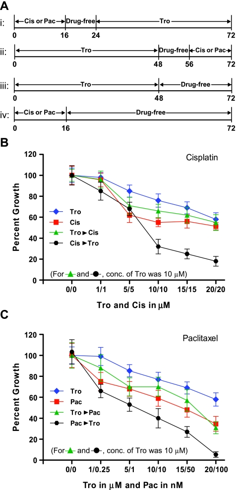
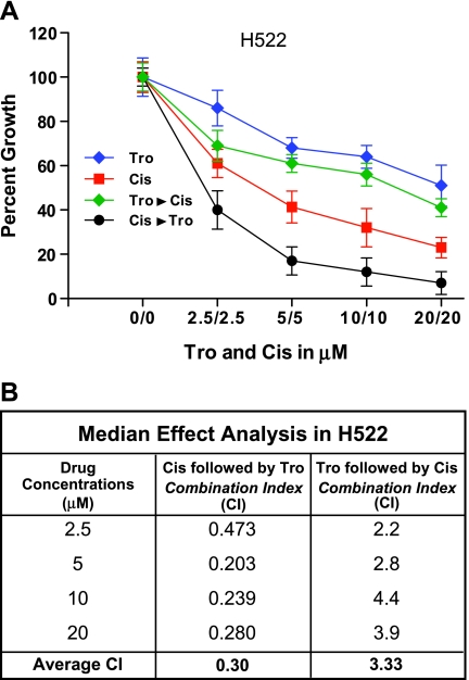
To assess the efficacy of combining Tro with either of the chemotherapeutic agents Cis or Pac to inhibit NSCLC cell growth, A549 cells were treated with various concentrations of Tro, Cis or Pac in four different combinations as described in the Materials and Methods section and Figure 1_A_. After 72 hours, cell growth was assessed by MTT assay. We observed that A549 cells treated with Cis or Pac and followed by Tro demonstrated maximum growth inhibition compared with individual drug alone (Figure 1, B and C). Interestingly, A549 cells that were first treated with Tro and then followed by Cis or Pac were not significantly different from the individual drug treatments, demonstrating a sequence-specific effect. Furthermore, we observed a similar sequence-specific effect on cell growth inhibition when Cis treatment was followed by Tro in another NSCLC-derived cell line, H522 (Figure 2_A_). This demonstrates that the effects we observed in A549 cells were not cell line specific.
Figure 1.
Sequence-specific potentiation of cisplatin- or paclitaxel-induced growth inhibition by troglitazone (Tro) in A549 cells. (A) Timeline of in vitro experiments: i, in studies of Tro after treatment, cells were treated with cisplatin (Cis) or paclitaxel (Pac) for 16 hours followed by an 8-hour drug-free washout and Tro for 48 hours; ii, in studies of Tro before treatment, cells were treated with Tro for 48 hours followed by an 8-hour drug-free washout and Cis or Pac for 16 hours; iii, in studies of Tro as a single agent, Tro was administered for 48 hours followed by 24 hours without drug; iv, in studies of Cis or Pac as single agents, they were administered for 16 hours followed by 56 hours without drug. In single-agent studies, cells were treated with Tro at concentrations of 0, 1, 5, 10, 15, or 20 _µ_M, Cis at concentrations of 0, 1, 5, 10, 15, or 20 _µ_M, or Pac at concentrations of 0, 0.25, 1, 10, 50, or 100 nM. In combination studies, cells were treated with 10 _µ_M Tro before or after various concentrations of Cis and Pac as indicated. (B) A549 cell growth kinetics in various combinations of Tro and Cis. (C) A549 cell growth kinetics in various combinations of Tro and Pac. Results shown are averages of six replicate experiments.
Figure 2.
Sequence-specific synergistic potentiation of cisplatin (Cis)-induced growth inhibition by troglitazone (Tro) in H522 cells. H522 cells were treated with Cis and/or Tro according to the protocol previously described for A549 cells. (A) Growth curves for treatment with Cis alone, Tro alone, Cis followed by Tro, and Tro followed by Cis. (B) CIs, derived from median effect analysis, for each drug concentration and order of administration. CI values < 1 indicate synergy and values > 1 indicate partial antagonism.
Median Effect Analysis Reveals Sequence-Specific Synergistic Interaction between Tro and Cis
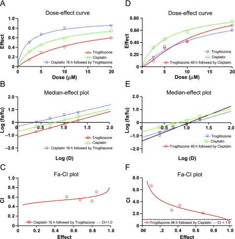
We then assessed the interaction between Cis and Tro by median effect analysis. For these studies, Tro and Cis were each administered in a 1:1 ratio at four fractions of their IC50 concentrations (10 µM for each). The respective dose-effect curves are displayed in Figure 3, A and D, and are linearized by log-log transformation in Figure 3, B and E. The straightness of the lines demonstrates that these data are suitable for median effect analysis, whereas the parallelism of the lines in Figure 3_B_ demonstrates that growth inhibition by Cis and by Tro after treatment are mutually exclusive. The fraction-effect versus CI plots (Figure 3, C and F) indicate the nature of the interaction between the drugs being tested. The CI, for the combination with Cis treatment followed by Tro, is consistently below 1 (Figure 3_C_), with an average of 0.59. The average CI of <1 demonstrates that the effects of the two drugs are synergistic. On the other hand, for the combination that has Tro treatment preceding the Cis treatment, the CI is consistently greater than 1 (Figure 3_F_), with an average of 3.62. The CI > 1 indicates that the interaction is partially antagonistic. Median effect analysis in H522 cells also demonstrated similar synergistic interaction when Cis is followed by Tro (average CI of 0.30) and not vice versa (average CI of 3.33; Figure 2_B_).
Figure 3.
Median effect analysis reveals synergistic interaction when cisplatin (Cis) treatment is followed by troglitazone (Tro) but partial antagonism when Tro treatment precedes cisplatin. After determining the IC50 for each drug, Tro and Cis were administered in a fixed ratio with two concentrations below and one concentration above their IC50 concentrations (10 _µ_M), in a total of four different ratios (2.5:2.5, 5:5, 10:10, and 20:20 in _µ_M). Cell proliferation was assessed by MTT assay after 72 hours. Results for Tro after treatment are displayed in panels (A), (B), and (C) and for Tro before treatment in panels (D), (E), and (F). Dose-effect curves for Cis as a function of Tro concentration are displayed in panels (A) and (D) and after log-log transformation in panels (B) and (E); panels (C) and (F) show the Fa-CI curves calculated using the median effect equation. Values < 1 indicate synergy and values > 1 indicate partial antagonism.
Cis or Pac Induces PPAR-γ Expression
In preliminary studies investigating the regulation of PPAR-γ expression in A549 cells, we found that inducing growth inhibition by serum deprivation up-regulated PPAR-γ expression (Figure 4). We therefore investigated the effect of inhibiting growth by exposure to Cis or Pac.We found that exposure to either Cis or Pac for 16 hours significantly up-regulated PPAR-γ expression as assessed by Western immunoblot analysis, with the extent of the increase being dose-dependent (Figure 4).
Figure 4.
Serum deprivation or exposure to either cisplatin (Cis) or paclitaxel (Pac) induces PPAR-γ expression. A549 cells were either cultured in serum-free medium for 72 hours (first column) or were exposed in the presence of serum to Cis (1 or 5 _µ_M), Pac (1 or 5 nM), or vehicle for 16 hours followed by 56 hours in drug-free medium with serum. At the end of 72 hours, PPAR-γ was assessed by Western immunoblot analysis. β-Actin expression is displayed as a loading control.
Tro or Pio Treatment Enhances the Effect of Cis on Growth of A549 Tumor Xenografts in SCID Mice
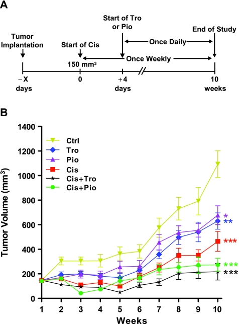
To determine whether the enhanced tumor suppression we observed with Tro in vitro could also be observed in vivo, we used a xenograft model in which NSCLC-derived A549 cells are injected into either side of the dorsal flank of SCID mice. After the tumors had reached a mean volume of approximately 150 mm3, mice were divided into six different groups. As summarized in Figure 5_A_, treatment with Cis (3 mg/kg) or vehicle once weekly by intraperitoneal injection was begun at this time for 3 groups of mice. Four days later, each of the 3 Cis treatment groups received either Tro (400 mg/kg), Pio (100 mg/kg), or vehicle daily by oral gavage. The remaining 3 groups were either untreated, or received Tro or Pio alone as described. All treatments were continued for 10 weeks, and tumor volumes were monitored by weekly measurements (Figure 5_B_). All three drugs as single agents significantly inhibited the growth of A549 xenografts. Consistent with our in vitro observations, combination treatments of either Cis and Tro or Cis and Pio were significantly more effective in inhibiting A549 xenografts than either drug alone.
Figure 5.
Combined treatment with cisplatin (Cis) and troglitazone (Tro) or pioglitazone (Pio) is more effective than any drug alone against tumor growth in a xenograft model. A549 cells were inoculated subcutaneously into the flanks of SCID mice. (A) Experimental design: After the tumor size had reached approximately 150 mm3, mice (six mice/group) were injected weekly with 3-mg/kg Cis and, beginning 4 days later, were treated daily with Tro (400 mg/kg), Pio (100 mg/kg), or vehicle by oral gavage. In other studies, mice were treated with Tro, Pio, or vehicle as described but received vehicle (for Cis) by intraperitoneal injection. (B) Tumor size was measured weekly for 10 weeks, beginning at the initiation of treatment, and tumor volume was calculated by averaging the largest and smallest radii and multiplying the cube of the result by π. *P < .05, **P < .01, and ***P < .001 compared with Ctrl at week 10 by analysis of variance.
Cis Up-regulates PPAR-γ Expression in Xenografts
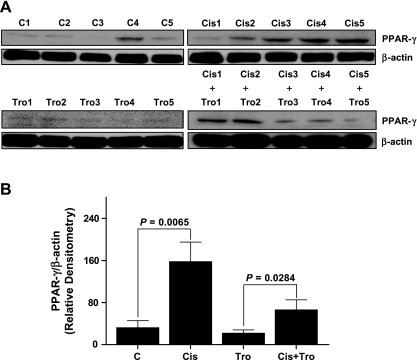
Similar to our in vitro observations, we determined whether Cis upregulated PPAR-γ expression in xenografts. For this, we assessed PPAR-γ expression in the tumor tissue lysates from A549 xenografts after indicated treatment regimens by Western immunoblot analysis. As shown in Figure 6, we observed significant up-regulation of PPAR-γ expression in the tumors from mice that received Cis treatment. Densitometric scans (Figure 6_B_) confirmed the significance of upregulation both by Cis alone and by the addition of Cis to Tro treatment. These observations further support the hypothesis that induction of PPAR-γ expression by chemotherapeutic drugs is important for potentiating the effect of subsequent treatment with PPAR-γ ligands.
Figure 6.
Cisplatin (Cis) up-regulates PPAR-γ expression in tumor xenografts. A549 cells were inoculated subcutaneously into the flanks of SCID mice as previously described. After 10 weeks of treatment with vehicle (C1-5), cisplatin (Cis1-5), troglitazone (Tro1-5), or Cis + Tro, mice were killed, and tumors were resected and assayed for PPAR-γ expression by Western immunoblot analysis. (A) Blots of PPAR-γ and the β-actin loading control are shown for each mouse. (B) Densitometric scans showing mean PPAR-γ expression, normalized to β-actin, for each condition.
Discussion
In this study, we demonstrate synergy between PPAR-γ ligands and two cancer chemotherapeutic agents of different classes, one platinum-based and the other a taxane. These effects were seen both in vitro, as inhibition of growth in two NSCLC-derived cell lines, and in vivo as inhibition of tumor growth in a xenograft model. Median effect analysis of varying doses of the drugs demonstrated true synergy rather than a mere additive effect. Perhaps the most striking aspect of our findings is that synergy was specific to the sequence in which the agents were administered, specifically, Cis followed by Tro and not vice versa. These results may be attributed to the fact that Cis or Pac treatment results in the up-regulation of PPAR-γ expression, which is expressed to varying degrees in both normal and tumor epithelia [19]. We observed a similar up-regulation of PPAR-γ expression during serum-deprivation-induced growth inhibition. These data suggest that during the observed synergistic interaction, the growth-inhibiting effect of chemotherapeutic agents potentiate the differentiation-inducing effects of PPAR-γ ligands by increasing the availability of their receptor. Consistently, we also observed up-regulation of PPAR-γ expression in all the Cis-treated tumor groups in the xenograft model. In addition, two different PPAR-γ ligands were equally effective in enhancing the effects of Cis in inhibiting tumor growth. This supports our conclusion that the observed synergy reflects activation of the increased levels of PPAR-γ. By contrast, a partial antagonistic effect was observed when the cells were treated with PPAR-γ ligand before the chemotherapeutic agent. This may be because PPAR-γ ligands inhibit cell proliferation, and cytotoxic agents such as Cis are less effective against nondividing cells.
Two recent studies have also reported apparently synergistic effects against NSCLC-derived cell lines and mouse xenografts after combination treatment with PPAR-γ ligands and chemotherapeutic agents. Girnun et al. [20] assessed the combination of the TZD rosiglitazone and the platinum-based drug carboplatin, whereas Fulzele et al. [21] studied the natural PPAR-γ ligand 15-deoxy-Δ12,14-prostaglandin J2 (15d-PGJ2) and the taxane docetaxel. Furthermore, Copland et al. [22] found that the novel PPAR-γ ligand RS5444 enhanced the activity of Pac against anaplastic thyroid carcinoma in vitro and in a xenograft model. However, this group found only additive antiproliferative effects, whereas Girnun et al. [20] did not statistically evaluate whether the enhanced effectiveness they observed was truly synergistic. Additionally, in all three studies, the drugs were administered concurrently, as opposed to the sequential treatment we followed. This allowed us to demonstrate that the effects of Tro and Cis are mutually exclusive.
Girnun et al. [20] attributed the synergy they observed to rosiglitazone-induced inhibition of metallothionein expression. Metallothioneins are proteins that bind heavy metals, including platinum, and thus cause sequestration of platinum-based drugs in cancer cells. This effect on metallothionein expression may also contribute to the synergy we observed. However, it accounts neither for our observation of a sequence-specific effect nor for the effect on activity of the non-platinum drug Pac. Nevertheless, it is entirely possible and perhaps likely that more than one mechanism is operating when PPAR-γ ligands and chemotherapeutic drugs are administered simultaneously.
In addition to enhancing the efficacy of general chemotherapeutics such as platinum-based drugs and taxanes, PPAR-γ ligands are also implicated in enhancing the efficacy of several targeted therapeutics. Lee et al. [23] showed that the PPAR-γ ligand rosiglitazone facilitated the antiproliferative effects of gefitinib, an inhibitor of the epidermal growth factor receptor tyrosine kinase. Similarly, Han and Roman [24] reported that the inhibitory effect of rosiglitazone on the growth of NSCLC-derived cell lines was facilitated by the chemotherapeutic agent rapamycin. Increased growth inhibition and apoptosis of A549 cells has also been demonstrated after combined treatment with MK886, an inhibitor of 5-lipoxygenase activator protein, the cyclooxygenase inhibitor indomethacin, and 15d-PGJ2 [25]. Furthermore, a novel PPAR-α/γ ligand has been reported to synergistically enhance the antiproliferative and proapoptotic effects of imatinib against chronic myeloid leukemia cell lines [26].
PPAR-γ/PPAR-γ ligands exert their antitumor effects on NSCLC cells directly as well by a variety of mechanisms. One effect involves suppression of the tumorigenic mediator prostaglandin E2. Studies have shown that up-regulation of PPAR-γ reduces synthesis of prostaglandin E2 by down-regulating cyclooxygenase-2 expression [27] while PPAR-γ ligands increase catabolism of the molecule by up-regulating 15-hydroxyprostaglandin dehydrogenase [28]. Treatment with PPAR-γ ligands also leads to increased expression of the growth arrest and DNA damage-inducible 153 (GADD153) gene, which is believed to mediate apoptosis [29]. Another mechanism by which PPAR-γ ligands may exert antitumor effects is through up-regulation of E-cadherin, which is thought to promote differentiation of NSCLC cells [30]. Furthermore, we have shown that PPAR-γ activation inhibits angiogenesis by blocking production of chemokines of the ELR+ CXC family [15] and produces cell cycle arrest by down-regulating cyclins D and E [14].We also observed sustained up-regulation of extracellular regulated kinase 1/2, which may initiate the pathway leading to differentiation.
In the context of the present study, the possibility that PPAR-γ ligands may be more effective against NSCLC than many other cancers is intriguing. As previously mentioned, a large epidemiological study has found that TZD use reduces the risk of lung cancer but not of colorectal or prostate cancer [16]. Lack of any effect of TZD use on risk of colon and prostate cancer, as well as breast cancer, has similarly been found in an even larger epidemiological study [31]. Conversely, the smaller study of Ramos-Nino et al. [32] found an increased overall cancer risk among TZD users. This study did not identify the risks for individual types of cancer, so possible implications for NSCLC are difficult to assess.
In conclusion, our results demonstrate that PPAR-γ ligands act synergistically with both the platinum-based chemotherapeutic agent Cis and the taxane Pac to inhibit the growth of NSCLC cells both in vitro and in vivo. Strikingly, this synergism is dependent on the sequence in which these drugs are administered, which presumably reflects the increased PPAR-γ expression observed after treatment with the chemotherapeutic agents. This finding raises the possibility that PPAR-γ ligands may be clinically useful not only in combination with Cis and Pac but also as salvage therapy after prior cytotoxic therapy has failed.
Abbreviations
15d-PGJ2
15-deoxy-Δ12,14-prostaglandin J2
Cis
cisplatin
NSCLC
non-small cell lung cancer
Pac
paclitaxel
Pio
pioglitazone
Tro
troglitazone
TZD
thiazolidinedione
Footnotes
1
Supported by Flight Attendant Medical Research Institute Grant N005884 (to V.G. K.) and National Institutes of Health Grants P50 HL60289 (to T.J. S.) and HL070068 (to R.C. R.).
References
- 1.Edwards BK, Brown ML, Wingo PA, Howe HL, Ward E, Ries LA, Schrag D, Jamison PM, Jemal A, Wu XC, et al. Annual report to the nation on the status of cancer, 1975–2002, featuring population-based trends in cancer treatment. J Natl Cancer Inst. 2005;97:1407–1427. doi: 10.1093/jnci/dji289. [DOI] [PubMed] [Google Scholar]
- 2.Bren-Mattison Y, Van Putten V, Chan D, Winn R, Geraci MW, Nemenoff RA. Peroxisome proliferator-activated receptor-gamma (PPAR(gamma)) inhibits tumorigenesis by reversing the undifferentiated phenotype of metastatic non-small-cell lung cancer cells (NSCLC) Oncogene. 2005;24:1412–1422. doi: 10.1038/sj.onc.1208333. [DOI] [PubMed] [Google Scholar]
- 3.Elstner E, Muller C, Koshizuka K, Williamson EA, Park D, Asou H, Shintaku P, Said JW, Heber D, Koeffler HP. Ligands for peroxisome proliferator-activated receptorgamma and retinoic acid receptor inhibit growth and induce apoptosis of human breast cancer cells in vitro and in BNX mice. Proc Natl Acad Sci USA. 1998;95:8806–8811. doi: 10.1073/pnas.95.15.8806. [DOI] [PMC free article] [PubMed] [Google Scholar]
- 4.Heaney AP, Fernando M, Yong WH, Melmed S. Functional PPAR-gamma receptor is a novel therapeutic target for ACTH-secreting pituitary adenomas. Nat Med. 2002;8:1281–1287. doi: 10.1038/nm784. [DOI] [PubMed] [Google Scholar]
- 5.Kubota T, Koshizuka K, Williamson EA, Asou H, Said JW, Holden S, Miyoshi I, Koeffler HP. Ligand for peroxisome proliferator-activated receptor gamma (troglitazone) has potent antitumor effect against human prostate cancer both in vitro and in vivo. Cancer Res. 1998;58:3344–3352. [PubMed] [Google Scholar]
- 6.Mueller E, Sarraf P, Tontonoz P, Evans RM, Martin KJ, Zhang M, Fletcher C, Singer S, Spiegelman BM. Terminal differentiation of human breast cancer through PPAR gamma. Mol Cell. 1998;1:465–470. doi: 10.1016/s1097-2765(00)80047-7. [DOI] [PubMed] [Google Scholar]
- 7.Clay CE, Namen AM, Atsumi G, Willingham MC, High KP, Kute TE, Trimboli AJ, Fonteh AN, Dawson PA, Chilton FH. Influence of J series prostaglandins on apoptosis and tumorigenesis of breast cancer cells. Carcinogenesis. 1999;20:1905–1911. doi: 10.1093/carcin/20.10.1905. [DOI] [PubMed] [Google Scholar]
- 8.Guan Y-F, Zhang Y-H, Breyer RM, Davis L, Breyer MD. Expression of peroxisome proliferator-activated receptor gamma (PPARgamma) in human transitional bladder cancer and its role in inducing cell death. Neoplasia. 1999;1:330–339. doi: 10.1038/sj.neo.7900050. [DOI] [PMC free article] [PubMed] [Google Scholar]
- 9.Li M, Lee TW, Yim AP, Mok TS, Chen GG. Apoptosis induced by troglitazone is both peroxisome proliferator-activated receptor-gamma- and ERK-dependent in human non-small lung cancer cells. J Cell Physiol. 2006;209:428–438. doi: 10.1002/jcp.20738. [DOI] [PubMed] [Google Scholar]
- 10.Vignati S, Albertini V, Rinaldi A, Kwee I, Riva C, Oldrini R, Capella C, Bertoni F, Carbone GM, Catapano CV. Cellular and molecular consequences of peroxisome proliferator-activated receptor-γ activation in ovarian cancer cells. Neoplasia. 2006;8:851–861. doi: 10.1593/neo.06433. [DOI] [PMC free article] [PubMed] [Google Scholar]
- 11.Yang F-G, Zhang Z-W, Xin D-Q, Shi C-J, Wu J-P, Guo Y-L, Guan Y-F. Peroxisome proliferator-activated receptor gamma ligands induce cell cycle arrest and apoptosis in human renal carcinoma cell lines. Acta Pharmacol Sin. 2005;26:753–761. doi: 10.1111/j.1745-7254.2005.00753.x. [DOI] [PubMed] [Google Scholar]
- 12.Zhang M, Zou P, Bai M, Jin Y, Tao X. Peroxisome proliferator-activated receptor-gamma activated by ligands can inhibit human lung cancer cell growth through induction of apoptosis. J Huazhong Univ Sci Technolog Med Sci. 2003;23:138–140. doi: 10.1007/BF02859937. [DOI] [PubMed] [Google Scholar]
- 13.Kawakami S, Arai G, Hayashi T, Fujii Y, Xia G, Kageyama Y, Kihara K. PPARgamma ligands suppress proliferation of human urothelial basal cells in vitro. J Cell Physiol. 2002;191:310–319. doi: 10.1002/jcp.10099. [DOI] [PubMed] [Google Scholar]
- 14.Keshamouni VG, Reddy RC, Arenberg DA, Joel B, Thannickal VJ, Kalemkerian GP, Standiford TJ. Peroxisome proliferator-activated receptor-γ activation inhibits tumor progression in non-small-cell lung cancer. Oncogene. 2004;23:100–108. doi: 10.1038/sj.onc.1206885. [DOI] [PubMed] [Google Scholar]
- 15.Keshamouni VG, Arenberg DA, Reddy RC, Newstead MJ, Anthwal S, Standiford TJ. PPAR-γ activation inhibits angiogenesis by blocking ELR+ CXC chemokine production in non-small cell lung cancer. Neoplasia. 2005;7:294–301. doi: 10.1593/neo.04601. [DOI] [PMC free article] [PubMed] [Google Scholar]
- 16.Govindarajan R, Ratnasinghe L, Simmons DL, Siegel ER, Midathada MV, Kim L, Kim PJ, Owens RJ, Lang NP. Thiazolidinediones and the risk of lung, prostate, and colon cancer in patients with diabetes. J Clin Oncol. 2007;25:1476–1481. doi: 10.1200/JCO.2006.07.2777. [DOI] [PubMed] [Google Scholar]
- 17.Mosmann T. Rapid colorimetric assay for cellular growth and survival: application to proliferation and cytotoxicity assays. J Immunol Methods. 1983;65:55–63. doi: 10.1016/0022-1759(83)90303-4. [DOI] [PubMed] [Google Scholar]
- 18.Chou T-C, Talalay P. Quantitative analysis of dose-effect relationships: the combined effects of multiple drugs or enzyme inhibitors. Adv Enzyme Regul. 1984;22:27–55. doi: 10.1016/0065-2571(84)90007-4. [DOI] [PubMed] [Google Scholar]
- 19.Subbarayan V, Xu X-C, Kim J, Yang P, Hoque A, Sabichi AL, Llansa N, Mendoza G, Logothetis CJ, Newman RA, et al. Inverse relationship between 15- lipoxygenase-2 and PPAR-γ gene expression in normal epithelia compared with tumor epithelia. Neoplasia. 2005;7:280–293. doi: 10.1593/neo.04457. [DOI] [PMC free article] [PubMed] [Google Scholar]
- 20.Girnun GD, Naseri E, Vafai SB, Qu L, Szwaya JD, Bronson R, Alberta JA, Spiegelman BM. Synergy between PPARgamma ligands and platinum-based drugs in cancer. Cancer Cell. 2007;11:395–406. doi: 10.1016/j.ccr.2007.02.025. [DOI] [PMC free article] [PubMed] [Google Scholar]
- 21.Fulzele SV, Chatterjee A, Shaik MS, Jackson T, Ichite N, Singh M. 15-Deoxy-Delta12,14-prostaglandin J2 enhances docetaxel anti-tumor activity against A549 and H460 non-small-cell lung cancer cell lines and xenograft tumors. Anticancer Drugs. 2007;18:65–78. doi: 10.1097/CAD.0b013e3280101006. [DOI] [PubMed] [Google Scholar]
- 22.Copland JA, Marlow LA, Kurakata S, Fujiwara K, Wong AK, Kreinest PA, Williams SF, Haugen BR, Klopper JP, Smallridge RC. Novel high-affinity PPARgamma agonist alone and in combination with paclitaxel inhibits human anaplastic thyroid carcinoma tumor growth via p21WAF1/CIP1. Oncogene. 2006;25:2304–2317. doi: 10.1038/sj.onc.1209267. [DOI] [PubMed] [Google Scholar]
- 23.Lee SY, Hur GY, Jung KH, Jung HC, Kim JH, Shin C, Shim JJ, In KH, Kang KH, Yoo SH. PPAR-γ agonist increase gefitinib's antitumor activity through PTEN expression. Lung Cancer. 2006;51:297–301. doi: 10.1016/j.lungcan.2005.10.010. [DOI] [PubMed] [Google Scholar]
- 24.Han S, Roman J. Rosiglitazone suppresses human lung carcinoma cell growth through PPARgamma-dependent and PPARgamma-independent signal pathways. Mol Cancer Ther. 2006;5:430–437. doi: 10.1158/1535-7163.MCT-05-0347. [DOI] [PubMed] [Google Scholar]
- 25.Avis I, Martinez A, Tauler J, Zudaire E, Mayburd A, Abu-Ghazaleh R, Ondrey F, Mulshine JL. Inhibitors of the arachidonic acid pathway and peroxisome proliferator-activated receptor ligands have superadditive effects on lung cancer growth inhibition. Cancer Res. 2005;65:4181–4190. doi: 10.1158/0008-5472.CAN-04-3441. [DOI] [PubMed] [Google Scholar]
- 26.Zang C, Liu H, Waechter M, Eucker J, Bertz J, Possinger K, Koeffler HP, Elstner E. Dual PPARalpha/gamma ligand TZD18 either alone or in combination with imatinib inhibits proliferation and induces apoptosis of human CML cell lines. Cell Cycle. 2006;5:2237–2243. doi: 10.4161/cc.5.19.3259. [DOI] [PubMed] [Google Scholar]
- 27.Bren-Mattison Y, Meyer AM, Van Putten V, Li H, Kuhn K, Stearman R, Weiser-Evans M, Winn RA, Heasley LE, Nemenoff R. Antitumorigenic effects of peroxisome proliferator-activated receptor-γ (PPARγ) in non-small cell lung cancer cells (NSCLC) are mediated by suppression of COX-2 via inhibition of NF-κB. Mol Pharmacol. 2008;73:709–717. doi: 10.1124/mol.107.042002. [DOI] [PubMed] [Google Scholar]
- 28.Hazra S, Batra RK, Tai HH, Sharma S, Cui X, Dubinett SM. Pioglitazone and rosiglitazone decrease prostaglandin E2 in non-small-cell lung cancer cells by up-regulating 15-hydroxyprostaglandin dehydrogenase. Mol Pharmacol. 2007;71:1715–1720. doi: 10.1124/mol.106.033357. [DOI] [PubMed] [Google Scholar]
- 29.Satoh T, Toyoda M, Hoshino H, Monden T, Yamada M, Shimizu H, Miyamoto K, Mori M. Activation of peroxisome proliferator-activated receptor-gamma stimulates the growth arrest and DNA-damage inducible 153 gene in non-small cell lung carcinoma cells. Oncogene. 2002;21:2171–2180. doi: 10.1038/sj.onc.1205279. [DOI] [PubMed] [Google Scholar]
- 30.Wick M, Hurteau G, Dessev C, Chan D, Geraci MW, Winn RA, Heasley LE, Nemenoff RA. Peroxisome proliferator-activated receptor-gamma is a target of nonsteroidal anti-inflammatory drugs mediating cyclooxygenase-independent inhibition of lung cancer cell growth. Mol Pharmacol. 2002;62:1207–1214. doi: 10.1124/mol.62.5.1207. [DOI] [PubMed] [Google Scholar]
- 31.Koro C, Barrett S, Qizilbash N. Cancer risks in thiazolidinedione users compared to other anti-diabetic agents. Pharmacoepidemiol Drug Saf. 2007;16:485–492. doi: 10.1002/pds.1352. [DOI] [PubMed] [Google Scholar]
- 32.Ramos-Nino ME, MacLean CD, Littenberg B. Association between cancer prevalence and use of thiazolidinediones: results from the Vermont Diabetes Information System. BMC Med. 2007;5:17. doi: 10.1186/1741-7015-5-17. [DOI] [PMC free article] [PubMed] [Google Scholar]
