Tools for Functional Postgenomic Analysis of Listeria monocytogenes (original) (raw)
Abstract
We describe the development of genetic tools for regulated gene expression, the introduction of chromosomal mutations, and improved plasmid transfer by electroporation in the food-borne pathogen Listeria monocytogenes. pIMK, a kanamycin-resistant, site-specific, integrative listeriophage vector was constructed and then modified for overexpression (pIMK2) or for isopropyl-β-d-thiogalactopyranoside (IPTG)-regulated expression (pIMK3 and pIMK4). The dynamic range of promoters was assessed by determining luciferase activity, P60 secretion, and internalin A-mediated invasion. These analyses demonstrated that pIMK4 and pIMK3 have a stringently controlled dynamic range of 540-fold. Stable gene overexpression was achieved with pIMK2, giving a range of expression for the three vectors of 1,350-fold. The lactococcal pORI280 system was optimized for the generation of chromosomal mutations and used to create five new prfA star mutants. The combination of pIMK4 and pORI280 allowed streamlined creation of “IPTG-dependent” mutants. This was exemplified by creation of a clean deletion mutant with deletion of the universally essential secA gene, and this mutant exhibited a rapid loss of viability upon withdrawal of IPTG. We also improved plasmid transfer by electroporation into three commonly used laboratory strains of L. monocytogenes. A 125-fold increase in transformation efficiency for EGDe compared with the widely used protocol of Park and Stewart (S. F. Park and G. S. Stewart, Gene 94:129-132, 1990) was observed. Maximal transformation efficiencies of 5.7 × 10⁶ and 6.7 × 10⁶ CFU per μg were achieved for EGDe and 10403S, respectively, with a replicating plasmid. An efficiency of 2 × 10⁷ CFU per μg is the highest efficiency reported thus far for L. monocytogenes F2365.
The low-G+C-content, gram-positive, food-borne pathogen Listeria monocytogenes causes the disease listeriosis, which can culminate in a gastroenteritic infection in healthy adults. However, in immunocompromised individuals it can lead to a meningitic infection due to uncontrolled intracellular replication within the cytoplasm of host cells (61, 67). In 2006, there were 1,583 reported cases of listeriosis in 25 member states and four other nonmember states (Iceland, Norway, Switzerland, and Bulgaria) of the European Union. Of the 120 cases of listeriosis related to outbreaks, 14.2% were fatal, giving this bacterium the unwanted record of being the leading cause of death from a zoonotic food-borne infection in 2006 (24). There has been a significant rise (59%) in the incidence of infections in the European Union due to L. monocytogenes over the past 5 years (24).
Due to the risk related to L. monocytogenes infections, understanding the molecular basis of virulence is of the utmost importance. In 2001, the genome sequence of the prototype L. monocytogenes serotype 1/2a strain EGDe (27) was published, which heralded the launch of the postgenomic era for L. monocytogenes research. Three years later the complete sequence of the epidemic serotype 4b clone F2365 was deciphered, along with 8× coverage of an additional serotype 1/2a and 4b strain (50). In the same year the partial sequence of a serotype 4b isolate from the Institute Pasteur was also published (19). More recently, a bank of 16 animal, food, and environmental L. monocytogenes isolates, including the widely used virulent 10403S strain, were sequenced by the BROAD Institute (http://www.broad.mit.edu). The resulting sequences should provide an invaluable source of information for identification of genetic determinants involved in the pathogenicity and environmental biology of the organism.
To capitalize on the enormous amount of data obtained from the sequencing projects, molecular tools for improved genetic manipulation of L. monocytogenes have been specifically developed for this organism (10, 13, 37) or adapted from tools for other low-G+C-content gram-positive bacteria (5, 57). L. monocytogenes is a genetically tractable organism with no restriction-modification barriers to hinder the uptake of foreign plasmid DNA. However, as this bacterium is not naturally competent (or the conditions for competence have not been identified), the low electroporation efficiency of the commonly used strains (EGDe and 10403S) has limited electroporation as a means of DNA transfer, particularly in instances where efficient delivery of DNA is a prerequisite (37). While conjugative DNA transfer is also applicable to L. monocytogenes, it is time-consuming compared to electroporation and requires the recipient strain to be marked with a selectable drug marker.
The ability to manipulate chromosomally encoded genes and to control gene expression within a bacterium is a fundamental tool for dissecting the biology of the organism. Overexpression systems in L. monocytogenes have been developed using increased copy numbers of a gene (supplied on a multicopy plasmid) and/or “constitutive” promoters to improve overall levels of gene expression (16, 18, 20, 69). However, problems can arise due to the burden of plasmid maintenance, which can decrease the growth rate and cause plasmid instability in the absence of antibiotic selection, which is especially important in animal models (2, 26). Inducible gene expression systems have previously been described for Listeria, based on the isopropyl-β-d-thiogalactopyranoside (IPTG)-inducible Lac system and the site-specific integrative vector pPL2 (17). Adaptation of the widely used PSPAC promoter from Bacillus subtilis phage SPO-1, which works well in other gram-positive bacteria (25, 39, 71), resulted in only low-level gene expression in L. monocytogenes (44; data not shown). Recently, in Mycobacterium smegmatis, the highly expressed ribosomal protein S1 promoter was modified to develop a conditional expression vector for complementation of an inactivated copy of the essential preprotein translocase, SecA (29).
Here, we describe protocols and tools for four aspects of L. monocytogenes genetic manipulation: (i) an improved electroporation protocol, (ii) a suite of site-specific integrative vectors created for complementation, overexpression, and IPTG-inducible gene expression, (iii) a lactococcal system adapted for rapid chromosomal mutagenesis, and (iv) a streamlined essential gene deletion protocol.
MATERIALS AND METHODS
Bacterial strains.
Escherichia coli strains XL1-Blue and DH10B (Table 1) were purchased from Stratagene and Invitrogen, respectively, and were made competent using the method of Sheng et al. (64). L. monocytogenes strains were obtained from Werner Goebel (EGDe), Jonathan Hardy (10403S), and Todd Ward (F2365). E. coli was routinely cultured in Luria-Bertani (LB) broth, and L. monocytogenes was routinely cultured in brain heart infusion broth (BHI) (Oxoid) with shaking at 200 rpm. Broth media were solidified with 1.5% agar (Merck). For antibiotic selection, the following concentrations were used: erythromycin, 250 μg/ml for E. coli and 5 μg/ml for L. monocytogenes; chloramphenicol, 10 μg/ml for E. coli and 7.5 μg/ml for L. monocytogenes; and kanamycin, 50 μg/ml for E. coli and L. monocytogenes. E. coli strain EC10B (a derivative of DH10B) was created by PCR amplification of the genomic region encompassing integrated kanamycin resistance/P23RepA genes (including 800 bp upstream and downstream from the site of integration) from E. coli strain EC101 (38). The PCR amplimer was concentrated and electroporated into electrocompetent DH10B, and colonies were selected on LB agar containing kanamycin as described by El Karoui et al. (23). EC10B is capable of replicating _repA_-negative plasmids (e.g., pORI19 or pORI280) and has the added advantage of producing high-quality plasmid DNA compared to the JM109 derivative (EC101). Reagents were purchased from Sigma Aldrich unless otherwise stated, and oligonucleotides were purchased from MWG. All cell culture reagents were purchased from Gibco.
TABLE 1.
Strains and plasmids
| Plasmid or strain | Description | Reference or source |
|---|---|---|
| Plasmids | ||
| pIMK | Site-specific listerial integrative vector, 5.1 kb, Kanr | This study |
| pIMK2 | Site-specific listerial integrative vector, constitutive overexpression, 6.2 kb, Kanr | This study |
| pIMK3 | Site-specific listerial integrative vector, high-level IPTG-controlled gene expression, 7.5 kb, Kanr | This study |
| pIMK4 | Site-specific listerial integrative vector, strictly IPTG-controlled gene expression, 7.5 kb, Kanr | This study |
| pPL2 | Site-specific listerial integrative vector, 6.1 kb, Cmr | 37 |
| pORI280 | RepA− gene replacement vector, constitutive lacZ, 5.3 kb, Emr | 40 |
| pPL2_lux_-Phelp | Site-specific listerial integrative vector, constitutive high-level luciferase expression, 11.8 kb, Cmr | 59 |
| pVE6007 | Temperature-sensitive helper plasmid, supplies RepA in trans | 47 |
| pNZ8048 | Lactococcal vector for nisin-inducible gene expression, 3 kb, Cmr | 48 |
| pIMK2_lux_ | Phelp-driven constitutive LuxABCDE overexpression, Kanr | This study |
| pIMK3_lux_ | IPTG-inducible high-level LuxABCDE expression, Kanr | This study |
| pIMK4_lux_ | IPTG-inducible strict LuxABCDE expression, Kanr | This study |
| pIMK2_p60_ | Phelp-driven constitutive P60 overexpression, Kanr | This study |
| pIMK3_p60_ | IPTG-inducible high-level P60 expression, Kanr | This study |
| pIMK4_p60_ | IPTG-inducible strict P60 expression, Kanr | This study |
| pIMK2_inlA_ | Phelp-driven constitutive InlA overexpression, Kanr | This study |
| pIMK3_inlA_ | IPTG-inducible high-level InlA expression, Kanr | This study |
| pIMK4_inlA_ | IPTG-inducible strict InlA expression, Kanr | This study |
| pIMK4_secA_ | IPTG-inducible strict SecA expression, Kanr | This study |
| p_hlyC_ | hlyA promoter fused to CAT gene, cloned into pIMK (AatII/NdeI) | This study |
| E. coli strains | ||
| XL1-Blue | Cloning host strain | Stratagene |
| DH10B | Cloning host strain | Invitrogen |
| EC101 | JM109 derivative, Kanr and RepA integrated in the glgB gene | 38 |
| EC10B | DH10B derivative, Kanr and RepA integrated in the glgB gene | This study |
| EC10B/pORI280Δ_p60_ | pORI280 containing 400-bp region on either side flanking p60 deletion (2 to 484 amino acids) | This study |
| EC10B/pORI280Δ_inlA_ | pORI280 containing 400-bp region on either side flanking inlA deletion (80 to 506 amino acids) | This study |
| EC10B/pORI280_prfA_* | pORI280 containing 400-bp region on either side flanking the mutated prfA nucleotide | This study |
| L. monocytogenes strains | ||
| EGDe | Wild-type serotype 1/2a strain, genome sequenced | 27 |
| 10403S | Wild-type serotype 1/2a strain, genome sequenced | 8 |
| F2365 | Wild-type serotype 4b strain, genome sequenced | 50 |
| EGDe::pIMK3 | EGDe transformed with vector only integrated at tRNAArg locus | This study |
| EGDe::pIMK2_lux_ | EGDe with Lux overexpressed from the Phelp promoter integrated at tRNAArg locus | This study |
| EGDe::pIMK3_lux_ | EGDe with Lux expressed from high-level IPTG-inducible promoter integrated at tRNAArg locus | This study |
| EGDe::pIMK4_lux_ | EGDe with Lux expressed from low-level IPTG-inducible promoter integrated at tRNAArg locus | This study |
| EGDeΔ_p60_ | EGDe with the entire p60 gene deleted (amino acids 2 to 484) | This study |
| EGDe::pIMK2_p60_ | EGDe with P60 overexpressed from he Phelp promoter integrated at tRNAArg locus | This study |
| EGDeΔ_p60_::pIMK2_p60_ | EGDeΔ_p60_ with P60 overexpressed from the Phelp promoter integrated at tRNAArg locus | This study |
| EGDeΔ_p60_::pIMK3_p60_ | EGDeΔ_p60_ with P60 expressed from high-level IPTG-inducible promoter integrated at tRNAArg locus | This study |
| EGDeΔ_p60_::pIMK4_p60_ | EGDeΔ_p60_ with P60 expressed from strict IPTG-inducible promoter integrated at tRNAArg locus | This study |
| EGDeΔ_inlA_ | EGDe with the E-cadherin interacting region of InlA deleted (amino acids 80 to 506) | This study |
| EGDeΔ_inlA_::pIMK2_inlA_ | EGDeΔ_inlA_ with InlA overexpressed from the Phelp promoter integrated at tRNAArg locus | This study |
| EGDeΔ_inlA_::pIMK3_inlA_ | EGDeΔ_inlA_ with InlA expressed from high-level IPTG-inducible promoter integrated at tRNAArg locus | This study |
| EGDeΔ_inlA_::pIMK4_inlA_ | EGDeΔ_inlA_ with InlA expressed from a strict IPTG-inducible promoter integrated at tRNAArg locus | This study |
| EGDeΔ_secA_::pIMK4_secA_ | EGDe with the entire secA gene deleted (amino acids 2 to 837), complemented with secA under the control of a strict IPTG-dependent promoter integrated at tRNAArg locus | This study |
For confirmation of pPL2/pIMK integration, a colony PCR (10) was preformed with primers PL95 and PL102 (37). When high-fidelity PCR was required for cloning, KOD Hotstart DNA polymerase (Merck) was employed with genomic DNA (GenElute bacterial genomic miniprep kit; Sigma) or plasmid DNA (Qiaprep spin miniprep kit; Qiagen) as the template at a concentration of 10 to 50 ng per 50-μl reaction mixture. For promoter synthesis, a gene tiling method was employed as described by Riedel et al. (59). The PCR mixtures contained 1 mM MgSO4, 500 nM of each primer, and 200 μM of each deoxynucleoside triphosphate. The PCR conditions consisted of an initial denaturation at 94°C for 2 min, followed by 29 cycles of 94°C for 30 s, 50°C for 15 s, and 68°C for 1 min/kb and then a final cycle of 94°C for 30 s, 50°C for 15 s, and 68°C for 2 min/kb.
Preparation of electrocompetent L. monocytogenes.
A shaken overnight BHI culture was diluted 1:100 in 500 ml of BHI containing 500 mM sucrose (autoclaved) (BHIS), resulting in an initial optical density at 600 nm (OD600) of 0.01 to 0.02, and then grown to an OD600 of 0.2 to 0.25, as determined with a Biophotometer (Eppendorf). Ampicillin was added to a final concentration of 10 μg/ml (100 μl of a 50-mg/ml solution, freshly constituted), and the culture was incubated with shaking for a further 2 h. During this period there was a doubling of the cell density. Cells were cooled on ice for 10 min and centrifuged (5,000 × g for 10 min at 4°C). Cell pellets were resuspended in 500 ml (total volume) of ice-cold sucrose-glycerol wash buffer (SGWB) (10% glycerol, 500 mM sucrose; pH adjusted to 7 with 100 mM NaOH; filter sterilized) by swirling on ice. Cells were centrifuged two more times; they were resuspended in 175 ml of SGWB after the first centrifugation and in 50 ml of SGWB after the second centrifugation. An optimized concentration of filter-sterilized lysozyme (40,000 to 45,000 U per mg; hen egg white; crystallized three times; Sigma) was added (for strains EGDe and 10403S, 10 μg/ml [50 μl of a 10-mg/ml solution, freshly constituted]; for F2365, 25 μg/ml) and incubated at 37°C for 20 min. Cells were centrifuged (3,000 × g for 10 min at 4°C) and resuspended in 20 ml SGWB. Cells were finally centrifuged, the final volume was adjusted to 2.5 ml by pipetting, and 50-μl aliquots were frozen at −80°C.
Electroporation of L. monocytogenes.
A 50-μl aliquot of electrocompetent cells was mixed with 1 μg of pellet paint-precipitated (Novagen) plasmid DNA and incubated on ice for 5 min. The mixture was transferred to a chilled 1-mm electroporation cuvette (Bio-Rad) and pulsed at 10 kV/cm, 400 Ω, and 25 μF. Time constants between 7 and 8 ms were observed with the protocol described above. To regenerate the cells, 1 ml of room temperature autoclaved BHI that was subsequently supplemented with 500 mM sucrose and then filer sterilized was pipetted immediately into the cuvette and incubated statically at 30°C for 1.5 h. Regenerated cells were diluted in BHIS and plated on BHI agar containing the selective antibiotic.
pORI280 chromosomal mutagenesis protocol.
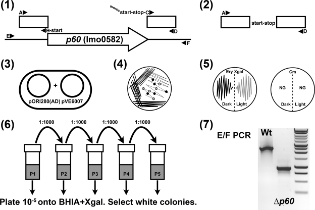
We used the pORI-based _repA_-negative plasmid system for rapid deletion or site-directed mutagenesis of Listeria genes (see Fig. 4 and 5) (41). Gene deletions were constructed by splice overlap extension (SOE) PCR (34) to generate two 400-bp fragments: one upstream including the ATG codon or a point mutation (AB product) and one downstream beginning from the stop codon (CD product) or directly after the point mutation (with a 20-bp tail on the C primer complementary to the B primer). The initial PCR products were diluted 1:20 in PCR-grade water, and 1 μl of each product was used as a template in a second round of PCR with the AD primers to generate an 800-bp product. The SOE PCR product was cloned into the multiple cloning site (MCS) of pORI280, and the sequence of the cloned product was verified (MWG Biotech, Germany) with MCS primers (forward primer TATCGATGCATGCCATGGTACC and reverse primer CGCCAGGGTTTTCCCAGTCACGAC). The plasmid was cotransformed into L. monocytogenes with the highly temperature-sensitive plasmid pVE6007 (47) supplying RepA in trans. Transformants were selected on BHI agar containing erythromycin at a concentration of 5 μg/ml and X-Gal (5-bromo-4-chloro-3-indolyl-β-d-galactopyranoside) (Calibochem, Merck) at a concentration of 100 μg/ml. For deletion of essential genes, the IPTG-inducible plasmid pIMK4 (containing the gene to be deleted) (see below) was also included in the electroporation mixture along with kanamycin at a concentration of 50 μg/ml. Plates were incubated at 30°C for 48 h.
FIG. 4.
Rapid gene deletion protocol for L. monocytogenes: the pORI280 system (41). The following steps are shown diagrammatically. In step 1, an SOE PCR product was generated with 400-bp upstream (AB to the start codon) and downstream (CD from the stop codon) fragments of the gene to be deleted. In step 2, the AB and CD fragments were joined by a second round of PCR with the A and D primers to form the AD product. This amplimer was cloned into pORI280, generating pORI280(AD). In step 3, pORI280(AD) and the RepA-supplying temperature-sensitive plasmid pVE6007 were cotransformed into the recipient electrocompetent L. monocytogenes strain and selected on BHI agar containing 5 μg/ml erythromycin and 100 μg/ml X-Gal at 30°C for 48 h. In step 4, single blue colonies were complex streaked on BHI agar containing erythromycin and X-Gal and then incubated at 37°C for 24 h. This step resulted in the loss of pVE6007 and then caused pORI280AD to integrate via the AB or CD crossover. In step 5, light and dark blue colonies arising from pORI280AD integration were patched onto both BHI agar containing 5 μg/ml erythromycin and BHI agar containing 7.5 μg/ml chloramphenicol (Cm) and then incubated at 37°C for 24 h. NG indicates no growth on chloramphenicol plates and loss of pVE6007. In step 6, a single light blue colony and a single dark blue colony were grown statically to stationary phase at 37°C. The cultures were diluted 1:1,000 in fresh BHI, and the process was repeated for five sequential passages. Each of the five passages was diluted 10−5, 100 μl was spread plated onto BHI agar containing X-Gal, and the plates were incubated at 37°C for 24 h. In step 7, white colonies were screened by colony PCR with the E and F primers as shown for step 1. Colonies were also tested for erythromycin sensitivity by patching for the loss of pORI280(AD), and the EF amplimer was sequenced. Wt, wild type.
FIG. 5.
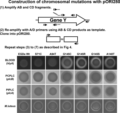
Site-directed chromosomal mutagenesis using pORI280: new prfA star mutants. The procedure described in the legend to Fig. 4 was used to create single nucleotide point mutations in the chromosome. In steps 1 and 2, SOE PCR was used to create directed nucleotide changes in a region of DNA, and both the B and C primers contained the desired mutations (indicated by three stars). However, in this case, genomic DNA from naturally induced prfA mutants was used as the template. PrfA mutants were generated from a hemolysin promoter fused to a chloramphenicol reporter gene (EGDe::p_hly_C), with selection pressure (BHI agar containing 50 μg/ml of chloramphenicol) to induce prfA mutations. Mutants were recreated using the pORI280 system in a fresh EGDe background. Five microliters of an overnight BHI culture of each recreated isolate was spotted onto BHI agar containing (i) 5% defibrinated sheep blood with 1 U/100 ml sphingomyelinase (Sigma), (ii) 4% lecithin (Oxoid) (PCPLC), (iii) 0.2% l-α-phosphatidylinositol (1 g dissolved in 25 ml distilled water, autoclaved, and added to 475 ml BHI agar at 50°C) (Sigma) (PIPLC), and (iv) 0.2% freeze-dried Micrococcus luteus cells (cell wall hydrolase activity) (Sigma). Plates were incubated for 48 h at 37°C. Wt, wild type.
The integration of pORI280 by single crossover was stimulated by picking a single blue colony from the transformation plate, streaking it to obtain single colonies onto BHI agar containing erythromycin plus X-Gal, and incubating the preparation at 37°C for 24 h. For deletion of essential genes, 1 mM IPTG was included in all agar plates and broth media. The loss of pVE6007 was confirmed by patching single colonies (both light and dark blue) onto BHI agar containing chloramphenicol (7.5 μg/ml) or BHI agar containing erythromycin (5 μg/ml). Single light and dark blue Cms clones were passaged statically at 37°C in BHI by diluting the cultures 1:1,000 for five successive passages. Cells from each passage were diluted 10−5 and plated onto BHI agar containing X-Gal, and white colonies were screened by colony PCR. For gene deletion screening, primers external to the SOE PCR product (primers E and F [see Fig. 4, steps 1 and 7]) were used. For point mutation screening, an external forward primer and an internal reverse primer with the mutation incorporated at the 3′ end were employed (primers E and F [see Fig. 5, step 1]). Putative deletion mutants were confirmed by sequencing the mutated region.
Construction of pIMK, a kanamycin site-specific integrative plasmid.
The Listeria vector pPL2 utilizes the site-specific integrase of the PSA phage to direct integration of the vector into the tRNAArg locus on the L. monocytogenes chromosome (between the lmo1240 and lmo1241 genes in EGDe) (37). The derivative vector pIMK was generated by SOE PCR to decrease the plasmid size and to change the antibiotic selection. Initially, three fragments were PCR amplified: (i) a 171-bp fragment encoding the pBluescriptII KS(+) MCS taken from pPL2 (primers IM203 and IM204 [Table 2]), (ii) the backbone region of pPL2 from nucleotide 1100 to nucleotide 4585 (GenBank accession number AJ417449 nucleotide numbering) (primers IM205 and IM211), and (iii) the aphA3 kanamycin resistance gene from pTV1-OK (primers IM207 and IM208) (30).
TABLE 2.
Oligonucleotides used in this study
| Primer (description)a | Oligonucleotide sequence (5′-3′)b | Restriction site |
|---|---|---|
| pIMK | ||
| IM203 (MCS T3 FWD) | AATTAACCCTCACTAAAGGGAAC | |
| IM204 (MCS T7 REV) | GACGTCGTAATACGACTCACTATAGGGC | AatII |
| IM205 (T7/1,100 bp O FWD) | AGTGAGTCGTATTACGACGTCCCAGGGCTTCCCGGTATCAAC | AatII |
| IM211 (4,585 bp REV) | CATATGATCATCATAATTCTGTCTCATTATATAAC | NdeI |
| IM207 (Kan FWD) | ATAAACCCAGCGAACCATTTGAGGTGATAGG | |
| IM208 (4,585 bp/Kan O FWD) | ATAATGAGACAGAATTATGATGATCATATGAAACATCAGAGTATGG | NdeI |
| pIMK2, pIMK3, and pIMK4 | ||
| IM236 (Phelp FWD) | ATATGAGCTCCATTATGCTTTGGCAGTTTATTCTTGACAT | SacI |
| IM214 (tiling FWD1) | GAATACCATAATTCCAATAGATATGAGCTCCATTATGCTT | SacI |
| IM215 (tiling FWD2) | TGGCAGTTTATTCTTGACATGTAGTGAGGGGGCTGGTATA | |
| IM216 (tiling FWD3) | ATCACATAAATTGTGAGCGCTCACAATTATAAAGCAAGCA | |
| IM217 (tiling FWD4) | TATAATATTGCGTTTCATCTTTAGAAGCGAATTTCGCCAA | |
| IM218 (tiling FWD5) | TATTATAATTATCAAAAGAGAGGGGTGGCAAACGGTATTT | |
| IM219 (tiling FWD6) | GGCATTATTAGGTTAAAAAATGTAGAAGGAGAGTGAAACC | |
| IM220 (tiling REV1) | TATATGGATCCTTTTCCATGGGTTTCACTCTCCTTCTACA | BamHI/NcoI |
| IM221 (tiling REV2) | TTTTTTAACCTAATAATGCCAAATACCGTTTGCCACCCCT | |
| IM222 (tiling REV3) | CTCTTTTGATAATTATAATATTGGCGAAATTCGCTTCTAA | |
| IM223 (tiling REV4) | AGATGAAACGCAATATTATATGCTTGCTTTATAATTGTGA | |
| IM224 (tiling REV5) | GCGCTCACAATTTATGTGATTATACCAGCCCCCTCACTAC | |
| IM225 (tiling REV6) | ATGTCAAGAATAAACTGCCAAAGCATAATGGAGCTCATAT | |
| IM327 (OID2 FWD) | ATATGAGCTCAATTGTGAGCGCTCACAATTC | SacI |
| IM200 (Phelp FWD) | AGATGTCGACGATCCCATTATGCTTTGGC | SalI |
| IM201 (lacI REV) | TATAGGTACCTTACTGCCCGCTTTCCAGTCGG | KpnI |
| IM202 (Phelp/lacI O FWD) | ATGTAGAAGGAGAGTGAAACCCATGAAACCAGTAACGTTATACGATGTCG | |
| IM212 (rrnB TT FWD) | ATATGACGTCGGGCCCTTTCGTCTTCAAGAATTAA | AatII |
| IM213 (rrnB TT REV) | TCAAGAGCTCGAATTCCGATCCCCAATTCC | SacI |
| Deletion mutant | ||
| IM463 (_inlA_-A FWD) | AACGCCATGGAGGATATCACTAAACGGCTCC | NcoI |
| IM464 (_inlA_-B REV) | CGTTGTAACTTGGTCTAGATCTGTTTGTG | |
| IM465 (_inlA_-C FWD) | AGATCTAGACCAAGTTACAACGAAAGAAACAACCAAAGAAGTGGAAG | |
| IM466 (_inlA_-D REV) | ATATCTGCAGCAAACGTTGCTGTATAGCTATTGG | PstI |
| IM467 (_inlA_-E out FWD) | TATATAGGAAAAATGTGCTGGAACG | |
| IM468 (_inlA_-F out REV) | TCCTTGATAGTCTACTGCTTGAGTCG | |
| IM330 (_p60_-A FWD) | TATACTGCAGTTCTATTATAGAATACCATAAACTCATC | PstI |
| IM079 (_p60_-B REV) | CATAAAACTCCTCTCTTTTTTCAGAAAATC | |
| IM331 (_p60_-C FWD) | AAAAAAGAGAGGAGTTTTATGTAATTAATAACTTAAAGTAACCTGTGG | |
| IM332 (_p60_-D REV) | TATATCTAGAAAGCCGTAGTAGAGTCATCTGC | XbaI |
| IM362 (_p60_-E out FWD) | ACTATTTTTCGATCATCATAATTCTGTCTC | |
| IM363 (_p60_-F out REV) | ATGCCTAGACTGTACCATTGAATTACG | |
| IM435 (prfA*145/148 FWD) | TATACTGCAGTAGAAACTAACGGGATAAAACC | PstI |
| IM436 (prfA*145/148 REV) | ATATTCTAGATTGCAGCTCTTCTTGGTGAAGC | XbaI |
| IM443 (prfA*71 FWD) | TTAACTGCAGAATAAATCCGTTTTTAAATATG | PstI |
| IM444 (prfA*71 REV) | TATATCTAGAATTTTTATACACGATAACTTTCTC | XbaI |
| IM447 (prfA*94 FWD) | AATCCTGCAGGTAAAAAACATCATTTAGCGTGAC | PstI |
| IM449 (prfA*94 REV) | ACATTCTAGATAAAACCATTCATCTAATTTAGG | XbaI |
| IM457 (_secA_-A FWD) | TATACTGCAGGAACAAAACAATTTTCATTGAAACC | PstI |
| IM458 (_secA_-B REV) | CATTTAAATTCTCCTCTACTGTTTTCGC | |
| IM459 (_secA_-C FWD) | ACAGTAGAGGAGAATTTAAATGTAGATTGAAACAATTTAAAAAGCGC | |
| IM460 (_secA_-D REV) | TATACCATGGTTAATATCTTTTTCTAATTCTTCTTG | NcoI |
| IM152 (_secA_-E out FWD) | AAAATGAAGGAGATTTTGATCTTGAAATTG | |
| IM153 (_secA_-E out REV) | ATGCACTAATAGTCGCCATGTAAGC | |
| Cloned genes | ||
| IM319 (LuxA FWD) | ATATCCATGGAATTTGGAAACTTTTTGCTTACATACC | NcoI |
| IM319a (LuxE REV) | ATATCTGCAGGATATCAACTATCAAACGCTTCG | PstI |
| IM333 (p60 FWD) | ATATCCATGGATATGAAAAAAGCAACTATCG | NcoI |
| IM334 (p60 REV) | ATATGTCGACAAACCTGTGAAGCGAACTGCTTGC | SalI |
| IM194 (inlA FWD) | ATATCCATGGGAAAAAAACGATATGTATGGTTG | NcoI |
| IM188 (inlA REV) | TTTTCTGCAGTTATTTACTAGCACGTGCTTTTTTAG | PstI |
| IM461 (secA FWD) | ATATCCATGGCTGGACTATTGAAAAAAATTTTTG | NcoI |
| IM462 (secA REV) | ATATCTGCAGTTATGCTTCTTTACCATGACAATTT | PstI |
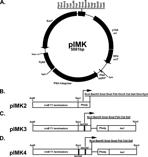
The MCS was amplified to include the T3 and T7 promoter primer binding sites and then joined to the backbone (1,100-bp end) of pPL2 by SOE PCR, thereby removing both chloramphenicol resistance genes. The complete vector fragment was constructed by joining the 3′ end of the kanamycin resistance gene to the free pPL2 backbone (4,585-bp end) of the previous PCR product. The amplimer was gel extracted and then phosphorylated with T4 polynucleotide kinase (NEB). The product was ligated with the LigaFAST rapid DNA ligation system (Promega) for 30 min at 22°C and transformed into DH10B, with selection on LB agar containing kanamycin at a concentration of 50 μg/ml. One clone was fully sequenced (pIMK; 5,081 bp). A plasmid schematic diagram (Fig. 1A) was drawn with an online plasmid drawing tool (http://bioinformatics.org/savvy/) and exported as an .svg file on an iBook (Apple) from the Opera web browser into Adobe Illustrator CS2 for further manipulation as an .svg file.
FIG. 1.
Plasmid maps of pIMK and promoter derivatives. (A) Plasmid pIMK was created by SOE PCR as described in Materials and Methods. This plasmid is based on the pPL2 vector (37), which replicates autonomously in E. coli but is unable to replicate in L. monocytogenes. Integration into the tRNAArg locus is directed by the PSA phage integrase under the control of the listerial P60 promoter and confers kanamycin resistance. For sequencing purposes, T3 and T7 primer binding sites are present before the KpnI restriction site and after the SacI restriction site, respectively. The restriction sites labeled on pIMK are unique. (B to D) Derivatives of pIMK were constructed to enable heterologous gene expression through in-frame cloning of promoterless genes via a unique NcoI site overlapping the start codon. Additional restriction sites used for cloning are underlined. (B) pIMK2 enables constitutive overexpression of genes from the synthetic Phelp promoter (59). (C) pIMK3. There is high-level, IPTG-controlled expression from the Phelp promoter. A consensus lacOid sequence (LacI binding site [gray box] [51]) was inserted between the PCP25 promoter (containing the −35 and −10 regions) and the downstream 5′ untranslated region of the hlyA gene. (D) pIMK4. There is strict IPTG-controlled expression from the pIMK3 promoter with an additional lacOid sequence (gray box) incorporated directly upstream of the PCP25 region. In both pIMK3 and pIMK4, the lacI repressor is expressed from the Phelp promoter, which is cloned between the SalI and KpnI restriction sites.
Construction of pIMK2, a _Listeria_-specific overexpression plasmid.
The rrnB T1 transcription terminators were PCR amplified (with IM212 and IM213) from pLIV2 (33) and cloned into the unique AatII/SacI restriction sites of pIMK, creating pIMK-rrnB. The Phelp promoter amplified from pPL2_lux_-Phelp (59) was cloned downstream into the SacI/BamHI restriction sites (with IM236 and IM220), creating pIMK2. A unique NcoI site overlapping the ATG of Phelp was introduced, facilitating direct, in-frame cloning of Listeria genes to obtain high-level expression.
Construction of pIMK3 and pIMK4, two IPTG-inducible plasmids.
The Phelp promoter was joined to the LacI repressor from pLIV2 by SOE PCR (with primers IM200 and IM108 (10) and primers IM201 and IM202) and then cloned into the SalI/KpnI restriction sites of pIMK-rrnB. The Phelp promoter was PCR amplified from an oligonucleotide tiling template (with oligonucleotides IM214 through IM225) (59) to include the consensus lacOid LacI repressor binding site (51), separating the mapped transcription start site of PCP25 and the 5′ untranslated region of hlyA. The product was cloned as a SacI/BamHI fragment, creating pIMK3. A second lacOid repressor was introduced upstream of the −35 region by PCR amplification of the pIMK3 promoter (using pIMK3 as the template and primers IM327 and IM220) and cloned as a SacI/BamHI fragment, generating pIMK4.
Overexpression and IPTG inducibility: vector testing.
To test the function of the IPTG-inducible (pIMK3 and pIMK4) and Phelp-driven overexpression (pIMK2) constructs, the following three regions were PCR amplified: luxABCDE from pPL2_lux_ (with IM319 and IM319a) (10), full-length P60 gene (with IM333 and IM334), and internalin A gene from EGDe genomic DNA (with IM194 and IM188). All three regions were cloned as NcoI/PstI fragments into the three vectors.
(i) Luciferase expression.
Overnight precultures of EGDe, EGDe::pIMK4_lux_, EGDe::pIMK3_lux_, and EGDe::pIMK2_lux_ were diluted 1:100 in fresh BHI containing different concentrations of IPTG and grown statically (200-μl aliquots in triplicate) at 37°C in either Costar black 96-well plates (incubated in an IVIS100 in vivo imager [Xenogen]) or Costar clear-bottom 96-well plates (incubated in a Spectramax M2 plate reader [Molecular Devices]). Readings were taken every 30 min for 10 h, with the IVIS100 acquiring luminescence readings for 5 min at a binning of 8 and the plate reader determining the OD600. As a control for luciferase expression and the effect on the growth rate, EGDe transformed with pIMK3 was included. There was a minimal effect on the growth rate due to luciferase expression, and for all samples EGDe::pIMK3 was used for subtraction of the background luminescence.
(ii) Western blotting of P60 secretion.
Overnight precultures of EGDe, EGDe Δ_p60_, EGDe Δ_p60_::pIMK4_p60_, EGDe Δ_p60_::pIMK3_p60_, EGDe Δ_p60_::pIMK2_p60_, and EGDe::pIMK2_p60_ were grown in filter-sterilized BHI at 37°C with shaking. Precultures were diluted 1:50 in 25 ml of fresh medium containing either no IPTG or 1 mM IPTG, grown as described above to exponential phase (OD600, 1.0), and harvested by centrifugation (7,000 × g for 10 min). The supernatant samples were subsequently processed as described previously (49).
(iii) Invasion assays.
The human colonic Caco-2 cell line (HTB-37; ATCC) was seeded at a density of 1 × 10⁵ cells per well in Primera 24-well tissue culture plates (Falcon) and grown to confluence at 37°C in the presence of 5% CO2 in Caco-2 medium (Dulbecco modified Eagle medium [DMEM] containing 10% fetal calf serum, 1% nonessential amino acids, and a penicillin/streptomycin mixture). On the day prior to use, the medium was changed to antibiotic-free Caco-2 medium and, before invasion, washed twice with 1 ml prewarmed DMEM. Overnight BHI precultures of EGDe, EGDeΔ_inlA_, EGDeΔ_inlA_::pIMK4_inlA_, EGDeΔ_inlA_::pIMK3_inlA_, and EGDe Δ_inlA_::pIMK2_inlA_ were diluted to obtain an initial OD600 of 0.1 (∼30-fold) in the presence of various concentrations of IPTG (0, 0.01, 0.1, 1, and 10 mM) and grown to exponential phase (OD600, 0.8 to 1.0). Cells were washed twice in prewarmed DMEM and diluted 1:100 to obtain an initial inoculum of 1 × 10⁷ CFU/ml (multiplicity of infection, 10). A 1-ml aliquot was added to the Caco-2 cells (in quadruplicate) and incubated for 1 h at 37°C in the presence of 5% CO2, and then each preparation was washed once with Dulbecco's phosphate-buffered saline (PBS) (Sigma) before it was overlaid with 1 ml of DMEM containing 10 μg/ml of gentamicin. After 1 h of incubation at 37°C in the presence of 5% CO2, the Caco-2 cells were washed twice with PBS before the monolayer was lysed with 1 ml of sterile distilled water. Cells were incubated for 1 h at 4°C, vortexed using the shake setting (Scientific Industries) for 15 s, and then diluted in PBS and plated onto BHI agar. Agar plates were incubated overnight at 37°C.
Nucleotide sequence accession number.
The nucleotide sequence of pIMK has been deposited in the EMBL nucleotide sequence database under accession number AM940000.
RESULTS
Construction of a novel site-specific integration vector.
pPL2, a site-specific integration vector developed in 2002, is based on the integrative mechanism of the PSA listeriophage (37). This vector provides a simple and effective tool for complementation of chromosomal gene deletions or for expression of heterologous proteins (6, 9, 42, 70). pPL2 is generally highly stable without antibiotic selection and functions in a broad range of L. monocytogenes strains. Here we attempted to further develop the system through a reduction in plasmid size, restriction site expansion, and a change in the antibiotic selection marker. From this starting point we constructed a suite of vectors for gene overexpression or IPTG-inducible gene expression.
After a series of SOE PCRs, we replaced both chloramphenicol acetyltransferase (CAT) markers of pPL2 with a single kanamycin resistance gene to create pIMK (Fig. 1A). In brief, the pBluescriptII KS MCS was joined to the E. coli origin of replication end of the pPL2 backbone (finishing with the PSA integrase), thereby removing the gram-positive and gram-negative CAT genes. The aphA3 resistance cassette (encoding kanamycin resistance) was amplified from pTV1-OK and joined to the PSA integrase end of the previous PCR product and circularized. This kanamycin-resistant vector is 1 kb smaller than pPL2 and is compatible with the systems currently used to create chromosomal mutations in L. monocytogenes (pMAD [4], pAUL-A [14], pGM [43], pLSV1 [54], pORI19 [57], and pKSV7 [66]), and it allows further manipulation with other plasmids based on alternative antibiotic selection (e.g., erythromycin, chloramphenicol, or tetracycline selection).
New IPTG-inducible and overexpression constructs.
We used the recently created, highly expressed Phelp promoter (59) to obtain three levels of gene expression; (i) constitutive overexpression (pIMK2) (Fig. 1B); (ii) high-level IPTG-inducible expression (pIMK3 [one lacOid site]) (Fig. 1C); and (iii) low-level, strictly controlled IPTG-inducible gene expression (pIMK4 [two lacOID sites]) (Fig. 1D). The rrnB transcription terminators were cloned upstream of the promoter to minimize readthrough, and one of the three promoters described above was cloned downstream. For both IPTG-inducible constructs, the Phelp promoter fused to the lacI repressor was cloned at the opposite end of the MCS, leaving a number of restriction sites for cloning purposes. A unique NcoI site was introduced that overlapped the ATG of Phelp, which allowed in-frame cloning of genes (without their associated ribosome binding sites [RBS]) for functional analysis.
We employed three assays to test the system: (i) a translational fusion with the gram-positive optimized luciferase operon from Photorhabdus luminescens; (ii) qualitative Western blotting of P60 secretion; and (iii) a quantitative screen of internalin A-mediated invasion of Caco-2 cells.
The levels of pIMK2_lux_-driven luciferase were equivalent to the Phelp-LUX expression data of Riedel et al. (59). The level of luminescence from the pIMK3_lux_ construct was 10-fold less than that from Phelp, with maximal induction occurring in media containing 1 mM IPTG. Luciferase production from EGDe::pIMK4_lux_ was at the limit of detection, so additional assays were performed to compare the levels of gene expression.
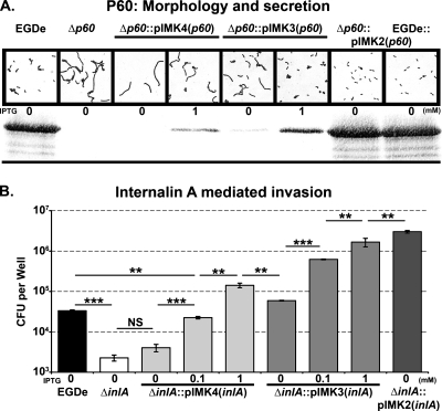
The cell wall hydrolytic protein P60 (encoded by the p60 gene, also known as cwhA or iap) is the most abundant protein secreted by L. monocytogenes in rich laboratory media and is essential for full virulence of the pathogen (42). To test the expression system, we cloned the full-length p60 gene behind the three promoters and assayed the levels of P60 expression in wild-type strain EGDe and the corresponding p60 deletion mutant transformed with the three plasmids. Previous attempts to complement an EGDe p60 deletion mutant proved to be difficult; one group had to repair the original deletion (54), while a second group was unable to obtain E. coli transformants of p60 in pPL2 (46). We found that a functional p60 gene could be successfully cloned into E. coli XL1-Blue; however, the growth of the strains harboring the gene was affected. Transformation of EGDeΔ_p60_ with the p60 gene expressed from pIMK2 resulted in a fully complemented strain (Fig. 2A). When the mutant was transformed with an IPTG-dependent plasmid (pIMK3_p60_ or pIMK4_p60_), overlapping levels of P60 expression were obtained, since fully induced pIMK4_p60_ produced levels greater than the levels produced by uninduced pIMK3_p60_. However, full complementation was not achieved. As determined by microscopy, the P60 deletion mutant exhibited a long-chain phenotype (Fig. 2A). Increasing the level of P60 expression resulted in incremental decreases in cell chain length (Fig. 2A). In the case of EGDeΔ_p60_::pIMK3_p60_ with 1 mM IPTG, single cells were observed; however, the morphology of these cells did not completely resemble the wild-type morphology. At this concentration there was still threefold less P60 expression than in wild-type strain EGDe and fourfold less than in the pIMK2 overexpression strains (quantified by NIH Image 1.63). This was manifested by curved single and paired cells instead of the wild-type rod-shaped single and paired cells (Fig. 2A).
FIG. 2.
Expression profiles of pIMK4, pIMK3 (IPTG inducible), and pIMK2 (overexpression) demonstrated by P60 secretion and InlA-mediated invasion. (A) Gram staining (top panel) and anti-P60-specific Western blotting (bottom panel) for the secreted proteins from BHI-grown, exponential-phase (OD600, 1.0) cells transformed with the pIMK derivatives containing p60. (B) Gentamicin protection assay for InlA-mediated invasion of Caco-2 cells. Strains transformed with the pIMK derivatives containing inlA were grown to exponential phase (OD600,1.0) in BHI containing various concentrations of IPTG and added to Caco-2 cells as described in Materials and Methods. Invasion was expressed as the total number of CFU recovered per well, and each mean and standard deviation are the results of one representative experiment performed in quadruplicate. Inclusion of IPTG in the preculture did not impact the invasion by EGDe. Statistical analyses were performed using the raw CFU counts and the Student t test, and P values less than 0.01 were considered significant (**, P < 0.005; ***, P < 0.001). NS indicates that the P value is above the level of significance.
A third quantitative assay was employed to ascertain the dynamic range of the newly created promoters. The abilities of L. monocytogenes to invade a human intestinal epithelial cell line (Caco-2) are mediated via the E-cadherin-internalin A interaction. The extent of invasion can be assessed by enumerating the intracellular survivors after a gentamicin protection assay. The invasion of EGDe was compared to the invasion of an inlA deletion strain and the invasion of the deletion mutant transformed with the inlA_-containing plasmids. As previously described (7), we observed that the ability of the inlA deletion mutant to invade the nonphagocytic Caco-2 cell line was significantly impaired (the yield was approximately 5% of the yield of wild-type survivors). The invasive ability could be restored following complementation with all three constructs. To test promoter expression, we used a low multiplicity of infection (10 bacteria per Caco-2 cell) to increase the dynamic range. The EGDeΔ_inlA::pIMK4_inlA_ strain exhibited low residual invasion in the absence IPTG; the level was 3.6 ± 2.92 log10 CFU per well, which was very similar to the value for the Δ_inlA_ strain (3.49 ± 2.5 log10 CFU per well) (Fig. 2B). Addition of 0.1 and 1 mM IPTG to the pregrowth medium stimulated invasion 5.5- and 35-fold, respectively. No further increase in invasion was observed with higher concentrations of IPTG (data not shown). The kinetics of invasion of wild-type strain EGDe or EGDeΔ_inlA_::pIMK2_inlA_ grown in the presence and in the absence 1 mM IPTG were very similar, suggesting that inclusion of IPTG in the preculture does not impact the invasion process.
The EGDeΔ_inlA_::pIMK3_inlA_ construct exhibited leaky expression in the absence of IPTG (as previously observed for P60 [Fig. 2A]); however, the level of invasion was lower than that observed for pIMK4_inlA_ with full induction (20.5-fold increase in invasion for pIMK3_inlA_ and 35-fold increase in invasion for pIMK4_inlA_ compared with uninduced pIMK4_inlA_). For the pIMK3_inlA_ construct, invasion was simulated 10.5-, 28-, and 37-fold using 0.1, 1, and 10 mM IPTG, respectively, compared to uninduced pIMK3_inlA_. A dynamic range of invasion of 540-fold was determined by dividing the invasion of pIMK3_inlA_ induced with 10 mM IPTG by the invasion of uninduced pIMK4_inlA_. Expression of inlA from the unmodified Phelp promoter (pIMK2) led to a further 2.5-fold stimulation of invasion compared with fully induced pIMK3_inlA_. The discrepancy in the difference between the maximal expression of pIMK2 and the expression of fully induced pIMK3 (luciferase, 10-fold; P60, 4-fold; and InlA, 2.5-fold) may have been due to additional processing components required for the correct localization of P60 or internalin A protein or possible saturation of the assay during gentamicin protection (increasing the multiplicity of infection did not increase the numbers of EGDeΔ_inlA_::pIMK2_inlA_ recovered from the Caco-2 cells [data not shown]). In conclusion, the constructs described here allow dramatic manipulation of L. monocytogenes gene expression.
Optimization of L. monocytogenes electroporation.
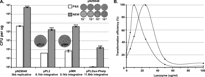
The electroporation method of Park and Stewart for the transformation of L. monocytogenes (53) has been cited in over 100 Listeria research papers (ISI Web of Knowledge). With the strain of L. monocytogenes used in the study of Park and Stewart (ATCC 23074), transformation efficiencies of up to 6.6 log10 CFU/μg of plasmid DNA were achieved. In our hands, EGDe, a commonly used strain of L. monocytogenes (27) whose genome has been sequenced, was poorly transformable. Maximal transformation efficiencies of 4.7 log10 CFU/μg were obtained with a 3-kb lactococcal shuttle vector, pNZ8048 (48) (Fig. 3A). When the site-specific integrative plasmids pPL2 (6.1 kb) (37) and pPL2_lux_-Phelp (11.8 kb) (59) were used, a drastic decrease in efficiency resulted in values of less than 100 and 0 to 2 CFU/μg, respectively (Fig. 3A). This poor transformation efficiency is consistent with the results of other workers (3, 12, 52), who took advantage of the mobilizable nature of pPL2 (rather than electroporation) to transfer the plasmid into EGDe and the recently sequenced strain 10403S.
FIG. 3.
Improved electroporation protocol for L. monocytogenes. Systematic changes to the protocol of Park and Stewart (53) (P&S) led to the development of the improved electroporation protocol (NEW) as described in Materials and Methods. A series of replicative (pNZ8048) and integrative (pPL2, pIMK, and pPL2_lux_-Phelp) plasmids were tested to compare the efficiency of the protocol of Park and Stewart and the efficiency of the new protocol. Most transformants were selected on BHI agar containing chloramphenicol at a concentration of 7.5 μg/ml; the only exception was pIMK, which was selected on medium containing kanamycin at a concentration of 50 μg/ml. The images in the top right of panel A show 10-μl spot dilutions (10−1, 10−2, 10−3, and 10−4) from pNZ8048 transformations for the protocol of Park and Stewart and the new protocol. Colonies obtained with the new protocol recovered faster than colonies obtained with the protocol of Park and Stewart, as shown by larger colonies. The images of plates (transformed with the new protocol) from pPL2 and pIMK transformations show the uniformity of the colony size for pIMK-containing cells compared to the pPL2-containing cells. Colonies were visible after overnight incubation of pIMK-transformed strains. Statistical analyses were preformed using the raw CFU counts and the Student t test, and P values less than 0.01 were considered significant (***, P < 0.001). (B) Relative contributions of lysozyme treatment for EGDe (circles) and F2365 (triangles) expressed as a percentage of the maximal transformation. The highest levels of transformation were obtained with 10 and 25 μg/ml lysozyme for EGDe and F2365, respectively.
We attempted to improve the transformation efficiency of EGDe through systematic changes in the protocol of Park and Stewart (53). In all initial transformations, 1 μg of ethanol-precipitated pNZ8048 was used. The protocol of Park and Stewart utilizes penicillin for 2 h with exponential phase cells grown in BHI containing 500 mM sucrose to protect against osmotic stress. Increasing the concentration of penicillin from 10 to 15 μg/ml or addition of 1% glycine (with 10 μg/ml of penicillin) decreased the number of transformants almost fivefold (to 4.0 log10 CFU/μg). We maintained the initial concentration of penicillin but subsequently changed the wash buffer from 1 mM HEPES (pH 7), 500 mM sucrose to the buffer described by Shepard and Gilmore (65) (SGWB). This resulted in a slight increase in transformation efficiency (1.3-fold), suggesting that the glycerol had an improved osmostabilizing role or the removal of HEPES had an effect. We then examined the effect of lysozyme on the washed cells, as described by Powell et al. (55) for electroporation of Lactococcus lactis. Using the original buffer, we were unable to obtain transformants with additional treatment with 10 to 100 μg/ml lysozyme at 37°C. However, with SGWB, transformants were obtained at all concentrations tested (Fig. 3B). Under these conditions a maximal transformation efficiency of 6.76 ± 5.61 log10 CFU/μg was obtained with 20 min of incubation in SGWB containing 10 μg/ml of lysozyme at 37°C. Increasing the concentration of lysozyme dramatically decreased the efficiency (Fig. 3B). When we used the optimized conditions for transformation of pPL2-based plasmids, we observed an improvement of at least 125-fold (Fig. 3A). In our hands, pPL2-transformed EGDe took 2 days to form colonies on BHI agar containing 7.5 μg/ml chloramphenicol, and a mixture of both wild-type and small colonies was consistently observed, suggesting that there was poor expression of the gram-positive cat gene in L. monocytogenes. However, EGDe transformed with the pIMK vector formed colonies that were a uniform size and were visible after overnight incubation. The electroporation parameters were also examined, and the results confirmed that an optimal pulse of 1 kV/cm, 400 V, and 25 μF works best, in agreement with the data of Park and Stewart (53). Electrocompetent cells prepared as described here can be stored at −80°C for over 6 months without a decrease in transformability. There was a marked decrease in transformation efficiency in cells washed with either (i) filter-sterilized SGWB with the pH not adjusted or (ii) autoclaved SGWB with the pH adjusted (data not shown). Experiments were subsequently conducted to determine the optimal concentration of lysozyme for treating other commonly used sequenced strains of L. monocytogenes, including 10403S (10 μg/ml) and F2365 (25 μg/ml) (Fig. 3B). Using pNZ8048, transformation efficiencies of 6.83 ± 5.97 and 7.31 ± 6.49 log10 CFU/μg were obtained for 10403S and F2365, respectively. A further slight improvement in transformation efficiency was observed if 50 μl rather than 100 μl of electrocompetent cells was used.
Gene deletion and site-directed chromosomal mutagenesis.
Listeria researchers have utilized a number of strategies and vectors to create chromosomal mutations (4, 11, 43, 54). Here we added to the repertoire through implementation of the pORI system originally developed with L. lactis (38). The pORI vectors have been thoroughly reviewed by Leenhouts et al. (41). Briefly, pORI vectors contain no origin of replication and require the replication initiation protein, RepA, to be supplied in trans for plasmid maintenance. In E. coli, a host strain containing the repA gene integrated into the chromosome is used for cloning purposes and plasmid propagation. For mutagenesis, the construct is transferred to the recipient strain with a temperature-sensitive helper plasmid expressing RepA. The deletion or mutagenesis proceeds through a forced single-crossover event due to a temperature shift (nonpermissive for the helper plasmid [36, 41]), followed by a period of nonselective growth that stimulates the loss of the integrated plasmid and results in the generation of either a wild-type or mutated sequence (theoretically at a ratio of 50:50).
Here we used a two-plasmid system consisting of pORI280, which contains a constitutively expressed lacZ gene that allows discrimination of strains containing the integration plasmid, and the highly temperature-sensitive RepA+ helper plasmid pVE6007. An SOE PCR deletion construct (containing fused regions from each side of the preferred deletion event) was cloned into pORI280 (Fig. 4 and 5) and transformed, with pVE6007, into the target strain. The initial integration event was selected by a shift to a nonpermissive temperature for pVE6007 replication. In EGDe, we found that pVE6007 was unable to replicate at 37°C; therefore, we could simplify the integration event by performing a complex streak from a single colony from the initial transformation plate onto BHI agar containing erythromycin (5 μg/ml) and X-Gal (100 μg/ml). This step had the additional benefit of decreasing the selection pressure for a dominant crossover (Fig. 4, AB versus CD) that could arise during growth in broth (through growth retardation) and thus improved the frequency of crossover identification (data not shown). Colonies from the plate were scored for loss of pVE6007 (Cms), and subsequently one light blue colony and one dark blue colony (usually corresponding to AD and CD crossovers) were passaged five times at 37°C. Diluted cells from each passage were plated onto BHI agar containing X-Gal, and white colonies were screened by colony PCR with primers E and F to identify mutants (Fig. 4). These steps were all optimized for creation of the previously described p60 and inlA deletion mutants. From the start of cloning to creation of the mutant, the procedure could routinely be completed in 10 to 14 days, and there was a reduction in the number of passages in rich media compared to the results with other vectors in our hands (data not shown).
The procedure described above can also be used for creation of point mutations, as shown in Fig. 5. We used the protocol described above to recreate a series of prfA “star” mutants (60). In brief, these mutants were isolated by fusing the hemolysin promoter to a CAT gene (p_hly_C) and plating on BHI agar containing chloramphenicol at a concentration of 50 μg/ml (the EGDe::p_hly_C strain is unable to grow at a chloramphenicol concentration greater than 7.5 μg/ml). The prfA gene was sequenced for all 20 spontaneously arising chloramphenicol-resistant colonies (from three separate experiments), all of which had point mutations in prfA. Only one (G145S) of the six prfA star mutants identified in this manner has been described previously (60). Two additional mutants were shown to have a mutation at the same nucleotide as G145S, resulting in introduction of a different amino acid (cysteine or asparagine instead of glycine). All six point mutations were reintroduced into the chromosome of EGDe using the protocol for gene deletion to create an allelic exchange (Fig. 5). The phenotypes of the three G145 mutants and the L148P mutant were identical in that they showed elevated secretion of HlyA, PlcA, and PlcB (Fig. 5). Two additional mutants (S71C and A94T) had less dramatic phenotypes for virulence gene expression, with only a slight increase in hemolysis on BHI agar containing sheep blood.
Essential gene deletion through “IPTG dependence” confirms that SecA is essential in EGDe.
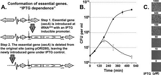
Since the majority of the gene deletion vectors currently used in Listeria research are based on selection with chloramphenicol or erythromycin, the kanamycin-based pIMK inducible vectors permit investigation of whether a particular gene is “essential.” Here we showed that the cytoplasmic preprotein chaperone, encoded by secA, which is essential in all bacteria examined previously (62), is also essential in L. monocytogenes. Initially, a promoterless copy of the secA gene was cloned into pIMK4 and electroporated (along with pORI280Δ_secA_ and pVE6007) into EGDe, providing a chromosomally located IPTG-inducible copy of secA. Then, the secA gene from the start codon (ATG) to the stop codon (TAA) was deleted at its original location, using the pORI280 protocol. After the transformation all plates and broth media had to contain 1 mM IPTG and 50 μg/ml kanamycin. The resulting strain had only one copy of secA, and this copy was under the control of the IPTG-inducible promoter, creating a conditional or IPTG-dependent mutant (Fig. 6A). This strain was assayed to determine (i) its ability to grow on agar plates containing different concentrations of IPTG and (ii) the effect of withdrawal of IPTG on growth in broth. As shown in Fig. 6C, cells were unable to form colonies at an IPTG concentration below 50 μM, and there was a progressive increase in colony size to the wild-type equivalent size at 500 μM IPTG. When IPTG was withdrawn from a broth culture, growth ceased within two generations, and then there was a marked loss of cell viability (Fig. 6B), confirming that secA is an essential gene in L. monocytogenes strain EGDe.
FIG. 6.
Essential gene deletion in L. monocytogenes: secA is an essential gene. The pORI system was combined with the strict IPTG-inducible promoter (pIMK4) for inactivation of an essential gene. The procedure is outlined in panel A for deletion of the essential preprotein translocase gene secA. (B) An overnight culture of EGDeΔ_secA_::pIMK4_secA_ (grown in BHI containing 1 mM IPTG) was washed three times with BHI and diluted 1:100 in fresh medium with (filled circles) or without (open circles) 1 mM IPTG. Cultures were grown at 37°C with shaking, and samples were taken at the indicated time points. Cells were then diluted in PBS, plated onto BHI agar containing 1 mM IPTG, and incubated overnight before colony enumeration. EGDeΔ_secA_::pIMK4_secA_ exhibited a wild-type growth profile (data not shown). (C) An overnight culture of EGDeΔ_secA_::pIMK4_secA_ (grown in BHI containing 1 mM IPTG) was diluted 10−5 and plated onto BHI agar containing different concentrations of IPTG, as indicated. The plates were incubated at 37°C for 24 h before visualization.
DISCUSSION
We describe construction of a number of tools for postgenomic functional analysis of L. monocytogenes. We focused on four aspects: (i) electroporation efficiency, (ii) development of integrative expression vectors, (iii) rapid and efficient construction of deletion mutants, and (iv) confirmation of essential genes.
Through systematic changes to the electroporation protocol of Park and Stewart (53), we improved the efficiency of plasmid transfer in EGDe at least 125-fold, making it possible to routinely obtain direct integration of large pPL2-based plasmids (Fig. 3A). We also describe the highest level of electroporation competence described thus far for an L. monocytogenes strain, 2 × 10⁷ CFU per μg for the serotype 4b isolate F2365. The enhancement of electroporation efficiency was due to a change in buffer, which improved cell integrity and allowed addition of a second cell wall-weakening step involving the enzymatic activity of lysozyme (Fig. 3B). The increase in competence of the sequenced strains made it easier to obtain transformants with larger plasmids and should enable new techniques, such as co-plasmid transformation, direct cloning of toxic genes, and genomic library construction, to be used directly for L. monocytogenes (32).
Serine and tyrosine phage integrases have been widely used for construction of site-specific integrative vectors (28). The integrase gene mediates crossover between the phage (attP) and bacterial (attB) attachment sites, disrupting the attB site on the chromosome, producing hybrid attBP/attPB at the junction sites. While numerous loci that can function for different integrases have been identified (22), there seems to be a preference for the targeting of conserved essential genes (e.g., the 3′ end of tRNA codons). As observed with the PSA integrase of pPL2, integration results in recreation of the tRNA gene by the hybrid attBP site due to a 17-bp region of homology within attP. Recently, the integration sites of an additional three listeriophages were identified (31), and each phage had a novel site on the L. monocytogenes chromosome. Replacement of the int gene and attP recognition region of the PSA phage with the integrase from phage A006, A500, or B054 may generate a vector which could integrate at a secondary site on the chromosome, akin to the pPL1 vector integrating at the comK locus. The use of multiple integrative vectors has been described for other bacteria for genetic complementation and expression of heterologous genes (35, 45). However, as shown by Corr et al. (15), at least two DNA fragments can be inserted into the MCS of a pPL2 derivative for heterologous gene expression (bacteriocin immunity expression and luciferase reporter genes in this instance).
pPL2-derived overexpression (pLOV [63]) and IPTG-inducible (pLIV2 [33] and pLIV3 [1]) vectors were recently developed. The expression of promoterless genes is driven from the enhanced Hyper-SPO1 promoter (56) containing the 5′ untranslated region of the L. monocytogenes hlyA gene, without the hlyA RBS. Genes are cloned containing their own RBS or a consensus RBS incorporated through oligonucleotide design. The two IPTG-inducible vectors are analogous to the two vectors described here, with one or two lacOid binding sites achieving stricter control of gene expression. The vectors described here for gene expression are an attractive alternative. The approach to cloning is streamlined as genes are expressed entirely from the plasmid machinery, there is no requirement for RBS inclusion in primer design, and therefore a gene can be cloned from a defined point (e.g., the start codon). This also allows direct comparison of levels of expression as identical constructs can be made in all three vectors. A dynamic range of invasion of 1,350-fold was determined for the three vectors (Fig. 2B). We have also used the pIMK2 system in Listeria to fully complement gene deletions by heterologous expression from the Phelp promoter (I. R. Monk, C. U. Riedel, P. G. Casey, C. Hill, and C. G. M. Gahan, unpublished).
Creation of clean markerless chromosomal mutations can be an arduous task, requiring the screening of thousands of colonies. Here we describe a protocol for chromosomal mutagenesis, which was developed for the pORI280 system but has also been applied to other thermosensitive plasmids, such as pKSV7. The procedure from the start of cloning to mutation confirmation can take as few as 10 days, and mutants are regularly obtained after the first passage at 30°C. In other protocols, the integration of the temperature-sensitive plasmid is stimulated by high-temperature incubation (at 39 to 43.5°C), often with passages at the high temperatures (11). This exposure to high temperature has been observed by others to stimulate spontaneous mutations in Listeria (68) and therefore should be avoided. Here we utilized a single temperature shift to 37°C, mediated by streaking a colony directly from the transformation plate for single colonies, and showed that this is sufficient to obtain plasmid integration. In addition, crossovers on both sides of the target site can be easily identified by the difference in blue coloration of a colony. The utility of the procedure was shown by the ability to generate gene deletions and single nucleotide changes in the chromosome (Fig. 4 and 5).
The first report of deletion of an essential gene in L. monocytogenes was published in 2006 (21). This was a noteworthy achievement, but the methodology was somewhat cumbersome as it involved the integration of a temperature-sensitive plasmid into lmo2537 (involved in teichoic acid synthesis), production of electrocompetent cells from the integrated strain, and then complementation with a temperature-sensitive IPTG-inducible vector, pLIV(lmo2537). The strain EGDe lmo2537_aph3_′containing pLIV(lmo2537) was unable to grow on BHI agar without IPTG supplementation. However, it did grow in BHI broth without IPTG, although there was a decrease in the final cell density. To show gene essentiality, we employed a streamlined approach facilitated by the pIMK4 vector. The deletion confirmed the essential nature of the secA gene, and there was a dramatic decrease in cellular viability upon withdrawal of IPTG (Fig. 6B). Improvements to the system would require cloning of an inducible counterselectable marker (58) into pORI280. This would improve the loss of the plasmid through the toxicity of the integrated plasmid when cultured on an appropriate substrate. Plasmid loss could be differentiated from spontaneous resistance by blue/white selection.
While this study was concerned primarily with tool development, several biologically relevant results are also reported here. We confirmed the essential nature of secA, we identified five additional PrfA “star” mutations, and we investigated the consequences of expressing inlA over a broad dynamic range for the invasion of Listeria. Lastly, we anticipate that the plasmids and methods outlined here will be a welcome addition to the postgenomic toolbox available to the Listeria research community.
Acknowledgments
We thank Darren Higgins for supplying plasmid pLIV2 and Christian Riedel for establishment of the Caco-2 cell line and for fruitful discussions.
We acknowledge funding received from the Irish Government under National Development Plan 2000-2006 and funding for the Alimentary Pharmabiotic Centre provided by the Science Foundation of Ireland Centres for Science Engineering and Technology (CSET) program.
Footnotes
▿
Published ahead of print on 25 April 2008.
REFERENCES
- 1.Alberti-Segui, C., K. R. Goeden, and D. E. Higgins. 2007. Differential function of Listeria monocytogenes listeriolysin O and phospholipases C in vacuolar dissolution following cell-to-cell spread. Cell. Microbiol. 9**:**179-195. [DOI] [PubMed] [Google Scholar]
- 2.Andersen, J. B., B. B. Roldgaard, A. B. Lindner, B. B. Christensen, and T. R. Licht. 2006. Construction of a multiple fluorescence labelling system for use in co-invasion studies of Listeria monocytogenes. BMC Microbiol. 6**:**86. [DOI] [PMC free article] [PubMed] [Google Scholar]
- 3.Archambaud, C., M. A. Nahori, J. Pizarro-Cerda, P. Cossart, and O. Dussurget. 2006. Control of Listeria superoxide dismutase by phosphorylation. J. Biol. Chem. 281**:**31812-31822. [DOI] [PubMed] [Google Scholar]
- 4.Arnaud, M., A. Chastanet, and M. Debarbouille. 2004. New vector for efficient allelic replacement in naturally nontransformable, low-GC-content, gram-positive bacteria. Appl. Environ. Microbiol. 70**:**6887-6891. [DOI] [PMC free article] [PubMed] [Google Scholar]
- 5.Begley, M., C. Hill, and C. G. Gahan. 2003. Identification and disruption of btlA, a locus involved in bile tolerance and general stress resistance in Listeria monocytogenes. FEMS Microbiol. Lett. 218**:**31-38. [DOI] [PubMed] [Google Scholar]
- 6.Begley, M., R. D. Sleator, C. G. Gahan, and C. Hill. 2005. Contribution of three bile-associated loci, bsh, pva, and btlB, to gastrointestinal persistence and bile tolerance of Listeria monocytogenes. Infect. Immun. 73**:**894-904. [DOI] [PMC free article] [PubMed] [Google Scholar]
- 7.Bergmann, B., D. Raffelsbauer, M. Kuhn, M. Goetz, S. Hom, and W. Goebel. 2002. InlA- but not InlB-mediated internalization of Listeria monocytogenes by non-phagocytic mammalian cells needs the support of other internalins. Mol. Microbiol. 43**:**557-570. [DOI] [PubMed] [Google Scholar]
- 8.Bishop, D. K., and D. J. Hinrichs. 1987. Adoptive transfer of immunity to Listeria monocytogenes. The influence of in vitro stimulation on lymphocyte subset requirements. J. Immunol. 139**:**2005-2009. [PubMed] [Google Scholar]
- 9.Brockstedt, D. G., M. A. Giedlin, M. L. Leong, K. S. Bahjat, Y. Gao, W. Luckett, W. Liu, D. N. Cook, D. A. Portnoy, and T. W. J. Dubensky. 2004. Listeria-based cancer vaccines that segregate immunogenicity from toxicity. Proc. Natl. Acad. Sci. USA 101**:**13832-13837. [DOI] [PMC free article] [PubMed] [Google Scholar]
- 10.Bron, P. A., I. R. Monk, S. C. Corr, C. Hill, and C. G. Gahan. 2006. Novel luciferase reporter system for in vitro and organ-specific monitoring of differential gene expression in Listeria monocytogenes. Appl. Environ. Microbiol. 72**:**2876-2884. [DOI] [PMC free article] [PubMed] [Google Scholar]
- 11.Cabanes, D., O. Dussurget, P. Dehoux, and P. Cossart. 2004. Auto, a surface associated autolysin of Listeria monocytogenes required for entry into eukaryotic cells and virulence. Mol. Microbiol. 51**:**1601-1614. [DOI] [PubMed] [Google Scholar]
- 12.Cabanes, D., S. Sousa, A. Cebria, M. Lecuit, F. Garcia-del Portillo, and P. Cossart. 2005. Gp96 is a receptor for a novel Listeria monocytogenes virulence factor, Vip, a surface protein. EMBO J. 24**:**2827-2838. [DOI] [PMC free article] [PubMed] [Google Scholar]
- 13.Cao, M., A. P. Bitar, and H. Marquis. 2007. A mariner-based transposition system for Listeria monocytogenes. Appl. Environ. Microbiol. 73**:**2758-2761. [DOI] [PMC free article] [PubMed] [Google Scholar]
- 14.Chakraborty, T., M. Leimeister-Wachter, E. Domann, M. Hartl, W. Goebel, T. Nichterlein, and S. Notermans. 1992. Coordinate regulation of virulence genes in Listeria monocytogenes requires the product of the prfA gene. J. Bacteriol. 174**:**568-574. [DOI] [PMC free article] [PubMed] [Google Scholar]
- 15.Corr, S. C., Y. Li, C. U. Riedel, P. W. O'Toole, C. Hill, and C. G. Gahan. 2007. Bacteriocin production as a mechanism for the anti-infective activity of Lactobacillus salivarius UCC118. Proc. Natl. Acad. Sci. USA 104**:**7617-7621. [DOI] [PMC free article] [PubMed] [Google Scholar]
- 16.Cotter, P. D., C. M. Guinane, and C. Hill. 2002. The LisRK signal transduction system determines the sensitivity of Listeria monocytogenes to nisin and cephalosporins. Antimicrob. Agents Chemother. 46**:**2784-2790. [DOI] [PMC free article] [PubMed] [Google Scholar]
- 17.Dancz, C. E., A. Haraga, D. A. Portnoy, and D. E. Higgins. 2002. Inducible control of virulence gene expression in Listeria monocytogenes: temporal requirement of listeriolysin O during intracellular infection. J. Bacteriol. 184**:**5935-5945. [DOI] [PMC free article] [PubMed] [Google Scholar]
- 18.Darji, A., T. Chakraborty, K. Niebuhr, N. Tsonis, J. Wehland, and S. Weiss. 1995. Hyperexpression of listeriolysin in the nonpathogenic species Listeria innocua and high yield purification. J. Biotechnol. 43**:**205-212. [DOI] [PubMed] [Google Scholar]
- 19.Doumith, M., C. Cazalet, N. Simoes, L. Frangeul, C. Jacquet, F. Kunst, P. Martin, P. Cossart, P. Glaser, and C. Buchrieser. 2004. New aspects regarding evolution and virulence of Listeria monocytogenes revealed by comparative genomics and DNA arrays. Infect. Immun. 72**:**1072-1083. [DOI] [PMC free article] [PubMed] [Google Scholar]
- 20.Dramsi, S., I. Biswas, E. Maguin, L. Braun, P. Mastroeni, and P. Cossart. 1995. Entry of Listeria monocytogenes into hepatocytes requires expression of inlB, a surface protein of the internalin multigene family. Mol. Microbiol. 16**:**251-261. [DOI] [PubMed] [Google Scholar]
- 21.Dubail, I., A. Bigot, V. Lazarevic, B. Soldo, D. Euphrasie, M. Dupuis, and A. Charbit. 2006. Identification of an essential gene of Listeria monocytogenes involved in teichoic acid biogenesis. J. Bacteriol. 188**:**6580-6591. [DOI] [PMC free article] [PubMed] [Google Scholar]
- 22.Dupont, L., B. Boizet-Bonhoure, M. Coddeville, F. Auvray, and P. Ritzenthaler. 1995. Characterization of genetic elements required for site-specific integration of Lactobacillus delbrueckii subsp. bulgaricus bacteriophage mv4 and construction of an integration-proficient vector for Lactobacillus plantarum. J. Bacteriol. 177**:**586-595. [DOI] [PMC free article] [PubMed] [Google Scholar]
- 23.El Karoui, M., S. K. Amundsen, P. Dabert, and A. Gruss. 1999. Gene replacement with linear DNA in electroporated wild-type Escherichia coli. Nucleic Acids Res. 27**:**1296-1299. [DOI] [PMC free article] [PubMed] [Google Scholar]
- 24.European Food Safety Authority. 2007. The community summary report on trends and sources of zoonoses, zoonotic agents, antimicrobial resistance and foodborne outbreaks in the European Union in 2006. Euro Food Safety Authority J. 130**:**1-310. [Google Scholar]
- 25.Fan, F., R. D. Lunsford, D. Sylvester, J. Fan, H. Celesnik, S. Iordanescu, M. Rosenberg, and D. McDevitt. 2001. Regulated ectopic expression and allelic-replacement mutagenesis as a method for gene essentiality testing in Staphylococcus aureus. Plasmid 46**:**71-75. [DOI] [PubMed] [Google Scholar]
- 26.Francis, K. P., D. Joh, C. Bellinger-Kawahara, M. J. Hawkinson, T. F. Purchio, and P. R. Contag. 2000. Monitoring bioluminescent Staphylococcus aureus infections in living mice using a novel luxABCDE construct. Infect. Immun. 68**:**3594-3600. [DOI] [PMC free article] [PubMed] [Google Scholar]
- 27.Glaser, P., L. Frangeul, C. Buchrieser, C. Rusniok, A. Amend, F. Baquero, P. Berche, H. Bloecker, P. Brandt, T. Chakraborty, A. Charbit, F. Chetouani, E. Couve, A. de Daruvar, P. Dehoux, E. Domann, G. Dominguez-Bernal, E. Duchaud, L. Durant, O. Dussurget, K. D. Entian, H. Fsihi, F. Garcia-del Portillo, P. Garrido, L. Gautier, W. Goebel, N. Gomez-Lopez, T. Hain, J. Hauf, D. Jackson, L. M. Jones, U. Kaerst, J. Kreft, M. Kuhn, F. Kunst, G. Kurapkat, E. Madueno, A. Maitournam, J. M. Vicente, E. Ng, H. Nedjari, G. Nordsiek, S. Novella, B. de Pablos, J. C. Perez-Diaz, R. Purcell, B. Remmel, M. Rose, T. Schlueter, N. Simoes, A. Tierrez, J. A. Vazquez-Boland, H. Voss, J. Wehland, and P. Cossart. 2001. Comparative genomics of Listeria species. Science 294**:**849-852. [DOI] [PubMed] [Google Scholar]
- 28.Groth, A. C., and M. P. Calos. 2004. Phage integrases: biology and applications. J. Mol. Biol. 335**:**667-678. [DOI] [PubMed] [Google Scholar]
- 29.Guo, X. V., M. Monteleone, M. Klotzsche, A. Kamionka, W. Hillen, M. Braunstein, S. Ehrt, and D. Schnappinger. 2007. Silencing Mycobacterium smegmatis by using tetracycline repressors. J. Bacteriol. 189**:**4614-4623. [DOI] [PMC free article] [PubMed] [Google Scholar]
- 30.Gutierrez, J. A., P. J. Crowley, D. P. Brown, J. D. Hillman, P. Youngman, and A. S. Bleiweis. 1996. Insertional mutagenesis and recovery of interrupted genes of Streptococcus mutans by using transposon Tn_917_: preliminary characterization of mutants displaying acid sensitivity and nutritional requirements. J. Bacteriol. 178**:**4166-4175. [DOI] [PMC free article] [PubMed] [Google Scholar]
- 31.Hain, T., S. S. Chatterjee, R. Ghai, C. T. Kuenne, A. Billion, C. Steinweg, E. Domann, U. Karst, L. Jansch, J. Wehland, W. Eisenreich, A. Bacher, B. Joseph, J. Schar, J. Kreft, J. Klumpp, M. J. Loessner, J. Dorscht, K. Neuhaus, T. M. Fuchs, S. Scherer, M. Doumith, C. Jacquet, P. Martin, P. Cossart, C. Rusniock, P. Glaser, C. Buchrieser, W. Goebel, and T. Chakraborty. 2007. Pathogenomics of Listeria spp. Int. J. Med. Microbiol. 297**:**541-557. [DOI] [PubMed] [Google Scholar]
- 32.Hain, T., S. Otten, U. von Both, S. S. Chatterjee, U. Technow, A. Billion, R. Ghai, W. Mohamed, E. Domann, and T. Chakraborty. 2008. Novel bacterial artificial chromosome vector pUvBBAC for use in studies of the functional genomics of Listeria spp. Appl. Environ. Microbiol. 74**:**1892-1901. [DOI] [PMC free article] [PubMed] [Google Scholar]
- 33.Henry, R., L. Shaughnessy, M. J. Loessner, C. Alberti-Segui, D. E. Higgins, and J. A. Swanson. 2006. Cytolysin-dependent delay of vacuole maturation in macrophages infected with Listeria monocytogenes. Cell. Microbiol. 8**:**107-119. [DOI] [PMC free article] [PubMed] [Google Scholar]
- 34.Horton, R. M., Z. L. Cai, S. N. Ho, and L. R. Pease. 1990. Gene splicing by overlap extension: tailor-made genes using the polymerase chain reaction. BioTechniques 8**:**528-535. [PubMed] [Google Scholar]
- 35.Kim, A. I., P. Ghosh, M. A. Aaron, L. A. Bibb, S. Jain, and G. F. Hatfull. 2003. Mycobacteriophage Bxb1 integrates into the Mycobacterium smegmatis groEL1 gene. Mol. Microbiol. 50**:**463-473. [DOI] [PubMed] [Google Scholar]
- 36.Kloosterman, T. G., J. J. Bijlsma, J. Kok, and O. P. Kuipers. 2006. To have neighbour's fare: extending the molecular toolbox for Streptococcus pneumoniae. Microbiology 152**:**351-359. [DOI] [PubMed] [Google Scholar]
- 37.Lauer, P., M. Y. Chow, M. J. Loessner, D. A. Portnoy, and R. Calendar. 2002. Construction, characterization, and use of two Listeria monocytogenes site-specific phage integration vectors. J. Bacteriol. 184**:**4177-4186. [DOI] [PMC free article] [PubMed] [Google Scholar]
- 38.Law, J., G. Buist, A. Haandrikman, J. Kok, G. Venema, and K. Leenhouts. 1995. A system to generate chromosomal mutations in Lactococcus lactis which allows fast analysis of targeted genes. J. Bacteriol. 177**:**7011-7018. [DOI] [PMC free article] [PubMed] [Google Scholar]
- 39.Lecointe, F., G. Coste, S. Sommer, and A. Bailone. 2004. Vectors for regulated gene expression in the radioresistant bacterium Deinococcus radiodurans. Gene 336**:**25-35. [DOI] [PubMed] [Google Scholar]
- 40.Leenhouts, K., G. Buist, A. Bolhuis, A. ten Berge, J. Kiel, I. Mierau, M. Dabrowska, G. Venema, and J. Kok. 1996. A general system for generating unlabelled gene replacements in bacterial chromosomes. Mol. Gen. Genet. 253**:**217-224. [DOI] [PubMed] [Google Scholar]
- 41.Leenhouts, K., G. Venema, and J. Kok. 1998. A lactococcal pWV01-based integration toolbox for bacteria. Methods Cell Sci. 20**:**35-50. [Google Scholar]
- 42.Lenz, L. L., S. Mohammadi, A. Geissler, and D. A. Portnoy. 2003. SecA2-dependent secretion of autolytic enzymes promotes Listeria monocytogenes pathogenesis. Proc. Natl. Acad. Sci. USA 100**:**12432-12437. [DOI] [PMC free article] [PubMed] [Google Scholar]
- 43.Li, G., and S. Kathariou. 2003. An improved cloning vector for construction of gene replacements in Listeria monocytogenes. Appl. Environ. Microbiol. 69**:**3020-3023. [DOI] [PMC free article] [PubMed] [Google Scholar]
- 44.Li, Z., X. Zhao, D. E. Higgins, and F. R. Frankel. 2005. Conditional lethality yields a new vaccine strain of Listeria monocytogenes for the induction of cell-mediated immunity. Infect. Immun. 73**:**5065-5073. [DOI] [PMC free article] [PubMed] [Google Scholar]
- 45.Luong, T. T., and C. Y. Lee. 2007. Improved single-copy integration vectors for Staphylococcus aureus. J. Microbiol. Methods 70**:**186-190. [DOI] [PMC free article] [PubMed] [Google Scholar]
- 46.Machata, S., T. Hain, M. Rohde, and T. Chakraborty. 2005. Simultaneous deficiency of both MurA and p60 proteins generates a rough phenotype in Listeria monocytogenes. J. Bacteriol. 187**:**8385-8394. [DOI] [PMC free article] [PubMed] [Google Scholar]
- 47.Maguin, E., P. Duwat, T. Hege, D. Ehrlich, and A. Gruss. 1992. New thermosensitive plasmid for gram-positive bacteria. J. Bacteriol. 174**:**5633-5638. [DOI] [PMC free article] [PubMed] [Google Scholar]
- 48.Mierau, I., and M. Kleerebezem. 2005. 10 years of the nisin-controlled gene expression system (NICE) in Lactococcus lactis. Appl. Microbiol. Biotechnol. 68**:**705-717. [DOI] [PubMed] [Google Scholar]
- 49.Monk, I. R., G. M. Cook, B. C. Monk, and P. J. Bremer. 2004. Morphotypic conversion in Listeria monocytogenes biofilm formation: biological significance of rough colony isolates. Appl. Environ. Microbiol. 70**:**6686-6694. [DOI] [PMC free article] [PubMed] [Google Scholar]
- 50.Nelson, K. E., D. E. Fouts, E. F. Mongodin, J. Ravel, R. T. DeBoy, J. F. Kolonay, D. A. Rasko, S. V. Angiuoli, S. R. Gill, I. T. Paulsen, J. Peterson, O. White, W. C. Nelson, W. Nierman, M. J. Beanan, L. M. Brinkac, S. C. Daugherty, R. J. Dodson, A. S. Durkin, R. Madupu, D. H. Haft, J. Selengut, S. Van Aken, H. Khouri, N. Fedorova, H. Forberger, B. Tran, S. Kathariou, L. D. Wonderling, G. A. Uhlich, D. O. Bayles, J. B. Luchansky, and C. M. Fraser. 2004. Whole genome comparisons of serotype 4b and 1/2a strains of the food-borne pathogen Listeria monocytogenes reveal new insights into the core genome components of this species. Nucleic Acids Res. 32**:**2386-2395. [DOI] [PMC free article] [PubMed] [Google Scholar]
- 51.Oehler, S., M. Amouyal, P. Kolkhof, B. von Wilcken-Bergmann, and B. Muller-Hill. 1994. Quality and position of the three lac operators of Escherichia coli define efficiency of repression. EMBO J. 13**:**3348-3355. [DOI] [PMC free article] [PubMed] [Google Scholar]
- 52.O'Neil, H. S., and H. Marquis. 2006. Listeria monocytogenes flagella are used for motility, not as adhesins, to increase host cell invasion. Infect. Immun. 74**:**6675-6681. [DOI] [PMC free article] [PubMed] [Google Scholar]
- 53.Park, S. F., and G. S. Stewart. 1990. High-efficiency transformation of Listeria monocytogenes by electroporation of penicillin-treated cells. Gene 94**:**129-132. [DOI] [PubMed] [Google Scholar]
- 54.Pilgrim, S., A. Kolb-Maurer, I. Gentschev, W. Goebel, and M. Kuhn. 2003. Deletion of the gene encoding p60 in Listeria monocytogenes leads to abnormal cell division and loss of actin-based motility. Infect. Immun. 71**:**3473-3484. [DOI] [PMC free article] [PubMed] [Google Scholar]
- 55.Powell, I. B., M. G. Achen, A. J. Hillier, and B. E. Davidson. 1988. A simple and rapid method for genetic transformation of lactic streptococci by electroporation. Appl. Environ. Microbiol. 54**:**655-660. [DOI] [PMC free article] [PubMed] [Google Scholar]
- 56.Quisel, J. D., W. F. Burkholder, and A. D. Grossman. 2001. In vivo effects of sporulation kinases on mutant Spo0A proteins in Bacillus subtilis. J. Bacteriol. 183**:**6573-6578. [DOI] [PMC free article] [PubMed] [Google Scholar]
- 57.Rea, R. B., C. G. Gahan, and C. Hill. 2004. Disruption of putative regulatory loci in Listeria monocytogenes demonstrates a significant role for Fur and PerR in virulence. Infect. Immun. 72**:**717-727. [DOI] [PMC free article] [PubMed] [Google Scholar]
- 58.Reyrat, J. M., V. Pelicic, B. Gicquel, and R. Rappuoli. 1998. Counterselectable markers: untapped tools for bacterial genetics and pathogenesis. Infect. Immun. 66**:**4011-4017. [DOI] [PMC free article] [PubMed] [Google Scholar]
- 59.Riedel, C. U., I. R. Monk, P. G. Casey, D. Morrissey, G. C. O'Sullivan, M. Tangney, C. Hill, and C. G. Gahan. 2007. Improved luciferase tagging system for Listeria monocytogenes allows real-time monitoring in vivo and in vitro. Appl. Environ. Microbiol. 73**:**3091-3094. [DOI] [PMC free article] [PubMed] [Google Scholar]
- 60.Ripio, M. T., G. Dominguez-Bernal, M. Lara, M. Suarez, and J. A. Vazquez-Boland. 1997. A Gly145Ser substitution in the transcriptional activator PrfA causes constitutive overexpression of virulence factors in Listeria monocytogenes. J. Bacteriol. 179**:**1533-1540. [DOI] [PMC free article] [PubMed] [Google Scholar]
- 61.Roberts, A. J., and M. Wiedmann. 2003. Pathogen, host and environmental factors contributing to the pathogenesis of listeriosis. Cell. Mol. Life Sci. 60**:**904-918. [DOI] [PMC free article] [PubMed] [Google Scholar]
- 62.Schmidt, M. G., and K. B. Kiser. 1999. SecA: the ubiquitous component of preprotein translocase in prokaryotes. Microbes Infect. 1**:**993-1004. [DOI] [PubMed] [Google Scholar]
- 63.Shen, A., H. D. Kamp, A. Grundling, and D. E. Higgins. 2006. A bifunctional O-GlcNAc transferase governs flagellar motility through anti-repression. Genes Dev. 20**:**3283-3295. [DOI] [PMC free article] [PubMed] [Google Scholar]
- 64.Sheng, Y., V. Mancino, and B. Birren. 1995. Transformation of Escherichia coli with large DNA molecules by electroporation. Nucleic Acids Res. 23**:**1990-1996. [DOI] [PMC free article] [PubMed] [Google Scholar]
- 65.Shepard, B. D., and M. S. Gilmore. 1995. Electroporation and efficient transformation of Enterococcus faecalis grown in high concentrations of glycine. Methods Mol. Biol. 47**:**217-226. [DOI] [PubMed] [Google Scholar]
- 66.Smith, K., and P. Youngman. 1992. Use of a new integrational vector to investigate compartment-specific expression of the Bacillus subtilis spoIIM gene. Biochimie 74**:**705-711. [DOI] [PubMed] [Google Scholar]
- 67.Vazquez-Boland, J. A., M. Kuhn, P. Berche, T. Chakraborty, G. Dominguez-Bernal, W. Goebel, B. Gonzalez-Zorn, J. Wehland, and J. Kreft. 2001. Listeria pathogenesis and molecular virulence determinants. Clin. Microbiol. Rev. 14**:**584-640. [DOI] [PMC free article] [PubMed] [Google Scholar]
- 68.Vega, Y., M. Rauch, M. J. Banfield, S. Ermolaeva, M. Scortti, W. Goebel, and J. A. Vazquez-Boland. 2004. New Listeria monocytogenes prfA* mutants, transcriptional properties of PrfA* proteins and structure-function of the virulence regulator PrfA. Mol. Microbiol. 52**:**1553-1565. [DOI] [PubMed] [Google Scholar]
- 69.Wei, Z., L. A. Zenewicz, and H. Goldfine. 2005. Listeria monocytogenes phosphatidylinositol-specific phospholipase C has evolved for virulence by greatly reduced activity on GPI anchors. Proc. Natl. Acad. Sci. USA 102**:**12927-12931. [DOI] [PMC free article] [PubMed] [Google Scholar]
- 70.Wong, K. K., and N. E. Freitag. 2004. A novel mutation within the central Listeria monocytogenes regulator PrfA that results in constitutive expression of virulence gene products. J. Bacteriol. 186**:**6265-6276. [DOI] [PMC free article] [PubMed] [Google Scholar]
- 71.Yansura, D. G., and D. J. Henner. 1984. Use of the Escherichia coli lac repressor and operator to control gene expression in Bacillus subtilis. Proc. Natl. Acad. Sci. USA 81**:**439-443. [DOI] [PMC free article] [PubMed] [Google Scholar]
