Regulation of the Cyclin B Degradation System by an Inhibitor of Mitotic Proteolysis (original) (raw)
Abstract
The initiation of anaphase and exit from mitosis depend on the anaphase-promoting complex (APC), which mediates the ubiquitin-dependent proteolysis of anaphase-inhibiting proteins and mitotic cyclins. We have analyzed whether protein phosphatases are required for mitotic APC activation. In Xenopus egg extracts APC activation occurs normally in the presence of protein phosphatase 1 inhibitors, suggesting that the anaphase defects caused by protein phosphatase 1 mutation in several organisms are not due to a failure to activate the APC. Contrary to this, the initiation of mitotic cyclin B proteolysis is prevented by inhibitors of protein phosphatase 2A such as okadaic acid. Okadaic acid induces an activity that inhibits cyclin B ubiquitination. We refer to this activity as inhibitor of mitotic proteolysis because it also prevents the degradation of other APC substrates. A similar activity exists in extracts of Xenopus eggs that are arrested at the second meiotic metaphase by the cytostatic factor activity of the protein kinase mos. In Xenopus eggs, the initiation of anaphase II may therefore be prevented by an inhibitor of APC-dependent ubiquitination.
INTRODUCTION
The activation of Cdc2 and possibly of other mitotic protein kinases is thought to be largely responsible for the structural reorganization of the cell during prophase and metaphase (reviewed byKing et al., 1994; Nigg et al., 1996). These events, in particular chromosome condensation and spindle assembly, are essential prerequisites for the following separation of sister chromatids in anaphase. However, the initiation of anaphase itself and the subsequent exit from mitosis appear to depend on the activation of a multisubunit ubiquitination complex, called the anaphase-promoting complex (APC)1 in vertebrates and yeast or the cyclosome in clams (Irniger et al., 1995; King et al., 1995; Sudakin et al., 1995; Peters et al., 1996;Zachariae et al., 1996). This complex catalyzes the assembly of polyubiquitin chains on several specific substrate proteins, which targets them for destruction by the 26S proteasome complex (reviewed byPeters, 1994; Coux et al., 1996).
The first event in mitosis that is known to depend on the APC is the initiation of sister chromatid separation at the metaphase-to-anaphase transition (Holloway et al., 1993; Irniger et al., 1995). Proteins that have to be destroyed to allow anaphase have recently been identified in budding yeast as the Pds1 protein and in fission yeast as Cut2 but are still unknown in other organisms (Cohen-Fix et al., 1996; Funabiki et al., 1996). The best studied substrates of the APC are the mitotic cyclins, the positive regulatory subunits of Cdc2. Synthesis of cyclins during interphase is necessary to allow activation of Cdc2 and entry into mitosis, whereas APC-mediated degradation of cyclins in anaphase results in inactivation of the kinase (reviewed by King et al., 1994). The degradation of cyclins requires a short sequence element found in the N-terminal portion of mitotic cyclins, called the destruction box (Glotzer et al., 1991). Ectopic expression of nondegradable cyclin mutants lacking the destruction box arrests cells at telophase, indicating that cyclin proteolysis is a mechanism that normally allows exit from mitosis (reviewed by King et al., 1994).
High levels of ubiquitinated cyclins can be observed in mitotic_Xenopus_ egg extracts where cyclins are degraded, but little or no cyclin ubiquitination is detectable in interphase extracts where cyclins are stable (Glotzer et al., 1991). This suggests that the cyclin ubiquitination reaction is regulated during the cell cycle, and that the activation of this reaction triggers cyclin proteolysis. The recent identification and purification of the enzymes required for cyclin B ubiquitination has allowed the reconstitution of destruction box- and mitosis-specific ubiquitination reactions in vitro (King et al., 1995; Sudakin et al., 1995;Aristarkhov et al., 1996; Peters et al., 1996; Yu_et al._, 1996). Besides cyclin B, ubiquitin, and ATP, this reaction requires the presence of APC, the ubiquitin-activating enzyme E1, and either one of two ubiquitin-conjugating enzymes (E2s), called UBCx and UBC4 in Xenopus. In this assay, APC isolated from mitotic Xenopus extracts has a higher specific ubiquitination activity than interphase APC, whereas the activities of the E1 and the E2 enzymes appear not to be regulated during the cell cycle. Similar results have been obtained for the clam cyclosome, which interacts with an E2 related to UBCx, called E2-C (Hershko et al., 1994; Sudakin et al., 1995; Aristarkhov et al., 1996). These results suggest that stimulation of the ubiquitination activity of APC may activate the mitotic degradation pathway, resulting in the initiation of anaphase, the inactivation of Cdc2, and other events that depend on APC-dependent proteolysis.
The initiation of anaphase is a “point of no return” whose timely and proper execution is important for the maintenance of genomic stability in proliferating cells. To avoid the unequal separation of sister chromatids, anaphase must not be initiated before all chromosomes are connected to the mitotic spindle. If APC is required for the initiation of anaphase, it therefore appears likely that the activation of APC is tightly controlled. Special control mechanisms may regulate the APC pathway in the spindle assembly checkpoint of somatic cells and the cytostatic factor (CSF) arrest of vertebrate eggs. In the first case entry into anaphase is delayed in response to a damaged or incompletely assembled spindle (for recent reviews, see Gorbsky, 1997;Sorger et al., 1997). In the second situation progression into the second meiotic anaphase is inhibited until fertilization occurs, a mechanism that allows the synchronization of the egg and the sperm cell cycle (reviewed by Sagata, 1996). In both the mitotic checkpoint and the CSF arrest, chromosome segregation and cyclin B proteolysis are inhibited (Pines and Hunter, 1989; Kung et al., 1990; Kobayashi et al., 1991), suggesting that the absence of APC-dependent proteolysis is responsible for the cell cycle arrest. The activation of mitogen-activated protein (MAP) kinase has been implicated in both types of arrest (Haccard et al., 1993; Kosako et al., 1994; Minshull et al., 1994;Takenaka et al., 1997; Wang et al., 1997). However, it is not known how APC-dependent proteolysis is prevented in these situations. The activation of the APC could be inhibited, as has recently been proposed for the CSF arrest (Abrieu et al., 1996). Alternatively, APC could be activated normally, but ubiquitin-dependent proteolysis could be prevented by an inhibitor present in CSF and checkpoint-arrested cells.
Little is also known about how APC activity is regulated during progression through a normal mitosis. Several observations suggest that mitotic phosphorylation of APC may be causally related to its activation (Hershko et al., 1994; Lahav-Baratz et al., 1995; Sudakin et al., 1995; Peters et al., 1996). However, in budding yeast and in mammalian somatic cells the cyclin degradation machinery has been found to be active also during G1 (Amon et al., 1994; Brandeis and Hunt, 1996), a phase of the cell cycle in which mitotic kinases are not active. This suggests that mechanisms other than mitotic phosphorylation also regulate the activity of the APC. One family of potential regulators of APC-dependent proteolysis has recently been identified as the WD40 repeat proteins Cdc20 and Hct1/Cdh1 in budding yeast and as fizzy and fizzy-related in Drosophila (Schwab_et al._, 1997; Sigrist and Lehner, 1997; Visintin et al., 1997).
Another enzyme that has been discussed as a potential mitotic activator of the APC is protein phosphatase 1 (PP1). Mutation of the catalytic subunit of PP1 results in anaphase defects in Drosophila and_Aspergillus_ and in budding and fission yeast, resembling the phenotype of APC mutants in these organisms (Doonan and Morris, 1989;Ohkura et al., 1989; Axton et al., 1990; Hisamoto_et al._, 1994). In fission yeast, suppressors of PP1 mutants have been found to genetically interact with APC subunits (Ishii_et al._, 1996). A cell cycle arrest at metaphase has also been observed in mammalian cells injected with neutralizing PP1 antibodies (Fernandez et al., 1992) and in cells treated with okadaic acid (OA), a specific inhibitor of protein phosphatase 2A (PP2A) and PP1 (Ghosh and Paweletz, 1992; Vandré and Wills, 1992). The microinjection of OA into starfish oocytes has furthermore been found to inhibit cyclin B proteolysis (Picard et al., 1989). Taken together, these results indicate a role for PP1 and/or PP2A in the metaphase-to-anaphase transition. However, it is not known whether phosphatase activity is required for the activation of the APC, for proper assembly of the mitotic spindle (whose failure could activate a mitotic checkpoint), or for other yet unknown events that are required for anaphase.
In the experiments described in this paper we have for the first time directly analyzed the possible role of PP1 and PP2A in the mitotic activation of the Xenopus APC. Our results do not support a role of these phosphatases in mitotic APC activation, suggesting that the metaphase arrest observed in PP1 mutants may be due to activation of a mitotic checkpoint or to other anaphase defects. However, we find that inhibition of PP2A stimulates an inhibitor of APC-dependent ubiquitination reactions. A similar inhibitory activity exists in extracts of CSF-arrested Xenopus eggs and may be responsible for the metaphase arrest of these cells.
MATERIALS AND METHODS
Preparation and Fractionation of Xenopus Egg Extracts
Interphase extracts were prepared as described (Murray, 1991), except that eggs were activated with the calcium ionophore A23187 (Calbiochem, La Jolla, CA) at a concentration of 0.4 μg/ml. Extracts were prepared 40–50 min after activation. Cycloheximide was added to 100 μg/ml to arrest the extract in interphase, and extracts were frozen in the presence of 200 mM sucrose. To generate mitotic extracts, we added a bacterially expressed nondegradable Δ90 fragment of sea urchin cyclin B (Glotzer et al., 1991) to interphase extracts at a concentration of 10 μg/ml. CSF extracts were prepared according to the method of Murray (1991).
For the generation of a mitotic high-speed supernatant (S100), 30 ml of mitotic extract were diluted 10-fold in buffer Q-A (20 mM Tris-HCl [pH 7.7], 100 mM KCl, 1 mM MgCl2, 0.1 mM CaCl2, 1 mM dithiotreitol) and centrifuged for 1 h at 100,000 ×g. To obtain fractions enriched in APC, the mitotic S100 was applied to a 65-ml Resource Q column (Pharmacia, Uppsala, Sweden) equilibrated with Q-A. Bound proteins were eluted with 5 column volumes of a linear salt gradient (0–500 mM KCl in Q-A) using a fast-performance liquid chromatography system (Pharmacia). Fractions (15 ml) were desalted on PD10 columns (Pharmacia), reconcentrated to 1 ml each in Centriprep-10 concentrators (Amicon, Beverly, MA), and analyzed by immunoblotting with Cdc27 antibodies. APC eluted between 300 and 370 mM KCl in Q-A. All steps were performed at 4°C, and APC fractions were stored at −70°C.
Protein Phosphatase Assays
PP1 and PP2A activities were measured in crude interphase extracts using the Life Technologies (Vienna, Austria) protein phosphatase assay kit following the manufacturer’s instructions. Briefly, 10 μl of extract prewarmed to room temperature were incubated for various periods with 5 μl of 32P-labeled glycogen phosphorylase A. The reaction was stopped by addition of 200 μl of ice-cold 20% (wt/vol) trichloroacetic acid, and the samples were incubated on ice for 10 min. After centrifugation at 14,000 × g for 10 min at 4°C, the amount of trichloroacetic acid–soluble 32Pi released from the substrate was determined by scintillation counting of 150 μl of the supernatant. Protein phosphatase activity was linear for 2.5 min. End point measurements were stopped after 1.5 min. In some experiments, the protein phosphatase inhibitors OA (1 mM in dimethylsulfoxide [DMSO]; Calbiochem), tautomycin (200 μM in DMSO; Calbiochem), or inhibitor 2 (I-2; 500 μM in Q-A buffer; kindly provided by R. Tournebize, European Molecular Biology Laboratory, Heidelberg, Germany) were added at various concentrations to the extracts. OA and tautomycin were added together with the substrate, whereas extracts were preincubated with I-2 for 10 min at room temperature before addition of the substrate. Control extracts were treated with DMSO or buffer.
Protein Kinase Assays
Histone H1 kinase assays were performed as described (Murray, 1991), except that reactions were incubated for 5 min at room temperature. Calf thymus H1 was obtained from Life Technologies. Kinase activity was determined by 10% SDS-PAGE and phosphorimaging.
In-gel kinase assays were done as described (Kameshita and Fujisawa, 1989; Gotoh et al., 1990) using myelin basic protein (Sigma, St. Louis, MO) as a substrate (0.2 mg/ml polymerized in 10% polyacrylamide gels). Samples of Xenopus extracts containing 10 μg of protein were analyzed. The electrophoretic mobility shift of MAP kinase caused by activating phosphorylation was followed by immunoblotting with Erk2 antibodies (Santa Cruz Biotechnology, Santa Cruz, CA) after separation of Xenopus extract samples by 15% SDS-PAGE.
To detect the electrophoretic mobility shift of Cdc25 caused by mitotic phosphorylation, [35S]methionine-labeled_Xenopus_ Xcdc25-1 (Kumagai and Dunphy, 1992) was prepared by coupled transcription–translation reactions in rabbit reticulocyte lysate (Promega, Madison, WI). The translation mix was diluted 1:20 in egg extracts, and Cdc25 was analyzed by 10% SDS-PAGE and phosphorimaging. The phosphorylation-induced electrophoretic mobility shift of Cdc27 was followed by immunoblotting after separation of extract samples by 10% SDS-PAGE. Cdc27 antibodies were kindly provided by C. Gieffers (Research Institute of Molecular Pathology, Vienna, Austria).
Protein Degradation Assays
For the generation of [35S]methionine-labeled substrates, 13–91prA (a fusion of the N-terminus of sea urchin cyclin B and protein A; Glotzer et al., 1991), budding yeast Pds1 (Cohen-Fix et al., 1996), and Xenopus geminin (kindly provided by T. McGarry) were generated using coupled transcription–translation reactions (see above). We refer to 13–91prA, which behaves like full-length cyclin B with respect to mitotic degradation (Glotzer et al., 1991), as35S-labeled cyclin B in the remainder of the text. For the generation of iodinated substrates, bacterially expressed recombinant fragments of sea urchin cyclin B, consisting of residues 13–110 (Holloway et al., 1993), and of Xenopus cyclin B1, consisting of the N-terminal 102 amino acids (kindly provided by H. Yu, Harvard Medical School, Boston, MA), were radiolabeled by the chloramine T procedure (Parker, 1990).
All degradation assays were carried out at room temperature. Interphase extracts were incubated with 10 μg/ml Δ90, 1.25 mg/ml bovine ubiquitin (Sigma), and 1/20 volume of translation mix containing35S-labeled APC substrates for various periods, and samples were analyzed by 10% SDS-PAGE and phosphorimaging. In some cases OA, tautomycin, I-2, the MAP kinase kinase inhibitor PD98059 (20 mM in DMSO; Calbiochem), and a fusion protein containing Xenopus mos and the maltose-binding protein E (malE-mos; Nebreda and Hunt, 1993) were added at different time points and concentrations. In the case of I-2, extracts were preincubated for 10 min with various concentrations at room temperature before Δ90 addition, and additional amounts of I-2 (to 1 μM each) were added every 15 min to compensate for inactivation of the inhibitor by phosphorylation. Bacterially expressed recombinant malE-mos was purified according to the method of Nebreda and Hunt (1993) and was used at a concentration of 10 or 20 μg/ml in extracts. PD98059 was used at a concentration of 400 μM.
Addition of 1 μM OA to our interphase extracts induced H1 kinase and Cdc25 and Cdc27 phosphorylation in the absence of Δ90 but failed to cause cyclin degradation. In this respect our results differ from previous observations made by Félix et al. (1990) andLorca et al. (1991), who report that OA alone is sufficient to activate cyclin B proteolysis. We presently do not know the reason for this discrepancy but suspect that it is caused by differences in the extract preparation protocols.
To follow cyclin degradation in extracts supplemented with mitotic APC fractions, extracts were immunodepleted of their endogenous APC (see below) and mixed with an ATP energy mix (7.5 mM creatine phosphate, 1 mM ATP, 1 mM MgCl2, 0.1 mM EGTA), 1.25 mg/ml ubiquitin, 1/5 volume of mitotic APC fraction (see above), and 0.6 μg/ml iodinated cyclin B. Samples were analyzed by 5–15% gradient SDS-PAGE and phosphorimaging.
Protein Ubiquitination Assays
To monitor steady-state levels of cyclin B–ubiquitin conjugates, extracts were incubated with 1.25 mg/ml ubiquitin and 0.6 μg/ml iodinated cyclin B. In some experiments the extracts contained 50 μg/ml proteasome inhibitor_N_-acetylleucylleucylnorleucinal (LLnL; 25 mg/ml in DMSO; Sigma; Rock et al., 1994). For the immunoprecipitation of APC and the depletion of extracts, 1 volume of Cdc27 antibodies was incubated with 2 volumes of protein A-Affiprep beads (Bio-Rad; Hercules, CA, USA) for 2 h at 4°C. After two washes with Tris-buffered saline (150 mM NaCl, 20 mM Tris-HCl [pH 8]) and two washes with extract buffer (10 mM HEPES-KOH [pH 7.7], 100 mM KCl, 1 mM MgCl2, 0.1 mM CaCl2), the beads were incubated in 5 volumes of extract for 2 h at 4°C and subsequently removed by centrifugation. For APC immunoprecipitation and ubiquitination assays, the beads were washed twice in extract buffer containing 400 mM KCl and 0.5% (vol/vol) Nonidet P-40 and four times in extract buffer. Ubiquitination assays were performed in a volume of 10 μl, containing 6 μl of APC beads and 4 μl of reaction mix containing bacterially expressed and purified wheat E1 (80 μg/ml),Xenopus UBC4 and UBCx (50 μg/ml each), 1.25 mg/ml ubiquitin, 30 U/ml rabbit muscle creatine phosphokinase (type I; Sigma), ATP energy mix, and 1.2 μg/ml iodinated cyclin B. All reactions were incubated at room temperature, and samples were analyzed by 5–15% SDS-PAGE and phosphorimaging.
Protein Deubiquitination Assays
Cyclin B–ubiquitin conjugates were generated by incubating iodinated cyclin B in ubiquitination assays containing immunoprecipitated mitotic APC as described above. After 50 min, APC beads were removed by centrifugation. The supernatant containing conjugates was mixed with a fourfold volume of various APC-depleted extracts containing 50 μg/ml LLnL. Samples were analyzed by 5–15% SDS-PAGE and phosphorimaging.
Other Methods
Immunoblotting was done on nitrocellulose filters, which were probed with affinity-purified Cdc27 antibodies at 0.75 μg/ml or Erk2 antibodies at 0.25 μg/ml. Immunoreactions were detected using the enhanced chemiluminescence system (Amersham International, Amersham, United Kingdom). Radiolabeled gels were analyzed on a Molecular Dynamics PhosphorImager and quantitated using the Imagequant program (Molecular Dynamics, Sunnyvale, CA).
RESULTS
Inhibitors of PP1 Do Not Block Mitotic Activation of the APC and Cyclin B Proteolysis
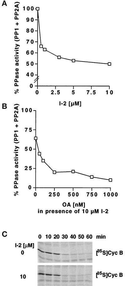
Concentrated interphase extracts prepared from activated_Xenopus_ eggs can be induced to enter a mitotic state by addition of a nondegradable version of cyclin B lacking the N-terminal 90 amino acids (Δ90), which binds to and activates endogenous Cdc2 (Glotzer et al., 1991). This in vitro system recapitulates a variety of events that occur during entry into mitosis in vivo, including activation of the APC and subsequent cyclin B ubiquitination and proteolysis. To address whether PP1 activity is required for these events, we analyzed the timing of cyclin B degradation in Δ90-containing Xenopus extracts in the presence of two different protein phosphatase inhibitors, I-2 and tautomycin. I-2 is a 23-kDa protein that binds to and inhibits the catalytic subunit of PP1 but does not significantly interfere with the activity of other known protein phosphatases (reviewed by Cohen, 1989). Tautomycin is a low molecular weight compound that inhibits PP1 with a 10-fold higher affinity than PP2A. We determined the efficacy of these inhibitors in_Xenopus_ extracts by measuring dephosphorylation of32P-labeled glycogen phosphorylase A, which is a substrate of both PP1 and PP2A. Titration of I-2 into Xenopus extracts showed that maximal inhibition of phosphatase activity to ∼50–60% was achieved between 3 and 10 μM (Figure1A), indicating that the majority of PP1 was inhibited under these conditions (for similar results, see Walker_et al._, 1992; Tournebize et al., 1997). 150 nM OA were required to inhibit 50% of the phosphatase activity remaining after treatment with 10 μM I-2 (Figure 1B). This dose of OA has previously been shown to block specifically PP2A activity in_Xenopus_ extracts (Félix et al., 1990), suggesting that the phosphatase activity remaining after I-2 treatment is largely due to PP2A.
Figure 1.
The PP1 inhibitor I-2 does not prevent activation of mitotic cyclin B proteolysis. (A) Dose–response curve showing the effect of different concentrations of I-2 on the dephosphorylation of32P-labeled glycogen phosphorylase A in_Xenopus_ interphase extracts. (B) Dose–response curve showing the effect of different concentrations of OA on the phosphatase activity in Xenopus interphase extracts containing 10 μM I-2 (determined as in A). (C) The stability of35S-labeled cyclin B was analyzed in extracts entering mitosis in the absence or presence of 10 μM I-2. Entry into mitosis was triggered by addition of nondegradable recombinant cyclin Δ90 at time zero, and samples taken at the indicated time points were analyzed by SDS-PAGE and phosphorimaging.
Cyclin B stability was analyzed in extracts by addition of trace amounts of in vitro-translated 35S-labeled cyclin B. When extracts were induced to enter mitosis in the presence of I-2, cyclin B degradation occurred on schedule, even if the inhibitor was present at doses as high as 10 μM (Figure 1C). This was not due to inactivation of I-2 in the mitotic extract, because the phosphatase activity did not increase during the course of the experiment. Similar results were obtained when tautomycin was added to extracts at concentrations that inhibit PP1 preferentially (up to 4 μM). These results suggest that the presence of I-2 or tautomycin does not interfere with activation of the APC. This was confirmed by immunoprecipitating APC from extracts treated with I-2 and measuring its cyclin B ubiquitination activity in a reconstituted system containing purified E1 and E2 enzymes (see below). APC isolated from I-2-treated extracts was as active as APC from untreated mitotic control extracts; see Figure 3, A and B, for a similar experiment). These results suggest that PP1 activity is not essential for mitotic activation of the APC and cyclin B proteolysis in_Xenopus_ extracts.
Figure 3.
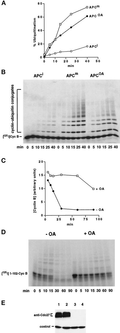
OA does not efficiently block the mitotic activation of the APC, but activates an IMP. (A and B) Time course showing the ability of different APC immunoprecipitates to ubiquitinate 125I-labeled-cyclin B 13–110 (Cyc B) in a reconstituted system containing purified E1, UBC4, and UBCx. APC was isolated from an interphase extract (APCi), from a mitotic Δ90 extract (APCm), and from an extract treated simultaneously with Δ90 and 1 μM OA (APCOA). Samples were analyzed by SDS-PAGE and phosphorimaging (B), and the ubiquitination activities were expressed as percentage of cyclin B converted into conjugates (A). (C and D) The stability of125I-labeled cyclin B1 1–102 (1–102-CycB) was analyzed in APC-depleted extracts supplemented with a mitotic APC fraction. Interphase extracts were either treated with Δ90 alone (−OA) or simultaneously with Δ90 and 1 μM OA (+OA) for 45 min before the APC depletion and reconstitution. Samples were analyzed as above (D), and cyclin B levels were quantitated (C). (E) Cdc27 immunoblot of the extracts used in C and D to control for the immunodepletion of APC. Lanes 1 and 3, Δ90 extract before and after depletion with Cdc27 antibodies, respectively; lanes 2 and 4, extract treated with Δ90 and OA at time zero, before and after Cdc27 immunodepletion, respectively. A protein band from a different portion of the same blot, which nonspecifically cross-reacts with Cdc27 antisera, is shown as a loading control.
Inhibitors of PP2A Activate an Inhibitor of Mitotic Proteolysis
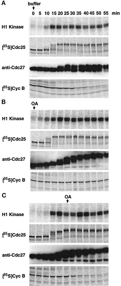
Different results were obtained when extracts were treated with OA, a substance that inhibits PP2A at a 100-fold lower dose than PP1 (Cohen, 1989). When 1 μM OA was added to extracts before mitotic activation by Δ90, no cyclin B degradation occurred during the time course of the experiment (Figure2B). This was not due to a failure of the extract to enter a mitotic state, because other mitotic events occurred on schedule: activation of Cdc2 (measured as histone H1 kinase activity), activation of Cdc25 (followed by a phosphorylation-induced electrophoretic mobility shift of35S-labeled Cdc25), and phosphorylation of APC subunits such as Cdc27 (detected as mobility shifts in immunoblots) all occurred normally or, if anything, slightly earlier in OA-treated extracts. However, cyclin B was degraded without delay when OA was added after the extract had entered a mitotic state (Figure 2C), suggesting that the presence of OA does not directly inhibit the cyclin B ubiquitination or degradation reactions. Titration of OA showed that 0.25 μM was sufficient to block cyclin B proteolysis if the drug was added before entry into mitosis. At this concentration primarily PP2A activity is inhibited in Xenopus extracts (Félix_et al._, 1990; Tournebize et al., 1997), suggesting that inhibition of this enzyme is responsible for the observed effect.
Figure 2.
OA inhibits cyclin B proteolysis if added before entry into mitosis. H1 kinase activity, the phosphorylation-induced mobility shift of 35S-labeled Cdc25 and of Cdc27, and the degradation of 35S-labeled cyclin B (Cyc B) were analyzed in extracts treated either with buffer (A) or with 1 μM OA (B and C), which was added either at time zero (B) or 25 min later (C). Δ90 was added to all reactions at time zero. Samples were taken at the indicated time points and analyzed by kinase assays and by SDS-PAGE followed by immunoblotting (anti-Cdc27) or phosphorimaging (H1 kinase, Cdc25, and Cyc B). The time points of buffer and OA addition are marked by arrows.
To analyze whether the stability of cyclin B after OA treatment was due to a failure to activate the APC, we isolated the complex from OA-treated extracts and from mitotic control extracts by immunoprecipitation. OA was present at 1 μM at every step during this procedure to rule out that PP2A-like activities could act on APC during or after immunoprecipitation. The cyclin B ubiquitination activity of APC was then determined in a reconstituted system containing purified E1 and E2 enzymes, ubiquitin, and an iodinated N-terminal fragment of either sea urchin or Xenopus cyclin B (Figure3, A and B). In several independent experiments, APC isolated from OA-treated extracts had between 65 and 75% of the activity of mitotic APC. We can therefore not exclude that OA partially interferes with APC activation. However, the high activity of APC isolated from OA-treated extracts made it unlikely that partial inhibition of APC activation accounted for the complete inhibition of cyclin B degradation in extracts.
We therefore wanted to test whether extracts treated with OA before entry into mitosis contained an activity that inhibited cyclin B ubiquitination and proteolysis. We first removed the endogenous APC from these extracts by immunodepletion (Figure 3E) and then supplemented them with a fraction enriched in mitotic APC. Although cyclin B was ubiquitinated and degraded efficiently in the reaction containing control mitotic extract, less ubiquitination and hardly any degradation occurred in the presence of the OA-treated extract (Figure3, C and D). Because equal amounts of mitotic active APC were present in both cases, this result suggests the presence of an inhibitory activity in the OA-treated extract. To test whether this activity could directly be associated with the APC, we washed APC immunoprecipitates obtained from OA-treated extracts only briefly with “physiological” extract buffer and compared its ubiquitination activity with APC washed with our standard immunoprecipitation buffer containing high concentrations of salts and detergents (see MATERIALS AND METHODS). APC washed only with extract buffer was not less but slightly more active than stringently washed APC. The inhibitory activity induced by OA is therefore either not directly associated with the APC, or it is lost during the immunoprecipitation procedure even under gentle washing conditions.
Inhibition of APC-dependent Proteolysis by OA Resembles the CSF Arrest of Xenopus Eggs
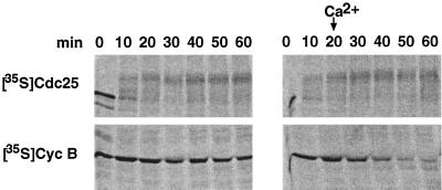
APC-dependent proteolysis is prevented by unknown mechanisms in the CSF-induced arrest of vertebrate eggs and in the spindle assembly checkpoint of somatic cells (see INTRODUCTION for references). The CSF arrest is overcome by a transient increase in cytoplasmic Ca2+ levels after fertilization, whereas the metaphase arrest caused by the spindle assembly checkpoint is insensitive to increases in the Ca2+ concentration (Minshull et al., 1994). When we added CaCl2 to extracts that had been treated with OA before entry into mitosis, cyclin B proteolysis was completely restored (Figure 4). This suggests that the OA treatment may mimic the CSF arrest but not the mitotic checkpoint.
Figure 4.
Inhibition of cyclin B proteolysis in OA-treated extracts is overcome by Ca2+ treatment. The electrophoretic mobility shift of 35S-labeled Cdc25 and the stability of35S-labeled cyclin B were analyzed in extracts entering mitosis, induced by addition of Δ90. The extracts contained 1 μM OA and were either treated with 0.5 mM CaCl2 after 20 min (right panel; indicated by arrows) or not (left panel). Samples were taken at the indicated time points and analyzed by SDS-PAGE and phosphorimaging.
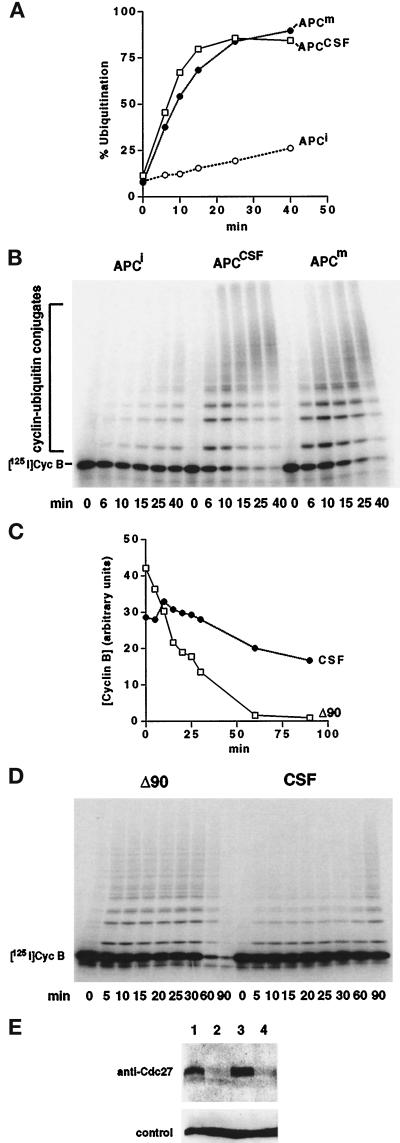
To further confirm this hypothesis we analyzed the regulation of APC in extracts prepared from CSF-arrested eggs. Cyclin B is not degraded in these extracts unless Ca2+ is added (Murray, 1991). Nevertheless, APC immunoprecipitated from CSF extracts had the same specific activity as mitotic APC when analyzed in cyclin B ubiquitination reactions containing purified E1 and E2 enzymes (Figure5, A and B), implying the presence of an inhibitor. This conclusion was further supported by reconstitution experiments in which we added fractions enriched in mitotic APC to either CSF or mitotic control extracts that had been depleted of their endogenous APC (Figure 5E). Cyclin B ubiquitination could be efficiently reconstituted in the mitotic extract but was much decreased in the CSF extract (Figure 5, C and D). Cyclin B proteolysis therefore appears to be prevented by an inhibitory activity present in CSF extracts that cannot be coimmunoprecipitated with the APC.
Figure 5.
An IMP exists in CSF extracts. (A and B) Time course showing the ability of different APC immunoprecipitates to ubiquitinate 125I-labeled-cyclin B 13–110 (Cyc B) in a reconstituted system containing purified E1, UBC4, and UBCx. APC was isolated from an interphase extract (APCi), from a CSF extract (APCCSF), and from a mitotic Δ90 extract (APCm). Samples were analyzed by SDS-PAGE and phosphorimaging (B), and the ubiquitination activities were expressed as percentage of cyclin B converted into conjugates (A). (C and D) The stability of 125I-labeled cyclin B 13–110 (Cyc B) was analyzed in APC-depleted Δ90 and CSF extracts supplemented with a mitotic APC fraction. Samples were analyzed as above (D), and cyclin B levels were quantitated (C). (E) Cdc27 immunoblot of the extracts used in C and D to control for the immunodepletion of APC. Lanes 1 and 2, Δ90 extract before and after depletion with Cdc27 antibodies, respectively; lanes 3 and 4, CSF extract before and after Cdc27 immunodepletion, respectively. A protein band from a different portion of the same blot, which nonspecifically cross-reacts with Cdc27 antisera, is shown as a loading control.
APC-dependent Ubiquitination Reactions Are Suppressed in CSF and in OA-treated Extracts
The inhibition of cyclin B proteolysis in CSF and OA-treated extracts could principally be due to an interference with any of the following three reactions: first, the cyclin B ubiquitination reaction could be inhibited; second, the rate with which cyclin B conjugates are deubiquitinated could be increased; and third, the proteasome-mediated degradation of ubiquitinated cyclin B could be inhibited. To distinguish between these three possibilities we first compared deubiquitination rates in different extracts. We ubiquitinated iodinated cyclin B in a reconstituted reaction containing APC and E1 and E2 enzymes. We then stopped the ubiquitination reaction by removing APC bound to antibody beads and added the radiolabeled conjugates to various extracts, which were previously depleted of their endogenous APC. The proteasome inhibitor LLnL was added to all extracts to rule out that the deubiquitination rates were affected by competing cyclin B proteolysis reactions. The conjugates were disassembled with similar kinetics (_t_½ = 4 min) in interphase extracts, mitotic Δ90 extracts, CSF extracts, or Δ90 extracts treated with OA before entry into mitosis. The inhibition of cyclin B proteolysis in CSF extracts and in OA-treated extracts is therefore not due to an increased deubiquitination rate.
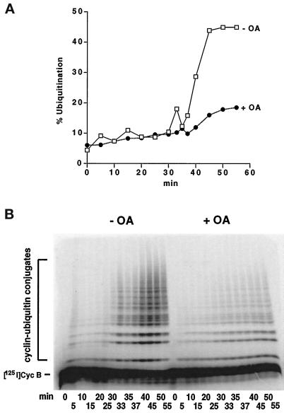
We then analyzed the steady-state levels of cyclin B–ubiquitin conjugates in mitotic Δ90 extracts in the presence or absence of OA and in CSF extracts. Although interphase extracts stimulated to enter mitosis by addition of Δ90 showed strong cyclin B ubiquitination activity after 30 min, much less ubiquitination activity was observed in extracts treated with OA before entry into mitosis (Figure6). The onset of cyclin B ubiquitination was not delayed in OA-containing extracts. This is consistent with the view that OA does not affect the kinetics of APC activation but instead induces an inhibitor of ubiquitination. Hardly any ubiquitination was observed in CSF extracts. These differences were detected in extracts containing proteasome inhibitors, ruling out that the reduced steady-state levels of cyclin B–ubiquitin conjugates were due to an increased proteasome activity. Taken together, these results suggest that in both OA-treated and CSF extracts inhibition of cyclin B proteolysis is due to suppression of the cyclin B ubiquitination reaction.
Figure 6.
The steady-state levels of cyclin B–ubiquitin conjugates are lowered in OA-treated extracts. (A and B) The conversion of 125I-labeled cyclin B 13–110 (Cyc B) into ubiquitin conjugates was followed in LLnL-treated extracts (50 μg/ml) induced to enter mitosis by addition of Δ90 in the absence (−OA) or presence (+OA) of 1 μM OA. Samples were taken at the indicated time points and analyzed by SDS-PAGE and phosphorimaging (B). The ubiquitination activity was expressed as the percentage of cyclin B converted into conjugates.
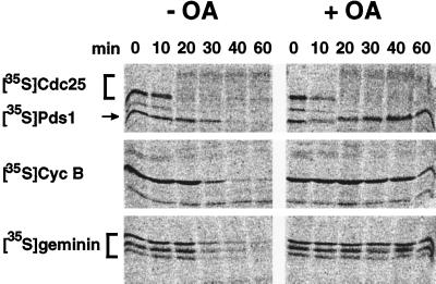
To test whether OA treatment specifically inhibits cyclin B proteolysis or also the degradation of other known APC substrates, we followed the stability of budding yeast Pds1 and of Xenopus geminin in extracts treated with OA before entry into mitosis. Both of these proteins are degraded in mitotic Xenopus extracts in a destruction box- and APC-dependent manner (Cohen-Fix et al., 1996; McGarry, personal communication). Both proteins were stabilized by OA (Figure 7), indicating that this treatment stimulates a general inhibitor of the APC pathway.
Figure 7.
OA stabilizes different APC substrates. The electrophoretic mobility shift of 35S-labeled Cdc25 caused by activating phosphorylation and the stability of35S-labeled cyclin B, 35S-labeled Pds1, or35S-labeled geminin were analyzed in extracts entering mitosis either in the presence (+OA) or absence of 1 μM OA (−OA). Δ90 cyclin was added to all reactions at time zero. Samples were withdrawn at the indicated time points and analyzed by SDS-PAGE and phosphorimaging.
Activation of MAP Kinase Can Block Cyclin B Proteolysis but Is Not Essential for the Inhibitory Effect of OA
Inhibition of PP2A by OA has been shown to activate the MAP kinase pathway in a variety of cell types from different organisms, including_Xenopus_ oocytes and extracts derived therefrom (Jessus_et al._, 1991; Shibuya et al., 1992). MAP kinase has also been implicated in mediating the CSF arrest (for references see INTRODUCTION). We therefore analyzed whether OA treatment of our egg extracts resulted in activation of MAP kinase and whether this was causally related to the inhibition of mitotic proteolysis. We found that OA activated a protein kinase in Xenopus extracts migrating at ∼42–44 kDa that could be detected in in-gel kinase assays using myelin basic protein as a substrate. Immunoblot analysis of portions of these gels showed that this kinase comigrated with MAP kinase. In immunoblotting experiments activation of MAP kinase could also be visualized by an electrophoretic mobility shift (Figure8) that is known to be caused by activating phosphorylation events (see, for example, Ferrell et al., 1991; Shibuya and Ruderman, 1993, and references therein). In both assays, OA treatment of extracts correlated with the activation of MAP kinase, always occurring 15–20 min after addition of the drug.
Figure 8.
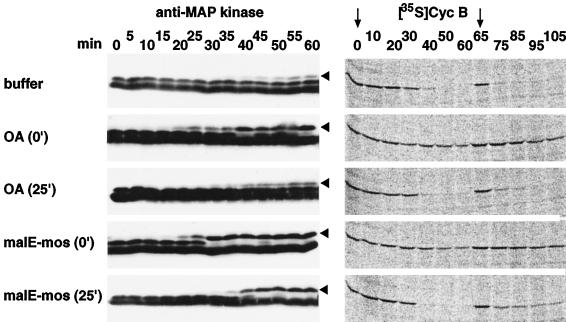
Activation of MAP kinase by OA and malE-mos correlates with the inhibition of cyclin B degradation. The electrophoretic mobility shift of MAP kinase that accompanies its activation and the stability of 35S-labeled cyclin B were examined in extracts entering mitosis, induced by addition of Δ90 at time zero. The extracts contained either 1 μM OA (added at time 0 or after 25 min), 20 μg/ml malE-mos (added at time 0 or after 25 min), or DMSO as a control (buffer). 35S-labeled cyclin B, Δ90, and ubiquitin were added a second time after 65 min. This was done to exclude the possibility that addition of OA or malE-mos after 25 min did not prevent cyclin B proteolysis, because the degradation reactions were completed before these reagents could activate MAP kinase. Samples were taken at the indicated time points and analyzed by SDS-PAGE and immunoblotting with Erk2 antibodies (anti-MAP kinase) or phosphorimaging (Cyc B). The slower-migrating bands representing active MAP kinase are indicated by arrowheads. The time points of35S-labeled cyclin B addition are marked by arrows.
It has recently been reported that activation of MAP kinase by the protein kinase mos, an activator of MAP kinase kinase (Nebreda and Hunt, 1993; Posada et al., 1993; Shibuya and Ruderman, 1993), can prevent cyclin B and cyclin A proteolysis in_Xenopus_ extracts (Abrieu et al., 1996; Jones and Smythe, 1996). In agreement with these reports we found that addition of a recombinant fusion protein containing the maltose-binding protein E and mos (malE-mos; Nebreda and Hunt, 1993) prevented cyclin B proteolysis if the mos protein was added together with Δ90 (Figure8), even though activation of Cdc25 and phosphorylation of Cdc27 were not affected. However, cyclin B proteolysis proceeded normally if mos was added 25 min after Δ90, similar to what we had observed with OA. Proteolysis also occurred when radiolabeled cyclin B was added at very late time points (65 min) to extracts that had been treated with OA or mos 25 min after addition of Δ90 (Figure 8). This rules out the possibility that there was simply not sufficient time for OA and mos to prevent cyclin B proteolysis when they were added at late time points in mitosis. Like OA, addition of mos caused activation of MAP kinase after a lag phase of 15–20 min (Figure 8).
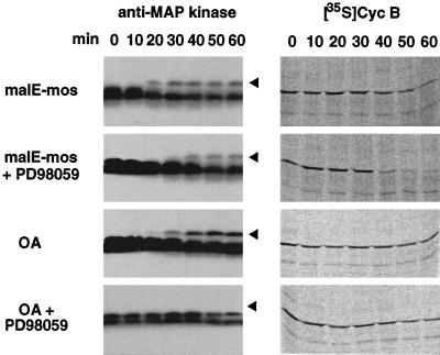
To address whether activation of MAP kinase is causally related to the inhibition of mitotic proteolysis, we treated Xenopus extracts with the inhibitor PD98059, which prevents activation of MAP kinase kinase (Dudley et al., 1995). In extracts treated with Δ90 and mos, this compound significantly delayed activation of MAP kinase as judged by electrophoretic mobility shift assays (Figure9). After this treatment no inhibition of cyclin proteolysis was observed, suggesting that the activation of MAP kinase is required for inhibition of cyclin B proteolysis induced by mos. However, cyclin B proteolysis still did not occur in extracts simultaneously treated with Δ90, PD98059 and OA, even though the activation of MAP kinase appeared to be completely suppressed under these conditions (Figure 9). Therefore, OA appears to be able to inhibit mitotic proteolysis without activation of MAP kinase.
Figure 9.
Inhibition of malE-mos- but not OA-induced MAP kinase activation allows cyclin B proteolysis. The electrophoretic mobility shift of MAP kinase caused by activating phosphorylation and the stability of 35S-labeled cyclin B were examined in extracts that were induced to enter mitosis by addition of Δ90 cyclin. The extracts contained either 20 μg/ml malE-mos or 1 μM OA and either 400 μM PD98059 or buffer. Samples were taken at the indicated time points and analyzed by SDS-PAGE and immunoblotting with Erk2 antibodies (anti-MAP kinase) or phosphorimaging (Cyc B). The positions of the slower-migrating bands representing active MAP kinase are indicated by arrowheads. Note that an activated form of MAP kinase cannot be detected in extracts treated with OA and PD98059.
DISCUSSION
The activity of the APC pathway that initiates the ubiquitin-dependent proteolysis of anaphase inhibiting proteins and of mitotic cyclins must be tightly controlled during progression through mitosis and meiosis, because otherwise unequal chromosome segregation could ensue. Correspondingly, the APC pathway must be inhibited in situations in which the initiation of anaphase must be delayed, as is the case in cells containing damaged mitotic spindles. Vertebrate eggs also appear to block progression through the second meiotic division by preventing APC-dependent proteolysis until fertilization occurs. However, little is known about the mechanisms that regulate the APC pathway.
Is PP1 Required for Mitotic Activation of the APC?
The initial goal of this study was to analyze whether protein phosphatases are required for the mitotic activation of the APC. This possibility was suggested by the genetic analysis of PP1, which, like the APC, is required for anaphase in several organisms (Doonan and Morris, 1989; Ohkura et al., 1989; Axton et al., 1990; Hisamoto et al., 1994). Using Xenopus egg extracts, which recapitulate the mitotic activation of cyclin B proteolysis in vitro, we found that APC activation can occur normally in the presence of PP1 inhibitors such as I-2 or low doses of tautomycin. Formally, it is difficult for us to exclude that residual or redundant phosphatase activities influenced the outcome of our experiments, as, for example, antibodies suitable for immunodepletion of PP1 are presently not available. However, our results clearly do not support a role for PP1 in the mitotic activation of the APC. The anaphase defects observed in fungal and fly PP1 mutants may therefore reflect a different requirement for this phosphatase, perhaps in allowing proper assembly of the mitotic spindle. Defects in spindle assembly caused by PP1 mutation could activate a mitotic checkpoint, resulting in indirect inhibition of APC-dependent proteolysis. This hypothesis is strongly supported by the recent observation that budding yeast PP1 mutants (glc7) do not arrest in mitosis if the mitotic checkpoint is inactivated by mutation of the kinetochore protein Ndc10 (Sassoon and Hyman, personal communication). PP1 could also be required for the coordination of spindle movements during anaphase. A role of PP1 in mitotic spindle function is supported by the observation that spindles appear morphologically abnormal in PP1 mutants of fission yeast Aspergillus and_Drosophila_ (Ohkura et al., 1988; Doonan and Morris, 1989; Axton et al., 1990) and by the recent finding that PP1 is required for spindle stability and elongation during anaphase in Xenopus extracts (Tournebize et al., 1997).
OA Activates an Inhibitor of Mitotic Proteolysis
Contrary to the results obtained with PP1 inhibitors, we observed that inhibition of PP2A by addition of OA or high doses of tautomycin to interphase extracts blocked the onset of APC-dependent ubiquitination and degradation reactions in extracts that were entering mitosis. Our results suggest that OA does not cause this effect by preventing the mitotic activation of the APC. Instead, this treatment appears to activate an inhibitor of cyclin B proteolysis that is not physically associated with the APC. We refer to this activity as inhibitor of mitotic (or meiotic) proteolysis (IMP) because it is also able to prevent the mitotic degradation of other APC substrates, such as Xenopus geminin and budding yeast Pds1. Activation of the IMP lowers the steady-state levels of cyclin B–ubiquitin conjugates without increasing the rate at which such conjugates are disassembled, suggesting that this inhibitor may function by preventing the assembly of poly-ubiquitin chains on APC substrates. So far, we have been unable to determine whether this is achieved by protecting substrates from ubiquitination or by inhibiting the APC, although our observation that the degradation of several APC substrates is affected would argue in favor of the latter hypothesis.
Inhibition of APC-dependent Proteolysis in the CSF Arrest
The inhibitory effect of OA on cyclin B proteolysis is sensitive to Ca2+. This observation raises the possibility that the OA treatment mimics the CSF arrest of vertebrate eggs, which is overcome by a transient increase in cytoplasmic Ca2+ levels after fertilization. Consistent with this view we found that CSF extracts also contain an activity that suppresses cyclin B ubiquitination, whereas APC isolated from these extracts is as active as mitotic APC in reconstituted cyclin B ubiquitination reactions. Unlike previously suspected (Abrieu et al., 1996), these results suggest that APC is activated normally in meiosis II. Its ubiquitination activity, however, seems to be suppressed by an inhibitor that may be responsible for the arrest at the second meiotic metaphase.
The meiosis-specific protein kinase mos is an essential component of CSF (O’Keefe et al., 1989; Sagata et al., 1989;Colledge et al., 1994; Hashimoto et al., 1994; reviewed by Sagata, 1996), and mos is thought to cause the metaphase arrest by activating MAP kinase (Haccard et al., 1993;Nebreda and Hunt, 1993; Posada et al., 1993; Shibuya and Ruderman, 1993; Kosako et al., 1994). We therefore tested whether the activation of MAP kinase was required to inhibit cyclin B proteolysis in Xenopus extracts treated either with purified mos kinase or with OA, both of which are able to activate MAP kinase. Consistent with two recent reports (Abrieu et al., 1996;Jones and Smythe, 1996), we found that mos was able to inhibit cyclin B proteolysis and that the activation of MAP kinase was required for this effect. As in OA-treated and CSF extracts, we found that an inhibitory activity not associated with the APC was responsible for preventing cyclin B proteolysis in mos-treated extracts. This activity was not MAP kinase itself, because purified active MAP kinase was not able to inhibit the cyclin B ubiquitination activity of purified APC (our unpublished results).
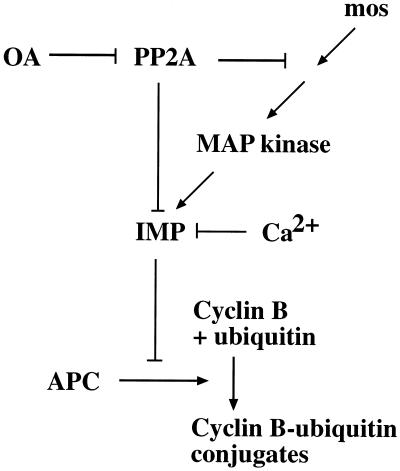
Contrary to the results obtained with mos, we found that OA prevented cyclin B proteolysis even under conditions in which the activation of MAP kinase appeared to be completely blocked. We presently do not know the reason for this difference between the effects of mos and OA. It is possible that OA prevents cyclin B proteolysis through pleiotropic effects that are not related to the CSF arrest. Alternatively, it is conceivable that OA is able to induce the same inhibitor of proteolysis found in CSF extracts. We favor this latter hypothesis based on the observation that the inhibitory activity in OA-treated extracts is Ca2+ sensitive, as are CSF and mos-treated extracts (see model shown in Figure 10). One possibility is that OA stimulates the inhibitor by activating multiple members of the MAP kinase family. OA has indeed been found to activate diverse members of the MAP kinase pathways (see, for example, Cano_et al._, 1995; Moriguchi et al., 1996), and the inhibitor PD98059 that we used in our experiments may not prevent the activation of all of these kinases. Alternatively, OA could activate the inhibitor of proteolysis independent of the MAP kinase pathway.
Figure 10.
Model for the mechanism of the CSF arrest and its artificial induction by OA. The model proposes that both mos and OA can activate an IMP, which prevents the initiation of anaphase II in unfertilized Xenopus eggs.
Is APC Regulated by Similar Mechanisms in the CSF and the Spindle Assembly Checkpoint?
The activation of MAP kinase has also been implicated in the mitotic spindle assembly checkpoint (Minshull et al., 1994;Takenaka et al., 1997; Wang et al., 1997). Presently, it is unknown whether the APC is also suppressed by an inhibitor in this situation, or whether APC-dependent proteolysis is prevented by other means. However, several observations suggest that different mechanisms may prevent anaphase in CSF-arrested eggs and in checkpoint-arrested somatic cells. Cyclin A is stable in_Xenopus_ CSF extracts or extracts treated with mos (Kobayashi_et al._, 1991; Jones and Smythe, 1996) but not in cells arrested by the spindle assembly checkpoint (Whitfield et al., 1990; Hunt et al., 1992; Minshull et al., 1994; Kramer and Peters, unpublished observation). If checkpoint-arrested cells contain a factor that inhibits APC-dependent ubiquitination reactions, this factor would therefore have different properties than the inhibitory activity found in CSF extracts. One protein that has been implicated in the spindle assembly checkpoint both in yeast and vertebrates is Mad2 (Li and Murray, 1991; Chen_et al._, 1996; Li and Benezra, 1996), which recently has been proposed to inhibit the APC by direct physical association (Li et al., 1997). Experiments by Chen et al. (1996) suggest, however, that Mad2 is not required for the CSF arrest of_Xenopus_ eggs. It is therefore unlikely that Mad2 is a component of the inhibitory activity detected in CSF extracts or in extracts treated with OA or mos.
Regulation of the Inhibitor of Mitotic Proteolysis
Mitotic cyclin B proteolysis can only be inhibited in_Xenopus_ extracts if OA or mos is added during entry into mitosis but not after a certain time point after entry into mitosis (Abrieu et al., 1996; Jones and Smythe, 1996; this study). This phenomenon was previously thought to reflect a role of mos in preventing activation of the cyclin B degradation system, whereas mos would not have any effect on the cyclin degradation system once it was activated (Abrieu et al., 1996). Our results suggest a different view: both OA and mos seem to stimulate an inhibitor of cyclin B ubiquitination, which, however, can only be activated up to a certain point in mitosis. Our reconstitution experiments show that, once activated, this inhibitor is able to suppress ubiquitination reactions catalyzed by active mitotic APC. This rules out that only the interphase and not the mitotic form of the APC is subject to inhibition. We do not know why activation of the inhibitor is only possible up to a certain time point, but timing experiments suggest that this event takes place after activation of Cdc2 and Cdc25, possibly around the time the APC becomes activated (Vorlaufer, unpublished observations). It is therefore possible that the inhibitor is modified directly or indirectly by Cdc2. Alternatively, activation of the APC may inactivate the inhibitor, perhaps by causing the ubiquitin-dependent proteolysis of the inhibitor itself.
In either case, our results predict that the relative timing of inhibitor activation and of Cdc2 and APC activation determines whether APC-dependent proteolysis will ensue or will be inhibited. This prediction is consistent with the situation in Xenopus eggs entering meiosis II, where Cdc2 is activated in the presence of active MAP kinase (reviewed by Sagata, 1996). In these cells MAP kinase may have activated the inhibitor of proteolysis by the time Cdc2 and APC are activated. Subsequently, the inhibitor may arrest the cell cycle at metaphase of meiosis II by preventing the degradation of Pds1/Cut2-like anaphase inhibitors until fertilization occurs.
ACKNOWLEDGMENTS
We thank M. Baccarini, M. Glotzer, K. Nasmyth, and W. Zachariae for helpful discussions and critical reading of the manuscript and C. Gieffers, S. Keyse, T. McGarry, A. Nebreda, E. Nishida, R. Tournebize, E. Wagner, and H. Yu for providing reagents. We are very grateful to G. Fang and M. Kirschner for sharing unpublished results about the partial purification of a CSF-specific inhibitor of APC-dependent ubiquitination, which may be identical to the inhibitory activity described in this paper. We are also grateful to R. King for communicating his observation that OA can inhibit cyclin B proteolysis and to T. McGarry, I. Sassoon, and A. Hyman for sharing unpublished results. This research was supported by a grant from the Austrian Industrial Research Promotion Fund (FFF 3/12801) to J.-M. P.
Footnotes
1
Abbreviations used: APC, anaphase-promoting complex; CSF, cytostatic factor; DMSO, dimethylsulfoxide; I-2, inhibitor 2; IMP, inhibitor of mitotic proteolysis; LLnL,_N_-acetylleucylleucylnorleucinal; MAP, mitogen-activated protein; OA, okadaic acid; PP1, protein phosphatase 1; PP2A, protein phosphatase 2A; UBC, ubiquitin-conjugating enzyme.
REFERENCES
- Abrieu A, Lorca T, Labbé J-C, Morin N, Keyse S, Dorée M. MAP kinase does not inactivate, but rather prevents the cyclin degradation pathway from being turned on in Xenopus egg extracts. J Cell Sci. 1996;109:239–246. doi: 10.1242/jcs.109.1.239. [DOI] [PubMed] [Google Scholar]
- Amon A, Irniger S, Nasmyth K. Closing the cell cycle circle in yeast: G2 cyclin proteolysis initiated at mitosis persists until the activation of G1 cyclins in the next cycle. Cell. 1994;77:1037–1050. doi: 10.1016/0092-8674(94)90443-x. [DOI] [PubMed] [Google Scholar]
- Aristarkhov A, Eytan E, Moghe A, Admon A, Hershko A, Ruderman JV. E2-C, a cyclin-selective ubiquitin carrier protein required for the destruction of mitotic cyclins. Proc Natl Acad Sci USA. 1996;93:4294–4299. doi: 10.1073/pnas.93.9.4294. [DOI] [PMC free article] [PubMed] [Google Scholar]
- Axton JM, Dombradi V, Cohen PT, Glover DM. One of the protein phosphatase 1 isoenzymes in Drosophila is essential for mitosis. Cell. 1990;63:33–46. doi: 10.1016/0092-8674(90)90286-n. [DOI] [PubMed] [Google Scholar]
- Brandeis M, Hunt T. The proteolysis of mitotic cyclins in mammalian cells persists from the end of mitosis until the onset of S-phase. EMBO J. 1996;15:5280–5289. [PMC free article] [PubMed] [Google Scholar]
- Cano E, Hazzalin CA, Kardalinou E, Buckle RS, Mahadevan LC. Neither ERK nor JNK/SAPK MAP kinase subtypes are essential for histone H3/HMG-14 phosphorylation or c-fos and c-jun induction. J Cell Sci. 1995;108:3599–3609. doi: 10.1242/jcs.108.11.3599. [DOI] [PubMed] [Google Scholar]
- Chen R-H, Waters JC, Salmon ED, Murray AW. Association of spindle assembly checkpoint component XMAD2 with unattached kinetochores. Science. 1996;274:242–246. doi: 10.1126/science.274.5285.242. [DOI] [PubMed] [Google Scholar]
- Cohen P. The structure and regulation of protein phosphatases. Annu Rev Biochem. 1989;58:453–508. doi: 10.1146/annurev.bi.58.070189.002321. [DOI] [PubMed] [Google Scholar]
- Cohen-Fix O, Peters J-M, Kirschner MW, Koshland D. Anaphase initiation in Saccharomyces cerevisiae is controlled by the APC-dependent degradation of the anaphase inhibitor Pds1p. Genes Dev. 1996;10:3081–3093. doi: 10.1101/gad.10.24.3081. [DOI] [PubMed] [Google Scholar]
- Colledge WH, Carlton MBL, Udy GB, Evans MJ. Disruption of c-mos causes parthenogenetic development of unfertilized mouse eggs. Nature. 1994;370:65–68. doi: 10.1038/370065a0. [DOI] [PubMed] [Google Scholar]
- Coux O, Tanaka K, Goldberg AL. Structure and functions of the 20S and 26S proteasomes. Annu Rev Biochem. 1996;65:801–847. doi: 10.1146/annurev.bi.65.070196.004101. [DOI] [PubMed] [Google Scholar]
- Doonan JH, Morris NR. The bimG gene of Aspergillus nidulans, required for completion of anaphase, encodes a homolog of mammalian phosphoprotein phosphatase 1. Cell. 1989;57:987–996. doi: 10.1016/0092-8674(89)90337-1. [DOI] [PubMed] [Google Scholar]
- Dudley DT, Pang L, Decker SJ, Bridges AJ, Saltiel AR. A synthetic inhibitor of the mitogen-activated protein kinase cascade. Proc Natl Acad Sci USA. 1995;92:7686–7689. doi: 10.1073/pnas.92.17.7686. [DOI] [PMC free article] [PubMed] [Google Scholar]
- Félix M-A, Cohen P, Karsenti E. Cdc2 H1 kinase is negatively regulated by a type 2A phosphatase in the Xenopus early embryonic cell cycle: evidence from the effects of okadaic acid. EMBO J. 1990;9:675–683. doi: 10.1002/j.1460-2075.1990.tb08159.x. [DOI] [PMC free article] [PubMed] [Google Scholar]
- Fernandez A, Brautigan DL, Lamb NJC. Protein phosphatase type 1 in mammalian cell mitosis: chromosomal localization and involvement in mitotic exit. J Cell Biol. 1992;116:1421–1430. doi: 10.1083/jcb.116.6.1421. [DOI] [PMC free article] [PubMed] [Google Scholar]
- Ferrell JE, Jr, Wu M, Gerhart JC, Martin GS. Cell cycle tyrosine phosphorylation of p34cdc2 and a microtubule-associated protein kinase homolog in Xenopus oocytes and eggs. Mol Cell Biol. 1991;11:1965–1971. doi: 10.1128/mcb.11.4.1965. [DOI] [PMC free article] [PubMed] [Google Scholar]
- Funabiki H, Yamano H, Kumada K, Nagao K, Hunt T, Yanagida M. Cut2 proteolysis required for sister-chromatid separation in fission yeast. Nature. 1996;381:438–441. doi: 10.1038/381438a0. [DOI] [PubMed] [Google Scholar]
- Ghosh S, Paweletz N. Okadaic acid inhibits sister chromatid separation in mammalian cell. Exp Cell Res. 1992;200:215–217. doi: 10.1016/s0014-4827(05)80091-6. [DOI] [PubMed] [Google Scholar]
- Glotzer M, Murray AW, Kirschner M. Cyclin is degraded by the ubiquitin pathway. Nature. 1991;349:132–138. doi: 10.1038/349132a0. [DOI] [PubMed] [Google Scholar]
- Gorbsky GJ. Cell cycle checkpoints: arresting progress in mitosis. Bioessays. 1997;19:193–197. doi: 10.1002/bies.950190303. [DOI] [PubMed] [Google Scholar]
- Gotoh Y, Nishida E, Yamashita T, Hoshi M, Kawakamii M, Sakai H. Microtubule-associated-protein (MAP) kinase activated by nerve growth factor and epidermal growth factor in PC12 cells. Eur J Biochem. 1990;193:661–669. doi: 10.1111/j.1432-1033.1990.tb19384.x. [DOI] [PubMed] [Google Scholar]
- Haccard O, Sarcevic B, Lewellyn A, Hartley L, Roy T, Izyumi E, Erikson E, Maller JL. Induction of metaphase arrest in cleaving Xenopus embryos by MAP kinase. Science. 1993;262:1262–1265. doi: 10.1126/science.8235656. [DOI] [PubMed] [Google Scholar]
- Hashimoto N, et al. Parthenogenetic activation of oocytes in c-mos-deficient mice. Nature. 1994;370:68–71. doi: 10.1038/370068a0. [DOI] [PubMed] [Google Scholar]
- Hershko A, Ganoth D, Sudakin V, Dahan A, Cohen LH, Luca FC, Ruderman JV, Eytan E. Components of a system that ligates cyclin to ubiquitin and their regulation by the protein kinase Cdc2. J Biol Chem. 1994;269:4940–4946. [PubMed] [Google Scholar]
- Hisamoto N, Sugimoto K, Matsumoto K. The Glc7 type 1 protein phosphatase of Saccharomyces cerevisiae is required for cell cycle progression in G2/M. Mol Cell Biol. 1994;14:3158–3165. doi: 10.1128/mcb.14.5.3158. [DOI] [PMC free article] [PubMed] [Google Scholar]
- Holloway SL, Glotzer M, King RW, Murray AW. Anaphase is initiated by proteolysis rather than by the inactivation of maturation promoting factor. Cell. 1993;73:1393–1402. doi: 10.1016/0092-8674(93)90364-v. [DOI] [PubMed] [Google Scholar]
- Hunt T, Luca FC, Ruderman JV. The requirements for protein synthesis and degradation, and the control of the destruction of cyclin A and cyclin B in the meiotic and mitotic cell cycles of the clam embryo. J Cell Biol. 1992;116:707–724. doi: 10.1083/jcb.116.3.707. [DOI] [PMC free article] [PubMed] [Google Scholar]
- Irniger S, Piatti S, Michaelis C, Nasmyth K. Genes involved in sister chromatid separation are needed for B-type cyclin proteolysis in budding yeast. Cell. 1995;81:269–278. doi: 10.1016/0092-8674(95)90337-2. [DOI] [PubMed] [Google Scholar]
- Ishii K, Kumada K, Toda T, Yanagida M. Requirement for PP1 phosphatase and 20S cyclosome/APC for the onset of anaphase is lessened by the dosage increase of a novel gene sds23+ EMBO J. 1996;15:6629–6640. [PMC free article] [PubMed] [Google Scholar]
- Jessus C, Rime H, Haccard O, Van Lint J, Goris J, Merlevede W, Ozon R. Tyrosine phophorylation of p34cdc2 and p42 during meiotic maturation of Xenopus oocyte. Antagonistic action of okadaic acid and 6-DMAP. Development. 1991;111:813–820. doi: 10.1242/dev.111.3.813. [DOI] [PubMed] [Google Scholar]
- Jones C, Smythe C. Activation of the Xenopus cyclin degradation machinery by full-length cyclin A. J Cell Sci. 1996;109:1071–1079. doi: 10.1242/jcs.109.5.1071. [DOI] [PubMed] [Google Scholar]
- Kameshita I, Fujisawa HA. A sensitive method for detection of calmodulin-dependent protein kinase II activity in sodium dodecyl sulfate-polyacrylamide gel. Anal Biochem. 1989;183:139–143. doi: 10.1016/0003-2697(89)90181-4. [DOI] [PubMed] [Google Scholar]
- King RW, Jackson PK, Kirschner MW. Mitosis in transition. Cell. 1994;79:563–571. doi: 10.1016/0092-8674(94)90542-8. [DOI] [PubMed] [Google Scholar]
- King RW, Peters J-M, Tugendreich S, Rolfe M, Hieter P, Kirschner MW. A 20S complex containing CDC27 and CDC16 catalyzes the mitosis-specific conjugation of ubiquitin to cyclin B. Cell. 1995;81:279–288. doi: 10.1016/0092-8674(95)90338-0. [DOI] [PubMed] [Google Scholar]
- Kobayashi H, Minshull J, Ford C, Golsteyn R, Poon R, Hunt T. On the synthesis and destruction of A- and B-type cyclins during oogenesis and meiotic maturation in Xenopus laevis. J Cell Biol. 1991;114:755–765. doi: 10.1083/jcb.114.4.755. [DOI] [PMC free article] [PubMed] [Google Scholar]
- Kosako H, Gotoh Y, Nishida E. Mitogen-activated protein kinase kinase is required for the mos-induced metaphase arrest. J Biol Chem. 1994;269:28354–28358. [PubMed] [Google Scholar]
- Kumagai A, Dunphy WG. Regulation of the cdc25 protein during the cell cycle in Xenopus extracts. Cell. 1992;70:139–151. doi: 10.1016/0092-8674(92)90540-s. [DOI] [PubMed] [Google Scholar]
- Kung AL, Sherwood SW, Schimke RT. Cell line-specific differences in the control of cell cycle progression in the absence of mitosis. Proc Natl Acad Sci USA. 1990;87:9553–9557. doi: 10.1073/pnas.87.24.9553. [DOI] [PMC free article] [PubMed] [Google Scholar]
- Lahav-Baratz S, Sudakin V, Ruderman JV, Hershko A. Reversible phosphorylation controls the activity of cyclosome-associated cyclin-ubiquitin ligase. Proc Natl Acad Sci USA. 1995;92:9303–9307. doi: 10.1073/pnas.92.20.9303. [DOI] [PMC free article] [PubMed] [Google Scholar]
- Li R, Murray AW. Feedback control of mitosis in budding yeast. Cell. 1991;66:519–531. doi: 10.1016/0092-8674(81)90015-5. [DOI] [PubMed] [Google Scholar]
- Li Y, Benezra R. Identification of a human mitotic checkpoint gene: hsMAD2. Science. 1996;274:246–248. doi: 10.1126/science.274.5285.246. [DOI] [PubMed] [Google Scholar]
- Li Y, Gorbea C, Mahaffey D, Rechsteiner M, Benezra R. MAD2 associates with the cyclosome/anaphase-promoting complex and inhibits its activity. Proc Natl Acad Sci USA. 1997;94:12431–12436. doi: 10.1073/pnas.94.23.12431. [DOI] [PMC free article] [PubMed] [Google Scholar]
- Lorca T, Fesquet D, Zindy F, Le Bouffant F, Cerruti M, Brechot C, Devauchelle G, Dorée M. An okadaic acid-sensitive phosphatase negatively controls the cyclin degradation pathway in amphibian eggs. Mol Cell Biol. 1991;11:1171–1175. doi: 10.1128/mcb.11.2.1171. [DOI] [PMC free article] [PubMed] [Google Scholar]
- Minshull JH, Sun H, Tonks NK, Murray AW. A MAP kinase-dependent spindle assembly checkpoint in Xenopus egg extracts. Cell. 1994;79:475–486. doi: 10.1016/0092-8674(94)90256-9. [DOI] [PubMed] [Google Scholar]
- Moriguchi T, Toyoshima F, Gotoh Y, Iwamatsu A, Irie K, Mori E, Kuroyanagi N, Hagiwara M, Matsumoto K, Nishida E. Purification and identification of a major activator for p38 from osmotically shocked cells. Activation of mitogen-activated protein kinase 6 by osmotic shock, tumor necrosis factor-alpha, and H2O2. J Biol Chem. 1996;271:26981–26988. doi: 10.1074/jbc.271.43.26981. [DOI] [PubMed] [Google Scholar]
- Murray AW. Cell cycle extracts. Methods Cell Biol. 1991;36:581–605. [PubMed] [Google Scholar]
- Nebreda AR, Hunt T. The c-mos proto-oncogene protein kinase turns on and maintains the activity of MAP kinase, but not MPF, in cell-free extracts of Xenopus oocytes and eggs. EMBO J. 1993;12:1979–1986. doi: 10.1002/j.1460-2075.1993.tb05847.x. [DOI] [PMC free article] [PubMed] [Google Scholar]
- Nigg EA, Blangy A, Lane HA. Dynamic changes in nuclear architecture during mitosis: on the role of protein phosphorylation in spindle assembly and chromosome segregation. Exp Cell Res. 1996;229:174–180. doi: 10.1006/excr.1996.0356. [DOI] [PubMed] [Google Scholar]
- O’Keefe SJ, Wolfes H, Kiessling AA, Cooper GM. Microinjection of antisense c-mos oligonucleotides prevents meiosis II in the maturing mouse egg. Proc Natl Acad Sci USA. 1989;86:7038–7042. doi: 10.1073/pnas.86.18.7038. [DOI] [PMC free article] [PubMed] [Google Scholar]
- Ohkura H, Adachi Y, Kinoshita N, Niwa O, Toda T, Yanagida M. Cold-sensitive and caffeine-supersensitive mutants of the Schizosaccharomyces pombe dis genes implicated in sister chromatid separation during mitosis. EMBO J. 1988;7:1465–1473. doi: 10.1002/j.1460-2075.1988.tb02964.x. [DOI] [PMC free article] [PubMed] [Google Scholar]
- Ohkura H, Kinoshita N, Miyatani S, Toda T, Yanagida M. The fission yeast dis2+ gene required for chromosome disjoining encodes one of two putative type 1 protein phosphatases. Cell. 1989;57:997–1007. doi: 10.1016/0092-8674(89)90338-3. [DOI] [PubMed] [Google Scholar]
- Parker CW. Radiolabeling of proteins. Methods Enzymol. 1990;182:721–737. doi: 10.1016/0076-6879(90)82056-8. [DOI] [PubMed] [Google Scholar]
- Peters J-M. Proteasomes: protein degradation machines of the cell. Trends Biochem Sci. 1994;19:377–382. doi: 10.1016/0968-0004(94)90115-5. [DOI] [PubMed] [Google Scholar]
- Peters J-M, King RW, Höög C, Kirschner MW. Identification of BIME as a subunit of the anaphase-promoting complex. Science. 1996;274:1199–1201. doi: 10.1126/science.274.5290.1199. [DOI] [PubMed] [Google Scholar]
- Picard A, Capony JP, Brautigan DL, Dorée M. Involvement of protein phosphatase 1 and 2A in the control of M phase-promoting factor activity in starfish. J Cell Biol. 1989;109:3347–3354. doi: 10.1083/jcb.109.6.3347. [DOI] [PMC free article] [PubMed] [Google Scholar]
- Pines J, Hunter T. Isolation of a human cyclin cDNA: evidence for cyclin mRNA and protein regulation in the cell cycle and for interaction with p34cdc2. Cell. 1989;58:833–846. doi: 10.1016/0092-8674(89)90936-7. [DOI] [PubMed] [Google Scholar]
- Posada J, Yew N, Ahn NG, Vande Woude GF, Cooper JA. Mos stimulates MAP kinase in Xenopus oocytes and activates a MAP kinase kinase in vitro. Mol Cell Biol. 1993;13:2546–2553. doi: 10.1128/mcb.13.4.2546. [DOI] [PMC free article] [PubMed] [Google Scholar]
- Rock KL, Gramm C, Rothstein L, Clark K, Stein R, Dick L, Hwang D, Goldberg AL. Inhibitors of the proteasome block the degradation of most cell proteins and the generation of peptides presented on MHC class I molecules. Cell. 1994;78:761–771. doi: 10.1016/s0092-8674(94)90462-6. [DOI] [PubMed] [Google Scholar]
- Sagata N, Watanabe N, Vande Woude GF, Ikawa Y. The c-mos proto-oncogene product is a cytostatic factor responsible for meiotic arrest in vertebrate eggs. Nature. 1989;342:512–518. doi: 10.1038/342512a0. [DOI] [PubMed] [Google Scholar]
- Sagata N. Meiotic metaphase arrest in animal oocytes: its mechanism and biological significance. Trends Cell Biol. 1996;6:22–28. doi: 10.1016/0962-8924(96)81034-8. [DOI] [PubMed] [Google Scholar]
- Schwab M, Schultze Lutum A, Seufert W. Yeast Hct1 is a regulator of Clb2 cyclin proteolysis. Cell. 1997;90:683–693. doi: 10.1016/s0092-8674(00)80529-2. [DOI] [PubMed] [Google Scholar]
- Shibuya EK, Polverino AJ, Chang E, Wigler M, Ruderman JV. Oncogenic ras triggers the activation of 42-kDa mitogen-activated protein kinase in extracts of quiescent Xenopus oocytes. Proc Natl Acad Sci USA. 1992;89:9831–9835. doi: 10.1073/pnas.89.20.9831. [DOI] [PMC free article] [PubMed] [Google Scholar]
- Shibuya EK, Ruderman JV. Mos induces the in vitro activation of mitogen-activated protein kinases in lysates of frog oocytes and mammalian somatic cells. Mol Biol Cell. 1993;4:781–790. doi: 10.1091/mbc.4.8.781. [DOI] [PMC free article] [PubMed] [Google Scholar]
- Sigrist SJ, Lehner CF. Drosophila fizzy-related down-regulates mitotic cyclins and is required for cell proliferation arrest and entry into endocycles. Cell. 1997;90:671–681. doi: 10.1016/s0092-8674(00)80528-0. [DOI] [PubMed] [Google Scholar]
- Sorger PK, Dobles M, Tournebize R, Hyman AA. Coupling cell division and cell death to microtubule dynamics. Curr Opin Cell Biol. 1997;9:807–814. doi: 10.1016/s0955-0674(97)80081-6. [DOI] [PubMed] [Google Scholar]
- Sudakin V, Ganoth D, Dahan A, Heller H, Hershko J, Luca FC, Ruderman JV, Hershko A. The cyclosome, a large complex containing cyclin-selective ubiquitination ligase activity, targets cyclins for destruction at the end of mitosis. Mol Biol Cell. 1995;6:185–198. doi: 10.1091/mbc.6.2.185. [DOI] [PMC free article] [PubMed] [Google Scholar]
- Takenaka K, Gotoh Y, Nishida E. MAP kinase is required for the spindle assembly checkpoint but dispensable for the normal M-phase entry and exit in Xenopus egg cell cycle extracts. J Cell Biol. 1997;136:1091–1098. doi: 10.1083/jcb.136.5.1091. [DOI] [PMC free article] [PubMed] [Google Scholar]
- Tournebize R, Andersen SSL, Verde F, Dorée M, Karsenti E, Hyman AA. Distinct roles for PP1 and PP2A-like phophatases in control of microtubule dynamics during mitosis. EMBO J. 1997;16:5537–5549. doi: 10.1093/emboj/16.18.5537. [DOI] [PMC free article] [PubMed] [Google Scholar]
- Vandré DD, Wills VL. Inhibition of mitosis by okadaic acid: possible involvement of a protein phosphatase 2A in the transition from metaphase to anaphase. J Cell Sci. 1992;101:79–91. doi: 10.1242/jcs.101.1.79. [DOI] [PubMed] [Google Scholar]
- Visintin R, Prinz S, Amon A. CDC20 and CDH1: a family of substrate-specific activators of APC-dependent proteolysis. Science. 1997;278:460–463. doi: 10.1126/science.278.5337.460. [DOI] [PubMed] [Google Scholar]
- Walker DH, DePaoli-Roach AA, Maller JL. Multiple roles for protein phosphatase 1 in regulating the Xenopus early embryonic cell cycle. Mol Biol Cell. 1992;3:687–698. doi: 10.1091/mbc.3.6.687. [DOI] [PMC free article] [PubMed] [Google Scholar]
- Wang XM, Zhai Y, Ferrell JE., Jr A role for mitogen-activated protein kinase in the spindle assembly checkpoint in XTC cells. J Cell Biol. 1997;137:433–443. doi: 10.1083/jcb.137.2.433. [DOI] [PMC free article] [PubMed] [Google Scholar]
- Whitfield WGF, Gonzalez C, Maldonado-Codina G, Glover DM. The A- and B-type cyclins of Drosophila are accumulated and destroyed in temporally distinct events that define separable phases of the G2-M transition. EMBO J. 1990;9:2563–2572. doi: 10.1002/j.1460-2075.1990.tb07437.x. [DOI] [PMC free article] [PubMed] [Google Scholar]
- Yu H, King RW, Peters J-M, Kirschner MW. Identification of a novel ubiquitin-conjugating enzyme involved in mitotic cyclin degradation. Curr Biol. 1996;6:455–466. doi: 10.1016/s0960-9822(02)00513-4. [DOI] [PubMed] [Google Scholar]
- Zachariae W, Shin TH, Galova M, Obermaier B, Nasmyth K. Identification of the anaphase-promoting complex of Saccharomyces cerevisiae. Science. 1996;274:1201–1204. doi: 10.1126/science.274.5290.1201. [DOI] [PubMed] [Google Scholar]
