Regulation of Articular Chondrocyte Proliferation and Differentiation by Indian Hedgehog and Parathyroid Hormone-related Protein (original) (raw)
. Author manuscript; available in PMC: 2009 Dec 1.
Published in final edited form as: Arthritis Rheum. 2008 Dec;58(12):3788–3797. doi: 10.1002/art.23985
Abstract
Objective
The chondrocytes of the epiphyseal growth zone are regulated by the Indian hedgehog (Ihh)-parathyroid hormone-related protein (PTHrP) axis. In weight-bearing joints, this growth zone comes to be subdivided by the secondary ossification center into distinct articular and growth cartilage structures. Here, we explored the cells of origin, localization, regulation of expression, and putative functions of Ihh and PTHrP in articular cartilage in the mouse.
Methods
We assessed Ihh and PTHrP expression in an allelic PTHrP-lacZ knockin mouse and several versions of PTHrP-null mice. Selected joints were unloaded surgically to examine load-induction of PTHrP and Ihh.
Results
The embryonic growth zone appears to serve as the source of PTHrP-expressing proliferative chondrocytes that populate both the forming articular cartilage and growth plate structures. In articular cartilage, these cells take the form of articular chondrocytes in the mid-zone. In PTHrP-knockout mice, mineralizing chondrocytes encroach upon developing articular cartilage but appear to be prevented from mineralizing the joint space by Ihh-driven surface chondrocyte proliferation. In growing and adult mice, PTHrP expression in articular chondrocytes is load-induced, and unloading is associated with rapid changes in PTHrP expression and articular chondrocyte differentiation.
Conclusion
We conclude that the PTHrP-Ihh axis participates in the maintenance of articular cartilage. Dysregulation of this system might contribute to the pathogenesis of arthritis.
INTRODUCTION
A growth zone at each end of a long bone drives linear growth. This zone is comprised of chondrocytes that move through an orderly differentiation program (round→flat→prehypertrophic→hypertrophic) that is regulated by the Indian hedgehog-parathyroid hormone-related protein (Ihh-PTHrP) feedback loop. Ihh is produced by the prehypertrophic chondrocytes and feeds back to control the proliferation of the round chondrocytes (RPCs) that lie early in the program. Ihh also stimulates RPC production of PTHrP, which acts as a brake on RPC differentiation but is not in itself a mitogen (1-5).
The ancient growth zone or chondroepiphysis in teleosts is a simple structure that ossifies directly by invasion of bone cells from the primary ossification center (6-9). The modern weight-bearing joint evolved from this primitive chondroepiphysis via the formation of a secondary ossification center and defined growth plate and articular cartilage structures (6-10). The embryonic growth zone of modern bipeds and tetrapods resembles the ancient chondroepiphysis and drives linear growth until the secondary center and growth plate form postnatally (1-5,11). It is this embryonic structure that has been so informative as regards the Ihh-PTHrP axis (1-5).
The PTHrP gene is tightly regulated, and its products exist at very low levels. We have developed a PTHrP-lacZ knockin mouse in which β-gal activity provides a five-fold or so enhancement in PTHrP detectability, facilitating its study in sites that were previously at the margin of detectability, such as articular cartilage (11,13). We report here that the RPC populations of both articular cartilage and the growth plate arise in part from the embryonic growth zone and that both are regulated by the Ihh-PTHrP axis. A preliminary report of some of these findings has been published (13).
MATERIALS AND METHODS
Animals and Procedures
The PTHrP-lacZ mouse was created by gene-replacement, so homozygous mice are PTHrP-null. We find no evidence of haploinsufficiency in the PTHrP-lacZ mouse itself (11-14). We also bred a so-called single-lacZ-allele PTHrP-null mouse in which the conventional gene-knockout construct provided the second null allele (14). Since this mouse bears only a single copy of the PTHrP-lacZ allele, any increase in β-gal activity in this system as compared to that in the heterozygous PTHrP-lacZ mouse provides a read-out of Ihh-driven PTHrP overexpression and/or increased RPC proliferation (1-5, 11).
Gestation was prolonged in selected mice (by progesterone i.p. at 30μg/gm/day from E17.5) to gain a day or so of additional developmental time in utero. The knee was unloaded by transecting the patellar ligament as well as the medial and lateral collateral ligaments and the ACL while leaving intact the PCL, menisci, and ligaments at the circumference of the knee; the knee was then splinted with plastic or plaster. Patella unloading was achieved simply by transecting the patellar ligament and leaving the patella in situ. In all experiments, we could not distinguish findings in the two genders and have included them together. The minimal number of replicates of all experiments was three.
Specimens
Gross anatomical specimens were prepared by partially clearing them after X-gal staining via a sequential trypsin-KOH protocol (12). Sections were prepared from decalcified specimens in paraffin or by the CryoJane frozen-section technique (which preserves full β-gal activity) (11, 12, 15). Although qualitative results are accurate, some of the indole β gal reaction product is lost to organic reagents during staining of paraffin sections; simple aqueous nuclear fast red counterstaining maintains most of this product. A heat-inactivation step virtually eliminates endogenous galactosidase activity (16). Immunohistochemical and in situ hybridization histochemical (ISHH) techniques were as described (11,13,17). Histomorphometry was performed in the Core Center of the Yale Center for Musculoskeletal Disorders using the Osteomeasure System in three 265μm2 fields from three mice (n=9 observations). BrdU (0.1mg/gm) was injected 1 hour prior to sacrifice and stained using a Chemicon kit (18).
RESULTS
PTHrP and PTH/PTHrP receptor expression during development
PTHrP is expressed in the round proliferative chondrocytes (RPCs) of the embryonic growth zone (chondroepiphysis) of the mouse as early as embryonic day 12.5 (E12.5) (1,2,11,13). Postnatally, the forming secondary ossification center subdivides this population into two subpopulations, one that comes to reside at the top of the chondrocyte columns as the so-called reserve zone and the other that remains in the original subarticular region (Figure 1A) (19). Both PTHrP-expressing subpopulations remain in the mouse indefinitely (11,13). The type 1 PTH/PTHrP receptor (PTH1R) is highly expressed in prehypertrophic chondrocytes that lie adjacent to the PTHrP-expressing RPCs throughout the forming endochondral bones (Figure 1A-E and Figure 2E&F).
Figure 1.
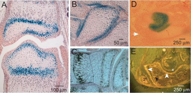
Figure 2.

Most endochondral bones develop by a process of segmentation of mesenchymal condensations. These segmentations correspond to future joints, and are specified by developmental regulatory molecules from the interzone (22-24). PTHrP had not been previously demonstrated to regulate articular chondrocytes, so we began by studying PTHrP and PTH1R expression at forming joint sites. We found PTHrP expression to be confined to these sites rather than to the ends of long bones per se, the latter having been thought previously to be the principal site of PTHrP expression (1-5,11,14,21). For example, carpals form via a single ossification center and have as many as six articulations, each of which expresses PTHrP, and at the forming elbow joint β-gal activity is present in the articular regions of the distal humerus and semilunar fossa but not at the olecranon (Figure 1B&D). In all of these locations, the receptor is expressed in prehypertrophic chondrocytes that lie immediately beneath the PTHrP-expressing articular chondrocytes (Figure 1C&E). Thus, PTHrP and its receptor are deployed at joint sites coincident with joint specification and always in a pattern in which the PTHrP-expressing cells lie at the articular surface and PTHrP receptor-expressing cells lie immediately subjacent.
If PTHrP were to retard chondrocyte differentiation in articular chondrocytes as it does in growth chondrocytes, this deployment of PTHrP and its receptor would exclude terminally differentiating hypertrophic chondrocytes from the articular cartilage and joint space, preventing their mineralization.
PTHrP regulation of hyaline chondrocytes
The chondrocytes of hyaline cartilage structures must remain undifferentiated in order to function, a phenomenon referred to as “maintenance”. The question posed here was whether PTHrP might regulate hyaline chondrocyte differentiation in a manner similar to its regulation of growth chondrocyte differentiation (1-5). There was a lead in this regard based on the fact that the PTHrP-null mouse dies at birth because of pathological mineralization of costal cartilage (14,21).
We carried out proof-of-principle studies of this question in two very simple permanent cartilagenous structures, nasal cartilage and costal cartilage. Both are composed of a tube of hyaline chondrocytes surrounded by a PTHrP-expressing perichondrium (11,13, see also Figure 2A).
In PTHrP-null mice at term, the hyaline chondrocytes of nasal cartilage were fully differentiated into prehypertrophic and hypertrophic chondrocytes, as evidenced histologically as well as by alkaline phosphatase and von Kossa staining (Figure 2A-D). There was also abundant collagen X and Ihh mRNA expression throughout the PTHrP-null nasal cartilage by ISHH, whereas none was seen in the PTHrP-lacZ or wild-type specimens (data not shown). In the single-allele PTHrP-null mouse, this increase in Ihh expression was reflected in a markedly enhanced periosteal β-gal signal, defining these cells as Ihh target cells (compare Figure 2A and B). Exactly the same findings were seen in costal cartilage (data not shown) (13).
Thus, these simple hyaline cartilage structures seem to be defended against pathological mineralization solely by the capacity of PTHrP to serve as a brake on hyaline chondrocyte differentiation. That is, PTHrP appears to normally regulate hyaline chondrocyte differentiation in these simple structures via circumferential paracrine signaling from the perichondrium to the PTH1R-bearing hyaline chondrocytes within. Ihh targets the perichondrial cells but not the chondrocytes themselves and therefore has no direct effects in these cells (in contrast to what is described below).
The next question concerned putative PTHrP regulation of articular chondrocytes. In addition to the ancient and embryonic iterations of the chondroepiphyseal growth zones, there exists a lesser-known iteration that occupies one end of the short bones of the hands and feet (8,9,20). Whether these short-bone chondroepiphyses represent a persistence of the primitive structure or evolved secondarily is unclear, but they phenocopy the ancient chondroepiphysis in every detail and might therefore serve as a model system of what a primitive joint structure might have been like. These simple structures contain a single subarticular population of PTHrP-expressing RPCs overlying subjacent populations of mineralizing chondrocytes and bone cells. These cells invade this zone from the primary ossification center via a process referred to as direct ossification (8,9).
The PTHrP-knockout mouse manifests a lethal chondrodysplasia that is driven by promiscuous chondrocyte differentiation and mineralization throughout the endochondral skeleton (1,11,14,21). The short-bone system in this mouse thus provided an opportunity to see if the chondroepiphysis and/or growth plates might be overrun by this pathological process more or less as seen in nasal and costal cartilage. To maximize the developmental time in which the joints might be at risk, we prolonged parturition to E20.5 with progesterone. Absent PTHrP, we found that both ends of the short bones mineralized prematurely via the direct pathway characteristic of the ancient process and that the secondary ossification centers and growth plates failed to form (Figure 2 G&H). Nevertheless, the residual chondroepiphyseal structures and cartilagenous joint spaces at both ends of the short bones were not invaded by mineralized cells (Figure 2H; see also Figure 3C).
Figure 3.
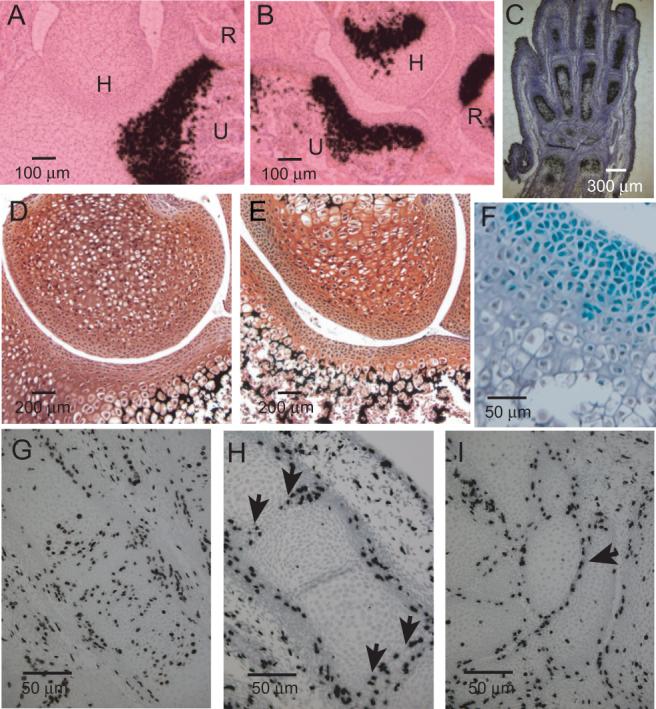
Thus, in the absence of PTHrP, the joints in short bones were threatened but not broached by mineralizing cells. We also looked at this question in more prototypical joints. In PTHrP-null mice, we observed that Ihh-expressing prehypertrophic chondrocytes and collagen X-expressing hypertrophic chondrocytes approached the joints at the elbow and in the digits but here too did not invade the joint space itself (Figure 3A,B&C). This same pattern was seen histologically in the form of hypertrophic cells that approached to within several cell layers of the joint in the PTHrP-null as compared to the PTHrP-lacZ specimen (Figure 3D&E).
Closer inspection of the PTHrP-null histological sections revealed that the surface articular chondrocyte population did not have the spindle-shaped morphology that typifies the normal lining cells (Figure 3D) but rather the prototypical appearance of RPCs (Figure 3E&F). Further, this RPC-like population appeared to be expanded as compared to that in the PTHrP-lacZ mouse, and the marked β-gal overexpression in the single-allele system clearly identified these RPC-like cells as Ihh target cells (Figure 3F).
Ihh is mitogenic for growth RPCs, whereas PTHrP maintains these cells in the cell cycle but is not itself a mitogen (1,2,4,5,23). Were Ihh to have the same mitogenic effect in young articular chondrocytes that it has in growth RPCs, it might be responsible for the dramatically different findings seen in articular versus costal/nasal cartilage in the PTHrP-null mice. We looked at this question by examining BrdU incorporation into the nuclei of chondrocytes in wild-type and single-allele PTHrP-null mice at E15.5 and 16.5, reasoning that such proliferation would be apparent several days in developmental time ahead of the established pattern seen at term (Figure 3E&F). In the wild-type specimens, we found a high rate of BrdU incorporation scattered throughout the developing carpals and digits, as expected at this age (Figure 3G), whereas in the PTHrP-null specimens a high rate of BrdU incorporation was confined to the chondrocytes at the articular margins of the MP joints and carpals (Figure 3H&I, respectively). This highly anatomical pattern of proliferation reflected premature chondrocyte differentiation in the PTHrP-null mice, as both structures exhibited a promiscuous advance of Ihh-expressing prehypertrophic chondrocytes to positions just subjacent to the articular margins (Figure 3C). Because of its anatomical simplicity, the central carpal bone lent itself to easy quantification on this regard (see legend Figure 3).
Thus, the Ihh-PTHrP feedback system appears to be fully deployed in young articular chondrocytes, and this regulatory system would seem to be particularly adept at protecting the forming joints in a combinatorial sense, as Ihh drives proliferation of undifferentiated chondrocytes flanking the joint, while PTHrP normally serves to prevent these cells from exiting the cell cycle.
Mechanical regulation
PTHrP is expressed in the load-bearing articular surfaces of the synovial joints from embryonic life through adulthood. The PTHrP gene is known to be mechanically-induced in a number of sites (12,25-27), so that this pattern suggested that mechanical loading might regulate PTHrP expression in articular cartilage as well. Since weight-bearing joints presumably would be loaded at baseline, we employed unloading techniques to look at this question. The femoropatellar joint was an attractive target because it is particularly heavily loaded in rodents that walk on a flexed knee (28) and because it is so simply unloaded by transecting the patellar ligament. We also unloaded the knee in separate experiments; since this method may have introduced a degree of joint instability as well, only short-term results are described here.
In femoropatellar surfaces unloaded for 7 days, there was a marked diminution in β-gal-expressing mid-zone chondrocytes, accompanied by an equally marked increase in subjacent alkaline phosphatase-expressing chondrocytes in the deep zone (Figure 4A&B). These changes were quantified by histomorphometry (see legend) We saw the same changes in the tibial plateaus of knees unloaded for three days (Figure 4C) as well as for as few as 24 hrs (not shown). The degree and the rapidity of the articular chondrocyte differentiation in the deep zone was corroborated by the expression of collagen X mRNA (Figure 5A&B). While the differentiating articular chondrocytes appeared to approximate the articular surface in the unloaded joints (Figure 4B&C), in none of the specimens did these cells broach the surface, nor did we see any evidence of chondrocyte degeneration. Thus, the unloading-associated changes in PTHrP expression and chondrocyte differentiation in articular cartilage seemed to be very rapid, were not progressive, and did not lead to degenerative findings. Since it is known that physiological physical forces are anabolic rather than catabolic for articular cartilage (29,30), our experimental findings very likely reflected the effects of an absence of physiological loading.
Figure 4.
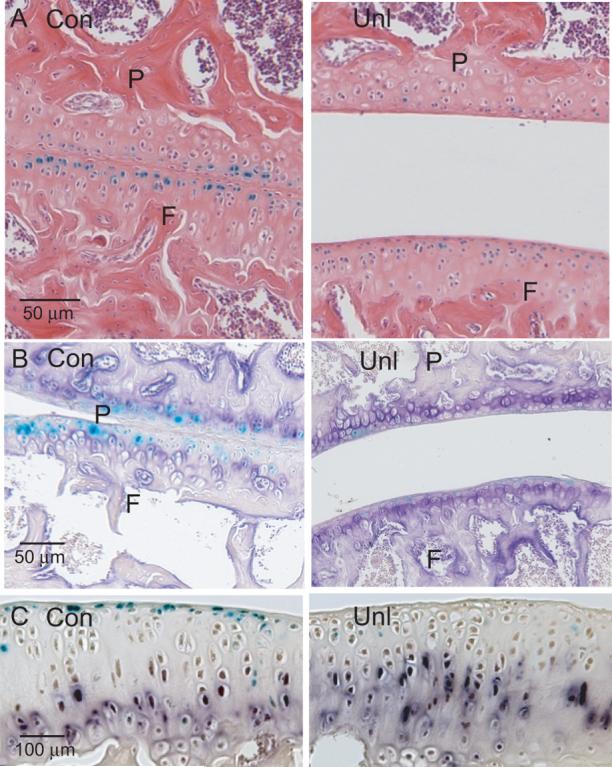
Figure 5.
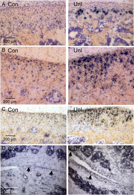
Ihh has also been reported to be load-induced (31,32), suggesting that unloading might reduce Ihh expression as well as that of PTHrP. On the other hand, the findings just described (the unloading-induced decrease in PTHrP expression and the increase in differentiating and likely Ihh-expressing chondrocytes) would argue for an unloading-associated increase rather than a decrease in Ihh expression. This point was examined by ISHH in both the unloaded patella and knee, each of which demonstrated a clear-cut increase in Ihh expression. This response was seen as early as three days of unloading and persisted for up to four weeks (Figure 5C&D).Thus, the PTHrP-Ihh regulatory system and its effect on chondrocyte differentiation clearly trumps any potential independent mechanical effect on Ihh expression in articular cartilage. The unusual aspect of Ihh signaling in these experiments was the absence of PTHrP expression in the face of the marked increase in Ihh expression, representing an uncoupling of the Ihh-PTHrP expression pattern that typifies regulation in growth chondrocytes (1-5).
DISCUSSION
The chondroepiphysis and growth chondrocytes
The ancestral chondroepiphysis arose some 425 million years ago (8,9). This structure was presumably multifunctional, serving as both a growth zone and as a limiting perimeter of ossification, and was very simple, having no capacity for continuous linear growth after ossification or for the formation of the subchondral structures of the modern joint (8,9). The chondroepiphysis of the short bones shares all of these features and presumably functions in much the same fashion (8-10,12). In contrast, the embryonic chondroepiphysis of land-mobile species has the developmental capacity to construct all of the structures of the modern weight-bearing joint (the bony epiphysis and metaphysis and defined articular and growth-plate structures), which together play a vital role in load transfer and distribution (30,33). Our data suggest that this embryonic structure serves as the source of PTHrP-expressing RPCs that are distributed to both growth plate and articular cartilage subpopulations (11,13). Other articular cells such as the lining cells appear to arise from the interzone (24,34-36).
PTHrP functions in growth cartilage
The best studied PTHrP function in the growth chondrocytes of the embryonic chondroepiphysis is its capacity to maintain RPCs in the cell cycle, thereby preventing their hypertrophic differentiation (1-5). PTHrP appears to act in the same fashion in the short-bone iteration of this structure and by inference possibly in its primitive iteration as well. In all three structures, this effect would serve not only to maintain a proliferative capacity to support growth but also protect the joint space from invasion by mineralizing chondrocytes and bone cells. In most locations, the embryonic chondroepiphysis comes to be subdivided by the secondary ossification center into an RPC population that remains in place as nascent articular chondrocytes and another that forms the so-called reserve zone above the chondrocyte columns (19). These evolutionary redeployments as well as the data provided here suggest several previously unrecognized candidate PTHrP functions. In the growth plate, these might include: 1) serving as the population of reserve chondrocytes that supplies recruits to the columns (19), 2) preventing this reserve RPC population from destruction by hypertrophic chondrocytes from above, and 3) delaying the approach of differentiating chondrocytes from the primary ossification center sufficiently to allow the secondary center to form. This synthesis simply assumes that in the growth plate PTHrP maintains chondrocytes in the cell cycle within its signaling range. These various candidate functions were presumably co-opted from the primitive chondroepiphysis with the evolution of the secondary center.
Articular Cartilage
Our group is not the first to identify PTHrP and/or Ihh in articular cartilage, but previous data did not address the potential function(s) of these molecules in articular chondrocytes (37-39). In general, our data indicate that PTHrP is not involved in joint specification but rather potentially in regulating the maintenance of articular chondrocytes.
Our genetic studies were carried out in two versions of PTHrP-null mice, the most informative being the single-allele PTHrP-null mouse. Absent PTHrP, we found that simple structures such as the nasal and costal cartilage were completely destroyed by mineralizing cells, whereas prehypertrophic and hypertrophic chondrocytes formed abnormally in PTHrP-null articular cartilage but did not broach the articular surface, implying a second protective mechanism at joint sites. As in prototypical growth cartilage, in nascent PTHrP-null articular cartilage there was a striking increase in Ihh expression, which induced a marked increase in lacZ/PTHrP expression in phenotypic RPCs at the articular surface. Also as seen in Ihh-stimulated growth RPCs, these surface articular chondrocytes were rapidly proliferating, creating an apparent moat which prevented hypertrophic chondrocytes from crossing. These data provide strong genetic evidence as to the function of the Ihh-PTHrP axis at the embryonic chondroepiphyseal stage but were limited in developmental time by the neonatal death of the PTHrP-null mouse. Attempts to extend these findings by conditional knockout techniques are in progress.
Our second category of evidence was functional. PTHrP has been known to be a mechanically-inducible gene since shortly after its discovery (26,40), and its expression in the contact regions at articular surfaces immediately suggested that it might be mechanically regulated in articular chondrocytes as well. Demonstrating this by unloading techniques was technically straightforward and revealed a pronounced unloading-induced decrease in PTHrP/lacZ expression in mid-zone articular chondrocytes as well as corresponding increases in the numbers of subjacent Ihh-, Col X-, and alkaline phosphatase-expressing prehypertrophic/hypertrophic chondrocytes, more or less as seen in the genetic systems. However, certain aspects of our results were not as might have been predicted. First among these was the rapidity of the reduction in both PTHrP expression and the corresponding differentiation of articular chondrocytes, such that the patterns we saw at 1-3 days and 4 weeks of unloading were basically the same. Certainly, our unloading techniques were not physiological, yet the rapidity of these responses was impressive. Second, based on the literature, there was as much reason to expect an unloading-associated decrease in Ihh (31,32) as in PTHrP expression, but it was clear that the release of articular chondrocytes from the PTHrP brake on their differentiation was the dominant response in these experiments. Third, given that Ihh is the principal regulator of PTHrP expression in growth chondrocytes (1-5), it was something of a surprise to see this regulation uncoupled in articular chondrocytes. Thus, in mature articular cartilage, the primary regulatory stimulus appears to mechanical force, with PTHrP being the primary responder. Finally, while Ihh is clearly in play in unloaded articular cartilage and therefore may act as a second line of defense as regards articular chondrocyte differentiation, we have not been able thus far to demonstrate Ihh-associated chondrocyte proliferation in adult mice. This question is being pursued via modified BrdU techniques. Since adult articular chondrocytes are regarded as mature cells that have a low rate of proliferation, it is entirely possible that Ihh elicits a unknown protective effect in adult articular cartilage that is non-proliferative.
Our findings on unloading presumably represent the mirror image of what would be seen in physiologically-loaded articular cartilage. Such loading would induce PTHrP expression in the sub-surface and mid-zone articular chondrocyte, maximal in the central contact areas of the joints. This response would maintain the undifferentiated articular chondrocytes and would also retard the differentiation of the high-abundance PTHrP receptor-expressing prehypertrophic cells in the deep zone, so that the overall response to loading would be to maintain the articular cartilage in its native state (30). Physiological unloading would loosen this control by some fractional amount and induce Ihh deployment as a classic chondrocyte differentiation feedback response. At the time we initiated our unloading studies, we were unacquainted with the extensive work of Brandt showing that immobilization-associated changes in articular cartilage are non-progressive and largely reversible with remobilization (29), and our results clearly reinforce his findings.
Ihh-PTHrP regulation in growth cartilage and articular cartilage
Figure 6 contains three panels that illustrate regulation by the Ihh-PTHrP system in the embryonic chondroepiphysis, growth plate, and articular cartilage. The putative functions of these molecules in these structures were described above, and the focus here is to contrast possible differences between Ihh/PTHrP developmental regulation versus how the system may operate in mature articular cartilage.
Figure 6.
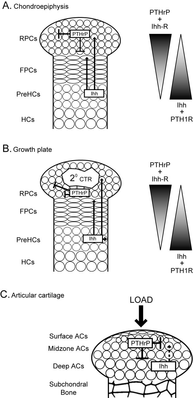
The master developmental regulatory cell in both the chondroepiphysis and growth plate is the prehypertrophic chondrocyte, and its principal product, Ihh, not only controls RPC proliferation and production of PTHrP but the early differentiation of RPCs into flat proliferative chondrocytes as well as induction of osteoblasts in adjacent structures (1-5). PTHrP serves in this system as the instrument by which Ihh regulates the flow of undifferentiated chondrocytes through the chondrocyte differentiation cascade. The essential features of this developmental regulation are that PTHrP and Ihh appear to be constitutive secretory messengers that likely operate via opposing gradients of high-abundance ligand- and receptor-expressing cells at the two ends of growth structures in question. As development compresses these gradients, the regulatory effects of the two molecules become increasingly powerful, examples in point being Ihh-driven RPC proliferation in the chondroepiphyses and in embryonic articular cartilage. Whether there may be additional regulatory modifiers involved in this axis is unclear (1-5, 41).
In contrast, in mature articular cartilage this axis appears to have been placed under the principal regulatory control of mechanical loading, with the primary responder being PTHrP and the chondrocyte differentiation cascade lying downstream. Here, the signaling distances are very compressed. This regulatory pattern probably evolved in the joints of the appendicular skeleton in shallow-water fish (6-10). This may correspond to the approximate period during which mechanical regulation of PTHrP expression in the tendon and ligament insertion sites also evolved (12).
Pathophysiological implications
To the extent that the Ihh-PTHrP axis may regulate the maintenance of articular chondrocytes in humans, this system may be relevant to the pathogenesis of mineralizing and/or degenerative forms of arthritis (42-46). There are two features of osteoarthritis to which this system might be of interest. One is the disordered differentiation and subsequent degeneration that is seen in the weight-bearing regions of articular cartilage. The other is the osteophyte, a frequent finding at the non-weight-bearing margins of osteoarthritic joints, a region that does not express PTHrP. Ihh and PTHrP may be deployed in some fashion in both of these locations and types of lesions, but how either molecule may relate to the pathogenesis of osteoarthritis is unknown.
ACKNOWLEDGEMENTS
We thank B. Dreyer and G. Liang for technical assistance, C. Coady and N. Troiano for assistance with histomorphometry and specialized bone stains, and Ann DeCosta for preparing the manuscript. All animal protocols were approved by the YACUC. Supported by NIH grants R01 DK62515, R01 DK48108, R21 DK75626, and P30 AR46032 (Yale Center for Musculoskeletal Disorders).
REFERENCES
- 1.Kronenberg HM. Developmental regulation of the growth plate. Nature. 2003;423:332–36. doi: 10.1038/nature01657. [DOI] [PubMed] [Google Scholar]
- 2.Vortkamp A, Lee K, Lanske B, Segre GV, Kronenberg HM, Tabin CJ. Regulation of rate of cartilage differentiation by Indian hedgehog and PTH-related protein. Science. 1996;273:613–22. doi: 10.1126/science.273.5275.613. [DOI] [PubMed] [Google Scholar]
- 3.Niswander L. Interplay between the molecular signals that control vertebrate limb development. Int J Dev Biol. 2002;46:877–81. [PubMed] [Google Scholar]
- 4.Long F, Zhang XM, Karp S, Yang Y, McMahon AP. Genetic manipulation of hedgehog signaling in the endochondral skeleton reveals a direct role in the regulation of chondrocyte proliferation. Development. 2001;128:5099–5108. doi: 10.1242/dev.128.24.5099. [DOI] [PubMed] [Google Scholar]
- 5.Kobasyashi T, Soegiarto DW, Yang Y, Lanske B, Schipani E, McMahon AP, Kronenberg HM. Indian hedgehog stimulates periarticular chondrocyte differentiation to regulate growth plate length independently of PTHrP. J Clin Invest. 2005;115:1734–42. doi: 10.1172/JCI24397. [DOI] [PMC free article] [PubMed] [Google Scholar]
- 6.Shubin NH. Origin of evolutionary novelty: Examples from limbs. J Morphol. 2002;252:15–28. doi: 10.1002/jmor.10017. [DOI] [PubMed] [Google Scholar]
- 7.Shubin NH, Daeschler EB, Jenkins FA., Jr The pectoral fin of Tiktaalik roseae and the origin of the tetrapod limb. Nature. 2006;440:764–71. doi: 10.1038/nature04637. [DOI] [PubMed] [Google Scholar]
- 8.Haines EW. The evolution of epiphyses and of endochondral bone. Biol Rev. 1942;17:267–91. (1942) [Google Scholar]
- 9.Reno PL, McBurney DL, Lovejoy CO, Horton WE., Jr Ossification of the mouse metatarsal: differentiation and proliferation in the presence/absence of a defined growth plate. Anat Rec Part A. 2006;288A:104–18. doi: 10.1002/ar.a.20268. [DOI] [PubMed] [Google Scholar]
- 10.Haines RW. Eudiathrodial joints in fishes. J Anat. 1942;76:12–19. [PMC free article] [PubMed] [Google Scholar]
- 11.Chen X, Macica CM, Dreyer BE, Hammond VE, Hens JR, Philbrick WM, Broadus AE. Initial characterization of PTH-related protein gene-driven lacZ expression in the mouse. J Bone Miner Res. 2006;20:113–23. doi: 10.1359/JBMR.051005. [DOI] [PubMed] [Google Scholar]
- 12.Chen X, Macica CM, Nasiri A, Judex S, Broadus AE. Mechanical regulation of PTHrP expression in entheses. Bone. 2007;41:752–59. doi: 10.1016/j.bone.2007.07.020. [DOI] [PMC free article] [PubMed] [Google Scholar]
- 13.Broadus AE, Macica CM, Chen X. PTHrP functional domain is at the gates of endochondral bones. Ann NY Acad Sci. 2007;1116:65–81. doi: 10.1196/annals.1402.061. [DOI] [PubMed] [Google Scholar]
- 14.Karaplis AC, Luz A, Glowacki J, Bronson RJ, Tybolewicz VLT, Kronenberg HM, Mulligan RC. Lethal skeletal dysplasis from targeted disruption of the parathyroid hormone-related peptide gene. Genes & Dev. 1994;8:277–89. doi: 10.1101/gad.8.3.277. [DOI] [PubMed] [Google Scholar]
- 15.Jiang XI, Kalajzic Z, Maye P, Braut A, Bellizzi J, Mina M, Rowe DW. Histological analysis of GFP expression in murine bone. J Histochem Cytochem. 2005;53:593–602. doi: 10.1369/jhc.4A6401.2005. [DOI] [PubMed] [Google Scholar]
- 16.Young DC, Kinsley SD, Ryan KA, Dutko FJ. Selective inactivation of eukaryotic β-galactosidase as reporter enzyme. Anal Biochem. 1993;215:24–39. doi: 10.1006/abio.1993.1549. [DOI] [PubMed] [Google Scholar]
- 17.Philbrick WM, Dreyer BE, Nakchbandi IA, Karaplis AC. Parathyroid hormone-related protein is required for tooth eruption. Proc Natl Acad Sci USA. 1998;95:11846–51. doi: 10.1073/pnas.95.20.11846. [DOI] [PMC free article] [PubMed] [Google Scholar]
- 18.Nolte T, Kaufmann W, Schorsch F, Soames T, Weber E. Standardized assessment of cell proliferation: The approach of the RITA-CEPA working group. Exp Tox Path. 2005;57:91–103. doi: 10.1016/j.etp.2005.06.002. [DOI] [PubMed] [Google Scholar]
- 19.Kember NF. Comparative patterns of cell division in epiphyseal cartilage plates in the rat. J Anat. 1972;111:137–142. [PMC free article] [PubMed] [Google Scholar]
- 20.Reno PL, Horton WE, Jr, Elsey RM, Lovejoy CO. Growth plate formation and development in alligator and mouse metapodials: Evolutionary and functional implications. J Exp Zoology (Mol. Dev. Evol.) 2007;308B:283–96. doi: 10.1002/jez.b.21148. [DOI] [PubMed] [Google Scholar]
- 21.Lanske B, Karaplis AC, Lee K, Luz A, Vortkamp A, Pirro A, Karperien M, Defize LHK, Ho C, Mulligan RC, Abou-Samra A-B, Jüppner H, Segre GV, Kronenberg HM. PTH/PTHrP receptor in early development and Indian hedgehog-regulated bone growth. Science. 1996;273:613–22. doi: 10.1126/science.273.5275.663. [DOI] [PubMed] [Google Scholar]
- 22.Storm EE, Huynh TV, Copeland NG, Jenkins NA, Kingsley DM, Lee S-J. Limb alterations in brachypodism mice due to mutations in a new member of the TGF-β superfamily. Nature. 1994;368:639–43. doi: 10.1038/368639a0. [DOI] [PubMed] [Google Scholar]
- 23.Brunet LJ, McMahon JA, McMahon AP, Harland RM. Noggin, cartilage morphogenesis, and joint formation in the mammalian skeleton. Science. 1998;280:1455–57. doi: 10.1126/science.280.5368.1455. [DOI] [PubMed] [Google Scholar]
- 24.Pacifici M, Koyama E, Iwamoto M. Mechanisms of synovial joint and articular cartilage formation: recent advances, but many lingering mysteries. Birth Defects Res. 2005;75:237–48. doi: 10.1002/bdrc.20050. [DOI] [PubMed] [Google Scholar]
- 25.Chen X, Macica CM, Ng KW, Broadus E. Stretch-induced PTH-related protein gene expression in osteoblasts. J Bone Min Res. 2005;20:1454–61. doi: 10.1359/jbmr.2005.20.8.1454. [DOI] [PubMed] [Google Scholar]
- 26.Clemens TL, Broadus AE. Physiologic actions of PTH and PTHrP: IV. Vascular, cardiovascular, and neurologic actions. In: Bilezikian JP, editor. The Parathyroids. 2nd ed. Acad Press; NY: 2001. pp. 261–274. [Google Scholar]
- 27.Tanaka N, Ohno S, Honda K, Tanimoto K, Doi T, Ohno-Nakahara M, Tafolla E, Kapilam S, Tanne K. Cyclic mechanical strain regulates the PTHrP expression in cultured chondrocytes via activation of the Ca2+ channel. J Dent Res. 2005;84:64–68. doi: 10.1177/154405910508400111. [DOI] [PubMed] [Google Scholar]
- 28.O'Connor KM. Unweighting accelerates tidemark advancement in articular cartilage at the knee joint of rats. J Bone Miner Res. 1997;12:580–589. doi: 10.1359/jbmr.1997.12.4.580. [DOI] [PubMed] [Google Scholar]
- 29.Brandt KD. Response of joint structures to inactivity and to reloading after immobilization. Arthritis Rheum. 2003;49:267–71. doi: 10.1002/art.11009. [DOI] [PubMed] [Google Scholar]
- 30.Carter DR, Beaupré GS, Wong M, Smith L, Andriacchi TP, Schurman DJ. The mechanobiology of articular cartilage development and degeneration. Clin Orthop Related Res. 2004;427S:S69–77. doi: 10.1097/01.blo.0000144970.05107.7e. [DOI] [PubMed] [Google Scholar]
- 31.Wu Q-Q, Zhang Y, Chen Q. Indian hedgehog is an essential component of mechanotransduction complex to stimulate chondrocyte proliferation. J Biol Chem. 2001;276:35290–96. doi: 10.1074/jbc.M101055200. [DOI] [PubMed] [Google Scholar]
- 32.Tang GH, Rabie AB, Hagg U. Indian hedgehog: a mechanotransduction mediator in condylar cartilage. J Dent Res. 2004;83:434–38. doi: 10.1177/154405910408300516. [DOI] [PubMed] [Google Scholar]
- 33.Hoshino A, Wallace WA. Impact-absorbing properties of the human knee. J. Bone Joint Surg. 1987;69:807–18. doi: 10.1302/0301-620X.69B5.3680348. [DOI] [PubMed] [Google Scholar]
- 34.Edwards CJ, Francis-West PH. Bone morphogenetic proteins in the development and healing of synovial joints. Seminars Arthritis and Rheum. 2001;31:33–42. doi: 10.1053/sarh.2001.24875. [DOI] [PubMed] [Google Scholar]
- 35.Hyde G, Dover S, Aszodi A, Wallis GA, Boot-Handford RP. Lineage tracing using matrilin-1 gene expression reveals that articular chondrocytes exist as the joint interzone forms. Dev Biol. 2007;304:825–33. doi: 10.1016/j.ydbio.2007.01.026. [DOI] [PMC free article] [PubMed] [Google Scholar]
- 36.Koyama E, Shibukawa Y, Nagayama M, Sugito H, Young B, Yuasa T, Okabe T, Ochiai T, Kamiya N, Roundtree RB, Kingsley DM, Iwamoto M, Enomoto-Iwamoto M, Pacifici M. A distinct cohort of progenitor cells participates in synovial joint and articular cartilage formation during mouse limb skeletogenesis. Dev Biol. 2008;316:62–73. doi: 10.1016/j.ydbio.2008.01.012. [DOI] [PMC free article] [PubMed] [Google Scholar]
- 37.Burton DW, Foster M, Johnson KA, Hiramoto M, Deftos LJ, Terkeltaub R. Chondrocyte calcium-sensing receptor expression is up-regulated in early guinea pig knee osteoarthritis and modulates PTHrP, MMP-13, and TIMP-3 expression. Osteoarth Cartil. 2005;13:395–404. doi: 10.1016/j.joca.2005.01.002. [DOI] [PubMed] [Google Scholar]
- 38.Semevolos SA, Nixon AJ, Fortier LA, Strassheim ML, Haupt J. Age-related expression of molecular regulators of hypertrophy and maturation in articular cartilage. J Orthop Res. 2006;24:1773–81. doi: 10.1002/jor.20227. [DOI] [PubMed] [Google Scholar]
- 39.Rabie ABM, Tang GH, Xiong H, Hägg U. PTHrP regulates chondrocyte maturation in condylar cartilage. J Dent Res. 2003;82:627–31. doi: 10.1177/154405910308200811. [DOI] [PubMed] [Google Scholar]
- 40.Thiede MA, Daifotis AG, Weir EC, Brines ML, Burtis WJ, Ikeda K, Dreyer BE, Garfield RE, Broadus A. Intrauterine occupancy controls expression of the parathyroid hormone-related peptide gene in preterm rat myometrium. Proc Natl Acad Sci USA. 1990;87:6969–73. doi: 10.1073/pnas.87.18.6969. [DOI] [PMC free article] [PubMed] [Google Scholar]
- 41.Alvarez J, Sohn P, Zeng X, Doetschman T, Robbins DJ, Serra R. TGFbeta2 mediates the effects of hedgehog on hypertrophic differentiation and PTHrP expression. Development. 2002;129:1913–24. doi: 10.1242/dev.129.8.1913. [DOI] [PubMed] [Google Scholar]
- 42.Ho AM, Johnson MD, Kingsley DM. Role of the mouse ank gene in control of tissue calcification and arthritis. Science. 2000;289:265–270. doi: 10.1126/science.289.5477.265. [DOI] [PubMed] [Google Scholar]
- 43.Zhang Y, Brown MA, Peach C, Russell G, Wordsworth BP. Investigation of the role of ENPPI and TNAP genes in chondrocalcinosis. Rheumatology. 2007;46:586–589. doi: 10.1093/rheumatology/kel338. [DOI] [PubMed] [Google Scholar]
- 44.Goldring MB. The role of chondrocyte in osteoarthritis. Arthritis. Rheum. 2000;43:1916–26. doi: 10.1002/1529-0131(200009)43:9<1916::AID-ANR2>3.0.CO;2-I. [DOI] [PubMed] [Google Scholar]
- 45.Sandell LJ, Aigner T. Articular cartilage and changes in arthritis an introduction: Cell biology of osteoarthritis. Arthritis Res. 2001;3:107–13. doi: 10.1186/ar148. [DOI] [PMC free article] [PubMed] [Google Scholar]
- 46.Drissi H, Zuscik M, Rosier R, O'Keefe R. Transcriptional regulation of chondrocyte maturation: Potential involvement of transcription factors in OA pathogenesis. Molec. Aspects of Med. 2005;26:169–79. doi: 10.1016/j.mam.2005.01.003. [DOI] [PubMed] [Google Scholar]