Microsatellite Instability Predicts Improved Response to Adjuvant Therapy With Irinotecan, Fluorouracil, and Leucovorin in Stage III Colon Cancer: Cancer and Leukemia Group B Protocol 89803 (original) (raw)
Abstract
Purpose
Colon cancers exhibiting DNA mismatch repair (MMR) defects demonstrate distinct clinical and pathologic features, including better prognosis and reduced response to fluorouracil (FU) –based chemotherapy. This prospective study investigated adjuvant chemotherapy containing FU and irinotecan in patients with MMR deficient (MMR-D) colon cancers.
Patients and Methods
Cancer and Leukemia Group B 89803 randomly assigned 1,264 patients with stage III colon cancer to postoperative weekly bolus FU/leucovorin (LV) or weekly bolus irinotecan, FU, and LV (IFL). The primary end point was overall survival; disease-free survival (DFS) was a secondary end point. Tumor expression of the MMR proteins, MLH1 and MSH2, was determined by immunohistochemistry (IHC). DNA microsatellite instability was also assessed using a panel of mono- and dinucleotide markers. Tumors with MMR defects were those demonstrating loss of MMR protein expression (MMR-D) and/or microsatellite instability high (MSI-H) genotype.
Results
Of 723 tumor cases examined by genotyping and IHC, 96 (13.3%) were MMR-D/MSI-H. Genotyping results were consistent with IHC in 702 cases (97.1%). IFL-treated patients with MMR-D/MSI-H tumors showed improved 5-year DFS as compared with those with mismatch repair intact tumors (0.76; 95% CI, 0.64 to 0.88 v 0.59; 95% CI, 0.53 to 0.64; P = .03). This relationship was not observed among patients treated with FU/LV. A trend toward longer DFS was observed in IFL-treated patients with MMR-D/MSI-H tumors as compared with those receiving FU/LV (0.57; 95% CI, 0.42 to 0.71 v 0.76; 95% CI, 0.64 to 0.88; P = .07; hazard ratio interaction between tumor status and treatment, 0.51; likelihood ratio P = .117).
Conclusion
Loss of tumor MMR function may predict improved outcome in patients treated with the IFL regimen as compared with those receiving FU/LV.
INTRODUCTION
Colorectal cancer (CRC) develops through accumulated genetic changes that produce loss of tumor suppressor genes and activation of oncogenes. In sporadic CRC, these changes occur through at least two distinct mechanisms. The first, termed chromosomal instability (CIN), is associated with loss of heterozygosity at multiple tumor suppressor loci, particularly 5q, 17p, and 18q.1 This type of genomic instability occurs in approximately 80% to 85% of sporadic CRC. A second subset demonstrates DNA mismatch repair (MMR) deficiency, characterized by an inability to repair single nucleotide DNA mismatches. This defect alters the size of di-, tri- and tetra-dinucleotide repeat sequences known as microsatellites, and this genomic instability is also termed microsatellite instability (MSI).2,3 Approximately 15% to 20% of sporadic CRCs demonstrate high levels of MSI (termed MSI-H). These CRCs may develop because MSI fosters inactivating mutations in tumor suppressors containing microsatellites, such as transforming growth factor β RII4 and BAX.5 In most sporadic cases, MSI occurs when the promoter region of the mismatch repair gene, MLH1, is silenced by CpG island hypermethylation.6,7
Identification of MSI requires DNA extraction from tumor and normal tissue, which can be technically challenging and time-consuming. Immunohistochemistry (IHC) can also detect loss of MMR protein expression, and IHC identification of MLH1 and MSH2, two MMR proteins most commonly lost in sporadic CRC, is a widely available clinical test to distinguish MMR deficient (MMR-D) from mismatch repair intact (MMR-I) tumors.8
Retrospective studies show that the clinical behavior of MMR-D/MSI-H CRCs is different than those without this characteristic. MMR-D/MSI-H tumors are more commonly located in the right colon and are more frequently poorly differentiated and mucin-containing.9–12 Despite these adverse histopathologic features, MMR-D/MSI-H tumors may have better treatment outcome than tumors with CIN. Studies comparing patients with MMR-D/MSI-H CRC with stage-matched controls show that patients with MMR-D/MSI-H tumors experience longer survival than those whose tumors lack this characteristic.13,14 A meta-analysis of 32 CRC studies estimated a combined hazard ratio (HR) for overall survival (OS) associated with MSI-H of 0.65 (95% CI, 0.59 to 0.71).14
A limited number of retrospective studies examined adjuvant chemotherapy for patients with stage II or III MSI-H CRC. These studies indicate that, unlike patients whose tumors demonstrate CIN, those with MSI-H tumors experience no benefit with regimens containing fluorouracil (FU).15–17 Cancer and Leukemia Group B (CALGB) 89803 was a phase III randomized trial comparing FU/leucovorin (LV) with irinotecan, FU, and LV (IFL) for postoperative adjuvant treatment of stage III or high-risk stage II colon cancer. This study prospectively determined whether patients with MMR-D/MSI-H tumors were more likely to achieve better outcome overall or were more likely to respond to IFL.
PATIENTS AND METHODS
Characteristics of Study Population
Patients with histologically confirmed stage III colon cancer were enrolled onto CALGB protocol 89803 (Table 1). All patients underwent complete surgical resection and started chemotherapy between postoperative days 21 to 56. The primary end point was OS; DFS was a secondary end point. Additional secondary aims addressed the relationship between tumor-associated risk factors and treatment outcome and required submission of paraffin blocks containing tumor and normal tissue to the CALGB Pathology Coordinating Office at Ohio State University. This protocol was reviewed by the institutional review board of each center, and all patients gave written informed consent before participation.
Table 1.
Characteristics of the Study Cohort
| Characteristic | All Protocol 89803 Patients (N = 1264) | Patients With Genotyping and IHC Agreement (n = 702) | Patients With MMR-D Tumors* (n = 96) | Patients With MMR-I Tumors* (n = 606) | P† | |||
|---|---|---|---|---|---|---|---|---|
| No. | % | No. | % | No. | % | No. | % | |
| Treatment | .729 | |||||||
| FU/LV | 629 | 49.8 | 348 | 49.6 | 46 | 47.9 | 302 | 49.8 |
| FU/LV + irinotecan | 635 | 50.2 | 354 | 50.4 | 50 | 52.1 | 304 | 50.2 |
| Age, years | .116 | |||||||
| Median | 61 | 62 | 66 | 61 | ||||
| Range | 21-85 | 24-85 | 24-85 | 24-85 | ||||
| Sex | .039 | |||||||
| Male | 702 | 55.5 | 384 | 54.7 | 43 | 44.8 | 341 | 56.3 |
| Female | 562 | 44.5 | 318 | 45.3 | 53 | 55.2 | 265 | 43.7 |
| Extracellular mucin | < .0001 | |||||||
| Yes | 125 | 9.9 | 68 | 9.7 | 27 | 28.1 | 41 | 6.8 |
| No | 1076 | 85.1 | 611 | 87.0 | 67 | 69.8 | 544 | 89.8 |
| Unknown | 63 | 5.0 | 23 | 3.3 | 2 | 8.7 | 21 | 3.5 |
| Site of tumor‡ | < .0001 | |||||||
| Proximal | 713 | 56.4 | 399 | 56.8 | 88 | 91.7 | 311 | 51.3 |
| Distal | 521 | 41.2 | 297 | 42.3 | 8 | 8.3 | 289 | 47.7 |
| Unknown | 30 | 2.4 | 6 | 0.9 | 0 | 0 | 6 | 1.0 |
| Tumor grade | < .0001 | |||||||
| Well differentiated | 70 | 5.5 | 32 | 4.6 | 2 | 2.1 | 30 | 5.0 |
| Moderately differentiated | 861 | 68.1 | 491 | 69.9 | 47 | 49.0 | 444 | 73.3 |
| Poorly differentiated or undifferentiated | 304 | 24.1 | 172 | 24.5 | 47 | 49.0 | 127 | 21.0 |
| Unknown | 29 | 2.3 | 5 | 0.7 | 0 | 0 | 5 | 0.8 |
| Lymph node status | ||||||||
| No. of positive nodes | ||||||||
| Median | 2 | 3 | 2 | 3 | ||||
| Range | 0-29 | 1-29 | 1-23 | 1-29 | .705 | |||
| Node ratio | .005 | |||||||
| Median | 0.23 | 0.22 | 0.18 | 0.23 | ||||
| Range | 0-1.3 | 0.01-1 | 0.03-1 | 0.01-1 | ||||
| Mean | 0.30 | 0.31 | 0.25 | 0.32 | ||||
| Standard deviation | 0.25 | 0.25 | 0.22 | 0.25 |
Trial Structure and Organization
This trial was conducted by CALGB with participation by the North Central Cancer Treatment Group, National Cancer Institute of Canada Clinical Trials Group, Eastern Cooperative Oncology Group, Southwest Oncology Group, and the National Cancer Institute Cancer Trials Support Unit. The CALGB data safety monitoring board reviewed safety data twice yearly and efficacy data at protocol-specified intervals in accordance with CALGB policies. The CALGB Statistical Center at Duke University in Durham, NC, maintained the clinical and laboratory database.
Treatment
After central registration, eligible patients were assigned by computer (randomized fixed block) to receive either IFL or FU/LV. The FU/LV group received the Roswell Park regimen, consisting of LV 500 mg/m2 intravenously over 2 hours, with a bolus of FU 500 mg/m2 by intravenous injection 1 hour after initiation of LV. Treatments were to be administered weekly for 6 consecutive weeks followed by a 2-week rest, for a total of four cycles or 32 weeks of therapy. The IFL group received irinotecan 125 mg/m2 over 90 minutes followed immediately by intravenous bolus injection of LV 20 mg/m2, then FU 500 mg/m2 also by intravenous bolus injection. Treatment was to be given for 4 consecutive weeks followed by a 2-week rest for five cycles or 30 weeks. Further details concerning the treatment portion of this trial have been published previously.18
Detection of Tumor MSI
Formalin-fixed, paraffin-embedded primary tumor and normal colon specimens were obtained for each case from the CALGB Pathology Coordinating Office. Central pathology review confirmed the histology of each specimen; blocks were sectioned at 4 μm for IHC and 10 μm for DNA extraction. Laboratory analysis was performed at Brigham and Women's Hospital in Boston, MA, without knowledge of the patient's clinical outcome.
IHC detected the presence of MLH1 and MSH2 proteins in primary tumor specimens. Positive controls were provided by examining staining of normal colonic mucosa from each case; tumors known to lack MLH1 or MSH2 were stained concurrently and served as negative controls. All cases were scored as positive (defined as ≥ 10% of tumor cells staining) or negative (< 10% tumor cells staining). Cases designated as MMR-D were those with a negative IHC score for either MLH1 or MSH2, and cases designated MMR-I retained expression of both proteins. Each case was scored by two independent experts in gastrointestinal pathology (C.C.C., H.P.H.). In cases of disagreement (four cases, 0.05%), a third reviewer (M.R.) examined the case to provide the final score.
DNA extracted from each tumor was amplified by standard polymerase chain reaction using microsatellite markers defined during the 1998 National Cancer Institute Workshop on Microsatellite Instability19: BAT25, BAT26, D17S250, D5S346, ACTC, D18S55, BAT40, D10S197, BAT34c4, and MycL. In most cases, normal control tissue was obtained from a separate, nontumor tissue block. When this was not possible, non-neoplastic control tissue was obtained by microdissection. Whenever necessary, microdissection was performed to enrich tumor specimens for neoplastic cells, ensuring a minimum of 60% tumor within the sample. Tumors were designated MSI-H if instability was identified at ≥ 50% of the loci screened, MSI-low (MSI-L) if at least one but ≤ 50% of the loci showed instability, and microsatellite stable (MSS) if all loci were stable. For analysis, MSI-L and MSS cases were combined (MSI-L/S). A minimum of five successfully amplified loci were required for classification.
Statistical Methods
The study goal was to determine whether tumor MMR status was associated with outcome for patients with stage III colon cancer. The primary end point was OS measured from entry onto the clinical trial until death from any cause. DFS was measured from study entry until documented progression of disease or death from any cause. A secondary goal was to determine the relationship between MMR status assigned by genotyping with that of IHC. Median follow-up was estimated among surviving patients. Relationships between tumor MMR status and clinical pathologic factors were studied using the χ2 and Satterthwaite t tests. The interaction between treatment arm and MMR status was tested using a Cox model, and the log-rank test was used for survival comparisons among categories defined by MMR status and treatment and within MMR and treatment subgroups. The proportional hazards model was used to make survival comparisons controlling for treatment and other clinicopathologic factors. Because of small sample sizes, only one additional prognostic variable was considered in each model simultaneously with treatment. The Kaplan-Meier method was used to estimate the DFS and OS curves and the 3-year and 5-year survival probabilities. Agreement between MMR status as determined by genotyping and IHC was estimated using the κ statistic. All outcome analyses were conducted on the subset of patients in which there was agreement between methods. Data were analyzed with continued follow-up for DFS and OS as of March 10, 2008. All statistical analyses were performed by CALGB statisticians.
RESULTS
Determination of Microsatellite Stability Status
Specimens were available for analysis from 986 (78%) of the 1,264 patients enrolled on CALGB 89803. Of these, 63 (6.4%) were insufficient for analysis as a result of lack of adequate tumor and/or normal tissue. IHC to detect MLH1 was successfully performed on 783 of 792 cases attempted. For MSH2, IHC was successfully performed on 782 of 792 cases attempted. MSI status by IHC was indeterminate on 1.1% of tumors. A total of 106 (13.4%) cases were MMR-D based on loss of MLH1 or MSH2, and 677 (85.5%) were MMR-I (Tables 2 and 3). MLH1 expression was lost in 102 of the tumors, and four lacked expression of MSH2. Polymerase chain reaction amplification of microsatellite loci was successfully performed on 846 of 923 cases attempted. Of these, 137 (14.8%) were designated MSI-H and 709 (76.8%) were MSI-L/S.
Table 2.
Characterization of Tumor Microsatellite Status
| Characteristic | Immunohistochemistry | Genotyping | ||
|---|---|---|---|---|
| No. | % | No. | % | |
| No. of cases attempted | 792 | 923 | ||
| Result | ||||
| MMR-D (MSI-H) | 106 | 13.4 | 137 | 14.8 |
| MMR-I (MSI-L/S) | 677 | 85.5 | 709 | 76.8 |
| Indeterminate | 9 | 1.1 | 77 | 8.3 |
Table 3.
Comparison of Analysis Methods
| Genotyping Result | Immunohistochemistry Result | |
|---|---|---|
| MMR-D | MMR-I | |
| MSI-H | 96 | 17 |
| MSI-L/S | 4 | 606 |
A total of 723 patients had tumors successfully scored by both IHC and DNA microsatellite analysis (Table 3). The methods were in agreement in 702 cases (97.1%), with most of the difference accounted for by tumors that were MSI-H by DNA analysis but MMR-I by IHC. The κ measure of agreement between the methods was estimated to be κ = 0.88 with 95% CI (0.84 to 0.93) indicating good to excellent agreement.20
Relationships Between Tumor MMR Status and Clinicopathologic Factors
Overall median follow-up was 6.65 years. No significant differences were found by age, sex, nodal status, site of primary, or primary tumor differentiation between patients with and without tumor samples for MMR analysis. Patients with MMR-D/MSI-H tumors had significantly more proximal tumors (χ2 P < .0001), poorly/undifferentiated tumors (χ2 _P_ < .0001), and tumors containing extracellular mucin (>50%; χ2 P < .0001). The nodal ratio (number of positive nodes divided by number of nodes sampled) was significantly lower among patients with MMR-D/MSI-H tumors (Satterthwaite t test P = .005), and women had more MMR-D/MSI-H tumors (χ2 P = .03). There were no significant differences between MMR-D and MMR-I tumors by age, treatment, or number of positive lymph nodes (Table 1).
Relationship Between Tumor MMR Status and Prognosis
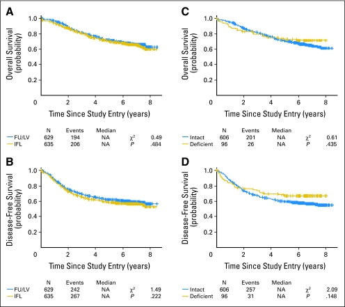
Analysis was conducted on data captured as of March 10, 2008, representing median follow-up of greater than 6.0 years; median OS and DFS have not yet been reached. The primary outcome was the difference in OS between patients receiving IFL and FU/LV. For the entire cohort, the probability of OS at 5 years was 0.70 with IFL and 0.72 with FU/LV, and the probability of DFS at 5 years was 0.59 for IFL and 0.62 for FU/LV (Fig 1).
Fig 1.
(A) Overall survival (OS) by treatment for the entire 89803 cohort (n = 1,264); (B) disease-free survival (DFS) by treatment for the entire 89803 cohort (n = 1,264); (C) OS by mismatch repair (MMR) status for all patients analyzed for MMR (n = 702); (D) DFS by MMR status for all patients analyzed for MMR (n = 702). FU, fluorouracil; LV, leucovorin; IFL, irinotecan, fluorouracil, and leucovorin; NA, not applicable.
We determined whether survival probability depended on MMR status, irrespective of treatment assignment (Table 4). For cases with IHC/genotyping agreement (n = 702), there was no difference in OS or DFS between patients with MMR-D/MSI-H and MMR-I/MSI-L/S tumors (Fig 1). The estimated HR for the OS comparison was 0.86 (95% CI, 0.57 to 1.29; log-rank P = .44). The 5-year probability of OS was 0.73 for MMR-D/MSI-H cases (95% CI, 0.64 to .82) and 0.71 for MMR-I/MSI-L/S cases (95% CI, 0.68 to 0.75). Similarly for DFS, the estimated HR was 0.61 (95% CI, 0.57 to 0.64; log-rank _P_= .15; Table 5). The estimated 5-year DFS was 0.67 for MMR-D/MSI-H cases (95% CI, 0.57 to 0.76) and 0.60 for MMR-I/MSI-L/S cases (95% CI, 0.56 to 0.64). Similar results were obtained by analyses using solely the IHC or the DNA genotyping results.
Table 4.
Effect of Tumor MSI Status on Treatment Outcome for OS
| Treatment Group | 5-Year OS | 95% CI | No. | Log-Rank P | HR | 95% CI |
|---|---|---|---|---|---|---|
| Prognostic analysis | ||||||
| All patients analyzed | 0.72 | 0.68 to 0.75 | 702 | |||
| All patients with MMR-D/MSI-H tumors | 0.73 | 0.64 to 0.82 | 96 | .44 | ||
| All patients with MMR-I/MSI-L/S tumors | 0.71 | 0.68 to 0.75 | 606 | 0.86 | 0.57 to 1.29 | |
| Predictive analysis: within treatment arm | ||||||
| Treated with FU/LV | ||||||
| Patients with MMR-D/MSI-H tumors | 0.67 | 0.53 to 0.81 | 46 | .88 | ||
| Patients with MMR-I/MSI-L/S tumors | 0.72 | 0.67 to 0.78 | 302 | 1.04 | 0.61 to 1.79 | |
| Treated with IFL | ||||||
| Patients with MMR-D/MSI-H tumors | 0.78 | 0.66 to 0.89 | 50 | .23 | ||
| Patients with MMR-I/MSI-L/S tumors | 0.70 | 0.65 to 0.76 | 304 | 0.69 | 0.37 to 1.28 | |
| Predictive analysis: within tumor subtype | ||||||
| Patients with MMR-D/MSI-H tumors | ||||||
| Treated with FU/LV | 0.67 | 0.53 to 0.81 | 46 | .27 | ||
| Treated with IFL | 0.78 | 0.65 to 0.89 | 50 | 0.65 | 0.30 to 1.41 | |
| Patients with MMR-I/MSI-L/S tumors | ||||||
| Treated with FU/LV | 0.72 | 0.67 to 0.78 | 302 | .74 | ||
| Treated with IFL | 0.70 | 0.65 to 0.76 | 304 | 0.95 | 0.72 to 1.26 |
Table 5.
Effect of Tumor MSI Status on Treatment Outcome for DFS
| Population | 5-Year DFS | 95% CI | No. | Log-Rank P | HR | 95% CI |
|---|---|---|---|---|---|---|
| All patients analyzed | 0.61 | 0.57 to 0.64 | 702 | |||
| Prognostic analysis | ||||||
| All patients with MMR-D/MSI-H tumors | 0.67 | 0.57 to 0.76 | 96 | .15 | ||
| All patients with MMR-I/MSI-L/S tumors | 0.60 | 0.56 to 0.64 | 606 | 0.77 | 0.53 to 1.12 | |
| Predictive analysis within treatment arm | ||||||
| FU/LV | ||||||
| Patients with MMR-D/MSI-H tumors | 0.57 | 0.42 to 0.71 | 46 | .80 | ||
| Patients with MMR-I/MSI-L/S tumors | 0.61 | 0.55 to 0.66 | 302 | 1.07 | 0.66 to 1.72 | |
| IFL | ||||||
| Patients with MMR-D/MSI-H tumors | 0.76 | 0.64 to 0.88 | 50 | .03 | ||
| Patients with MMR-I/MSI-L/S tumors | 0.59 | 0.53 to 0.64 | 304 | 0.53 | 0.29 to 0.96 | |
| Predictive analysis within tumor subtype | ||||||
| MMR-D/MSI-H tumors | ||||||
| FU/LV | 0.57 | 0.42 to 0.71 | 46 | .07 | ||
| IFL | 0.76 | 0.64 to 0.88 | 50 | 0.52 | 0.25 to 1.07 | |
| MMR-I/MSI-L/S tumors | ||||||
| FU/LV | 0.61 | 0.55 to 0.66 | 302 | .93 | ||
| IFL | 0.59 | 0.53 to 0.64 | 304 | 1.01 | 0.79 to 1.29 |
Predictive Value of MMR Status
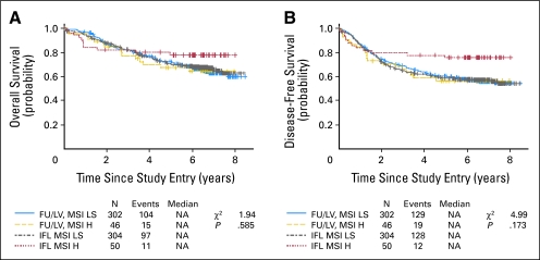
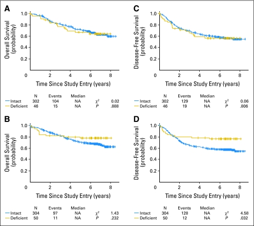
Tumor MMR status did not predict OS, either for analysis comparing MMR-D/MSI-H to MMR-I/MSI-L/S tumors within each treatment arm (combined prognostic and predictive analysis) or for a predictive analysis comparing treatment with FU/LV to treatment with IFL for each genomic category (Table 4; Fig 2). Patients treated with IFL whose tumors were MMR-D/MSI-H had better 5-year DFS than those whose tumors were MMR-I/MSI-L/S, with HR of 0.76 (95% CI, 0.64 to 0.88) versus 0.59 (95% CI, 0.53 to 0.64), respectively (P = .03). This relationship was not observed for patients treated with FU/LV (Table 5; Appendix Fig A1, online only). In a predictive analysis, DFS for patients with MMR-D/MSI-H tumors was compared among those receiving FU/LV and IFL. This showed a trend toward improved DFS for patients treated with IFL, with an HR of 0.76 (95% CI, 0.64 to 0.88) for IFL and 0.57 (95% CI, 0.42 to 0.71) for FU/LV (P = .07). Patients whose tumors were MMR-I/MSI-L/S did not show a similar trend (Table 5; Appendix Fig A2, online only). Finally, there was a trend toward longer DFS among patients with MMR-D tumors treated with IFL versus no difference in DFS by MMR status among patients treated with FU/LV (HR interaction, 0.51; likelihood ratio P = .117). The analyses reported were conducted for cases with genotype and IHC agreement (n = 702). Each analysis was also performed using genotype-only and IHC-only tumor characterizations. The results for both OS and DFS were essentially unchanged (data not shown).
Fig 2.
(A) Overall survival for mismatch repair (MMR) status by treatment groups (n = 702); (B) disease-free survival for MMR status by treatment groups (n = 702). FU, fluorouracil; LV, leucovorin; MSI LS, microsatellite instability low/stable; MSI H, microsatellite instability high; IFL, irinotecan, fluorouracil, and leucovorin; NA, not applicable.
Among patients with MMR-D/MSI-H tumors, a multivariable analysis was conducted to determine the relative significance of treatment arm (IFL; FU/LV) with each of the following prognostic variables: sex (male/female), age at study entry, extracellular mucin (≤ 50%; > 50%), tumor location (distal/proximal), differentiation (moderate/well; poor/undifferentiated), and nodal ratio. Treatment arm remained marginally associated with DFS in this subgroup in the presence of each of the following prognostic factors: sex (treatment arm P = .088), age (treatment arm P = .064), extracellular mucin (treatment arm P = .069), tumor location (treatment arm P = .077), tumor differentiation (treatment arm P = .087), and nodal ratio (treatment arm P = .039).
DISCUSSION
This large prospective study indicates that FU/LV alone may not be the optimal regimen for adjuvant treatment of stage III colon cancers that demonstrate MMR-D. However, although this study provides insight regarding the clinical behavior of MMR-D/MSI-H colon cancer, neither FU/LV alone nor IFL is the standard treatment regimen in this setting. Current data indicate that 6 months of postoperative FU, LV, and oxaliplatin (FOLFOX) achieves the best result after surgery for stage III colon cancer.21 Therefore, these results cannot currently be used to refine patient care.
Substantial data, largely from retrospective trials, indicate that patients with MMR-D/MSI-H colon cancers achieve improved survival when compared with those with MMR-I/MSI-L/S tumors.13,14 In retrospective reports, the effect is modest, with a 10% increase in DFS and no increase in OS for the MMR-D/MSI-H cases. A possible explanation involves the inclusion of FU in both treatment arms. Some studies suggest that FU adversely affects outcome for MSI-H tumors, and the preponderance of data indicate that FU is at best ineffective in this form of the disease.15–17 It is therefore possible that equalization of outcome for the MSI-H and non-MSI-H tumors occurred in our study because FU had a negative impact on the MSI-H tumors that was overcome by adding irinotecan. Because we see a trend (P = .07) but not a statistically significant association for the MMR-D/MSI-H subset, it is also possible that FU treatment negates an improved outcome in these patients, with no differential effect of irinotecan.
Several lines of evidence indicate that MSI-H tumors should respond to treatment with irinotecan. In both cultured cells22,23 and xenografted human CRCs,24 tumors with MSI-H were more sensitive to irinotecan than those with intact MMR. Small retrospective studies of rectal cancer25 and metastatic CRC26 found that MSI-H tumor status predicted favorable outcome after irinotecan therapy. The mechanism underlying increased sensitivity of MSI-H tumors to irinotecan is not well understood. Irinotecan inhibits the catalytic function of topoisomerase-I by stabilizing covalent complexes formed between DNA and this enzyme.27 This interaction impedes the DNA-relegation process, producing single-strand breaks that are converted into double-strand breaks after replication fork collision.28 Because double-strand DNA breaks are lethal if not repaired before mitosis, any process that inhibits the efficiency of DNA repair, including loss of MMR proteins, may potentiate tumor cell death. MSI-H tumors also demonstrate multiple defects in genes containing microsatellite repeats. These genes include those governing signal transduction (eg, transforming growth factor β RII29), apoptosis (eg, BAX,5 caspase 530), DNA repair (eg, hMSH6, MBD431), protein modification (eg, SEC63, OGT32), or transcriptional activation (eg, TCF433). It is possible that it is not the MMR defect itself, but rather loss of one or more of these genes, which causes increased irinotecan chemosensitivity.
The primary analysis from CALGB 89803 showed that addition of irinotecan to weekly bolus FU/LV did not improve DFS or OS in stage III disease, but did increase lethal and nonlethal toxicity. In the PETACC-3 trial, 2,333 patients with stage III colon cancer were randomly assigned to adjuvant treatment with biweekly infusional FU, either alone or with biweekly irinotecan.34 There was no significant difference in 3-year DFS, the prespecified primary end point (59.9% for FU/LV, 62.9% for FU plus irinotecan, P = .1). Although this infusional FU plus irinotecan combination did not show the unacceptable toxicity levels seen with the bolus IFL regimen of CALGB 89803, it failed to demonstrate a significant DFS or OS advantage for the combination.
This analysis, of a prospective secondary end point of CALGB 89803, shows that DFS may depend on the MMR status of the primary tumor. Among the subset of patients receiving IFL, those with MMR-D/MSI-H tumors had significantly longer DFS. A significant difference observed for the DFS end point but not the OS end point may be due to the smaller sample size for OS and the potential influence of subsequent therapy on OS. The DFS end point may also better reflect the most immediate impact of the protocol treatment. A strength of this study is that patients were treated on a controlled clinical trial and observed prospectively; a drawback is that the low prevalence of MMR-D/MSI-H tumors did not allow sufficient power to detect more than a trend in the treatment by MMR status interaction with the available sample size.
We conclude that MMR-D/MSI-H colon cancer may be a clinically distinct subset of stage III disease, with differential response to adjuvant chemotherapy. Differences in the treatment regimens tested and standard practice mean that this observation could not immediately be translated into clinical practice. It is therefore essential that we conduct new research to determine whether MMR determination can be used to select optimal chemotherapy. One study to consider is a randomized trial of patients with stage III MMR-D/MSI-H tumors, one arm of which eliminates FU/LV from the adjuvant regimen. Another issue is whether MMR status predicts a differential response to FOLFOX. In light of recent reports of increased chronic neurotoxicity with FOLFOX,35 an adjuvant trial of FOLFOX versus irinotecan and oxaliplatin for patients with stage III MMR-D/MSI-H tumors may also be indicated.
Acknowledgment
We thank Stephen N. Thibodeau, MD, for assistance in interpreting study results.
Appendix
The following institutions participated in this study: Baptist Cancer Institute Community Clinical Oncology Program, Memphis, TN (Lee S. Schwartzberg, MD, supported by Grant No. CA71323); Christiana Care Health Services Inc Community Clinical Oncology Program, Wilmington, DE (Stephen Grubbs, MD, supported by Grant No. CA45418); Dana-Farber Cancer Institute, Boston, MA (Eric P. Winer, MD, supported by Grant No. CA32291); Dartmouth Medical School–Norris Cotton Cancer Center, Lebanon, NH (Marc S. Ernstoff, MD, supported by Grant No. CA04326); Duke University Medical Center, Durham, NC (Jeffrey Crawford, MD, supported by Grant No. CA47577); Georgetown University Medical Center, Washington, DC (Minetta C. Liu, MD, supported by Grant No. CA77597); Cancer Centers of the Carolinas, Greenville, SC (Jeffrey K. Giguere, MD, supported by Grant No. CA291650; Long Island Jewish Medical Center, Lake Success, NY (Kanti R. Rai, MD, supported by Grant No. CA11028); Massachusetts General Hospital, Boston, MA (Jeffrey W. Clark, MD, supported by Grant No. CA12449); Memorial Sloan-Kettering Cancer Center, New York, NY (Clifford A. Hudis, MD, supported by Grant No. CA77651); Missouri Baptist Medical Center, St Louis, MO (Alan P. Lyss, MD); Mount Sinai Medical Center, Miami, FL (Rogerio C. Lilenbaum, MD, supported by Grant No. CA45564); Mount Sinai School of Medicine, New York, NY (Lewis R. Silverman, MD, supported by Grant No. CA04457); Nevada Cancer Research Foundation Community Clinical Oncology Program, Las Vegas, NV (John A. Ellerton, MD, supported by Grant No. CA35421); North Shore–Long Island Jewish Medical Center, Manhasset, NY (Daniel R. Budman, MD, supported by Grant No. CA35279); Rhode Island Hospital, Providence, RI (William Sikov, MD, supported by Grant No. CA08025); Roswell Park Cancer Institute, Buffalo, NY (Ellis Levine, MD, supported by Grant No. CA02599); Southeast Cancer Control Consortium Inc Community Clinical Oncology Program, Goldsboro, NC (James N. Atkins, MD, supported by Grant No. CA45808); State University of New York Upstate Medical University, Syracuse, NY (Stephen L. Graziano, MD, supported by Grant No. CA21060); The Ohio State University Medical Center, Columbus, OH (Clara D. Bloomfield, MD, supported by Grant No. CA77658); University of California at San Diego, San Diego, CA (Barbara A. Parker, MD, supported by Grant No. CA11789); University of California at San Francisco, San Francisco, CA (Alan P. Venook, MD, supported by Grant No. CA60138); University of Chicago, Chicago, IL (Gini Fleming, MD, supported by Grant No. CA41287); University of Illinois MBCCOP, Chicago, IL (Lawrence E. Feldman, MD, supported by Grant No. CA74811); University of Iowa, Iowa City, IA (Daniel A. Vaena, MD, supported by Grant No. CA47642); University of Maryland Greenebaum Cancer Center, Baltimore, MD (Martin Edelman, MD, supported by CA31983 University of Massachusetts Medical School, Worcester, MA (William V. Walsh, MD, supported by Grant No. CA37135); University of Minnesota, Minneapolis, MN (Bruce A. Peterson, MD, supported by Grant No. CA16450); University of Missouri/Ellis Fischel Cancer Center, Columbia, MO (Michael C Perry, MD, supported by Grant No. CA12046); University of Nebraska Medical Center, Omaha, NE (Anne Kessinger, MD, supported by Grant No. CA77298); University of North Carolina at Chapel Hill, Chapel Hill, NC (Thomas C. Shea, MD, supported by Grant No. CA47559); University of Tennessee Memphis, Memphis, TN (Harvey B. Niell, MD, supported by Grant No. CA47555); University of Vermont, Burlington, VT (Hyman B. Muss, MD, supported by Grant No. CA77406); Wake Forest University School of Medicine, Winston-Salem, NC (David D. Hurd, MD, supported by Grant No. CA03927); Walter Reed Army Medical Center, Washington, DC (Thomas Reid, MD, supported by Grant No. CA26806); Washington University School of Medicine, St Louis, MO (Nancy Bartlett, MD, supported by Grant No. CA77440); Weill Medical College of Cornell University, New York, NY (John Leonard, MD, supported by Grant No. CA07968).
Fig A1.
(A) Overall survival (OS) by mismatch repair (MMR) status among patients treated with fluorouracil/leucovorin (FU/LV; n = 348); (B) OS by MMR status among patients treated with irinotecan, FU, and LV (IFL; n = 354); (C) disease-free survival (DFS) by MMR status among patients treated with FU/LV (n = 348); (D) DFS by MMR status among patients treated with IFL (n = 354). NA, not applicable.
Fig A2.
(A) Overall survival (OS) by treatment among patients with mismatch repair deficient (MMR-D) tumors (n = 96); (B) disease-free survival (DFS) by treatment among patients with MMR-D tumors (n = 96); (C) OS by treatment among patients with mismatch repair intact (MMR-I) tumors (n = 606); (D) DFS by treatment among patients with MMR-I tumors (n = 606). NA, not applicable.
Footnotes
Supported in part by Grant No. CA31946 (to the Cancer and Leukemia Group B; Richard L. Schilsky, MD, Chair) and Grant No. CA33601 (to the Cancer and Leukemia Group B Statistical Center; Stephen George, PhD) from the National Cancer Institute.
The content of this article is solely the responsibility of the authors and does not necessarily represent the official views of the National Cancer Institute.
Authors' disclosures of potential conflicts of interest and author contributions are found at the end of this article.
Clinical Trials repository link available on JCO.org.
AUTHORS' DISCLOSURES OF POTENTIAL CONFLICTS OF INTEREST
Although all authors completed the disclosure declaration, the following author(s) indicated a financial or other interest that is relevant to the subject matter under consideration in this article. Certain relationships marked with a “U” are those for which no compensation was received; those relationships marked with a “C” were compensated. For a detailed description of the disclosure categories, or for more information about ASCO's conflict of interest policy, please refer to the Author Disclosure Declaration and the Disclosures of Potential Conflicts of Interest section in Information for Contributors.
Employment or Leadership Position: Mark Redston, Ameripath (C) Consultant or Advisory Role: Richard M. Goldberg, Pfizer Inc (C); Leonard B. Saltz, Pfizer (C), Genentech (C), Imclone (C), Merck (C) Stock Ownership: None Honoraria: Monica M. Bertagnolli, Pfizer Inc, Metamark, Inc; Richard M. Goldberg, Pfizer Inc Research Funding: Monica M. Bertagnolli, Pfizer Inc; Richard M. Goldberg, Pfizer Inc; Leonard B. Saltz, Pfizer Inc, Imclone, Bristol-Myers Squibb Co, Merck, Amgen Inc, Bayer Pharmaceuticals, Roche, Taiho Expert Testimony: Richard M. Goldberg, Pfizer Inc (C) Other Remuneration: None
AUTHOR CONTRIBUTIONS
Conception and design: Monica M. Bertagnolli, Donna Niedzwiecki, Robert J. Mayer, Richard M. Goldberg, Robert S. Warren
Administrative support: Monica M. Bertagnolli, Beatrice Damas, Richard M. Goldberg
Provision of study materials or patients: Scott D. Jewell, Richard M. Goldberg, Leonard B. Saltz
Collection and assembly of data: Donna Niedzwiecki, Hejin P. Hahn, Margaret Hall, Beatrice Damas, Robert S. Warren, Mark Redston
Data analysis and interpretation: Monica M. Bertagnolli, Donna Niedzwiecki, Carolyn C. Compton, Margaret Hall, Robert J. Mayer, Richard M. Goldberg, Leonard B. Saltz, Robert S. Warren, Mark Redston
Manuscript writing: Monica M. Bertagnolli, Donna Niedzwiecki, Margaret Hall, Scott D. Jewell, Leonard B. Saltz
Final approval of manuscript: Monica M. Bertagnolli, Donna Niedzwiecki, Margaret Hall, Robert J. Mayer, Richard M. Goldberg, Leonard B. Saltz, Robert S. Warren, Mark Redston
REFERENCES
- 1.Lengauer C, Kinzler KW, Vogelstein B. Genetic instability in colorectal cancers. Nature. 1997;386:623–627. doi: 10.1038/386623a0. [DOI] [PubMed] [Google Scholar]
- 2.Kuismanen SA, Holmberg MT, Salovaara R, et al. Genetic and epigenetic modification of MLH1 accounts for a major share of microsatellite-unstable colorectal cancers. Am J Pathol. 2000;156:1773–1779. doi: 10.1016/S0002-9440(10)65048-1. [DOI] [PMC free article] [PubMed] [Google Scholar]
- 3.Perucho M. Cancer of the microsatellite mutator phenotype. Biol Chem. 1996;377:675–684. [PubMed] [Google Scholar]
- 4.Parsons R, Myeroff LL, Liu B, et al. Microsatellite instability and mutations of the transforming growth factor beta type II receptor gene in colorectal cancer. Cancer Res. 1995;58:5548–5557. [PubMed] [Google Scholar]
- 5.Rampino N, Yamamoto H, Ionov Y, et al. Somatic frameshift mutations in the BAX gene in colon cancers of microsatellite mutator phenotype. Science. 1997;275:967–969. doi: 10.1126/science.275.5302.967. [DOI] [PubMed] [Google Scholar]
- 6.Cunningham MJ, Christensen ER, Tester DJ, et al. Hypermethylation of the hMLH1 promoter in colon cancer with microsatellite instability. Cancer Res. 1998;58:3455–3460. [PubMed] [Google Scholar]
- 7.Veigl ML, Kasturi L, Olechnoqicz J, et al. Biallelic inactivation of hMLH1 by epigenetic gene silencing, a novel mechanism causing human MSI cancers. Proc Natl Acad Sci U S A. 1998;95:8698–8702. doi: 10.1073/pnas.95.15.8698. [DOI] [PMC free article] [PubMed] [Google Scholar]
- 8.Shia J, Elleis NA, Klimstra DS. The utility of immunohistochemical detection of DNA mismatch repair gene proteins. Virchows Arch. 2004;445:431–441. doi: 10.1007/s00428-004-1090-5. [DOI] [PubMed] [Google Scholar]
- 9.Ionov Y, Peinado MA, Malkhosyan S, et al. Ubiquitous somatic mutations in simple repeated sequences reveal a new mechanism for colonic carcinogenesis. Nature. 1993;363:558–561. doi: 10.1038/363558a0. [DOI] [PubMed] [Google Scholar]
- 10.Jass JR, Do KA, Simms LA, et al. Morphology of sporadic colorectal cancer with DNA replication errors. Gut. 1998;42:673–679. doi: 10.1136/gut.42.5.673. [DOI] [PMC free article] [PubMed] [Google Scholar]
- 11.Thibodeau SN, Bren G, Schaid D. Microsatellite instability in cancer of the proximal colon. Science. 1993;260:816–819. doi: 10.1126/science.8484122. [DOI] [PubMed] [Google Scholar]
- 12.Lothe RA, Peltomaki P, Meling GI, et al. Genomic instability in colorectal cancer: Relationship to clinicopathological variables and family history. Cancer Res. 1993;53:5849–5852. [PubMed] [Google Scholar]
- 13.Gryfe R, Kim H, Hsieh ET, et al. Tumor microsatellite instability and clinical outcome in young patients with colorectal cancer. N Engl J Med. 2000;342:69–77. doi: 10.1056/NEJM200001133420201. [DOI] [PubMed] [Google Scholar]
- 14.Popat S, Hubner R, Houlston RS. Systematic review of microsatellite instability and colorectal cancer prognosis. J Clin Oncol. 2005;23:609–618. doi: 10.1200/JCO.2005.01.086. [DOI] [PubMed] [Google Scholar]
- 15.Ribic CM, Sargent DJ, Moore MJ, et al. Tumor microsatellite-instability status as a predictor of benefit from fluorouracil-based adjuvant chemotherapy for colon cancer. N Engl J Med. 2003;349:247–257. doi: 10.1056/NEJMoa022289. [DOI] [PMC free article] [PubMed] [Google Scholar]
- 16.Kim GP, Colangelo LH, Wieand HS, et al. Prognostic and predictive roles of high-degree microsatellite instability in colon cancer: A National Cancer Institute-National Surgical Adjuvant Breast and Bowel Project Collaborative Study. J Clin Oncol. 2007;25:767–772. doi: 10.1200/JCO.2006.05.8172. [DOI] [PubMed] [Google Scholar]
- 17.Barratt PL, Seymour MT, Stenning SP, et al. DNA markers predicting benefit from adjuvant fluorouracil in patients with colon cancer: A molecular study. Lancet. 2002;360:1381–1391. doi: 10.1016/s0140-6736(02)11402-4. [DOI] [PubMed] [Google Scholar]
- 18.Saltz L, Niedzwiecki D, Hollis D, et al. Irinotecan fluorouracil plus leucovorin is not superior to fluorouracil plus leucovorin alone as adjuvant treatment for stage III colon cancer: Results of CALGB 89803. J Clin Oncol. 2007;25:3456–3461. doi: 10.1200/JCO.2007.11.2144. [DOI] [PubMed] [Google Scholar]
- 19.Boland CR, Thibodeau SN, Hamilton SR, et al. The international workship on microsatellite instability and RER phenotypes in cancer detection and familial predisposition. Cancer Res. 1998;58:5248–5257. [PubMed] [Google Scholar]
- 20.Fleiss JL. Statistical Methods for Rate and Proportions. ed 2. New York, NY: John Wiley & Sons; 1981. [Google Scholar]
- 21.Wolpin BM, Mayeyerhardt JA, Mamon HJ, et al. Adjuvant treatment of colorectal cancer. CA Cancer J Clin. 2007;57:168–185. doi: 10.3322/canjclin.57.3.168. [DOI] [PubMed] [Google Scholar]
- 22.Magrini R, Bhonde MR, Hanski M-L, et al. Cellular effects of CPT-11 on colon carcinoma cells: Dependence on p53 and hMLH1 status. Int J Cancer. 2002;101:23–31. doi: 10.1002/ijc.10565. [DOI] [PubMed] [Google Scholar]
- 23.Jacob S, Aguado M, Fallik D, et al. The role of the DNA mismatch repair system in the cytotoxicity of the topoisomerase inhibitors camptothecin and etoposide to human colorectal cancer cells. Cancer Res. 2001;61:6555–6562. [PubMed] [Google Scholar]
- 24.Bras-Gonçalves RA, Rosty C, Soulie P, et al. Sensitivity of CPT-11 of xenografted human colorectal cancers as a function of microsatellite instability and p53 status. Br J Cancer. 2000;82:913–923. doi: 10.1054/bjoc.1999.1019. [DOI] [PMC free article] [PubMed] [Google Scholar]
- 25.Charara M, Edmonston TB, Burkholder S, et al. Microsatellite status and cell cycle associated markers in rectal cancer patients undergoing a combined regimen of 5-FU and CPT-11 chemotherapy and radiotherapy. Anticancer Res. 2004;24:3161–3167. [PubMed] [Google Scholar]
- 26.Fallik D, Vorrini F, Boige V, et al. Microsatellite instability is a predictive factor of the tumor response to irinotecan in patients with advanced colorectal cancer. Cancer Res. 2003;63:5738–5744. [PubMed] [Google Scholar]
- 27.Hsiang Y-H, Lihou MG, Liu LF. Arrest of replication forks by drug-stabilized topoisomerase I-DNA clevable complexes as a mechanism of cell killing by camptothecin. Cancer Res. 1989;49:5077–5082. [PubMed] [Google Scholar]
- 28.Vamvakas S, Vock EH, Lutz WK. On the role of DNA double-strand breaks in toxicity and carcinogenesis. Crit Rev Toxicol. 1997;27:155–174. doi: 10.3109/10408449709021617. [DOI] [PubMed] [Google Scholar]
- 29.Markowitz S, Wang J, Myeroff L, et al. Inactivation of the type II TGF-beta receptor in colon cancer cells with microsatellite instability. Science. 1995;268:1336–1338. doi: 10.1126/science.7761852. [DOI] [PubMed] [Google Scholar]
- 30.Schwartz S, Jr, Yamamoto H, Navarro M, et al. Frameshift mutations at mononucleotide repeats in caspase-5 and other target genes in endometrial and gastrointestinal cancer of the microsatellite mutator phenotype. Cancer Res. 1999;59:2995–3002. [PubMed] [Google Scholar]
- 31.Malkhosyan S, Rampino N, Yamamoto H, et al. Frameshift mutator mutations. Nature. 1996;382:499–500. doi: 10.1038/382499a0. [DOI] [PubMed] [Google Scholar]
- 32.Woerner SM, Gebert JF, Yuan YP, et al. Systematic identification of genes with coding microsatellites mutated in DNA mismatch repair-deficient cancer cells. Int J Cancer. 2001;93:12–19. doi: 10.1002/ijc.1299. [DOI] [PubMed] [Google Scholar]
- 33.Duval A, Gayet J, Zhou XP, et al. Frequent frameshift mutations of the TCF-4 gene in colorectal cancers with microsatellite instability. Cancer Res. 1999;59:4213–4215. [PubMed] [Google Scholar]
- 34.Van Cutsem E, Labianca R, Hossfeld D, et al. Randomized phase III trial comparing infused irinotecan/5-fluorouracil (5-FU)/folinic acid (IF) versus 5-FU/FA (F) in stage III colon cancer patients (pts) (PETACC 3). J Clin Oncol. 2005;23(suppl):3s. abstr LBA8. [Google Scholar]
- 35.Petrioli R, Pasucci A, Francini E, et al. Neurotoxicity of FOLFOX-4 as adjuvant treatment for patients with colon and gastric cancer: A randomized study of two different scheduled of oxaliplatin. Cancer Chemother Pharmacol. 2008;61:105–111. doi: 10.1007/s00280-007-0454-3. [DOI] [PubMed] [Google Scholar]


