Inhibition of proteasomal degradation by the Gly-Ala repeat of Epstein–Barr virus is influenced by the length of the repeat and the strength of the degradation signal (original) (raw)
Abstract
The Gly-Ala repeat (GAr) of the Epstein–Barr virus nuclear antigen-1 is a transferable element that inhibits in cis ubiquitin/proteasome-dependent proteolysis. We have investigated this inhibitory activity by using green fluorescent protein-based reporters that have been targeted for proteolysis by N end rule or ubiquitin-fusion degradation signals, resulting in various degrees of destabilization. Degradation of the green fluorescent protein substrates was inhibited on insertion of a 25-aa GAr, but strongly destabilized reporters were protected only partially. Protection could be enhanced by increasing the length of the repeat. However, reporters containing the Ub-R and ubiquitin-fusion degradation signals were degraded even in the presence of a 239-aa GAr. In accordance, insertion of a powerful degradation signal relieved the blockade of proteasomal degradation in Epstein–Barr virus nuclear antigen-1. Our findings suggest that the turnover of natural substrates may be finely tuned by GAr-like sequences that counteract targeting signals for proteasomal destruction.
Ubiquitin-proteasome-dependent proteolysis is the main source of peptides that are presented at the cell surface in association with MHC class I molecules (reviewed in ref. 1). Endogenously expressed proteins of cellular or foreign origin are marked for degradation by covalent linkage of multiple ubiquitin molecules that serve as a recognition signal for a multicatalytic complex, the proteasome, that progressively degrades the substrate into small peptides (reviewed in ref. 2). Various signals predisposing proteins for ubiquitination and degradation have been identified such as the destruction box, the PEST sequence, and the N end rule and ubiquitin-fusion degradation (UFD) signals (3–6). Peptides derived from viral proteins loaded onto MHC class I molecules are recognized by specific cytotoxic T lymphocytes and trigger elimination of the infected cell. The degradation and presentation of viral proteins are obvious targets for viruses in their effort to counteract the host's immune response. Numerous viral strategies have been identified that frustrate and abrogate these processes (7–10). Hence, it is not surprising that the first and hitherto only identified signal that overrides degradation signals and thereby protects proteins from proteasomal processing is from viral origin.
The Epstein–Barr virus (EBV) nuclear antigen 1 (EBNA1) is one of nine viral proteins expressed in latently infected EBV-transformed immunoblasts (7). EBNA1 is indispensable for the virus, because it safeguards the maintenance of the viral episomes in proliferating infected cells and is also the only viral protein that is regularly expressed in all EBV-associated malignancies. Cytotoxic T lymphocytes specific for antigenic peptides derived from EBNA1 have been demonstrated at relatively high frequency during primary EBV infection and in healthy virus carriers (11), suggesting that EBNA1 could provide an important rejection target. However, these cytotoxic T lymphocytes do not recognize EBV-infected cells or cells infected with EBNA1 encoding recombinant vaccinia or adenoviruses, and require sensitization by exogenous antigen or synthetic peptides. We have previously shown that the failure to recognize endogenously expressed EBNA1 can be attributed to the presence of a long Gly-Ala repeat (GAr) that prevents antigen presentation in vivo (12) and protects EBNA1 from ubiquitin/proteasome-dependent proteolysis in vitro (13). Thus, the GAr may contribute to the immune evasion of EBV-infected cells by excluding an important potential target from antigen presentation.
The mechanism by which the GAr inhibits the ubiquitin/proteasomal pathway is still poorly understood. An interesting feature of the GAr is the capacity to act as transferable element, a property shared with many known protein degradation signals. Insertion of the GAr abolished the presentation of epitopes from another EBV protein, EBNA4 (12), and prevented tumor necrosis factor-α-induced degradation of the NF-κB inhibitor IκBα (14), confirming that the phenomenon is not restricted to viral proteins. GAr-containing proteasomal substrates were ubiquitinated efficiently, indicating that the GAr acts downstream of this proteasome-targeting step. Synthetic Gly-Ala polypeptides have no stable conformation in solution, and the presence of the repeat did not influence the folding and thermal stability of IκBα chimeras (15). This result, together with the finding that an 8-aa GAr is sufficient to prevent the degradation of IκBα, suggests that the repeat may interact with an as-yet unidentified component of the degradation pathway.
In this study, we have exploited the transferable property of the GAr to investigate whether similar inhibitory effects can be achieved depending on the nature and strength of the signal that targets the substrate for ubiquitin/proteasome-dependent proteolysis. We demonstrate that, although the GAr acts in a length-dependent manner on different types of degradation signal, the effect can be overcome by strong signals that induce the rapid clearance of the ubiquitinated substrates.
Materials and Methods
Reagents and Antibodies
The proteasome inhibitors carboxybenzyl-leucyl-leucyl-leucinal (MG132) and carboxybenzyl-leucyl-leucyl-leucine vinyl sulfone (Z-L3-VS) were obtained from Peptides Institute (Herrsching, Germany) and donated by H. L. Ploegh (Harvard Medical School, Cambridge, MA), respectively. A green fluorescent protein (GFP)-specific rabbit serum was purchased from Molecular Probes. A polyclonal rabbit serum specific for the GAr (peptide p107) was produced as described (16).
Plasmid Construction.
Construction of the different Ub-X-GFP reporters with a red-shifted variant of GFP (EGFP-N1 vector, CLONTECH) has been reported (17), and 25-aa GAr fragments were generated by annealing partially overlapping complementary oligonucleotides that were then filled by Klenow large fragment (Amersham Pharmacia). The fragment was digested with _Ssp_BI and cloned in the unique _Ssp_BI restriction site positioned at the 3′ end of the GFP ORF. The full-length GAr encoding a 239-aa polypeptide was obtained by PCR amplification of the cloned repeat with the sense primer 5′-AGCTGTACATCGGATCCACCCACGGTGGAACAG-3′ (_Ssp_BI restriction site, underlined) and the antisense primer 5′-TATGCGGCCGCTTATGCAGAATTCCTGCAGCCCCGGCCT-3′ (_Not_I restriction site, underlined; stop codon, in bold). The EBNA1 coding sequence of the prototype B95.8 EBV strain was PCR amplified with the sense primer 5′-GCGAAGCTTGGATCCAATGCTTGACGAGGGGCCAGGTA-3′ (_Hin_dIII, underlined; _Bam_HI site double underlined; start codon EBNA1 bold) and antisense primer 5′-CGTCCATGGTTATCACCCCCTCTT-3′ (_Nco_I site underlined) that introduced a 5′ _Bam_HI cleavage site. The _Hin_dIII and _Nco_I sites were used to replace the 5′ end of the EBNA1 ORF with the modified sequence. The introduced _Bam_HI site allowed exchanging the GFP and EBNA1 ORFs in the Ub-X-GFP plasmid. The 25-aa repeat and each of the cloned PCR fragments were checked by sequencing except for the full-length GAr where the linker region was sequenced and presence of the repeat was confirmed by Western blotting with the anti p107 serum.
Expression Analysis.
The human cervical carcinoma line HeLa was cultured in Iscove's modified Eagle's medium supplemented with 10% (vol/vol) FCS (Life Technologies, Grand Island, NY). Transient transfections were performed with Lipofectamine (Life Technologies), and expression was detected after 48 h by Western blotting, flow cytometry, or fluorescence microscopy. For Western blot analysis, lysates of 105 cells were fractionated by SDS/10% PAGE and transferred to Protan BA85 nitrocellulose filters (Schleicher & Schuell). After blocking for 1 h in PBS supplemented with 5% (wt/wt) skim milk and 0.1% Tween-20, the filters were incubated for 1 h with the primary antibody and for 1 h with peroxidase-conjugated goat anti-rabbit serum. Complexes were visualized by enhanced chemiluminescence (ECL, Amersham Pharmacia). Flow cytometry was performed with a FACSort flow cytometer (Becton Dickinson), and data were analyzed with cellquest software. For fluorescence microscopy, the cells were grown and transfected on coverslips. Cells expressing the GFP constructs were fixed with 4% (wt/wt) paraformaldehyde in PBS. Ub-X-EBNA1 constructs were visualized by anticomplement immunofluorescence staining (18) with a healthy human serum with high antibody titers to all EBNAs. The samples were examined with a LEITZ-BMRB fluorescence microscope (Leica, Heidelberg, Germany) with a bandpass FITC filter setting. Images were captured with a Hamamatsu 4800 cooled charge-coupled device camera (Hamamatsu, Osaka, Japan) and processed with adobe photoshop software.
Results
GAr Length-Dependent Stabilization of Ub-X-GFP Reporters Containing Different Types of Degradation Signals.
N end rule and UFD-targeted GFP reporters were used to investigate the capacity of the GAr to overcome different types of targeting signals for ubiquitin-proteasome dependent proteolysis. N end rule and UFD signals determine the rate of polyubiquitination and thereby the sensitivity of a protein to proteasomal degradation (3, 6). We have previously shown that the proteasome-dependent turnover of GFPs containing N end rule or UFD signals varies depending on the identity of the amino acid exposed after cleavage of the N-terminal ubiquitin by ubiquitin hydrolases and on the rate of this cleavage (17). Thus, although the control Ub-M-GFP is not targeted for degradation, N end rule substrate Ub-R-GFP and UFD substrate UbG76V-GFP are highly unstable proteins that are barely detected in transfected cells unless accumulation is induced by treatment with proteasome inhibitors. The UFD substrate Ub-P-GFP is expressed at intermediate levels because of accumulation of the stable P-GFP after inefficient cleavage of the N-terminal ubiquitin.
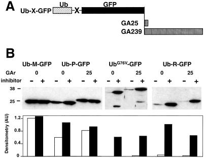
Ub-X-GFP/GA25 chimeras were constructed by inserting a 25-aa GAr (15) at the C terminus of the reporters (Fig. 1A), and their turnover was investigated in transiently transfected cells by Western blot analysis. As previously shown, the steady-state expression of Ub-P-GFP, UbG76V-GFP, and Ub-R-GFP was reduced significantly compared with the stable control Ub-M-GFP, whereas a strong increase was induced by treatment with the proteasome inhibitors Z-L3-VS (Fig. 1B) and MG132 (not shown). Insertion of the GAr resulted in stabilization of the weakly destabilized Ub-P-GFP. In repeated experiments, the steady-state expression of Ub-P-GFP/GA25 was higher than the control Ub-P-GFP, and no further increase could be induced by treatment with Z-L3-VS. In contrast, GA25 did not seem to affect the turnover of the strongly destabilized UbG76V-GFP and Ub-R-GFP. The Ub-R-GFP/GA25 and UbG76V-GFP/GA25 polypeptides were hardly detected in transfected cells, whereas significant accumulation was induced by treatment with Z-L3-VS, confirming that these chimeras are still targeted for proteasomal degradation.
Figure 1.
Effect of GA25 on the degradation of the Ub-X-GFP reporters. (A) GAr polypeptides of 25 and 239 amino acids were inserted at the C terminus of GFP reporters that had been targeted for ubiquitin-proteasome-dependent degradation by insertion of N end rule or UFD degradation signals. (B Upper) Lysates of HeLa cells transfected with the Ub-M-GFP, Ub-P-GFP, UbG76V-GFP, and Ub-R-GFP plasmids and their GA25-containing counterparts were analyzed in Western blots with a polyclonal anti-GFP antibody. The transfected cells were preincubated for 10 h without (−) or with (+) the proteasome inhibitor Z-L3-VS (10 μM). Molecular mass markers are indicated on the left. (B Lower) Densitometric analysis of the Western blot is presented (AU, arbitrary units).
We then asked whether the strongly destabilized UbG76V-GFP and Ub-R-GFP could be protected by the GAr of a natural EBV isolate. To this end, chimeric reporters were constructed by insertion of the 239-aa repeat from the B95.8 EBV strain (Fig. 1A). Western blot analysis revealed a dramatic increase in the steady-state expression of both reporters compared with their GAr-less counterparts, with levels of expression comparable or even higher than those detected with the control unmodified GFP (Fig. 2). Blockade of proteasomal degradation by treatment with Z-L3-VS did not result in further accumulation.
Figure 2.
Effect of GA239 on the strongly destabilized UbG76V-GFP and Ub-R-GFP. Lysates of HeLa cells expressing the UbG76V-GFP and Ub-R-GFP reporters and their GA239-containing counterparts were analyzed in Western blots with a polyclonal anti-GFP antibody. The transfected cells were incubated for 10 h without (−) or with (+) 10 μM Z-L3-VS. The GFP products are indicated.
Quantification of GAr Activity in Living Cells.
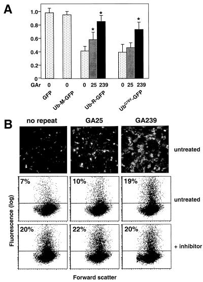
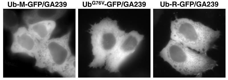
The GFP reporters allow accurate quantification of expression levels in living cells and analysis of protein localization (17). To this end, the expression of UbG76V-GFP and Ub-R-GFP chimeras containing GAr domains of different length was investigated in untreated and proteasome-inhibitor-treated cells by flow cytometry and fluorescence microscopy. Reproducible differences in the percentage of fluorescent cells were observed in cells transfected with the control GFP, the stable Ub-M-GFP, and the destabilized UbG76V-GFP and Ub-R-GFP. These differences were abolished by treatment with proteasome inhibitors (not shown and Fig. 3B), confirming that, in the absence of the inhibitors, the fluorescence is below detection levels in the majority of cells expressing the destabilized reporters. The ratio between the percentage of fluorescent cells in the absence and presence of proteasome inhibitor was therefore used as a measure of proteasomal degradation. This ratio was close to 1 in cells expressing the unmodified GFP and Ub-M-GFP, confirming that these proteins are not targeted for proteasomal degradation (Fig. 3A), and ≈0.4 in transfectants expressing UbG76V-GFP and Ub-R-GFP. Insertion of the GA25 resulted in a small but significant stabilization of Ub-R-GFP (paired t test; P < 0.05; Fig. 3A), whereas the effect was below the limits of significance in UbG76V-GFP. A substantial stabilization of both reporters was achieved by insertion of GA239, but the protection was not complete, suggesting that these full-length GAr-containing substrates are still degraded by the proteasome (Fig. 3A). The length dependence of the effect is clearly illustrated by the flow cytometry data and low-magnification micrographs of cells transfected with Ub-R-GFP, Ub-R-GFP/GA25, and Ub-R-GFP/GA239 (Fig. 3B). Inspection of the micrographs at higher magnification demonstrated that, although GA25-containing chimeras are homogeneously distributed throughout the cells (not shown), chimeras containing GA239 were localized in the cytoplasm, as expected for large proteins lacking a nuclear localization signal (Fig. 4). More importantly, neither the stable nor the destabilized Ub-X-GFP chimeras gave a punctate cytoplasmic fluorescence. Thus, the stabilizing effect of the highly hydrophobic GAr domain cannot be attributed to the formation of large aggregates that could render the reporters inaccessible to the proteasome.
Figure 3.
Fluorimetric quantification of the inhibitory activity of the GAr. (A) Flow cytometric analysis of HeLa cells transfected with GFP-, Ub-M-GFP-, UbG76V-GFP-, and Ub-R-GFP-expressing plasmids and their GA25 or GA239 counterparts. Data are expressed as the ratio between the number of fluorescent cells in untreated samples and samples treated with 10 μM Z-L3-VS. Values shown are means ± SD from three experiments. *, Significantly different from the corresponding construct without GAr (paired t test; P < 0.05). (B) Representative low-magnification micrographs (Top) and flow cytometric analyses (Middle and Bottom) of HeLa cells expressing Ub-R-GFP, Ub-R-GFP/GA25, and Ub-R-GFP/GA239. The cells were incubated for 10 h without (Middle) and with 10 μM Z-L3-VS (Bottom). The percentages of positive cells are indicated.
Figure 4.
Cell distribution of GA239-harboring Ub-X-GFPs. Fluorescence micrographs of HeLa cells expressing the Ub-M-GFP/GA239, UbG76V-GFP/GA239, and Ub-R-GFP/GA239 chimeras. Magnification: ×200.
A Strong Degradation Signal Targets EBNA1 for Proteasomal Degradation.
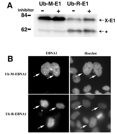
The observation that the full-length GAr can only partially override the strong UFD and N end rule degradation signals suggests that EBNA1 itself may be converted into a substrate of the proteasome. To test this possibility, EBNA1 was targeted for ubiquitin/proteasome-dependent proteolysis by insertion of the N end rule signal Ub-R- and, as a control, Ub-M-. Western blot analysis of lysates from HeLa cells expressing these Ub-X-EBNA1 with a GAr specific antiserum demonstrated predominant polypeptides of size corresponding to X-EBNA1, indicating that the chimeras are cleaved efficiently by ubiquitin hydrolases (Fig. 5A). The steady-state expression of Ub-R-EBNA1 was significantly lower compared with that of Ub-M-EBNA1. Moreover, treatment with Z-L3-VS or MG132 (not shown) resulted in accumulation of Ub-R-EBNA1, whereas the expression of Ub-M-EBNA1 remained unchanged (Fig. 5A). Notably, an additional polypeptide of approximately 60 kDa was detected by the GAr-specific serum in cells expressing the Ub-R-EBNA1. This polypeptide was not accumulated in cells treated with proteasome inhibitors, suggesting that it may be derived from a distinct cleavage event. The expression of Ub-M-EBNA1 and Ub-R-EBNA1 was restricted to the nucleus (Fig. 5B), excluding the possibility that proteasomal degradation may be induced by redistribution of the EBNA1 expressed as a ubiquitin fusion protein.
Figure 5.
A strong N end rule signal targets EBNA1 for proteasomal degradation. (A) HeLa cells transfected with the Ub-M-EBNA1 or Ub-R-EBNA1 plasmids were preincubated for 10 h without (−) or with (+) 10 μM Z-L3-VS and then analyzed in Western blots with a GAr-specific antibody. Bands corresponding in size to the cleaved X-EBNA1 are indicated. The asterisk indicates an additional GAr-containing polypeptide that is mainly detected in Ub-R-EBNA1-expressing cells. Molecular mass markers are indicated on the left. (B) Anticomplement immunofluorescence (Left) and Hoechst (Right) staining of HeLa cells expressing Ub-M-EBNA1 and Ub-R-EBNA1. Magnification: ×200.
Discussion
In the present study, we confirm the _cis_-inhibitory activity of the EBV GAr on ubiquitin/proteasome-dependent proteolysis by demonstrating that the degradation of N end rule and UFD substrates can be blocked by the viral repeat. The degradation pathways followed by N end rule and UFD substrates deviate from each other at each identified step. These substrates are ubiquitinated by different ubiquitin conjugases and ligases (3, 6, 19), rely differently on chaperones once ubiquitinated (3), and interact differently with one of the subunits of the 19S regulator (20). Thus, the stabilization of GFPs targeted by structurally similar but functionally different degradation signals is a clear illustration of the broad applicability of the GAr as a _cis_-acting inhibitor of proteasomal degradation.
Quantification of the GFP reporters allowed a more detailed analysis of the effect of the GAr. We have found that substrates that are targeted efficiently for proteasomal degradation are rescued only partially by the repeat. Although increasing the length of the repeat from 25 to 239 amino acids could enhance the protective effect, GA239-containing reporters were still degraded by the proteasome. To our knowledge, ours is the first report of proteasomal degradation of GAr-containing proteins, which demonstrates that the proteasome is capable of attacking these substrates. Nevertheless, the GAr seems to exert a fine tuning effect on the turnover of proteasomal substrates depending on the strength of their degradation signal. A clear illustration of this phenomenon is provided by the Ub-P-GFP and UbG76V-GFP reporters, which are targeted for degradation with different efficiency by the same type of UFD signal, caused by production of the stable P-GFP by slow cleavage of ubiquitin in Ub-P-GFP (3). Insertion of GA25 in Ub-P-GFP resulted in full protection, whereas a repeat of the same length had no measurable effect on the strongly destabilized UbG76V-GFP substrate. Furthermore, although the influence of GA25 on UFD-harboring substrates seemed to be minimal, a small but significant protection was accomplished in the strongly destabilized N end rule substrate Ub-R-GFP. It is tempting to speculate that such a sophisticated regulatory mechanism could have a counterpart in natural proteasome substrates where similar sequences may delay degradation. Indeed, repeated sequences that resemble the GAr are found in many cellular proteins, including transcription factors and cell-cycle regulators (A. Sharipo and M.G.M., unpublished observation).
We have previously shown that an 8-aa GAr was sufficient to block the degradation of IκBα (14), independent of its localization and without affecting the folding properties of the substrate (15), suggesting that the GAr may function as a recognition signal. Our present observation that much longer repeats are required for stabilization of certain substrates can be accommodated in a model in which multiple sequentially positioned recognition signals may tighten the interaction between the GAr and a putative binding partner. An analogous situation is observed in the targeting of ubiquitinated substrates to the proteasome. Here, a large number of recognition signals, provided by the polyubiquitin tree, are required to establish an interaction sufficiently strong to initiate progressive degradation (21). For the GAr, it seems that the strength of the degradation signals plays a pivotal role in determining the number of repeats required for protection. In addition, the type or position of the degradation signal within the target protein may also be important, because IκBα, which is fully protected by the GA8, is degraded with a very short half-life after signal-dependent targeting.
In line with the residual degradation of the Ub-R-GFP and UbG76V-GFP reporters containing the full-length GAr, we have found that EBNA1 itself can be converted into a degradable proteasome substrate once provided with a strong degradation signal. It should be stressed that the endogenous signal present in EBNA1 is sufficient for proteasomal targeting on removal of the GAr (refs. 12 and 13 and A. Sharipo and M.G.M., unpublished data). EBNA1 homologues from the primate rhesus and baboon herpesviruses Papio and Baboon contain GAr-like sequences; however, unlike EBNA1, they are still subject to proteasomal processing, because the repeats do not abrogate the presentation of MHC class I restricted epitopes from these proteins (22). However, the degradation of these molecules has not been tested in the natural host species, and adaptive changes of the putative partners involved in the protective effects cannot be excluded.
Collectively, these findings demonstrate that the GAr and GAr-like sequences do not have a digital effect, changing the fate of the protein from degradation to protection. It seems more likely that the net effect of the repeats will be determined by several parameters such as their length and amino acid composition versus the strength and type of the counteracting degradation signal. We have shown that an important effect of the repeats is the prolongation of protein half-life (13, 14), which is likely to have major consequences on protein expression and gene transcription in different types of virus-infected cells. Although this prolongation may be the primary effect of the repeat in the context of the virus, an important complementary function of the EBV GAr may be to abrogate the presentation of the endogenous EBNA1 (12, 23). This function would also justify the presence of variable but always relatively long GAr in different EBV isolates, because only sufficiently long GAr would allow a complete obstruction of antigen processing.
Acknowledgments
This work was supported by grants awarded by the Swedish Cancer Society, the Swedish Foundation of Strategy Research, and the Petrus and Augusta Hedlund Foundation, Stockholm, Sweden. N.P.D. and S.H. are supported by fellowships awarded by the European Commission Training and Mobility Program on “The central role of the ubiquitin proteasome system in regulatory processes involved in immunological, inflammatory, endocrinological, and malignant disorders” (contract no. ERBFMRXCT960026).
Abbreviations
GAr
Gly-Ala repeat
EBV
Epstein–Barr virus
EBNA
EBV nuclear antigen
GFP
green fluorescent protein
UFD
ubiquitin-fusion degradation
Footnotes
Article published online before print: Proc. Natl. Acad. Sci. USA, 10.1073/pnas.140217397.
Article and publication date are at www.pnas.org/cgi/doi/10.1073/pnas.140217397
References
- 1.Rock K L, Goldberg A L. Annu Rev Immunol. 1999;17:739–779. doi: 10.1146/annurev.immunol.17.1.739. [DOI] [PubMed] [Google Scholar]
- 2.Hershko A, Ciechanover A. Annu Rev Biochem. 1998;67:425–479. doi: 10.1146/annurev.biochem.67.1.425. [DOI] [PubMed] [Google Scholar]
- 3.Johnson E S, Ma P C, Ota I M, Varshavsky A. J Biol Chem. 1995;270:17442–17456. doi: 10.1074/jbc.270.29.17442. [DOI] [PubMed] [Google Scholar]
- 4.Hershko A. Curr Opin Cell Biol. 1997;9:788–799. doi: 10.1016/s0955-0674(97)80079-8. [DOI] [PubMed] [Google Scholar]
- 5.Rechsteiner M, Rogers S W. Trends Biochem Sci. 1996;21:267–271. [PubMed] [Google Scholar]
- 6.Varshavsky A. Proc Natl Acad Sci USA. 1996;93:12142–12149. doi: 10.1073/pnas.93.22.12142. [DOI] [PMC free article] [PubMed] [Google Scholar]
- 7.Masucci M G, Ernberg I. Trends Microbiol. 1994;2:125–130. doi: 10.1016/0966-842x(94)90599-1. [DOI] [PubMed] [Google Scholar]
- 8.Ploegh H L. Science. 1998;280:248–253. doi: 10.1126/science.280.5361.248. [DOI] [PubMed] [Google Scholar]
- 9.Hill A, Jugovic P, York I, Russ G, Bennink J, Yewdell J, Ploegh H, Johnson D. Nature (London) 1995;375:411–415. doi: 10.1038/375411a0. [DOI] [PubMed] [Google Scholar]
- 10.Collins K L, Chen B K, Kalams S A, Walker B D, Baltimore D. Nature (London) 1998;391:397–401. doi: 10.1038/34929. [DOI] [PubMed] [Google Scholar]
- 11.Blake N, Lee S, Redchenko I, Thomas W, Steven N, Leese A, Steigerwald-Mullen P, Kurilla M G, Frappier L, Rickinson A. Immunity. 1997;7:791–802. doi: 10.1016/s1074-7613(00)80397-0. [DOI] [PubMed] [Google Scholar]
- 12.Levitskaya J, Coram M, Levitsky V, Imreh S, Steigerwald-Mullen P M, Klein G, Kurilla M G, Masucci M G. Nature (London) 1995;375:685–688. doi: 10.1038/375685a0. [DOI] [PubMed] [Google Scholar]
- 13.Levitskaya J, Sharipo A, Leonchiks A, Ciechanover A, Masucci M G. Proc Natl Acad Sci USA. 1997;94:12616–12621. doi: 10.1073/pnas.94.23.12616. [DOI] [PMC free article] [PubMed] [Google Scholar]
- 14.Sharipo A, Imreh M, Leonchiks A, Imreh S, Masucci M G. Nat Med. 1998;4:939–944. doi: 10.1038/nm0898-939. [DOI] [PubMed] [Google Scholar]
- 15.Leonchiks A, Liepinsh E, Barishev M, Sharipo A, Masucci M G, Otting G. FEBS Lett. 1998;440:365–369. doi: 10.1016/s0014-5793(98)01488-4. [DOI] [PubMed] [Google Scholar]
- 16.Dillner J, Sternas L, Kallin B, Alexander H, Ehlin-Henriksson B, Jornvall H, Klein G, Lerner R. Proc Natl Acad Sci USA. 1984;81:4652–4656. doi: 10.1073/pnas.81.15.4652. [DOI] [PMC free article] [PubMed] [Google Scholar]
- 17.Dantuma N P, Lindsten K, Glas R, Jellne M, Masucci M G. Nat Biotechnol. 2000;18:538–543. doi: 10.1038/75406. [DOI] [PubMed] [Google Scholar]
- 18.Reedman B M, Klein G. Int J Cancer. 1973;11:499–520. doi: 10.1002/ijc.2910110302. [DOI] [PubMed] [Google Scholar]
- 19.Koegl M, Hoppe T, Schlenker S, Ulrich H D, Mayer T U, Jentsch S. Cell. 1999;96:635–644. doi: 10.1016/s0092-8674(00)80574-7. [DOI] [PubMed] [Google Scholar]
- 20.van Nocker S, Sadis S, Rubin D M, Glickman M, Fu H, Coux O, Wefes I, Finley D, Vierstra R D. Mol Cell Biol. 1996;16:6020–6028. doi: 10.1128/mcb.16.11.6020. [DOI] [PMC free article] [PubMed] [Google Scholar]
- 21.Thrower J S, Hoffman L, Rechsteiner M, Pickart C M. EMBO J. 2000;19:94–102. doi: 10.1093/emboj/19.1.94. [DOI] [PMC free article] [PubMed] [Google Scholar]
- 22.Blake N W, Moghaddam A, Rao P, Kaur A, Glickman R, Cho Y G, Marchini A, Haigh T, Johnson R P, Rickinson A B, et al. J Virol. 1999;73:7381–7389. doi: 10.1128/jvi.73.9.7381-7389.1999. [DOI] [PMC free article] [PubMed] [Google Scholar]
- 23.Mukherjee S, Trivedi P, Dorfman D M, Klein G, Townsend A. J Exp Med. 1998;187:445–450. doi: 10.1084/jem.187.3.445. [DOI] [PMC free article] [PubMed] [Google Scholar]


