Ectopic expression of B-lymphoid kinase in cutaneous T-cell lymphoma (original) (raw)
Abstract
B-lymphoid kinase (Blk) is exclusively expressed in B cells and thymocytes. Interestingly, transgenic expression of a constitutively active form of Blk in the T-cell lineage of mice results in the development of T-lymphoid lymphomas. Here, we demonstrate nuclear factor–kappa B (NF-κB)–mediated ectopic expression of Blk in malignant T-cell lines established from patients with cutaneous T-cell lymphoma (CTCL). Importantly, Blk is also expressed in situ in lesional tissue specimens from 26 of 31 patients with CTCL. Already in early disease the majority of epidermotropic T cells express Blk, whereas Blk expression is not observed in patients with benign inflammatory skin disorders. In a longitudinal study of an additional 24 patients biopsied for suspected CTCL, Blk expression significantly correlated with a subsequently confirmed diagnosis of CTCL. Blk is constitutively tyrosine phosphorylated in malignant CTCL cell lines and spontaneously active in kinase assays. Furthermore, targeting Blk activity and expression by Src kinase inhibitors and small interfering RNA (siRNA) inhibit the proliferation of the malignant T cells. In conclusion, this is the first report of Blk expression in CTCL, thereby providing new clues to the pathogenesis of the disease.
Introduction
Cutaneous T-cell lymphomas (CTCL) are the most frequent primary lymphomas of the skin, with mycosis fungoides (MF) being the most prevalent clinical form.1 In early disease stages, which can last several years, MF presents as flat erythematous skin patches resembling inflammatory diseases such as allergic contact dermatitis, eczema, or psoriasis. In later stages, MF lesions gradually form plaques and overt tumors and may disseminate to lymph nodes and internal organs. The early skin lesions contain numerous inflammatory cells, including a large quantity of T cells with a normal phenotype as well as a small population of T cells with abnormal morphology and a malignant phenotype. T cells with a malignant phenotype are characterized by epidermotropism and are preferentially present in the upper parts of the skin, whereas T cells with a normal phenotype primarily are detected in the lower portions of the dermis. The epidermal T cells are sometimes found in patterns of Pautrier microabscesses, which are collections of T cells adherent to dendritic processes of Langerhans cells. During disease development, the epidermotropism is gradually lost concomitant with an increase in malignant, and a decrease in nonmalignant, infiltrating T cells. The etiology of CTCL remains poorly understood, and occupational exposures, infectious agents, and genetic mutations have been proposed as etiological factors, but no evidence of causation has been provided.2–6 However, already in early disease stages, the transcription factor nuclear factor kappa B (NF-κB) has been shown to be constitutively active in the malignant T cells of patients with CTCL where it promotes proliferation and cell survival.7–9 The malignant T cells also show aberrant hyperactivation of the Janus kinase 3 (Jak3)/signal transducer and activator of transcription 3 (Stat3) pathway, which protects them from apoptosis and is a marker of resistance to therapy.10–13 It has been hypothesized that the aberrant activation of NF-κB and the Jak3/Stat3 pathway are key events in the development of CTCL.7–10
Early diagnosis of CTCL has important consequences concerning therapeutic options and determination of prognosis.14 Currently, it is primarily based on clinical observations and histologic examinations of cutaneous biopsies as well as additional laboratory tests such as analysis of T-cell receptor (TCR) clonality by polymerase chain reaction (PCR). Unfortunately, early diagnosis of CTCL has proven difficult because of the great clinical, pathologic, and histologic resemblance to benign inflammatory skin diseases and because inflammatory skin disorders can be associated with clonal TCR rearrangements.15–18
In humans, the Src family kinases (SFKs) of nonreceptor protein tyrosine kinases classically consists of 8 members: c-Src, Fyn, Lck, c-Yes, Fgr, Hck, Lyn, and B-lymphoid kinase (Blk).19 Blk is exclusively expressed in B cells and thymocytes but not in mature T cells.20–22 Besides a role of Blk in B-cell receptor (BCR) signaling and B-cell development, the cellular functions of Blk are poorly defined.23–26 Interestingly, transgenic mice expressing a constitutively active form of Blk in the T-cell lineage develop clonal T-lymphoid lymphomas.27 Furthermore, expression of constitutively active Blk in the murine B-cell linage results in increased proliferation of progenitor B cells and, eventually, development of B-lymphoid lymphomas.25,27 The oncogenic capacity of Blk is kinase dependent because lymphoid tumors do not develop in mice expressing a catalytically inactive form of Blk.27 However, so far, evidence that Blk is involved in the pathogenesis of human cancer has not been provided. Here, we report on an ectopic expression of Blk in the majority of patients with CTCL and provide evidence that Blk functions as an oncogene enhancing proliferation of the malignant T cells.
Methods
Antibodies and reagents
Antibodies against Blk (C-20), Erk1/2, Fgr, Lyn, NF-κB p65, and Cdk2 were from Santa Cruz Biotechnology (Santa Cruz, CA). Antibodies against Stat3, NF-κB p105/p50, IκB-α, Csk, and Fyn were from Cell Signaling Technology (Beverly, MA) and the phosphotyrosine monoclonal antibody (mAb; AG410) from Upstate Biotechnology (Charlottesville, VA). The phospho-Stat3 (Y705) mAb was from nanoTools (Denzlingen, Germany), and the Lck mAb was from Becton Dickinson (Franklin Lakes, NJ). The goat anti–human IgM F(ab′)2 fragments were from Jackson ImmunoResearch (West Grove, PA). The Lck inhibitor (LckI), the NF-κB activation inhibitor (NF-κBI), Jak3 inhibitor II (Jak3I; WHI-P154), JSH-23, Mg-132, and PD98059 were from Calbiochem (San Diego, CA). Sp600125 (JNKI) and Tyrphostin Ag1478 were from Alexis (Laufelfigen, Switzerland), PP1 from BIOMOL Research Laboratories (Plymouth Meeting, PA), and DMSO (dimethyl sulfoxide) from Sigma-Aldrich (St Louis, MO).
Cell lines and cell culture
The malignant T-cell lines MyLa2000 (MF2000), MF3675, and PB2B and the nonmalignant T-cell lines MF1850, MF1885, MF1928, and MySi were obtained from patients with MF.28–31 The Jurkat T-cell line, J-Tag,32 and the B-cell line, Ramos 2G6,33 have been described elsewhere. Psor-2 is a T-cell line obtained from a punch biopsy of a patient with psoriasis vulgaris.30 The cells were cultured as previously described.12,34,35
RNA purification and reverse transcriptase PCR
RNA purification and reverse transcriptase PCR (RT-PCR) were performed as described previously,34 using the primer sets shown in Table S1 (available on the Blood website; see the Supplemental Materials link at the top of the online article).
Protein extraction and Western blotting
The cells were initially lysed in cytoplasmic lysis buffer (Table S2) for 15 minutes. Next, Nonidet P-40 (NP-40) was added, the samples were incubated for 15 minutes, and the supernatants (cytoplasmic lysates) were harvested. Nuclear lysis buffer (Table S2) was then added to the pellets, and the samples were centrifuged for 15 minutes. Finally, the supernatants (nuclear lysates) and cytoplasmic lysates were subjected to Western blotting (WB). Whole-cell lysates and WB were performed as described previously.12 Quantifications of band intensities were performed with the use of ImageMaster Totallab version 2.0 (GE Healthcare, Little Chalfont, United Kingdom).
Transient transfections
Transient transfections were essentially performed as described previously,10 using 0.5 nmol Blk or nontargeting ON-TARGETplus SMARTpool small interfering RNA (siRNA; Dharmacon, Chicago, IL) and 2 × 10⁶ cells.
Patients
Thirty-one patients with different stages of MF were included in the retrospective in situ analysis. All patients were tended by the Cutaneous Lymphoma Clinic at the Department of Dermatology, University of Würzburg. Clinical stage IA MF was grouped as early-stage CTCL and higher clinical stages (IB-IVB) as late-stage CTCL. Both paraffin-embedded and cryopreserved tissue samples were used for analysis. All samples were obtained by surgical excision for diagnostic purposes and immediately after excision were fixed in formalin or stored in liquid nitrogen. As control, tissue samples from patients with inflammatory skin diseases (ie, allergic and atopic dermatitis) were used. A second series of 24 patients were chosen for whom the clinical suspicion of CTCL could neither be confirmed nor confuted by the initial biopsy, and for whom the clinical follow-up was available for at least 12 months. For 13 of these patients the diagnosis of CTCL was subsequently confirmed. For statistical analysis, a 1-tailed Fisher exact test was used with the a priori hypothesis that the frequency of patients positive for Blk who would develop CTCL was larger than the frequency of patients negative for Blk who would develop CTCL. Only tissue sections not needed for diagnosis were used for these analyses. Informed consent was obtained from all patients in accordance with the Declaration of Helsinki before any of these measures, which were approved by the Institutional Review Board of the University of Würzburg.
Immunohistochemistry
Paraffin-embedded tissue sections were deparaffinized in xylol, rehydrated in graded alcohol, and washed with aqua bidestilata (Aqb). For antigen retrieval of Blk after formalin fixation, slides were overlaid with prewarmed antigen retrieval solution pH6 (S1699; DAKO, Hamburg, Germany), followed by an incubation in saturated steam for 20 minutes. After cooling to room temperature, slides were washed with Aqb. For staining, slides were incubated with polyclonal rabbit anti-Blk antibody (C-20) at a final dilution of 1:800 for 1 hour, washed with buffer once, and subjected to the Multi Link Biotin Kit (K0690; DAKO) for 25 minutes. Afterward, slides were again washed with buffer. Endogenous peroxidase was blocked with Peroxidase Blocking solution (K0690; DAKO) for 10 minutes. After additional washing, slides were subjected to streptavidin–horseradish peroxidase (HRP) for 25 minutes (K0690; DAKO) followed by washing. Next, slides were stained with Vector Nova Red for 15 minutes. After one wash with Aqb, slides were counterstained with hemalum, washed with Aqb, and overlaid in grading alcohol, xylol, and finally mounted in Hypermount (Thermo Electron, Waltham, MA). A semiquantitative scoring system was used for evaluating the staining results. Validation of the data were made by 2 independent investigators. Each section was assigned to one of the following percentage categories: less than 1% (negative), less than 10% (+), less than 30% (++), less than 60% (+++), or less than 90% (++++). The microscope used was an Olympus BX 40 (Olympus, Tokyo, Japan) with Olympus Plan 20/0.40 numeric aperture (NA; total magnification: 200×) and Olympus Plan 40/0.65 NA (total magnification: 400×) objective lenses. An Olympus Color View III camera and Olympus Europe Cell D 5.1 software (Olympus Deutschland, Hamburg, Germany) were used for image acquisition and processing.
Luciferase assay
MF2000 cells were transiently cotransfected with 6 μg pGL3 (Promega, Madison, WI), pGL3-NF-κB (generous gift from Dr Mohamed Oukka,36 Center for Neurologic Diseases, Brigham and Women's Hospital and Harvard Medical School, Cambridge, MA) or pGL3-VEGF (generous gift from Dr Hua Yu,37 Immunology Program, H. Lee Moffitt Cancer Center and Research Institute, Department of Oncology, University of South Florida College of Medicine, Tampa, FL) and 1.5 μg Renilla luciferase vector (pRL-CMV; Promega). After 48 hours, the luciferase activities were determined with the use of the Dual-Luciferase Reporter Assay (Promega).
Oligoprecipitation
Cells were incubated for 3 hours with TNF-α (Leinco Technologies, St Louis, MO), inhibitors, or vehicle, and 100 μL nuclear lysates were prepared. Then, 450 μL oligo binding buffer (Table S2) was added, and the samples were precleared with avidin-agarose-beads followed by overnight incubation with 250 pmol biotinylated oligonucleotides (bio-oligos) with the sequence 5′-bio-AAAACCAGTGAGGCTGAAAGAACGGCT-3′. The bio-oligo–protein complexes were precipitated by incubation with avidin-agarose-beads, washed with binding buffer, and analyzed by WB.
Immunoprecipitation
Blk was immunoprecipitated from cell lysates with the use of the ExactaCruz F kit from Santa Cruz Biotechnology.
Blk autophosphorylation kinase assay
Initially, MF2000 cells were incubated with 10 μM LckI for 30 minutes. Then, they were lysed, and Blk was immunoprecipitated from the lysates. The precipitated Blk was washed in lysis buffer and 1× reaction-buffer (Table S2) before the addition of 5 μL 10× reaction buffer, 10 μL kinase buffer (Table S2), 2 μM LckI, or 1 ng/μL constitutive active Blk (Upstate Biotechnology) as indicated and H2O for a total volume of 50 μL/sample. Subsequently, the samples were incubated at 30°C for different time intervals and analyzed by WB.
3-(4,5-Dimethylthiazol-2-yl)-2,5-diphenyltetrazolium bromide
3-(4,5-Dimethylthiazol-2-yl)-2,5-diphenyltetrazolium bromide assay was performed as described previously.38
[3H]-thymidine incorporation assay
[3H]-thymidine incorporation assays were performed as described previously.35
Flow cytometry
Cells were washed in PBS and incubated in fluorescence-activated cell sorting (FACS) binding buffer (Table S2) containing 3 μg/mL 7-amino-actinomycin D (7-AAD; Sigma-Aldrich) for 20 minutes in the dark. Finally, the cells were resuspended in FACS binding buffer and analyzed on a FACSCalibur with the use of CellQuest software (Becton Dickinson).
Results
Selective expression of Blk in malignant CTCL cell lines
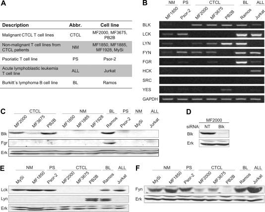
Initially, we investigated whether Blk was expressed in malignant CTCL cell lines (Figure 1A). In Figure 1, RT-PCR (Figure 1B) and WB analyses (Figure 1C,E,F) of malignant (MF2000, MF3675, PB2B) and nonmalignant (MF1850, MySi, MF1928, MF1885) T-cell lines derived from patients with CTCL, a T-cell line established from a patient with psoriasis vulgaris (Psor-2), a malignant B-cell line from Burkitt lymphoma (Ramos), and a malignant T-cell line from a patient with acute lymphoblastic leukemia (Jurkat) are shown. To our surprise, expression of Blk mRNA and protein was detected in all of the malignant CTCL cell lines (Figure 1B,C). In contrast, Blk was not expressed in any of the nonmalignant T-cell lines or the psoriatic T-cell line (Figure 1B,C). As expected, Blk was detected both by RT-PCR and WB in Ramos cells (Figure 1B,C). Blk is expressed in thymocytes22; therefore, it was not surprising that Blk mRNA was detected in the immature T-cell line Jurkat (Figure 1B). Indeed, it has previously been reported that Jurkat cells express low levels of Blk mRNA.22,39 However, protein expression of Blk was not observed in the Jurkat cells (Figure 1C). Confirming the identity of the band recognized as Blk, transfection of the malignant CTCL cell lines with Blk-specific siRNA induced a selective down-regulation of the band, whereas the expression of Erk remained unaffected (Figure 1D; data not shown). Finally, Blk could be cloned from the malignant CTCL cell lines, and, of notice, no mutations were found by sequence analysis (data not shown).
Figure 1.
Malignant CTCL cell lines selectively express Blk. (A) Descriptions and abbreviations (Abbr.) of the cell lines used in the study. (B) RT-PCR and (C,E,F) WB analyses of the expression of SFKs in the cell lines shown in panel A. (D) MF2000 cells were transiently transfected with nontargeting (NT) or Blk-specific siRNA, and the expression of Blk and Erk was determined by WB 48 hours after transfection.
The SFKs are differentially expressed in the lymphoid lineage. B cells preferentially express Blk, Lyn, and Fyn, whereas T cells primarily express Lck and Fyn.19 The ectopic expression of Blk prompted us to investigate whether other SFKs exhibited an atypical expression pattern in malignant CTCL cells. Lck expression has previously been shown to be absent in the MF2000 cell line.40 In agreement, we did not detect Lck on either mRNA or protein level in MF2000 cells, or the 2 other malignant CTCL cell lines MF3675 and PB2B (Figure 1B,E). In contrast, Lck was found to be expressed in the nonmalignant T-cell lines (Figure 1B,E). Lyn mRNA was detected in most of the cell lines (Figure 1B) but only expressed on the protein level in PB2B and Ramos cells (Figure 1E). Even though Fyn mRNA was detected in 2 of the 3 malignant CTCL cell lines (Figure 1B), the protein expression of Fyn was severely down-regulated or absent in all of the malignant CTCL cell lines compared with the nonmalignant T-cell lines (Figure 1F). Expectedly, expression of Fgr, Hck, or Src was not observed in either nonmalignant or malignant CTCL cell lines (Figure 1B,C; data not shown) and Yes mRNA and protein were barely detectable (Figure 1B; data not shown). Collectively, these results show that malignant CTCL cell lines selectively express Blk, whereas the expression of the 2 predominant T-cell SFKs, Lck and Fyn, is absent or severely down-regulated.
Ectopic expression of Blk in CTCL lesions
Having established that Blk is expressed in malignant but not nonmalignant T cells derived from CTCL or psoriasis lesions, we addressed whether we could detect Blk expression in situ in CTCL lesions. Accordingly, we performed immunohistochemical stainings of lesional tissue specimens obtained from 31 patients diagnosed with MF using an anti-Blk antibody. This analysis showed Blk expression in 26 of the 31 patients (Table 1; for detailed information see Table S3). In all positive cases, the expression of Blk was restricted to the cytoplasmic compartment (Table S3). As exemplified in Figure 2, Blk expression was found in both early- and late-stage CTCL lesions. In early lesions, the expression of Blk was primarily present in the epidermotropic T cells. Indeed, most T cells characterized by epidermotropism were Blk positive (Figure 2; data not shown). Collectively, Blk expression was detected in 8 of 11 early lesions and 18 of 20 late lesions (Table 1). Importantly, Blk expression was not detected in T-cell infiltrates of a variety of benign inflammatory skin diseases, ie, allergic and atopic eczema (data not shown).
Table 1.
Immunohistochemical stainings of skin specimens from patients diagnosed with early- or late-stage MF using an anti-Blk antibody
| Early stage* | Late stage† | Total | |
|---|---|---|---|
| Blk positive | 8 | 18 | 26 |
| Blk negative | 3 | 2 | 5 |
| Total | 11 | 20 | 31 |
Figure 2.
Ectopic expression of Blk in patients with early- and late-stage CTCL. Immunohistologic detection of Blk in early (left) and late (right) lesions of CTCL. Representative sections are depicted at original magnification ×20 and ×40.
A second series of 24 patients were chosen for whom the clinical suspicion of CTCL could neither be confirmed nor confuted by the initial biopsy and for whom the clinical follow-up was available for at least 12 months. Blk expression was observed in specimens from 11 of 13 patients in whom the diagnosis of CTCL was subsequently confirmed. In contrast, only in 2 of 11 patients who did not develop clinically verified CTCL within the follow-up period, we detected Blk expression (Table 2). Accordingly, there was a significant correlation between Blk expression and the development of clinically verified CTCL (P < .002). The sensitivity and positive predictive value (PPV) in the study population were 85%, and the specificity and negative predictive value (NPV) were 82%.
Table 2.
Longitudinal study of patients admitted on clinical suspicion of CTCL
| Patients who developed CTCL | Patients who did not develop CTCL | Total | |
|---|---|---|---|
| Blk positive | 11 | 2 | 13 |
| Blk negative | 2 | 9 | 11 |
| Total | 13 | 11 | 24 |
Expression of Blk is induced by NF-κB
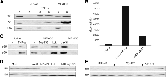
In murine B cells the transcription factors NF-κB and Pax-5 (BSAP) have been identified as the major regulators of Blk transcription.39,41,42 CTCL cell lines expressed both of the NF-κB subunits, p65 and p50, with the latter being overexpressed, compared with nonmalignant T-cell lines (Figure S1). In contrast, no expression of Pax-5 mRNA was detected (data not shown), suggesting that NF-κB is a candidate transcription factor in CTCL. To evaluate whether NF-κB was constitutively active in malignant CTCL cells and, hence, localized in the nucleus, nuclear and cytoplasmic cell extracts were analyzed by WB. In unstimulated Jurkat cells, used as a negative control, both p65 and p50 were almost exclusively found in the cytoplasm (Figure 3A). As expected, treatment of the cells with TNF-α led to a decrease in IκB-α levels and translocalization of p65 and p50 to the nucleus (Figure 3A). Importantly, the majority of both p65 and p50 was constitutively present in the nuclear fractions of the malignant CTCL cell line, MF2000, whereas IκB-α was detected only in the cytoplasm (Figure 3A). Likewise, nuclear localization of p65 was also observed in immunohistochemical analyses of MF2000 cells (Figure S2). To substantiate these findings, we established a reporter assay using MF2000 cells transfected with a luciferase reporter construct containing no insert (vector control), a NF-κB consensus binding site, or the proximal VEGF promoter (positive control). As shown in Figure 3B, the NF-κB reporter construct gave rise to a high spontaneous increase in luciferase activity compared with the vector control. These findings confirm previous reports that NF-κB is constitutively active in CTCL.7,8
Figure 3.
Constitutive NF-κB activity correlates with Blk expression. (A) WB analysis of p65, p50, and IκB-α in cytoplasmic (c) and nuclear (n) extracts of Jurkat T cells either not stimulated (−) or stimulated for 1 hour with 100 ng/mL TNF-α and unstimulated (−) malignant CTCL cells (MF2000). (B) MF2000 cells were cotransfected with a luciferase reporter construct containing either no insert (pGL3), a NF-κB consensus binding site (pGL3-NF-κB), or the proximal VEGF promoter (pGL3-VEGF) and a Renilla luciferase vector. At 48 hours after transfection the cells were lysed, and the luciferase activities were determined. The coexpressed Renilla luciferase activity was used for normalization of transfection efficiency. The experiment was performed with triplicate cultures and is representative of 3 independent experiments. (C) Jurkat, MF2000 (malignant), and MF1850 (nonmalignant) cells were incubated for 3 hours with TNF-α (100 ng/mL), Mg-132 (10 μM), LckI (10 μM), Jak3I (40 μg/mL), or vehicle (−) as shown. Then, DNA binding p65 and p50 were affinity purified from nuclear lysates using bio-oligos with a sequence identical to a putitative NF-κB binding site in the Blk promoter. Finally, the levels of precipitated p65 and p50 were analyzed by WB. (D,E) MF2000 cells were incubated for 24 hours in media (Med.) containing vehicle (−), Jak3I (40 μg/mL), NF-κBI (100 nM), LckI (10 μM), JNKI (5 μM), Ag1478 (200 ng/mL), or increasing concentrations (→) of the NF-κB inhibitors JSH-23 (10, 20, 40 μM) and Mg-132 (0.4, 4, 40 μM). Subsequently, the expression of Blk and Erk was determined by WB.
In murine B cells NF-κB p65/p50 heterodimers have been shown to induce Blk expression by binding a region from −65 to −45 in the Blk promoter.42 To address whether NF-κB could bind the human Blk promoter, we constructed a bio-oligo with a sequence identical to the region −411 to −385 in the human Blk promoter which contains a putative NF-κB binding site, and we investigated whether it could precipitate p65 and p50 from nuclear extracts of Jurkat and MF2000 cells. No binding of either p65 or p50 to the bio-oligo was observed in nuclear lysates of Jurkat cells without stimulation (Figure 3C). On stimulation with TNF-α, binding of both p65 and p50 was observed (Figure 3C). Importantly, p65 and p50 spontaneously bound the bio-oligo in malignant (MF2000) but not nonmalignant (MF1850) T cells (Figure 3C). Treatment of the malignant cells with a selective SFK inhibitor (LckI) or a Jak3 inhibitor (Jak3I) did not decrease the binding, whereas an inhibitor of NF-κB (Mg-132) almost completely abrogated the binding of both p65 and p50 (Figure 3C). To determine whether the constitutive NF-κB activity correlated with the expression of Blk, malignant CTCL cells were treated with an inhibitor of NF-κB transcriptional activation (NF-κBI), Jak3I, LckI, an inhibitor of c-Jun N-terminal kinase (JNKI), or an inhibitor of epidermal growth factor receptor (Ag1478), and the expression of Blk was investigated by WB. As seen from Figure 3D, NF-κBI almost completely blocked the expression of Blk. Furthermore, 2 other inhibitors of NF-κB (JSH-23 and Mg-132) also mediated a dose-dependent down-regulation of Blk expression (Figure 3E). LckI also moderately inhibited the Blk expression, whereas the other inhibitors, including Jak3I, did not (Figure 3D). Thus, NF-κB is capable of binding the Blk promoter, and the constitutive activity of NF-κB correlates with the expression of Blk in malignant CTCL cells.
Blk is constitutively active
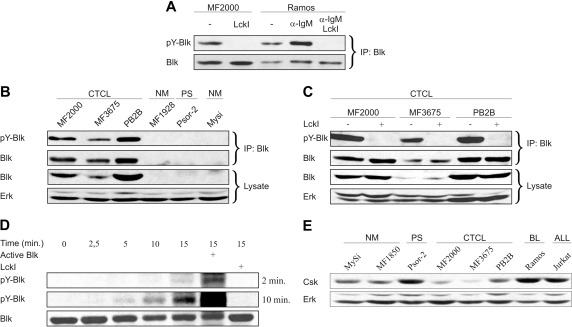
Because the oncogenic capacity of Blk requires that it is enzymatically active,27 it was important to know if Blk was persistently activated in malignant CTCL cells. It has previously been shown that the activity of Blk is positively correlated with the level of tyrosine phosphorylation.23,24 In agreement, the level of tyrosine-phosphorylated (pY)–Blk was increased on BCR stimulation of the malignant B-cell line, Ramos, but abrogated in BCR-stimulated Ramos cells simultaneously exposed to LckI (Figure 4A). Importantly, Blk was highly tyrosine-phosphorylated in all of the 3 malignant CTCL cell lines examined, and treatment with LckI completely abrogated the spontaneous phosphorylation (Figure 4A-C). In agreement with our previous findings, Blk was not detected in the nonmalignant T-cell lines or a psoriatic T-cell line even after immunoprecipitation (Figure 4B).
Figure 4.
Blk is constitutively active in malignant CTCL cell lines. (A) Malignant CTCL cells (MF2000) and Ramos B cells were incubated without (−) or with LckI (10 μM) for 30 minutes. Then, 20 μg/mL goat anti–human IgM F(ab′)2 fragments (α-IgM) were added as indicated, and the cells were incubated for 5 minutes further. Subsequently, the cells were lysed, and Blk was immunoprecipitated (IP) from the total cell lysates. Finally, the levels of pY-Blk and Blk were determined by WB using phosphotyrosine and Blk-specific antibodies, respectively. (B) Blk was immunoprecipitated from total cell lysates of malignant (CTCL) and nonmalignant (NM) cell lines as well as from a psoriatic T-cell line (PS). Both total cell lysates (Lysate) and immunoprecipitated Blk (IP: Blk) were analyzed by WB using antibodies against phosphotyrosine (pY-Blk), Blk, and Erk. (C) Malignant CTCL cell lines were incubated for 30 minutes with LckI (10 μM) or vehicle (−). Then immunoprecipitated Blk and respective total cell lysates were analyzed by WB. (D) MF2000 cells were incubated with LckI (10 μM) for 30 minutes and lysed, and Blk was immunoprecipitated from the total cell lysates. The precipitated Blk was incubated for different time intervals (minutes) in kinase buffer without or with LckI (2 μM) and constitutive active Blk (1 ng/μL) as indicated. The levels of pY-Blk and Blk were analyzed by WB. For pY-Blk, films exposed for 2 or 10 minutes are shown. (E) WB analysis of Csk and Erk expression in the indicated cell lines.
In some human tumors and tumor-derived cell lines aberrant SFK activity is linked to the overexpression of SFKs and the resulting autophosphorylation.43 Because LckI totally abrogated the tyrosine phosphorylation of Blk, it was possible that the aberrant activity was caused by autophosphorylation. We investigated, therefore, whether Blk precipitated from MF2000 cells spontaneously autophosphorylated in vitro. MF2000 cells were treated with LckI, and Blk was, subsequently, immunoprecipitated from total cell lysates. The precipitated Blk was incubated in kinase buffer for different time intervals with or without a constitutively active form of Blk or LckI serving as positive and negative controls, respectively. Finally, the levels of pY-Blk and Blk were determined by WB. As shown in Figure 4D, there was a time-dependent increase in pY-Blk despite similar levels of total Blk. The increased level of pY-Blk was reflected by a decreased migration of Blk in the gel. Addition of constitutively active Blk led to a further increase in the level of pY-Blk, whereas addition of LckI completely blocked the autophosphorylation (Figure 4D). Of notice, no other SFKs were found to coimmunoprecipitate with Blk (data not shown).
Normally, negative regulators of SFK activity, such as c-terminal Src kinase (Csk), continuously keep the SFKs in check to avoid abnormal levels of SFK activity. Thus, Csk-negative cells exhibit high levels of autophosphorylation and constitutive activation of SFKs.44,45 A previous study has shown that the enzymatic activity of membrane-bound Csk is suppressed in tumor cells from patients with CTCL.46 Furthermore, we found that the expression of Csk was down-regulated (albeit to a different extend) in the malignant CTCL cell lines relative to other T-cell lines (Figure 4E). Collectively, these findings suggest that Blk is constitutively active in malignant CTCL cells and, further, that the aberrant activity is caused by autophosphorylation promoted by high abnormal expression of Blk combined with down-regulation of negative regulators of SFK activity such as Csk.
Blk is not involved in the constitutive activation of Stat3
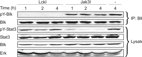
Constitutive activation of the Jak3/Stat3 signaling pathway is a hallmark of CTCL and has been hypothesized to drive the malignant transformation.10–12 SFKs can directly phosphorylate and activate Stat3, both solely or in cooperation with Jaks.47 Therefore, we investigated whether Blk was involved in the constitutive activation of Stat3 in CTCL. MF2000 cells were incubated for different time intervals with LckI, Jak3I, or vehicle, and the tyrosine phosphorylation of Stat3 and Blk was determined by WB. As expected, LckI completely blocked the phosphorylation of Blk, and Jak3I inhibited the phosphorylation of Stat3 (Figure 5). However, LckI did not influence the level of pY-Stat3, and, likewise, Jak3I did not reduce the level of pY-Blk (Figure 5). Finally, Blk could not coprecipitate Stat3, or vice versa, in coimmunoprecipitation experiments (data not shown).
Figure 5.
Constitutive activation of Stat3 in CTCL is Blk independent. Malignant T cells (MF2000) were incubated for different time intervals (hours) with LckI (10 μM), Jak3I (40 μg/mL), or vehicle (−). Subsequently, immunoprecipitated Blk (IP: Blk) and respective total cell lysates (Lysate) were analyzed by WB using antibodies against phosphotyrosine, Blk, pY-Stat3, Stat3, and Erk.
Blk promotes the proliferation of malignant CTCL cells
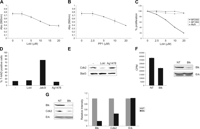
The SFKs regulate a plethora of fundamental cellular processes involved in carcinogenesis, including adhesion, invasiveness, angiogenesis, proliferation, survival, and differentiation.19,48 The specific molecular and cellular processes regulated by Blk are poorly defined, but expression of a constitutively activated form of Blk in the murine B-cell lineage leads to a significant increase in proliferation, and a slight decrease in cell death, of progenitor B cells.25 Therefore, to address the function of Blk in CTCL pathogenesis, we focused on the possible role of Blk in the proliferation and survival of the malignant T cells. Thus, malignant CTCL cells were incubated with different concentrations of the SFK inhibitors LckI and PP1. Then, after 48 hours the number of viable cells was estimated by MTT assay. Treatment of MF2000 cells with LckI and PP1 resulted in a dose-dependent relative decrease in viable cells, with approximately 51% viable cells at 20 μM compared with the vehicle-treated controls (Figure 6A,B). To substantiate these findings, malignant and nonmalignant T cells were incubated for 48 hours with different concentrations of LckI, and the proportion of dividing cells was determined by [3H]-thymidine incorporation. Not surprisingly, LckI induced a dose-dependent reduction in [3H]-thymidine incorporation of the malignant CTCL cells compared with vehicle. In contrast, essentially no reduction of [3H]-thymidine incorporation was observed in the nonmalignant cell lines treated with the same concentrations of LckI (Figure 6C). The decrease in viable and dividing cells after Src kinase inhibition could be a consequence of decreased proliferation, increased cell death, or both. Therefore, we determined the proportion of dead cells subsequent to treatment with LckI, Jak3I (positive control), Ag1478 (negative control), or vehicle. Expectedly, incubation of malignant CTCL cells with Jak3I led to a large increase in the proportion of dead cells (Figure 6D). In contrast, treatment with LckI only resulted in a minute increase in the proportion dead cells compared with vehicle or Ag1478 (Figure 6D). Furthermore, LckI did not induce surface phosphatidylserine (PS) expression or Poly (ADP-ribose) polymerase (PARP) cleavage within 48 hours in MF2000 cells (data not shown). Finally, the inhibition of proliferation by LckI was accompanied by a reduction in the levels of the positive regulator of cell cycle progression, cyclin-dependent kinase 2 (Cdk2; Figure 6E). Collectively, these results show that suppression of SFK activity selectively inhibits the proliferation of malignant CTCL cells.
Figure 6.
Blk promotes the proliferation of malignant CTCL cells. (A,B) Malignant T cells (MF2000; 4 × 10³) were incubated with the given concentrations of (A) LckI or (B) PP1 for 48 hours, and the proportion of viable cells was determined by MTT assay as measured by the absorbance (Abs) at 560 nm. MTT determinations were performed with triplicate cultures, and the error bars represent SDs. (C) Malignant (MF2000) or nonmalignant (MF1850, MySi) T cells (4 × 10³) were cultured for 48 hours with the indicated concentrations of LckI. Then [3H]-thymidine was added, and the cells were cultured for 20 hours, before determination of [3H]-thymidine incorporation. The [3H]-thymidine incorporation was determined as mean counts per minute (CPM) of triplicate cultures. The percentage of proliferation of each cell line was calculated as the mean CPM for cells treated with a given concentration of LckI divided with the mean CPM for cells treated with vehicle. (D) MF2000 cells were incubated for 48 hours with vehicle (−), LckI (10 μM), Jak3I (40 μg/mL), or Ag1478 (200 ng/mL). Subsequently, the cells were stained with 7-AAD, and the percentage of 7-AAD–positive cells was determined by flow cytometry. (E) MF2000 cells were incubated with vehicle (−), LckI (10 μM), or Ag1478 (200 ng/mL) for 24 hours, and the levels of Cdk2 and Stat3 were examined by WB. (F) MF2000 cells were transiently transfected with nontargeting (NT) or Blk-specific siRNA and cultured with [3H]-thymidine. After 24 hours, the cells were harvested, and the [3H]-thymidine incorporation (left), as well as the levels of Blk and Erk expression (right), was determined. The [3H]-thymidine incorporation is expressed as the mean CPM of 6 replicate cultures, and the error bars represent SDs. (G) MF2000 cells were transiently transfected with NT or Blk-specific siRNA. At 24 hours after transfection, the cells were lysed, and the lysates were analyzed by WB (left). The band intensities from the WB were quantified, and the relative band intensities between MF2000 cells transfected with NT and Blk-specific siRNA are depicted (right). The data are representative of at least 3 independent experiments.
We previously found that, in addition to Blk, low levels of Fyn and Yes were also present in the malignant CTCL cells (Figure 1A,E; data not shown). Consequently, the antiproliferative effect of SFK inhibitors could be due to inhibition of Blk-independent kinase activity. To address this issue, malignant CTCL cells were transiently transfected with nontargeting or Blk-specific siRNA, and the proliferation was estimated by [3H]-thymidine incorporation assays. In line with our previous findings, selective down-regulation of Blk expression led to a decrease in the proliferation of the malignant T cells (Figure 6F). As observed with the SFK inhibitors, down-regulation of Blk led to a decrease in Cdk2 expression (Figure 6G) but did not induce significant levels of cell death in MF2000 cells (data not shown). In conclusion, these results support the notion that Blk promotes the proliferation of malignant CTCL cells.
Discussion
Transgenic expression of a constitutively active form of Blk in the T-cell lineage of mice results in the development of clonal T-lymphoid lymphomas.27 However, so far, Blk expression has not been described in T cells from patients with T-cell neoplasms. In the present study, we show the expression of Blk in CTCL cell lines in vitro as well as in CTCL lesions in situ. In contrast, Blk expression was not detected in a variety of benign inflammatory skin diseases. Moreover, in a longitudinal study of an additional 24 patients admitted on clinical suspicion of CTCL, but for whom the clinical suspicion of CTCL could neither be confirmed nor confuted by the initial biopsy, Blk expression in these early lesions significantly correlated with the development of clinically verified CTCL. Thus, specimens from 11 of 13 patients who developed clinically verified CTCL were Blk positive, whereas only 2 of 11 patients who did not develop clinically verified CTCL were Blk positive (P > .002). Accordingly, the PPV in the study population was 85% and the NPV was 82%. However, because MF is a low-grade lymphoma, the follow-up period of 12 months is not sufficient to completely rule out subsequent development of overt CTCL. Therefore, the present study calls for new long-term prospective studies for a definitive evaluation of the strength of Blk as an early diagnostic marker. It is also important to notice that even though Blk expression was found in the large majority of patients with CTCL (in total 84%), not all patients were Blk positive. We were not able to assign Blk expression to any specific subgroups of patients, and further investigations are needed to elucidate whether Blk is associated with specific clinical subgroups of CTCL. Nevertheless, the present work suggests that Blk expression might serve as a diagnostic marker with a capacity to discriminate between early CTCL and benign inflammatory skin diseases in patients with clinical suspicion of CTCL.
Our findings indicate that the ectopic expression of Blk in CTCL is induced by constitutive activation of the NF-κB pathway. These findings are in agreement with a previous report showing activation of NF-κB in CTCL lesions in 22 (92%) of 24 patients already in early disease stages.7 NF-κB has been shown to promote the proliferation and survival of malignant CTCL cells.8,9 Apoptosis induced by inhibition of NF-κB is associated with an up-regulation of Bax dimers and a decrease of survivin.9 In contrast, the molecular mechanisms involved in NF-κB–induced proliferation have not been identified. The present findings imply that NF-κB–induced proliferation of malignant CTCL cells is mediated, at least in part, by Blk-dependent mechanisms. Given the ubiquitous expression and pleiotropic functions of NF-κB, Blk could potentially provide a more selective target for CTCL therapy directed at proliferative inhibition of the malignant T cells.
In some human tumors and tumor-derived cell lines aberrant SFK activity is linked to overexpression of SFKs.43 Our findings that Blk is able to autophosphorylate and that SFK inhibitors block the constitutive tyrosine phosphorylation of Blk in cell lines, as well as the autophosphorylation in vitro, suggest that Blk is constitutively active and that the aberrant activity is induced by autophosphorylation. The observations that the endogenous SFK inhibitor, Csk, is down-regulated in the malignant T-cell lines and that the activity of membrane-bound Csk is suppressed in tumor cells from patients with CTCL support this hypothesis.46 It is possible, however, that other SFKs or non-SFK tyrosine kinases also play a role in the constitutive activation of Blk.
The Jak3/Stat3 signaling pathway is constitutively active in CTCL.10–12 Given the link between SFKs and Stat3 activation in some tumors,47 it was obvious to address whether Blk was involved in the aberrant activation of Stat3. However, blocking Blk activity by SFK inhibitors had no influence on the constitutive activation of Stat3. Likewise, inhibition of the Jak3/Stat3 pathway by chemical inhibitors, Stat3-specific siRNA (data not shown) or a dominant-negative form of Stat3 (data not shown) had no effect on Blk expression or activity, indicating that the Blk and Jak3/Stat3 signaling pathways are parallel in CTCL. Supporting this conclusion, we were unable to coprecipitate Jak3 and Stat3 with Blk and vice versa.
Several functions have been assigned to SFKs in neoplastic transformation and metastasis.19,48 In particular, expression of a constitutively active form of Blk in the murine B-cell linage results in increased proliferation of progenitor B cells.19,25 Accordingly, we show that Blk inhibition by selective SFK inhibitors or siRNA inhibits the spontaneous proliferation of malignant CTCL cells without inducing apoptosis. In contrast, LckI did not suppress the proliferation of nonmalignant T-cell lines in concentrations that severely inhibited Blk activity and proliferation of the malignant T cells. The SFKs Lck and Fyn can mediate induction of both proliferation and apoptosis in T cells, and T cells that express constitutive active Lck undergo apoptosis on TCR crosslinking.49 Accordingly, down-regulation of Lck and Fyn expression/activity could protect the malignant T cells from apoptosis. Because Blk exhibits overlapping as well as distinct substrate specificities compared with other SFKs,50 we speculate that selective expression of Blk might promote the proliferation of the malignant T cells without increasing their susceptibility to apoptosis.
In conclusion, the present study indicates that ectopic expression of Blk is a characteristic feature of CTCL. Moreover, we provide evidence that Blk is involved in the malignant proliferation and could be an important diagnostic marker discriminating early CTCL from nonmalignant inflammatory skin disorders.
Figure S1
Supplementary PDF file available online.
Figure S2
Supplementary PDF file available online.
Table S1
Supplementary PDF file available online.
Table S2
Supplementary PDF file available online.
Table S3
Supplementary PDF file available online.
[Supplemental Tables and Figures]
Acknowledgments
We thank Claudia Siedel for her excellent technical assistance and Eva-Bettina Bröcker for critical discussions during the evaluation of the immunohistochemistry slides. We also thank K. Kaltoft for providing us with the MyLa cell lines.
This work was supported in part by research funding from The Danish Research Councils, The Danish Cancer Society, The Danish Cancer Research Foundation, The Lundbeck Foundation, The Novo Nordic Foundation, Fabrikant Vilhelm Pedersen og Hustrus Mindelegat, The Neye Foundation, The University of Copenhagen, The Deutsche Forschungsgemeinschaft (KFO124; J.C.B.), and the National Cancer Institute (grant CA89194; M.A.W.). T.K. is a PhD candidate at the University of Copenhagen, and this work is submitted in partial fulfillment of the requirement for the PhD.
Footnotes
The online version of this article contains a data supplement.
The publication costs of this article were defrayed in part by page charge payment. Therefore, and solely to indicate this fact, this article is hereby marked “advertisement” in accordance with 18 USC section 1734.
Authorship
Contribution: T.K., P.L., J.C.B., and N.Ø. designed the research; T.K., C.S.V.-K., A.W., and H.K. performed the research; T.K., C.S.V.-K., A.W., H.K., K.W.E., Q.Z., M.A.W., C.G., E.R., J.C.B., and N.Ø. analyzed data; and T.K., M.A.W., J.C.B., and N.Ø. wrote the paper.
Conflict-of-interest disclosure: The authors declare no competing financial interests.
Correspondence: Niels Ødum; Institute of International Health, Immunology and Microbiology, University of Copenhagen, Blegdamsvej 3c, 2200 Copenhagen, Denmark; e-mail: ndum@sund.ku.dk.
References
- 1.Willemze R, Jaffe ES, Burg G, et al. WHO-EORTC classification for cutaneous lymphomas. Blood. 2005;105:3768–3785. doi: 10.1182/blood-2004-09-3502. [DOI] [PubMed] [Google Scholar]
- 2.Kim EJ, Hess S, Richardson SK, et al. Immunopathogenesis and therapy of cutaneous T cell lymphoma. J Clin Invest. 2005;115:798–812. doi: 10.1172/JCI24826. [DOI] [PMC free article] [PubMed] [Google Scholar]
- 3.Girardi M, Heald PW, Wilson LD. The pathogenesis of mycosis fungoides. N Engl J Med. 2004;350:1978–1988. doi: 10.1056/NEJMra032810. [DOI] [PubMed] [Google Scholar]
- 4.Edelson RL. Cutaneous T cell lymphoma: the helping hand of dendritic cells. Ann N Y Acad Sci. 2001;941:1–11. [PubMed] [Google Scholar]
- 5.Trautinger F, Knobler R, Willemze R, et al. EORTC consensus recommendations for the treatment of mycosis fungoides/Sezary syndrome. Eur J Cancer. 2006;42:1014–1030. doi: 10.1016/j.ejca.2006.01.025. [DOI] [PubMed] [Google Scholar]
- 6.Woetmann A, Lovato P, Eriksen KW, et al. Nonmalignant T cells stimulate growth of T-cell lymphoma cells in the presence of bacterial toxins. Blood. 2007;109:3325–3332. doi: 10.1182/blood-2006-04-017863. [DOI] [PMC free article] [PubMed] [Google Scholar]
- 7.Izban KF, Ergin M, Qin JZ, et al. Constitutive expression of NF-kappa B is a characteristic feature of mycosis fungoides: implications for apoptosis resistance and pathogenesis. Hum Pathol. 2000;31:1482–1490. doi: 10.1053/hupa.2000.20370. [DOI] [PubMed] [Google Scholar]
- 8.Sors A, Jean-Louis F, Pellet C, et al. Down-regulating constitutive activation of the NF-kappaB canonical pathway overcomes the resistance of cutaneous T-cell lymphoma to apoptosis. Blood. 2006;107:2354–2363. doi: 10.1182/blood-2005-06-2536. [DOI] [PubMed] [Google Scholar]
- 9.Sors A, Jean-Louis F, Begue E, et al. Inhibition of IkappaB kinase subunit 2 in cutaneous T-cell lymphoma down-regulates nuclear factor-kappaB constitutive activation, induces cell death, and potentiates the apoptotic response to antineoplastic chemotherapeutic agents. Clin Cancer Res. 2008;14:901–911. doi: 10.1158/1078-0432.CCR-07-1419. [DOI] [PubMed] [Google Scholar]
- 10.Sommer VH, Clemmensen OJ, Nielsen O, et al. In vivo activation of STAT3 in cutaneous T-cell lymphoma. Evidence for an antiapoptotic function of STAT3. Leukemia. 2004;18:1288–1295. doi: 10.1038/sj.leu.2403385. [DOI] [PubMed] [Google Scholar]
- 11.Zhang Q, Wang HY, Woetmann A, et al. STAT3 induces transcription of the DNA methyltransferase 1 gene (DNMT1) in malignant T lymphocytes. Blood. 2006;108:1058–1064. doi: 10.1182/blood-2005-08-007377. [DOI] [PMC free article] [PubMed] [Google Scholar]
- 12.Krejsgaard T, Vetter-Kauczok CS, Woetmann A, et al. Jak3- and JNK-dependent vascular endothelial growth factor expression in cutaneous T-cell lymphoma. Leukemia. 2006;20:1759–1766. doi: 10.1038/sj.leu.2404350. [DOI] [PubMed] [Google Scholar]
- 13.Fantin VR, Loboda A, Paweletz CP, et al. Constitutive activation of signal transducers and activators of transcription predicts vorinostat resistance in cutaneous T-cell lymphoma. Cancer Res. 2008;68:3785–3794. doi: 10.1158/0008-5472.CAN-07-6091. [DOI] [PubMed] [Google Scholar]
- 14.Dummer R, Assaf C, Bagot M, et al. Maintenance therapy in cutaneous T-cell lymphoma: who, when, what? Eur J Cancer. 2007;43:2321–2329. doi: 10.1016/j.ejca.2007.06.015. [DOI] [PubMed] [Google Scholar]
- 15.Pimpinelli N, Olsen EA, Santucci M, et al. Defining early mycosis fungoides. J Am Acad Dermatol. 2005;53:1053–1063. doi: 10.1016/j.jaad.2005.08.057. [DOI] [PubMed] [Google Scholar]
- 16.Glusac EJ. Criterion by criterion, mycosis fungoides. Am J Dermatopathol. 2003;25:264–269. doi: 10.1097/00000372-200306000-00014. [DOI] [PubMed] [Google Scholar]
- 17.Olsen E, Vonderheid E, Pimpinelli N, et al. Revisions to the staging and classification of mycosis fungoides and Sezary syndrome: a proposal of the International Society for Cutaneous Lymphomas (ISCL) and the cutaneous lymphoma task force of the European Organization of Research and Treatment of Cancer (EORTC). Blood. 2007;110:1713–1722. doi: 10.1182/blood-2007-03-055749. [DOI] [PubMed] [Google Scholar]
- 18.Keehn CA, Belongie IP, Shistik G, Fenske NA, Glass LF. The diagnosis, staging, and treatment options for mycosis fungoides. Cancer Control. 2007;14:102–111. doi: 10.1177/107327480701400203. [DOI] [PubMed] [Google Scholar]
- 19.Thomas SM, Brugge JS. Cellular functions regulated by Src family kinases. Annu Rev Cell Dev Biol. 1997;13:513–609. doi: 10.1146/annurev.cellbio.13.1.513. [DOI] [PubMed] [Google Scholar]
- 20.Dymecki SM, Niederhuber JE, Desiderio SV. Specific expression of a tyrosine kinase gene, blk, in B lymphoid cells. Science. 1990;247:332–336. doi: 10.1126/science.2404338. [DOI] [PubMed] [Google Scholar]
- 21.Drebin JA, Hartzell SW, Griffin C, Campbell MJ, Niederhuber JE. Molecular cloning and chromosomal localization of the human homologue of a B-lymphocyte specific protein tyrosine kinase (blk). Oncogene. 1995;10:477–486. [PubMed] [Google Scholar]
- 22.Islam KB, Rabbani H, Larsson C, Sanders R, Smith CI. Molecular cloning, characterization, and chromosomal localization of a human lymphoid tyrosine kinase related to murine Blk. J Immunol. 1995;154:1265–1272. [PubMed] [Google Scholar]
- 23.Burkhardt AL, Brunswick M, Bolen JB, Mond JJ. Anti-immunoglobulin stimulation of B lymphocytes activates src-related protein-tyrosine kinases. Proc Natl Acad Sci U S A. 1991;88:7410–7414. doi: 10.1073/pnas.88.16.7410. [DOI] [PMC free article] [PubMed] [Google Scholar]
- 24.Saouaf SJ, Mahajan S, Rowley RB, et al. Temporal differences in the activation of three classes of non-transmembrane protein tyrosine kinases following B-cell antigen receptor surface engagement. Proc Natl Acad Sci U S A. 1994;91:9524–9528. doi: 10.1073/pnas.91.20.9524. [DOI] [PMC free article] [PubMed] [Google Scholar]
- 25.Tretter T, Ross AE, Dordai DI, Desiderio S. Mimicry of pre-B cell receptor signaling by activation of the tyrosine kinase Blk. J Exp Med. 2003;198:1863–1873. doi: 10.1084/jem.20030729. [DOI] [PMC free article] [PubMed] [Google Scholar]
- 26.Saijo K, Schmedt C, Su IH, et al. Essential role of Src-family protein tyrosine kinases in NF-kappaB activation during B cell development. Nat Immunol. 2003;4:274–279. doi: 10.1038/ni893. [DOI] [PubMed] [Google Scholar]
- 27.Malek SN, Dordai DI, Reim J, Dintzis H, Desiderio S. Malignant transformation of early lymphoid progenitors in mice expressing an activated Blk tyrosine kinase. Proc Natl Acad Sci U S A. 1998;95:7351–7356. doi: 10.1073/pnas.95.13.7351. [DOI] [PMC free article] [PubMed] [Google Scholar]
- 28.Kaltoft K, Bisballe S, Dyrberg T, et al. Establishment of two continuous T-cell strains from a single plaque of a patient with mycosis fungoides. In Vitro Cell Dev Biol. 1992;28A:161–167. doi: 10.1007/BF02631086. [DOI] [PubMed] [Google Scholar]
- 29.Kaltoft K, Hansen BH, Thestrup-Pedersen K. Cytogenetic findings in cell lines from cutaneous T-cell lymphoma. Dermatol Clin. 1994;12:295–304. [PubMed] [Google Scholar]
- 30.Kaltoft K, Hansen BH, Pedersen CB, Pedersen S, Thestrup-Pedersen K. Common clonal chromosome aberrations in cytokine-dependent continuous human T-lymphocyte cell lines. Cancer Genet Cytogenet. 1995;85:68–71. doi: 10.1016/0165-4608(95)00118-2. [DOI] [PubMed] [Google Scholar]
- 31.Wasik MA, Seldin DC, Butmarc JR, et al. Analysis of IL-2, IL-4 and their receptors in clonally-related cell lines derived from a patient with a progressive cutaneous T-cell lymphoproliferative disorder. Leuk Lymphoma. 1996;23:125–136. doi: 10.3109/10428199609054811. [DOI] [PubMed] [Google Scholar]
- 32.Geisler C, Scholler J, Wahi MA, Rubin B, Weiss A. Association of the human CD3-zeta chain with the alpha beta-T cell receptor/CD3 complex. Clues from a T cell variant with a mutated T cell receptor-alpha chain. J Immunol. 1990;145:1761–1767. [PubMed] [Google Scholar]
- 33.Siegel JP, Mostowski HS. A bioassay for the measurement of human interleukin-4. J Immunol Methods. 1990;132:287–295. doi: 10.1016/0022-1759(90)90040-3. [DOI] [PubMed] [Google Scholar]
- 34.Krejsgaard T, Gjerdrum LM, Ralfkiaer E, et al. Malignant Tregs express low molecular splice forms of FOXP3 in Sezary syndrome. Leukemia. 2008;22:2230–2239. doi: 10.1038/leu.2008.224. [DOI] [PubMed] [Google Scholar]
- 35.Eriksen KW, Lovato P, Skov L, et al. Increased sensitivity to interferon-alpha in psoriatic T cells. J Invest Dermatol. 2005;125:936–944. doi: 10.1111/j.0022-202X.2005.23864.x. [DOI] [PubMed] [Google Scholar]
- 36.Bettelli E, Dastrange M, Oukka M. Foxp3 interacts with nuclear factor of activated T cells and NF-kappa B to repress cytokine gene expression and effector functions of T helper cells. Proc Natl Acad Sci U S A. 2005;102:5138–5143. doi: 10.1073/pnas.0501675102. [DOI] [PMC free article] [PubMed] [Google Scholar]
- 37.Niu G, Wright KL, Huang M, et al. Constitutive Stat3 activity up-regulates VEGF expression and tumor angiogenesis. Oncogene. 2002;21:2000–2008. doi: 10.1038/sj.onc.1205260. [DOI] [PubMed] [Google Scholar]
- 38.Nielsen M, Kaestel CG, Eriksen KW, et al. Inhibition of constitutively activated Stat3 correlates with altered Bcl-2/Bax expression and induction of apoptosis in mycosis fungoides tumor cells. Leukemia. 1999;13:735–738. doi: 10.1038/sj.leu.2401415. [DOI] [PubMed] [Google Scholar]
- 39.Lin YH, Shin EJ, Campbell MJ, Niederhuber JE. Transcription of the blk gene in human B lymphocytes is controlled by two promoters. J Biol Chem. 1995;270:25968–25975. doi: 10.1074/jbc.270.43.25968. [DOI] [PubMed] [Google Scholar]
- 40.Brockdorff J, Nielsen M, Kaltoft K, et al. Lck is involved in interleukin-2 induced proliferation but not cell survival in human T cells through a MAP kinase-independent pathway. Eur Cytokine Netw. 2000;11:225–231. [PubMed] [Google Scholar]
- 41.Zwollo P, Desiderio S. Specific recognition of the blk promoter by the B-lymphoid transcription factor B-cell-specific activator protein. J Biol Chem. 1994;269:15310–15317. [PubMed] [Google Scholar]
- 42.Zwollo P, Rao S, Wallin JJ, Gackstetter ER, Koshland ME. The transcription factor NF-kappaB/p50 interacts with the blk gene during B cell activation. J Biol Chem. 1998;273:18647–18655. doi: 10.1074/jbc.273.29.18647. [DOI] [PubMed] [Google Scholar]
- 43.Dehm SM, Bonham K. SRC gene expression in human cancer: the role of transcriptional activation. Biochem Cell Biol. 2004;82:263–274. doi: 10.1139/o03-077. [DOI] [PubMed] [Google Scholar]
- 44.Hata A, Sabe H, Kurosaki T, Takata M, Hanafusa H. Functional analysis of Csk in signal transduction through the B-cell antigen receptor. Mol Cell Biol. 1994;14:7306–7313. doi: 10.1128/mcb.14.11.7306. [DOI] [PMC free article] [PubMed] [Google Scholar]
- 45.Imamoto A, Soriano P. Disruption of the csk gene, encoding a negative regulator of Src family tyrosine kinases, leads to neural tube defects and embryonic lethality in mice. Cell. 1993;73:1117–1124. doi: 10.1016/0092-8674(93)90641-3. [DOI] [PubMed] [Google Scholar]
- 46.Fargnoli MC, Edelson RL, Berger CL, et al. Diminished TCR signaling in cutaneous T cell lymphoma is associated with decreased activities of Zap70, Syk and membrane-associated Csk. Leukemia. 1997;11:1338–1346. doi: 10.1038/sj.leu.2400745. [DOI] [PubMed] [Google Scholar]
- 47.Ingley E, Klinken SP. Cross-regulation of JAK and Src kinases. Growth Factors. 2006;24:89–95. doi: 10.1080/08977190500368031. [DOI] [PubMed] [Google Scholar]
- 48.Summy JM, Gallick GE. Src family kinases in tumor progression and metastasis. Cancer Metastasis Rev. 2003;22:337–358. doi: 10.1023/a:1023772912750. [DOI] [PubMed] [Google Scholar]
- 49.Di Somma MM, Nuti S, Telford JL, Baldari CT. p56lck plays a key role in transducing apoptotic signals in T cells. FEBS Lett. 1995;363:101–104. doi: 10.1016/0014-5793(95)00292-h. [DOI] [PubMed] [Google Scholar]
- 50.Malek SN, Desiderio S. SH2 domains of the protein-tyrosine kinases Blk, Lyn, and Fyn(T) bind distinct sets of phosphoproteins from B lymphocytes. J Biol Chem. 1993;268:22557–22565. [PubMed] [Google Scholar]
Associated Data
This section collects any data citations, data availability statements, or supplementary materials included in this article.
Supplementary Materials
Figure S1
Supplementary PDF file available online.
Figure S2
Supplementary PDF file available online.
Table S1
Supplementary PDF file available online.
Table S2
Supplementary PDF file available online.
Table S3
Supplementary PDF file available online.
[Supplemental Tables and Figures]
