Red blood cell-mimicking synthetic biomaterial particles (original) (raw)
Abstract
Biomaterials form the basis of current and future biomedical technologies. They are routinely used to design therapeutic carriers, such as nanoparticles, for applications in drug delivery. Current strategies for synthesizing drug delivery carriers are based either on discovery of materials or development of fabrication methods. While synthetic carriers have brought upon numerous advances in drug delivery, they fail to match the sophistication exhibited by innate biological entities. In particular, red blood cells (RBCs), the most ubiquitous cell type in the human blood, constitute highly specialized entities with unique shape, size, mechanical flexibility, and material composition, all of which are optimized for extraordinary biological performance. Inspired by this natural example, we synthesized particles that mimic the key structural and functional features of RBCs. Similar to their natural counterparts, RBC-mimicking particles described here possess the ability to carry oxygen and flow through capillaries smaller than their own diameter. Further, they can also encapsulate drugs and imaging agents. These particles provide a paradigm for the design of drug delivery and imaging carriers, because they combine the functionality of natural RBCs with the broad applicability and versatility of synthetic drug delivery particles.
Keywords: biomimetic, drug delivery, erythrocyte, imaging, nanotechnology
Biomaterials provide a technological platform for launching biomedical applications in drug delivery, medical imaging, and regenerative medicine (1, 2). Several biomaterials including polymeric nanoparticles and liposomes have been developed for applications in drug delivery, some of which are already available in the market (3–5). These biomaterials enhance the therapeutic benefit of drugs via sustained release, reduced side-effects, and effective targeting (6). Various innovative strategies have been designed and implemented to optimize materials used for drug delivery (7, 8). These include synthesis of polymers to improve biocompatibility (9), fabrication of particles with various morphologies to control pharmacokinetics (10–12), modification of particle surface with polyethylene glycol to improve circulation (13), and functionalization of particles with peptides (14) and aptamers (15) for targeted drug delivery.
While synthetic biomaterials used for drug delivery have been significantly advanced in terms of functionality and diversity, they fail to match the complexity and sophistication routinely exhibited by innate biological entities. In this context, red blood cells (RBCs), the most abundant cells in blood, represent a remarkably engineered biological entity designed for complex biological functionality including oxygen delivery (16). RBCs possess unique physical and chemical properties in terms of size, shape, flexibility, and chemical composition, all of which are essential to their biological functions (17, 18). Inspired by the unique ability of these cells to perform complex tasks and motivated by the need to design particles that adopt the sophistication exhibited by biological entities, we sought to design synthetic carriers that mimic the key structural attributes of RBCs including size, shape, and mechanical properties, yet offer engineering control required in synthetic carriers. Herein, we report the synthesis, initial characterization, and illustration of biomedical applications of RBC-like particles. These particles provide a path to bridge the gap between synthetic materials and biological entities.
Results and Discussion
The structure of RBCs is characterized by several unique properties including biconcave discoidal shape and mechanical flexibility that have so far been unmatched by synthetic particles, which are typically spherical and stiff. Unique structural properties of RBCs allow them to routinely pass through ultrathin capillaries smaller than their own diameter and sinusoidal slits in the spleen. The biconcave discoidal shape also provides a favorable surface area-to-volume ratio and allows RBCs to undergo marked deformations while maintaining a constant surface area (18). The unique morphological properties of RBCs are achieved by a well-orchestrated series of biochemical events. RBCs originate as spherical reticulocytes, which make a transition into the biconcave shape during maturation over a period of 2–3 days (19).
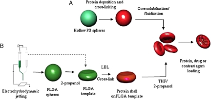
Recreation of the complex morphology of RBCs in a synthetic system has proved challenging using currently established techniques (20). We adopted a biomimetic strategy to prepare RBC-shaped particles. In nature, initial spherical reticulocytes, which have an elastic modulus of ≈3 MPa undergo a 100- to 1,000-fold reduction in elastic modulus and simultaneous change in shape to form discoidal RBCs (21). Mimicking the genesis of mature RBCs, we start with spherical polymeric particles, for example, polystyrene microspheres with high elastic modulus, and use them as a template to induce the change in shape and mechanical properties to form RBC-like particles (Fig. 1). Changing the shape of a solid polystyrene microparticle into an RBC-shaped object, however, is quite challenging. We hypothesized that hollow polystyrene particles, upon solvent or heat-induced fluidization, can collapse into an RBC shape. For this purpose, hollow polystyrene spheres (1-μm diameter, 400-nm shell thickness) were used. Although polystyrene, in its own right, should not be considered a biocompatible polymer, the commercial availability of hollow polystyrene spheres makes them excellent model particles, which can serve as a starting point. Layer-by-layer (LbL) self-assembly technique was used to electrostatically deposit cationic and anionic polymers on the particle surface (22). Initially, BSA and poly(allylamine hydrochloride) (PAH) were chosen as the polyanion and polycation, respectively. The stepwise adsorption of BSA and PAH onto template particles is mediated by hydrophobic and electrostatic interactions. After the adsorption of multiple layers, the shell was cross-linked using glutaraldehyde to provide stability to the particles. The template core was then exposed to tetrahydrofuran (THF) to induce collapse and formation of RBC-shaped particles (Fig. 2A). The collapse is induced by two factors; fluidization and partial solubilization of the polymer core and the build-up of an osmotic gradient across the shell due to the presence of solvent on the outside and water on the inside of the shell.
Fig. 1.
Synthesis technique of RBC-mimicking particles. (A) RBC-shaped particles prepared from hollow PS template. Complementary layers of proteins and polyelectrolytes were deposited by LbL technique on the template surface followed by cross-linking of the layers to increase stability. PS core was dissolved to yield RBC-shaped particles, which can be loaded with therapeutic and imaging agents. (B) Biocompatible RBC-mimicking particles prepared from PLGA template particles. PLGA RBC-shaped templates were synthesized by incubating spheres synthesized from electrohydrodynamic jetting in 2-propanol. LbL coating on template, protein cross-linking, and dissolution of template core yielded biocompatible sRBCs.
Fig. 2.
SEM micrographs of RBC-mimicking particles synthesized using hollow PS template particles. (A) BSA/PAH was deposited on template particles by LbL technique, and the layers were cross-linked. Particles were exposed to THF to yield sRBCs. Inset shows close up. (B) Hb/PSS-based sRBCs prepared by LbL technique. (C) sRBCs prepared by adsorption of Hb on template particles. (Scale bars, 1 μm.) (Inset, 500 nm.)
We next prepared RBC-like particles of similar morphology, but comprised of proteins innate to RBCs, such as hemoglobin (Hb), which is the main constituent of RBCs and is approximately 92% by dry weight (23). Hb is a tetramer with each chain noncovalently bound to each other. The protein further carries one heme group, to which oxygen and other small molecules can bind reversibly. In this case, poly(4-styrene sulfonate) (PSS) and Hb were used as complementary polyelectrolytes for the LbL assembly to yield RBC-shaped particles (Fig. 2B). Alternatively, Hb was adsorbed on to the surface of the template particles, cross-linked with glutaraldehyde followed by the dissolution of the core. The morphology of the particles was found to be similar to those of the LbL particles (Fig. 2C). The methods described here yield soft and synthetic RBC-mimicking particles, which we refer to as sRBCs with the recognition that these particles mimic key but not necessarily all features of natural RBCs.
Having demonstrated the feasibility of preparing sRBCs using hollow polystyrene templates, we sought to address two challenges that are associated with the use of polystyrene as a template. The size of RBC-like particles fabricated from PS templates was limited by the commercial availability of 1-μm hollow particles as opposed to natural RBCs, which are ≈7 μm in diameter. Moreover, PS is not biocompatible, and hence any residual polymer will have the potential to render the particles nonbiocompatible. To address these challenges, polystyrene was replaced by poly(lactic acid-co-glycolide) (PLGA). PLGA is biocompatible and biodegradable, and the size of PLGA particles can be controlled during particle synthesis (9). We first prepared RBC-shaped template PLGA particles (7 ± 2 μm). For this purpose, spherical PLGA particles of appropriate sizes were prepared using the electrohydrodynamic jetting process (24), and these particles were incubated in 2-propanol to induce formation of RBC-shaped PLGA template particles (Fig. 3A). The precise reason why incubation of PLGA particles with 2-propanol induces formation of RBC-shaped particles is unclear, although it may possibly originate from partial fluidization of PLGA due to 2-propanol and subsequent particle collapse. Smaller template particles (3 ± 1.5 μm) were also prepared using the same technique to illustrate the control over size using PLGA particles. These templates were used to yield soft, protein-based biocompatible particles using the modified LbL technique described above. Because PSS is not biocompatible, it was also replaced with BSA in the shell. Nine alternate layers of either Hb/BSA or PAH/BSA were assembled on the templates, the layers were cross-linked, and the underlying PLGA core was removed using 1:2 2-propanol:THF to form sRBCs (Fig. 3B, PAH/BSA sRBCs; see SI Text and Fig. S1 for images of sRBCs made from Hb/BSA). The choice of solvent was important, and deviation from this solvent mixture led either to incomplete dissolution (excess 2-propanol) or complete collapse (excess THF). sRBCs synthesized by this method demonstrate close resemblance to natural RBCs (Fig. 3 B, sRBCs, and C, mouse RBCs).
Fig. 3.
SEM images of biocompatible sRBCs. (A) RBC-shaped PLGA templates fabricated by electrohydrodynamic jetting. (B) Biocompatible sRBCs prepared from PLGA template particles by LbL deposition of PAH/BSA and subsequent dissolution of the polymer core. (C) Cross-linked mouse RBCs. sRBCs demonstrate striking resemblance to the natural counterparts. Insets show close up images. (Scale bars, 5 μm.) (Insets, 2 μm.)
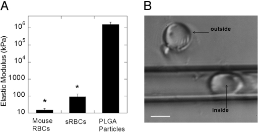
sRBCs were found to be flexible owing to the dissolution of the template PLGA core, which leaves behind a soft protein shell (Fig. 4A). The elastic modulus of sRBCs was measured using atomic force microscopy (AFM). AFM has been previously used to measure elastic modulus of soft materials, such as LbL films, hollow protein particles, and platelets, and a wide range of elastic moduli have been reported for LbL structures in the range of 10 kPa to >100 MPa depending on several parameters including the template/shell materials, shell density, shell cross-linking, and pH, among many others (25–27). The elastic modulus of sRBCs was obtained from force-indentation curves obtained by inducing deformations comparable to the capsule wall thickness, where the elastic response is expected. The typical loading-unloading cycle used for this study and the corresponding force curves obtained for sRBCs can be found in the SI Text and Fig. S2. The elastic modulus of sRBCs (92.8 ± 42 kPa) was found to be four orders of magnitude lower than that of PLGA template particles (1.6 ± 0.6 GPa) and of the same order of magnitude as that of natural RBCs. The elastic modulus of mouse RBCs was found to be 15.2 ± 3.5 kPa, which is consistent with the values reported in literature (21). Further studies are required to facilitate a detailed comparison of various mechanical properties of sRBCs and natural RBCs; however, the data in Fig. 4A clearly indicate that sRBCs are far closer to natural RBCs than to routine polymer particles with respect to mechanical properties.
Fig. 4.
Mechanical properties of sRBCs measured using AFM. (A) Comparison of elastic modulus of sRBCs with mouse RBCs and PLGA particles (*, P < 0.001, n = 5). (B) sRBCs (7 ± 2 μm) flowing through glass capillary (5-μm inner diameter). The image also shows a particle outside the capillary. (Scale bar, 5 μm.)
The flexibility of sRBCs (7 ± 2 μm) was confirmed by flowing them through narrow glass capillaries (5-μm inner diameter) and visualizing the stretching (Fig. 4B, two sRBCs, one inside the capillary and one outside the capillary). Whereas the particle outside the capillary is symmetric and circular, the particle inside the capillary is stretched due to flow (Fig. 4B). The average aspect ratio of stretching was found to be 170 ± 20% (n = 20). See Fig. S3 for more images of particles flowing through the capillary. Further, particles were able to regain their discoidal shape upon exiting the capillary, confirming the reversible nature of the shape deformation. Thus, similar to their natural counterpart, sRBCs maintain the ability to flow through channels smaller than their resting diameter and stretch in response to flow. Further detailed studies of the kinetics of shape transition while passing through the capillaries are necessary to gain further insight into the mechanical flexibility of sRBCs.
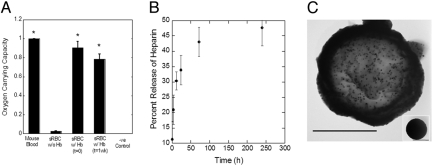
sRBCs reported in this study have numerous biomedical applications. Because the primary function of natural RBCs is to deliver oxygen to the various tissues of the body, we assessed the ability of sRBCs to bind oxygen (Fig. 5A). Cross-linking and exposure to solvent during particle preparation leads to deactivation of Hb, thereby limiting its oxygen carrying capacity (Fig. 5A, sRBC without Hb). To enhance oxygen carrying capacity of sRBCs, particles were further fortified with additional, uncross-linked Hb (see Materials and Methods). This procedure resulted in high oxygen binding levels (Fig. 5A, sRBC with Hb, t = 0) compared to the positive control, which was mouse blood. Approximately 90% of this oxygen carrying capacity was retained even after 1 week (Fig. 5A, sRBC with Hb, t = 1 week). Included is a negative control, BSA-coated particles, which showed no ability to bind oxygen [Fig. 5A, (−) control]. See Fig. S4 for visual confirmation of oxygen carrying capacity.
Fig. 5.
Biomedical applications of sRBCs. (A) Oxygen carrying capacity of sRBCs demonstrated based on the chemiluminescence reaction of luminol. Cross-linking and exposure to the organic solvent reduces the oxygen carrying capacity, but coating the sRBCs with uncross-linked Hb increased the oxygen-binding capacity to levels comparable to mouse blood (S-RBC, t = 0). Ninety percent of oxygen carrying capacity was retained after 1 week (S-RBC, t = 1 wk). BSA-coated particles were included as negative control (*, P < 0.01, n = 3). (B) Controlled release of radiolabeled heparin from sRBCs over a period of 10 days (n = 5). (C) TEM micrograph showing encapsulation of 30 nm iron oxide nanoparticles in RBC-shaped PLGA templates. The Inset shows PLGA particles loaded with iron oxide nanoparticles before conversion into RBC-like templates. (Scale bars, 1 μm.)
sRBCs are also excellent candidates for delivery of drugs, especially in the vascular compartment. These particles can be loaded with drugs by incubation in solutions containing the drug. A model molecule, Texas-Red-conjugated dextran (3 and 10 kDa molecular weight) was loaded into the sRBCs by direct incubation. Both molecules penetrated in the interior of the sRBCs. Dextran was subsequently released from these particles in a controlled manner (see SI Text and Fig. S5). Once the release of dextran was confirmed, controlled release of a therapeutic drug heparin (10–15 kDa) was tested. Heparin is widely used as an anti-coagulant for the treatment of thrombosis (28). Parenteral administration of heparin can result in severe side effects such as heparin-induced thrombocytopenia, elevation of serum aminotransferase levels, hyperkalemia, alopecia, and osteoporosis (29). The sRBCs showed high amounts of heparin loading (70 μg heparin per mg particles) and continuous release over a period of several days in vitro (Fig. 5B).
sRBCs also have potential applications in medical imaging. For example, iron oxide nanocrystals with an average diameter of 30 nm were encapsulated inside the PLGA particles prepared via electrohydrodynamic jetting. Incorporation of iron oxide nanoparticles makes particles suitable as contrast agents for magnetic resonance imaging (MRI) (30). An important requirement for this use is homogenous dispersion of the iron oxide nanocrystals. As shown in Fig. 5C, transmission electron microscopy (TEM) images show well-distributed iron oxide particles in the PLGA matrix. The Inset shows TEM image of a spherical PLGA particle before shape modification. Magnetic particles are currently being developed for a wide spectrum of applications such as MRI contrast agents for diseases, such as atherosclerotic plaque, targeted therapeutic delivery, and hyperthermia treatment for cancerous tumors (31). The interior of the particles described here can be further engineered by the formation of separate compartments using electrohydrodynamic co-jetting process (24). At the same time, the surface can be engineered by adsorption of additional proteins such as CD47, a ubiquitous self-marker expressed on the surface of RBCs or modification of the particle surface with hydrophilic polymers, such as PEG, depending on the application.
In addition to preparing particles that mimic the shape and properties of healthy RBCs, the technique reported here can also be used to design particles that mimic the shape and properties of diseased cells. For example, hereditary elliptocytosis is a disease that leads to the formation of elliptical RBCs (32), a shape that can be mimicked in our method (see SI Text and Fig. S6). Other examples of diseased conditions where the shape of RBCs is altered include spherocytosis and sickle-cell anemia. Such disease cell mimicking particles can serve as synthetic models to help elucidate the effect of transformation in physical properties of RBCs in these disease conditions.
Drug delivery carriers, which mimic the structural and functional properties of RBCs, have the potential to address some of the key challenges faced by current drug delivery carriers. The results presented here demonstrate synthetic mimicry of many key attributes of RBCs including the size, shape, elastic modulus, ability to deform under flow, and oxygen-carrying capacity. In addition, we report incorporation of additional functionalities such as therapeutic and diagnostic agents in these carriers, which enable further capabilities. Upon further confirmation of RBC-mimicry through in vivo experiments focused on circulation and biocompatibility, the particles reported here may open opportunities in drug delivery, medical imaging, and the establishment of improved disease models.
Materials and Methods
Materials.
PSS (Mw ≈70 kDa), PAH (Mw ≈50 kDa), BSA, human Hb, PLGA with a lactide:glycolide ratio of 85:15 (Mw = 40–75 kDa), chloroform, N,N-dimethylformamide, heparin, luminol, sodium perborate, sodium carbonate, 2-propanol, toluene, PBS tablets, sodium citrate, and poly(vinyl alcohol) (PVA fully hydrolyzed) were obtained from Sigma–Aldrich. Polybead hollow microspheres (5.21% solids, 1 μm in diameter) were purchased from Polysciences. Texas-Red-conjugated dextran (Mw = 3 kDa, 10 kDa) and anti-fade agent were purchased from Invitrogen. THF, mineral oil, and glycerol were purchased from EMD Biosciences. Solvable was purchased from Perkin-Elmer. Dialysis cassettes (MWCO = 2500) were purchased from Thermo Scientific. Syringes (1 mL) were purchased from BD and 23 guage, 1.5-inch-long single capillary stainless steel tip was from EFD. Iron oxide nanoparticles of 30-nm diameter suspended in chloroform with oleic acid stabilization were purchased from Ocean Nanotech. Filters (5–8 μm) were purchased from Millipore.
Preparation of RBC-Mimicking Particles.
LbL assembly was used to electrostatically adsorb proteins or polyelectrolytes (PEs) on the surface of hollow polystyrene particles. Proteins or PEs were incubated with template particles at a concentration of 2 mg/mL in 0.5 M NaCl solution for 20 min on a shaker plate at 350 rpm, followed by three washings (centrifugation and resuspension) in 0.5 M NaCl. For example, step-wise shell formation composed of BSA and polycation PAH was performed until four bilayers were deposited onto the polybead hollow microparticles (108 particles/mL) (BSA/PAH)4. Alternate layers of Hb and PSS were also used to construct the shell of RBC-mimicking particles. Next, the layers were cross-linked using the following procedure. Five hundred microliters 2.5% glutaraldehyde solution in 0.2 M sodium cacodylate buffer were added to protein-coated microparticles and left to incubate on a shaker plate for 1 h. Next, the particles were sonicated, and a stop solution of 30 mM sodium borohydride was added to the particle solution for 30 min followed by three wash steps with 0.01 M PBS. The particle solution was placed in a dialysis cassette in 0.01 M PBS. After the first hour in dialysis, 400 mL fresh 0.01 M PBS were added to the reservoir. After 24 h in the dialysis cassette, the particle solution was removed and centrifuged. To dissolve the polymeric core, cross-linked particles were incubated with THF, vortexed, and then sonicated. Template polymeric particles were dissolved in THF for approximately12 h. Polybead oligomers were removed by washing with 1 mL THF two times (vortexed and centrifuged). Particles were then washed four times with 1 mL 0.5 M NaCl followed by overnight dialysis in 0.5 M NaCl. Particles were finally resuspended in either 0.5 M NaCl or PBS or deionized water to ensure complete removal of the solvent from the particles.
sRBCs were also prepared by deposition of only Hb layers by incubating the particles with 2 mg/mL Hb for 4 h on a shaker plate followed by cross-linking using 5% glutaraldehyde as mentioned above. The polymeric template was then dissolved using THF. For chemiluminescence experiments, sRBCs were fortified with Hb by incubation with Hb solution (2 mg/mL) for 1 h. For sustained oxygen carrying capacity experiments, sRBCs fortified with Hb were washed three times with PBS and incubated in PBS for 7 days. The particles were washed again with PBS before the oxygen carrying capacity was determined.
Similar procedure was adopted for the fabrication of particles from PLGA template particles. Nine alternate layers of Hb/BSA or PAH/BSA were deposited, and the layers were cross-linked using glutaraldehyde. A mixture of THF and 2-propanol of varying concentrations (10:1, 5:1, 2:1, and 1:1) was used to dissolve the template PLGA particles.
Synthesis of PLGA Particles.
The experimental setup used for electrohydrodynamic jetting is described elsewhere (33). Briefly, a 4.5% (wt/wt) solution of PLGA in 97:3 (by volume) CHCl3: DMF was drawn in a syringe and pumped at 0.7 mL/h via a syringe pump (KDS100; KD Scientific). A single capillary was connected to the tip of the syringe and further attached to the cathode of a high-voltage supply (Gamma High Voltage Source). The voltage was controlled in the range of 5.7–6 kV. A square piece of aluminum foil was used as the anode, which also acted as a collecting substrate. The distance between the electrodes was maintained in the range of 25–30 cm.
PLGA RBC-Shaped Template Particles.
The particles obtained by electrohydrodynamic jetting were harvested from the substrate and incubated for 12 h in 2-propanol at room temperature (1 mL 2-propanol/2 mg particles). The particles were then centrifuged and resuspended in DI water containing 0.01% Tween-20. Alternatively, collapsed template particles were prepared by the use of higher flow rates during electrohydrodynamic jetting.
Iron Oxide Nanoparticle Encapsulation in PLGA RBC-Shaped Particles.
For encapsulation of iron oxide nanoparticles into the PLGA particles, electrohydrodynamic jetting was carried out using a 3.8 wt% PLGA in 95:5 CHCl3:DMF (by vol), and 30 nm iron oxide nanoparticles with oleic acid surface stabilization constituted ≈12% by weight of total PLGA. Flow rates from 0.08–0.1 mL/h and voltages in the range of 3.9–4.5 kV were used.
Mouse RBCs.
Mouse blood was obtained by cardiac puncture, collected in heparinized tubes and diluted in 4% sodium citrate buffer (pH 7.4). The RBCs were isolated by centrifugation at 100 × g for 3 min. These were then used for the chemiluminescence experiments in appropriate concentrations. For scanning electron microscopy (SEM), the cells were cross-linked using 2% glutaraldehyde for 2 h and washed with sodium citrate buffer.
Methods of Characterizing sRBCs.
These methods (confocal microscopy, electron microscopy, AFM, capillary flow experiments, chemiluminescence, and controlled release) are described in the SI Text.
Supplementary Material
Supporting Information
Acknowledgments.
We thank Dr. Alejandro Bonilla for assistance with atomic force microscopy and Mansi Seth and Prof. Gary Leal for providing capillaries and assistance with capillary flow experiments. This work was supported by the National Heart Lung and Blood Institute's Program of Excellence in Nanotechnology Grant 1UO1 HL080718, The National Center for Research Resources shared instrumentation Grant 1S10RR017753–01 was used for confocal laser-scanning microscopy, and the American Cancer Society Grant 08–1556 (to J.L.).
Note Added in Proof.
The authors point to a recent report by Haghgooie et al. (34) on synthesis of RBC-inspired, deformable soft hydrogel particles using stop flow lithography, which was published after the submission of this manuscript.
Footnotes
The authors declare no conflict of interest.
This article is a PNAS Direct Submission.
References
- 1.Langer R, Peppas N. Advances in biomaterials, drug delivery, and bionanotechnology. AIChE J. 2003;49:2990–3006. [Google Scholar]
- 2.Stupp S. Biomaterials for regenerative medicine. MRS Bull. 2005;30:546–553. [Google Scholar]
- 3.Farokhzad O, Langer R. Nanomedicine: Developing smarter therapeutic and diagnostic modalities. Adv Drug Deliv Rev. 2006;58:1456–1459. doi: 10.1016/j.addr.2006.09.011. [DOI] [PubMed] [Google Scholar]
- 4.Northfelt D, et al. Pegylated-liposomal doxorubicin versus doxorubicin, bleomycin, and vincristine in the treatment of AIDS-related Kaposi's sarcoma: Results of a randomized phase III clinical trial. J Clin Oncol. 1998;16:2445–2451. doi: 10.1200/JCO.1998.16.7.2445. [DOI] [PubMed] [Google Scholar]
- 5.Hawkins M, Soon-Shiong P, Desai N. Protein nanoparticles as drug carriers in clinical medicine. Adv Drug Deliv Rev. 2008;60:876–885. doi: 10.1016/j.addr.2007.08.044. [DOI] [PubMed] [Google Scholar]
- 6.Langer R. Drug delivery and targeting. Nature. 1998;392(6679) Suppl:5–10. [PubMed] [Google Scholar]
- 7.Mitragotri S, Lahann J. Physical approaches to biomaterial design. Nat Mater. 2009;8:15–23. doi: 10.1038/nmat2344. [DOI] [PMC free article] [PubMed] [Google Scholar]
- 8.Alexis F, Pridgen E, Molnar L, Farokhzad O. Factors affecting the clearance and biodistribution of polymeric nanoparticles. Mol Pharm. 2008;5:505–515. doi: 10.1021/mp800051m. [DOI] [PMC free article] [PubMed] [Google Scholar]
- 9.Soppimath K, Aminabhavi T, Kulkarni A, Rudzinski W. Biodegradable polymeric nanoparticles as drug delivery devices. J Control Release. 2001;70:1–20. doi: 10.1016/s0168-3659(00)00339-4. [DOI] [PubMed] [Google Scholar]
- 10.Geng Y, et al. Shape effects of filaments versus spherical particles in flow and drug delivery. Nat Nanotechnol. 2007;2:249–255. doi: 10.1038/nnano.2007.70. [DOI] [PMC free article] [PubMed] [Google Scholar]
- 11.Champion J, Mitragotri S. Role of target geometry in phagocytosis. Proc Natl Acad Sci USA. 2006;103:4930–4934. doi: 10.1073/pnas.0600997103. [DOI] [PMC free article] [PubMed] [Google Scholar]
- 12.Gratton S, et al. The effect of particle design on cellular internalization pathways. Proc Natl Acad Sci USA. 2008;105:11613–11618. doi: 10.1073/pnas.0801763105. [DOI] [PMC free article] [PubMed] [Google Scholar]
- 13.Moghimi S, Hunter A, Murray J. Long-circulating and target-specific nanoparticles: Theory to practice. Pharmacol Rev. 2001;53:283–318. [PubMed] [Google Scholar]
- 14.Arap W, Pasqualini R, Ruoslahti E. Cancer treatment by targeted drug delivery to tumor vasculature in a mouse model. Science. 1998;279:377–380. doi: 10.1126/science.279.5349.377. [DOI] [PubMed] [Google Scholar]
- 15.Farokhzad O, et al. Nanoparticle-aptamer bioconjugates a new approach for targeting prostate cancer cells. Cancer Res. 2004;64:7668–7672. doi: 10.1158/0008-5472.CAN-04-2550. [DOI] [PubMed] [Google Scholar]
- 16.Surgenor D. The Red Blood Cell. 2nd Ed. New York, NY: Academic; 1974. pp. 13–24. [Google Scholar]
- 17.Discher D, Mohandas N, Evans E. Molecular maps of red cell deformation: Hidden elasticity and in situ connectivity. Science. 1994;266:1032–1035. doi: 10.1126/science.7973655. [DOI] [PubMed] [Google Scholar]
- 18.Mohandas N, Chasis J. Red blood cell deformability, membrane material properties and shape: Regulation by transmembrane, skeletal and cytosolic proteins and lipids. Semin Hematol. 1993;30:171–192. [PubMed] [Google Scholar]
- 19.Waugh R, Mantalaris A, Bauserman R, Hwang W, Wu J. Membrane instability in late-stage erythropoiesis. Blood. 2001;97:1869–1875. doi: 10.1182/blood.v97.6.1869. [DOI] [PubMed] [Google Scholar]
- 20.Champion JA, Katare YK, Mitragotri S. Particle shape: A new design parameter for micro- and nanoscale drug delivery carriers. J Controlled Release. 2007;121(1–2):3–9. doi: 10.1016/j.jconrel.2007.03.022. [DOI] [PMC free article] [PubMed] [Google Scholar]
- 21.Fung Y. Biomechanics: Mechanical Properties of Living Tissues. New York, NY: Springer; 1993. [Google Scholar]
- 22.Duan L, et al. Hemoglobin protein hollow shells fabricated through covalent layer-by-layer technique. Biochem Biophys Res Commun. 2007;354:357–362. doi: 10.1016/j.bbrc.2006.12.223. [DOI] [PubMed] [Google Scholar]
- 23.Stadler A, et al. Hemoglobin dynamics in red blood cells: Correlation to body temperature. Biophys J. 2008;95:5449–5461. doi: 10.1529/biophysj.108.138040. [DOI] [PMC free article] [PubMed] [Google Scholar]
- 24.Roh K, Martin D, Lahann J. Biphasic Janus particles with nanoscale anisotropy. Nat Mater. 2005;4:759–763. doi: 10.1038/nmat1486. [DOI] [PubMed] [Google Scholar]
- 25.Lulevich V, Andrienko D, Vinogradova O. Elasticity of polyelectrolyte multilayer microcapsules. J Chem Phys. 2004;120:3822. doi: 10.1063/1.1644104. [DOI] [PubMed] [Google Scholar]
- 26.Radmacher M, Fritz M, Kacher C, Cleveland J, Hansma P. Measuring the viscoelastic properties of human platelets with the atomic force microscope. Biophys J. 1996;70:556–567. doi: 10.1016/S0006-3495(96)79602-9. [DOI] [PMC free article] [PubMed] [Google Scholar]
- 27.Picart C, Senger B, Sengupta K, Dubreuil F, Fery A. Measuring mechanical properties of polyelectrolyte multilayer thin films: Novel methods based on AFM and optical techniques. Colloids Surf A Physicochem Eng Asp. 2007;303:30–36. [Google Scholar]
- 28.Gutowska A, Bae Y, Feijen J, Kim S. Heparin release from the thermosensitive hydrogels. J Control Release. 1992;22:95–104. [Google Scholar]
- 29.Walenga J, Bick R. Heparin-induced thrombocytopenia, paradoxical thromboembolism, and other side effects of heparin therapy. Med Clin North Am. 1998;82:635–658. doi: 10.1016/s0025-7125(05)70015-8. [DOI] [PubMed] [Google Scholar]
- 30.Pouliquen D, Le Jeune J, Perdrisot R, Ermias A, Jallet P. Iron oxide nanoparticles for use as an MRI contrast agent: Pharmacokinetics and metabolism. Magn Reson Imaging. 1991;9:275–283. doi: 10.1016/0730-725x(91)90412-f. [DOI] [PubMed] [Google Scholar]
- 31.Neuberger T, Schöpf B, Hofmann H, Hofmann M, von Rechenberg B. Superparamagnetic nanoparticles for biomedical applications: Possibilities and limitations of a new drug delivery system. J Magn Magn Mater. 2005;293:483–496. [Google Scholar]
- 32.Palek J. Hereditary elliptocytosis, spherocytosis and related disorders: Consequences of a deficiency or a mutation of membrane skeletal proteins. Blood Rev. 1987;1:147–168. doi: 10.1016/0268-960x(87)90031-2. [DOI] [PubMed] [Google Scholar]
- 33.Bhaskar S, Roh K, Jiang X, Baker G, Lahann J. Spatioselective modification of bicompartmental polymer particles and fibers via Huisgen 1, 3-dipolar cycloaddition. Macromol Rapid Commun. 2008;29:1655–1660. [Google Scholar]
- 34.Haghgooie R, Toner M, Doyle P. Squishy non-spherical hydrogel microparticles. Macromol Rapid Comm. 2009 Sep 18; doi: 10.1002/marc.200900302. 10.1002/marc.200900302. [DOI] [PMC free article] [PubMed] [Google Scholar]
Associated Data
This section collects any data citations, data availability statements, or supplementary materials included in this article.
Supplementary Materials
Supporting Information


