Polarization of Chemoattractant Receptor Signaling During Neutrophil Chemotaxis (original) (raw)
. Author manuscript; available in PMC: 2010 Feb 16.
Abstract
Morphologic polarity is necessary for chemotaxis of mammalian cells. As a probe of intracellular signals responsible for this asymmetry, the pleckstrin homology domain of the AKT protein kinase (or protein kinase B), tagged with the green fluorescent protein (PHAKT-GFP), was expressed in neutrophils. Upon exposure of cells to chemoattractant, PHAKT-GFP is recruited selectively to membrane at the cell’s leading edge, indicating an internal signaling gradient that is much steeper than that of the chemoattractant. Translocation of PHAKT-GFP is inhibited by toxin-B from Clostridium difficile, indicating that it requires activity of one or more Rho guanosine triphosphatases.
Neutrophils and other motile cells respond to a chemoattractant gradient by rapidly adopting a polarized morphology, with distinctive leading and trailing edges oriented with respect to the gradient (1). Actin is polymerized preferentially at the leading edge (1, 2), even in quite shallow chemoattractant gradients (~1 to 2% change in concentration across one cell diameter) (1). The remarkable asymmetry of newly polymerized actin suggests that the neutrophil can greatly amplify the much smaller asymmetry of the extracellular signal detected by chemoattractant receptors. Amplification of the internal signaling asymmetry, relative to the external gradient of chemoattractant, must take place at a step between activation of these receptors and the actin polymerization machinery, because the receptors remain uniformly distributed across the cell surface during chemotaxis (3, 4). To explore the mechanism of asymmetry, we used a fluorescent probe of the spatial distribution of an intermediate intracellular signal. We find that this mechanism depends on activities of one or more Rho guanosine triphosphatases (GTPases) and probably also requires activation of phosphatidylinositol 3-kinase (PI3K).
Chemotaxis of a soil amoeba, Dictyostelium discoideum, is accompanied by asymmetric recruitment to the cell surface of two GFP-tagged signal transduction proteins, the cytosolic regulator of adenylyl cyclase (CRAC) (5) and the pleckstrin homology (PH) domain of the AKT protein kinase (PHAKT) (6). In this slime mold, asymmetry of the internal chemotactic signal does not require polymerization of actin (5). Although the βγ subunit of a guanine nucleotide binding protein (G protein) (Gβγ) is required for chemotaxis of Dictyostelium and for recruiting both probes of receptor activity to cell membranes, it is not clear whether Gβγ serves as an asymmetrically distributed binding site for either probe (5, 6).
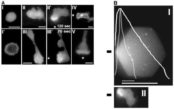
Here, we stably expressed PHAKT-GFP in an immortalized mammalian cell line, HL-60, which can be induced to differentiate into neutrophil-like cells (7, 8). PHAKT-GFP, localized mostly in the cytoplasm of unstimulated differentiated HL-60 cells (Fig. 1A, I), translocated to the plasma membrane when the cells were exposed to a uniform concentration (100 nM) of either of two neutrophil chemoattractants, _N_-formyl-Met-Leu-Phe (f MLP) (Fig. 1A, I′) and C5a (see below; Fig. 4B, VII). This translocation, seen in virtually every cell [96%, Web figure 4D (9)], was rapid and transient, reaching a peak after ~30 s and decreasing over the ensuing 2 min [supplemental figures and videos show the time course of PHAKT-GFP translocation (9)].
Fig. 1.
Translocation of PHAKT-GFP to the plasma membrane of neutrophil-differentiated HL-60 cells. (A) PHAKT-GFP–expressing cells (I through III), C5aR-GFP–expressing cells (IV), and GFP-expressing cells (V) were differentiated to neutrophil-like cells (7) and plated on glass cover slips as described (4). Cells were stimulated either with a uniform increase in _f_MLP concentration, from 0 (I) to 100 nM (I′: 60 s post-stimulation) or with a point source of _f_MLP (1 μM) delivered by a Femtotip micropipette (10) (II′, III′, IV, and V), whose position is indicated by an asterisk. Panels II and III correspond, respectively, to cells shown in panels II′ and III′, but before stimulation with the micropipette. Images were recorded in the fluorescein isothiocyanate (FITC) channel every 5 s as described (4). Each result was reproduced in at least 10 experiments (11). Bars, 10 μm. Videos of a time course and asymmetric translocation of PHAKT-GFP are available at (9). (B) Extracellular gradient generated with the micropipette (I), relative to the intracellular gradient of PHAKT-GFP recruitment (II). For the experiment in (I), a Femtotips micropipette filled with sulforhodamine, a fluorescent dye, was lowered onto a cover slip overlaid with the buffer used for cell stimulation (4, 12); the image was taken in the rhodamine channel (4) 2 min after repositioning the micropipette in a field lacking dye fluorescence. The micropipette tip was located just outside the illumination field, at the left of the hexagon. The solid white line in (I) indicates the intensity of dye fluorescence ( _y_-axis) along the _x_-axis at the level of the black horizontal bar shown to the left of the figure; this curve presumably reflects the shape of a gradient of chemoattractant of similar molecular size, such as _f_MLP. The _x_-axis indicates distance across the microscopic field (solid white scale bar, 50 μm). Fluorescence intensities at the ends of this solid scale bar in (I) were 1900 and 300 (arbitrary units, after subtracting a background value of 400, obtained outside the hexagon). The dashed white curve in (I) indicates the intensity of PHAKT-GFP fluorescence measured across a diameter of the neutrophil shown in (II), depicted at the same scale as in (I). This cell was exposed to _f_MLP supplied by a micropipette; the diameter across which PHAKT-GFP fluorescence was measured is positioned at the level of the black horizontal bar in (II). The 15-μm dashed white line in (I) represents the intensity of PHAKT-GFP recruitment across this diameter; fluorescence intensities at the two ends of this line were 900 versus 200 (arbitrary units, after subtracting a background of 100 units measured outside the cell boundary). Maximum fluorescence intensity for rhodamine decreases to half its value over a distance of 30 μm, while maximum fluorescence intensity for PHAKT-GFP decreases to half its value over 5 μm.
Fig. 4.
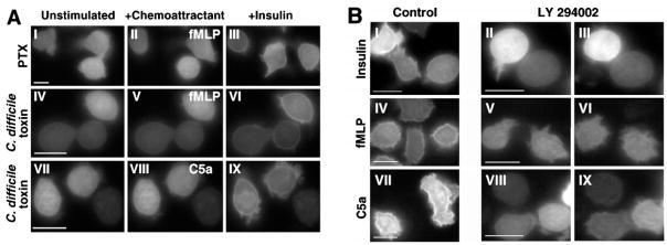
PHAKT-GFP translocation in cells treated with (A) PTX or C. difficile toxin-B and (B) a PI3K inhibitor, LY 294002 (14). (A) Neutrophil-differentiated cells (7), plated on glass cover slips (4), were stimulated sequentially with _f_MLP and insulin (panels II and III and panels V and VI, respectively) or C5a and insulin (panels VIII and IX, respectively). _f_MLP or C5a was added first, removed 2 min later, and replaced with insulin, then cells were stimulated for a further 3 min. Panel I through III: treatment with PTX (1 μg/ml). Panels IV through IX: treatment with C. difficile toxin-B (90 μg/ml). Panels I, IV, and VII: cells before stimulation with agonists (zero time). Images were recorded as described in the legend of Fig. 1. Stimulation times with the indicated agonists were as follows: II: 65 s; III: 115 s; V: 67 s; VI: 178 s; VIII: 39 s; IX: 161 s. Bars, 10 μm. (B) Effect of LY 294002 on chemoattractant- and insulin-induced plasma membrane translocation of PHAKT-GFP in neutrophil-differentiated HL-60 cells. Panels I, IV, and VII: untreated cells stimulated with insulin, _f_MLP, and C5a, respectively. Panels II, V, and VIII: unstimulated cells treated with LY 294002 (100 μM in panel II; 300 μM in panels V and VIII). Panels III, VI, and IX show responses of the same cells to insulin, _f_MLP, or C5a, respectively, in the presence of LY 294002 (100 μM in panel III; 300 μM in panels VI and IX). Images were recorded as described in the legend of Fig. 1. Stimulation times with agonists were as follows: I: 183 s; IV: 46 s; VII: 39 s; III: 199 s; VI: 61 s; IX: 41 s. For each treatment, the result shown is representative of at least eight different stimulation sessions performed on at least two different batches of neutrophil-differentiated HL-60 cells. Bars, 10 μm. Two additional panels of this figure (9) show the effect of 100 μM LY 294002 on chemoattractant-induced plasma membrane translocation of PHAKT-GFP (Web figure 4C) and a histogram (Web figure 4D) of percent cells responding to all three agonists tested in the presence of C. difficile toxin or LY 294002 at 100 or 300 μM.
In a gradient of f MLP, supplied by a nearby micropipette (10), PHAKT-GFP was recruited exclusively to the parts of a cell’s surface that received the strongest stimulation (Fig. 1A, II′ and III′). Indeed, translocation of PHAKT-GFP tightly accompanied actin polymerization and formation of a pseudopod at the leading edge (11) [for videos of this figure, see (9)]. Enrichment of PHAKT-GFP fluorescence at the leading edge contrasted with the uniform distribution of a plasma membrane marker, a GFP-tagged chemoattractant receptor for C5a (C5aR-GFP) (4) expressed in HL-60 cells (Fig. 1A, IV) and the exclusively cytosolic signal seen in HL-60 cells expressing GFP alone (Fig. 1A, V) (11). The internal gradient of PHAKT-GFP distribution is steeper than that of the extracellular stimulus that elicited it (Fig. 1B). From experiments with a fluorescent dye, sulforhodamine (12), we estimate that Femtotips micropipettes generate gradients that are reproducibly linear and rather shallow (~15% decrease in maximum dye concentration per 10 μm) (Fig. 1B, I). We estimate that the gradient of internal cellular signal was at least six times steeper than that of the chemoattractant itself (Fig. 1B, I and II). The asymmetry of the distribution of PHAKT-GFP probably reflects a parallel asymmetry of signals responsible for restricting actin polymerization to the cells’ leading edge.
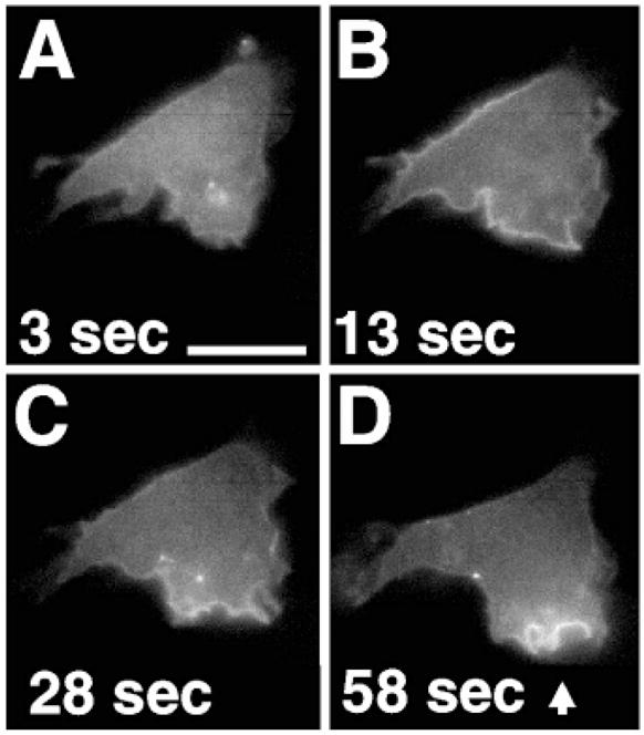
Neutrophils also polarize their morphology, albeit in random directions, when exposed to a uniformly increased concentration of chemoattractant (1, 2, 4). Such a uniform increase in f MLP concentration similarly induced asymmetric recruitment of PHAKT-GFP to the pseudopod (morphologic leading edge) in about 50% of polarizing cells (13). Recruitment of PHAKT-GFP correlated with the direction of membrane protrusion and the underlying actin polymerization, as revealed by the ruffled leading edge [Fig. 2, A through D; for a video of this figure, see (9)]. These observations show the intrinsic capacity of neutrophils to create asymmetric internal signals, not only in shallow chemoattractant gradients, but even in the presence of a uniform concentration of chemoattractant.
Fig. 2.

(A through D) Asymmetric translocation of PHAKT-GFP at the plasma membrane of neutrophil-differentiated HL-60 cells during polarization in response to a uniform increase in chemoattractant concentration. Differentiated cells (7) were plated on glass cover slips as described (4). Cells were stimulated with 100 nM _f_MLP at time 0 and images were then recorded as described in the legend of Fig. 1. The arrow in (D) points to the advancing leading edge of the cell. Uniform stimulation was assessed in 13 different sessions, recording the behavior of 68 cells (13). Bar, 10 μm. A video of this experiment is available at (9).
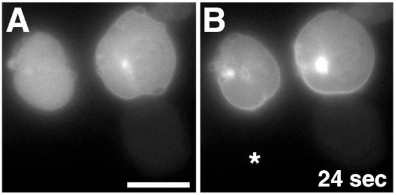
The close temporal and spatial association of actin-containing ruffles and pseudopods with PHAKT-GFP fluorescence raised the possibility that actin polymerization is necessary for translocation of PHAKT-GFP to the plasma membrane of HL-60 cells. In these mammalian cells—as in Dictyostelium (5)—this was not the case, however. Exposure of HL-60 cells to latrunculin-B (14), a toxin that sequesters monomeric actin, caused depolymerization of the dynamic actin cytoskeleton, producing a rounded morphology within 3 to 5 min (Fig. 3A). These cells still recruited PHAKT-GFP asymmetrically to the face closest to a pipet containing f MLP (Fig. 3B). Thus, the signaling machinery of neutrophils, like that of Dictyostelium (5), can amplify the external signaling gradient independently of actin polymerization.
Fig. 3.

Asymmetric translocation of PHAKT-GFP in latrunculin-B–treated neutrophil-differentiated HL-60 cells. Differentiated cells (7) were plated on glass cover slips as described (4). Cells were then pretreated with 20 μg/ml latrunculin-B for 10 min (A) (14) and then stimulated, in the continued presence of the toxin, with a point source of _f_MLP (10 μM) delivered from a micropipette (10) [(B), asterisk]. Images were recorded as described in the legend of Fig. 1. Cells showed asymmetric recruitment biased by the micropipette’s position in five of nine stimulation sessions performed under these conditions. Bar, 10 μm.
Because small GTPases of the Rho family mediate certain neutrophil responses to f MLP (15, 16) and play important roles in relaying signals to the actin cytoskeleton (17), we investigated whether Rho GTPases are required for recruitment of PHAKT-GFP to the neutrophil plasma membrane. A toxin from Clostridium difficile inactivates all three Rho GTPases—Rac, Cdc42, and Rho—by glucosylating a conserved amino acid in the effector domain (18). This toxin (14) induced a round morphology in HL-60 cells (Fig. 4A, IV and VII) and markedly inhibited actin polymerization in response to f MLP (19). The toxin also blocked f MLP- and C5a-induced translocation of PHAKT-GFP and membrane ruffling in most cells (Fig. 4A, V and VIII, respectively). A few cells [~9 to 20%, Web figure 4D (9)] showed detectable (but limited) translocation of PHAKT-GFP. In contrast, in the absence of toxin treatment a uniform concentration of f MLP or C5a induced robust PHAKT-GFP translocation and ruffling in virtually every cell [Fig. 1A, I′; Fig. 4B, IV and VII; Web figure 4D (9)]. Cells treated with C. difficile toxin were not simply unable to respond to extracellular stimuli: subsequent exposure of toxin-treated, f MLP- and C5a-resistant cells to insulin induced translocation of PHAKT-GFP [Fig. 4A, VI and IX, respectively; Web figure 4D (9)], although the ruffling response to insulin was inhibited (Fig. 4A) (20). At lower toxin concentrations (3.8 to 50 μg/ml), inhibition of the membrane ruffling response to f MLP varied widely from cell to cell; under these conditions, f MLP induced PHAKT-GFP translocation preferentially in those cells that showed the ruffling response, suggesting that PHAKT-GFP translocation tightly accompanies activation of Rho GTPases. In none of the conditions we tested (varying toxin concentrations and incubation times) did the toxin affect insulin-induced PHAKT-GFP translocation (19). Pertussis toxin (14) (PTX) blocked both PHAKT-GFP translocation and morphologic responses to f MLP, indicating that these events were mediated by Gi, a pertussis toxin–sensitive G protein [Fig. 4A, II; Web figure 4D (9)]. Conversely, PTX did not prevent responses to insulin [Fig. 4A, III; Web figure 4D (9)].
The PH domain of AKT binds with high affinity to 3′-phosphorylated lipid products of PI3K (21), a well-documented mediator of many actions of insulin (22, 23). At 100 μM, a specific PI3K inhibitor, LY 294002 (14) prevented insulin-induced recruitment of PHAKT-GFP to the plasma membrane (Fig. 4B, III) but did not efficiently block translocation triggered by f MLP or C5a [Web figures 4C and 4D (9)]. At a higher concentration of LY 294002 (300 μM), translocation induced by either f MLP or C5a was almost totally abolished in most cells [Fig. 4B, VI and IX, respectively; Web figure 4D (9)]. Point mutations in the PH domain of AKT (K20A and R25C), previously shown to impair translocation of PHAKT in response to PI3K activation (24) also blocked or severely impaired PHAKT-GFP translocation in HL-60 cells stimulated with f MLP (19). The corresponding residues in a PHAKT analog, PHBTK, interact with the 5-phosphate and 3-phosphate of inositol (1,3,4,5)P4 (25). Thus, the activity of at least one Rho GTPase and lipid products of PI3K seem to be required for the translocation of PHAKT-GFP in neutrophil-differentiated HL-60 cells.
Chemoattractant receptors can activate at least both class 1A and class 1B PI3K’s in neutrophils, whereas the insulin receptor only activates class 1A PI3K (23, 26, 27). This could explain differing sensitivities of these receptor-induced responses to the PI3K inhibitor. Recent studies in transgenic knockout mice found that chemoattractant-induced formation of 3′-phosphorylated lipids, activation of AKT, and chemotaxis of neutrophils depend entirely on p110γ, the only known PI3K of class 1B (28). Similarly, p110γ may be necessary for PHAKT recruitment to the plasma membrane; if so, our experiments with the PI3K inhibitor suggest that chemoattractant-induced translocation of PHAKT only requires activity of a small fraction of the HL-60 cell’s complement of p110γ. PI3K’s of class 1A are activated mainly through the recruitment of their regulatory subunit, p85, to the plasma membrane (23), whereas p110γ is activated directly by βγ subunits liberated from activated heterotrimeric G proteins (23, 26). Thus, it is possible that PHAKT-GFP translocation at the leading edge of motile neutrophils reflect spatially restricted activation of heterotrimeric G proteins. Our results with the toxin suggest, however, that amplification of the PHAKT recruitment requires an intermediate pathway dependent on activity of one or more Rho GTPases.
Our results do not identify the specific Rho GTPase(s) responsible for f MLP-induced recruitment of PHAKT-GFP to membranes of HL-60 cells. Although it is likely that recruitment requires more than one Rho GTPase, we speculate that Cdc42 plays a special role in determining the asymmetry of the f MLP response. Mutant forms of this GTPase or its exchange factor cause yeast (29), macrophages (30), and T lymphocytes (31) to lose their normal ability to polarize selectively toward an extracellular stimulus; such cells orient in random directions instead, like ships with broken compasses. We imagine that Cdc42 constitutes a key element of the neutrophil “compass,” which directs asymmetric translocation of PHAKT-GFP (Figs. 1 through 3) and asymmetric polymerization of actin (1, 2) at the cell’s leading edge in a gradient of chemoattractant.
Supplementary Material
Supplemental figures
Supplemental movie 1
Supplemental movie 2
Supplemental movie 3
Supplemental movie 4
Supplemental movie 5
Supplemental movie 6
References and Notes
- 1.Zigmond SH. J Cell Biol. 1977;75:606. doi: 10.1083/jcb.75.2.606. [DOI] [PMC free article] [PubMed] [Google Scholar]; Devreotes PN, Zigmond SH. Annu Rev Cell Biol. 1988;4:649. doi: 10.1146/annurev.cb.04.110188.003245. [DOI] [PubMed] [Google Scholar]; Cassimeris L, Zigmond SH. Semin Cell Biol. 1990;1:125. [PubMed] [Google Scholar]; Caterina MJ, Devreotes PN. FASEB J. 1991;5:3078. [PubMed] [Google Scholar]; Downey GP. Curr Opin Immunol. 1994;6:113. doi: 10.1016/0952-7915(94)90042-6. [DOI] [PubMed] [Google Scholar]
- 2.Weiner OD, et al. Nature Cell Biol. 1999;1:75. doi: 10.1038/10042. [DOI] [PMC free article] [PubMed] [Google Scholar]
- 3.Xiao Z, et al. J Cell Biol. 1997;139:365. doi: 10.1083/jcb.139.2.365. [DOI] [PMC free article] [PubMed] [Google Scholar]
- 4.Servant G, Weiner OD, Neptune ER, Sedat JW, Bourne HR. Mol Biol Cell. 1999;10:1163. doi: 10.1091/mbc.10.4.1163. [DOI] [PMC free article] [PubMed] [Google Scholar]
- 5.Parent CA, et al. Cell. 1998;95:81. doi: 10.1016/s0092-8674(00)81784-5. [DOI] [PubMed] [Google Scholar]; Parent CA, Devreotes PN. Science. 1999;284:765. doi: 10.1126/science.284.5415.765. [DOI] [PubMed] [Google Scholar]
- 6.Meili R, et al. EMBO J. 1999;18:2092. doi: 10.1093/emboj/18.8.2092. [DOI] [PMC free article] [PubMed] [Google Scholar]
- 7.Culture and differentiation of HL-60 cells with 1.3% dimethyl sulfoxide (DMSO) were performed as described (4).
- 8.The fusions of the PH domains of the protein kinase B (PKB/AKT) kinase and the C5aR with GFP are already described [ Varnai P, Balla T. J Cell Biol. 1998;143:501. doi: 10.1083/jcb.143.2.501.] (4). HL-60 cells were transfected by electroporation. Stable cell lines were generated with 1 mg/ml active G-418. Further details of these methods are available at (9).
- 9.Supplemental material is available at www.sciencemag.org/feature/data/1044239.shl.
- 10.Microscopic analysis of HL-60 cells was performed at room temperature as already described (4). For point-source stimulation, custom-made micropipets with an opening of 0.5 ± 0.2 μm (Eppendorf Femtotips; Fisher Scientific, Pittsburgh, PA) were used. Chemoattractant was back loaded, and air bubbles were pushed out with a microinjection device. The micropipette was placed at the desired coordinates, and a chemotactic gradient was generated with passive diffusion from the tip.
- 11.Seventeen different sessions of point-source stimulation were performed with PHAKT-GFP expressing HL-60 cells. In these experiments, 15 of 23 cells migrating toward the micropipette showed translocation at the leading edge. Failure to detect recruitment in a minority of the polarizing cells probably indicates a threshold phenomenon. That is, the stimulation received by individual cells exposed to the pipette is weaker than that received by cells in a uniform concentration (100 nM) of _f_MLP, which causes recruitment in 96% of cells [Web figure 4D (9)]. Eleven different sessions of point-source stimulation were performed with GFP-expressing HL-60 cells. In these experiments, as expected, we could not detect any increase of the fluorescent signal at the leading edge of 30 cells migrating toward the micropipette. The C5a receptor-GFP chimera (C5aR-GFP) was shown previously to behave as a spatially unbiased probe of plasma membrane concentration when expressed in a neutrophil-like cell line, PLB-985 (4). Similarly, we detected no apparent increase of C5aR-GFP fluorescence at the leading edge of HL-60 cells.
- 12.To determine the apparent shape of the chemotactic gradient generated by the Femtotips, we used a fluorescent dye of molecular weight similar to f MLP (MW: 437.6): sulforhodamine 101 (MW: 606.7; Molecular Probes, Eugene, OR). Two different Femtotips were tested and similar gradients were observed. IVE software [ Chen H, Hughes DD, Chan TA, Sedat JW, Agard DA. J Struct Biol. 1996;116:56. doi: 10.1006/jsbi.1996.0010.] was used to calculate fluorescence intensity along the lines indicated for the fluorescent ligand and PHAKT-GFP recruitment (Fig. 1B).
- 13.Virtually all differentiated HL-60 cells showed recruitment of PHAKT-GFP to the plasma membrane [96%; Web figure 4D (9)] when exposed to a uniform increase in f MLP concentration (from 0 to 100 nM f MLP), but only ~50% of the cells polarized morphologically within the first 4 min of stimulation. Of the cells that did polarize, ~50% (that is, ~25% of the entire population) showed periods of asymmetric recruitment of PHAKT-GFP to ruffles at the cell’s protruding edge.
- 14.Treatment of HL-60 cells with toxins and inhibitor were as follows: latrunculin-B (Calbiochem, La Jolla, CA) was added on plated cells at a final concentration of 20 μg/ml, and cells were incubated for 5 to 10 min at room temperature. PTX (List Biological Laboratories, Campbell, CA), which acts by inhibiting signal transduction by Gi proteins, was added directly to the cell culture medium a final concentration of 1 μg/ml, and cells were incubated at 37¡C for a period of 16 to 22 hours. For LY 294002 (Calbiochem, La Jolla, CA) treatment, a stock solution (100×) was prepared in 100% DMSO just before use. Cells plated on glass cover slips as described (4), were incubated for 20 min at room temperature with a freshly diluted solution (100 to 300 μM) of LY 294002 in modified Hank’s balanced salt solution (4). Clostridium difficile toxin-B (TechLab, Blacksburg, VA), was added directly to the cell culture medium at a final concentration of 90 μg/ml. Cells were incubated at 37¡C with the toxin for not less than 2 hours and not more than 4 hours, a period in which the best inhibition of _f_MLP-induced ruffling and actin polymerization responses was observed without excessive loss of cell viability. Further experimental details for treatments with LY 294002 and C. difficile toxin are available at (9).
- 15.Abo A, et al. Nature. 1991;353:668. doi: 10.1038/353668a0. [DOI] [PubMed] [Google Scholar]; Knaus UG, et al. Science. 1991;254:1512. doi: 10.1126/science.1660188. [DOI] [PubMed] [Google Scholar]; Cox D, et al. J Exp Med. 1997;186:1487. doi: 10.1084/jem.186.9.1487. [DOI] [PMC free article] [PubMed] [Google Scholar]; Bokoch GM. Blood. 1995;86:1649. [PubMed] [Google Scholar]
- 16.Chemotaxis, F-actin generation, and superoxide production induced by chemoattractants are markedly impaired in neutrophils lacking one GTPase, Rac2 [ Roberts AW, et al. Immunity. 1999;10:183. doi: 10.1016/s1074-7613(00)80019-9.].
- 17.Hall A. Annu Rev Cell Biol. 1994;10:31. doi: 10.1146/annurev.cb.10.110194.000335. [DOI] [PubMed] [Google Scholar]; Zigmond SH, et al. J Cell Biol. 1997;138:363. doi: 10.1083/jcb.138.2.363. [DOI] [PMC free article] [PubMed] [Google Scholar]; Zigmond SH, et al. J Cell Biol. 1998;142:1001. doi: 10.1083/jcb.142.4.1001. [DOI] [PMC free article] [PubMed] [Google Scholar]; Ma L, et al. J Cell Biol. 1998;140:1125. doi: 10.1083/jcb.140.5.1125. [DOI] [PMC free article] [PubMed] [Google Scholar]; Ma L, Rohatgi R, Kirschner MW. Proc Natl Acad Sci USA. 1998;95:15362. doi: 10.1073/pnas.95.26.15362. [DOI] [PMC free article] [PubMed] [Google Scholar]; Hall A. Science. 1998;279:509. doi: 10.1126/science.279.5350.509. [DOI] [PubMed] [Google Scholar]; Mackay DJ, Hall A. J Biol Chem. 1998;273:20685. doi: 10.1074/jbc.273.33.20685. [DOI] [PubMed] [Google Scholar]; Bi E, Zigmond SH. Curr Biol. 1999;9:R160. doi: 10.1016/s0960-9822(99)80102-x. [DOI] [PubMed] [Google Scholar]
- 18.Sehr P, et al. Biochemistry. 1998;37:5296. doi: 10.1021/bi972592c. [DOI] [PubMed] [Google Scholar]; Just I, et al. Nature. 1995;375:500. doi: 10.1038/375500a0. [DOI] [PubMed] [Google Scholar]
- 19.Servant G, et al. unpublished data. [Google Scholar]
- 20.Preliminary experiments show that PHAKT-GFP–ex-pressing HL-60 cells also migrated toward a micropipette delivering a point source of insulin. However, under these conditions we have not been able to detect translocation of PHAKT-GFP to the plasma membrane.
- 21.James SR, et al. Biochem J. 1996;315:709. doi: 10.1042/bj3150709. [DOI] [PMC free article] [PubMed] [Google Scholar]; Stokoe D, et al. Science. 1997;277:567. doi: 10.1126/science.277.5325.567. [DOI] [PubMed] [Google Scholar]; Franke TF, et al. Science. 1997;275:665. doi: 10.1126/science.275.5300.665. [DOI] [PubMed] [Google Scholar]; Frech M, et al. J Biol Chem. 1997;272:8474. doi: 10.1074/jbc.272.13.8474. [DOI] [PubMed] [Google Scholar]; Klippel A, et al. Mol Cell Biol. 1997;17:338. doi: 10.1128/mcb.17.1.338. [DOI] [PMC free article] [PubMed] [Google Scholar]; Coffer PJ, Jin J, Woodgett JR. Biochem J. 1998;335:1. doi: 10.1042/bj3350001. [DOI] [PMC free article] [PubMed] [Google Scholar]; Isakoff SJ, et al. EMBO J. 1998;17:5374. doi: 10.1093/emboj/17.18.5374. [DOI] [PMC free article] [PubMed] [Google Scholar]; Kavran JM, et al. J Biol Chem. 1998;273:30497. doi: 10.1074/jbc.273.46.30497. [DOI] [PubMed] [Google Scholar]; Watton SJ, Downward J. Curr Biol. 1999;9:433. doi: 10.1016/s0960-9822(99)80192-4. [DOI] [PubMed] [Google Scholar]
- 22.Ruderman NB, et al. Proc Natl Acad Sci USA. 1990;87:1411. doi: 10.1073/pnas.87.4.1411. [DOI] [PMC free article] [PubMed] [Google Scholar]
- 23.Wymann MP, Pirola L. Biochim Biophys Acta. 1998;1436:127. doi: 10.1016/s0005-2760(98)00139-8. [DOI] [PubMed] [Google Scholar]
- 24.Isakoff SJ, et al. EMBO J. 1998;17:5374. doi: 10.1093/emboj/17.18.5374. [DOI] [PMC free article] [PubMed] [Google Scholar]
- 25.Baraldi E, et al. Structure. 1999;7:449. doi: 10.1016/s0969-2126(99)80057-4. [DOI] [PubMed] [Google Scholar]
- 26.Stephens LR, et al. Cell. 1997;89:105. doi: 10.1016/s0092-8674(00)80187-7. [DOI] [PubMed] [Google Scholar]
- 27.Ptasznik A, et al. J Biol Chem. 1996;271:25204. doi: 10.1074/jbc.271.41.25204. [DOI] [PubMed] [Google Scholar]
- 28.Sasaki T, et al. Science. 2000;287:1040. doi: 10.1126/science.287.5455.1040. [DOI] [PubMed] [Google Scholar]; Li Z, et al. Science. 2000;287:1046. doi: 10.1126/science.287.5455.1046. [DOI] [PubMed] [Google Scholar]; Hirsch E, et al. Science. 2000;287:1049. doi: 10.1126/science.287.5455.1049. [DOI] [PubMed] [Google Scholar]
- 29.Yeast cells expressing an exchange factor for Cdc42 that is unable to bind to the G-protein βγ subunit are unable to orient toward a pheromone gradient [ Nern A, Arkowitz RA. Nature. 1998;391:195. doi: 10.1038/34458.].
- 30.Bac1.2F5 macrophages microinjected with a dominant-negative Cdc42 mutant are able to migrate but do not polarize in the direction of the chemotactic gradient [ Allen WE, et al. J Cell Biol. 1998;141:1147. doi: 10.1083/jcb.141.5.1147.].
- 31.Expression of either dominant-positive or dominant-negative Cdc42 mutants render cells of a T cell hybridoma, 2B4, unable to polarize their actin cytoskeleton toward the antigen-presenting cell [ Stowers L, Yelon D, Berg LJ, Chant J. Proc Natl Acad Sci USA. 1995;92:5027. doi: 10.1073/pnas.92.11.5027.].
- 32.We thank C. Bargmann, D. Stokoe, and A. Weiss for critical reading of the manuscript and M. Rosa for support. Supported in part by NIH grants GM-27800 and CA-54427 (H.R.B.), and GM-25101 (J.W.S.). G.S. is a medical Research Council of Canada Postdoctoral Fellow, and O.D.W. is a Howard Hughes Medical Institute Predoctoral Fellow.
Associated Data
This section collects any data citations, data availability statements, or supplementary materials included in this article.
Supplementary Materials
Supplemental figures
Supplemental movie 1
Supplemental movie 2
Supplemental movie 3
Supplemental movie 4
Supplemental movie 5
Supplemental movie 6

