BTBR Ob/Ob Mutant Mice Model Progressive Diabetic Nephropathy (original) (raw)
Abstract
There remains a need for robust mouse models of diabetic nephropathy (DN) that mimic key features of advanced human DN. The recently developed mouse strain BTBR with the ob/ob leptin-deficiency mutation develops severe type 2 diabetes, hypercholesterolemia, elevated triglycerides, and insulin resistance, but the renal phenotype has not been characterized. Here, we show that these obese, diabetic mice rapidly develop morphologic renal lesions characteristic of both early and advanced human DN. BTBR ob/ob mice developed progressive proteinuria beginning at 4 weeks. Glomerular hypertrophy and accumulation of mesangial matrix, characteristic of early DN, were present by 8 weeks, and glomerular lesions similar to those of advanced human DN were present by 20 weeks. By 22 weeks, we observed an approximately 20% increase in basement membrane thickness and a >50% increase in mesangial matrix. Diffuse mesangial sclerosis (focally approaching nodular glomerulosclerosis), focal arteriolar hyalinosis, mesangiolysis, and focal mild interstitial fibrosis were present. Loss of podocytes was present early and persisted. In summary, BTBR ob/ob mice develop a constellation of abnormalities that closely resemble advanced human DN more rapidly than most other murine models, making this strain particularly attractive for testing therapeutic interventions.
Diabetic nephropathy (DN) is the largest single cause of ESRD in the United States, accounting for nearly half of the patients who enter the dialysis patient population each year and currently accounting for 45% of prevalent kidney failure in the United States.1–4 Although both type 1 and type 2 diabetes lead to DN, the current epidemic of DN is due to type 2 diabetes; however, understanding the mechanisms that produce the constellation of clinical and pathologic alterations that define DN in humans remains very incomplete, in part because clinical DN is a slowly progressive disease, and relevant animal models that produce this constellation of pathologic and clinical abnormalities have important limitations. Mice rendered hyperglycemic by administration of streptozotocin (STZ) or through genetic predisposition such as the db/db mouse can develop some features of DN, most notably glomerular mesangial expansion, but do so only over prolonged periods and do not progress to ESRD.5–9 Most murine models to date have failed to develop reliably marked mesangial expansion or the distinctive nodular glomerulosclerosis with mesangiolysis or hyalinosis characteristic of human disease. Furthermore, the extent of loss or preservation of podocytes, currently accepted to be important in human DN, is generally unknown in each of these models.10 Thus, the availability of a murine model of DN that better resembles its human counterpart and in particular develops glomerular lesions of podocyte loss, mesangiolysis, and severe sclerosis would be a new and significant resource for mechanistic investigations of DN and to test potential therapeutic interventions.
A mouse model of insulin resistance that develops in the progeny of the BTBR (black and tan, brachyuric) mouse strain crossed with C57BL/6 mice has been characterized by Attie and colleagues.11, 12 BTBR mice are naturally hyperinsulinemic when compared with C57BL/6 mice, and BTBR × C57BL/6 F1 mice are substantially insulin resistant.11, 12 Mice homozygous for the ob/ob mutation lack the hormone leptin. When this mutation is on the C57BL/6 background, mice become obese but are only mildly hyperglycemic and do not develop renal lesions characteristic of human diabetes. When the ob/ob mutation is placed on a BTBR background, the mice are initially insulin resistant with elevated insulin levels and pancreatic islet hypertrophy and have marked hyperglycemia by 6 weeks of age.11–15 The C57BL/6 and BTBR strains, when made obese by introduction of the ob/ob mutation, differ significantly in their diabetes susceptibility; C57BL/6 ob/ob mice are insulin resistant but relatively diabetes resistant, whereas BTBR ob/ob mice are insulin resistant and develop severe diabetes.13 BTBR ob/ob mice maintain sustained hyperglycemia (blood glucose 350 to 400 mg/dl) and are largely resistant to the blood glucose–lowering effect of insulin administration. Although there are some sex differences in the diabetic disease manifestations, particularly in the early course of the disease, both sexes are ultimately affected by severe diabetes. In this study, we characterize the development of DN in both the male and female BTBR ob/ob mice.
Results
Blood and Urine Parameters
Both male and female BTBR ob/ob mice have significantly increased blood glucose levels and body weight detectable at 8 weeks when compared with heterozygous BTBR ob+/− and BTBR wild-type (WT) littermates (Table 1, Supplemental Figure 1). Male mice progressed to somewhat higher blood glucose levels, averaging 399.0 ± 38.8 mg/dl at 22 weeks compared with female mice with an average of 333.0 ± 46.3 mg/dl (Table 1). Compared with obese C57BL/6 ob/ob mice, BTBR ob/ob mice have significantly higher blood glucose levels (Table 1). Despite the early hyperglycemia, the growth rate of BTBR ob/ob mice was similar to that of nondiabetic C57BL/6 ob/ob mice (data not shown). Serum triglyceride, cholesterol, and blood urea nitrogen (BUN) levels were elevated in both male and female BTBR ob/ob mice compared with WT littermates, although serum creatinine levels measured by both colorimetric and HPLC methods were not significantly different (Table 1).
Table 1.
Representative laboratory data for BTBR ob/ob mice and C57BL/6 control mice all between 20 and 22 weeks of age (n = 6)
| Parameter | Female | |||
|---|---|---|---|---|
| C57BL/6 | C57BL/6 ob/ob | BTBR | BTBR ob/ob | |
| Glucose (mg/dl) | 135.0±7.5 | 159.5±7.2 | 133±10.2 | 333±46.3a,b |
| BUN (mg/dl) | 35.8±8.0 | 26±2.9 | 27.9±3.0 | 29.0±7.0 |
| Creatinine (HPLC; mg/dl) | ND | ND | 0.211±0.024 | 0.256±0.069 |
| Cholesterol (mg/dl) | 53.5±6.4 | 95.8±8.2c | 93.8±5.5b | 151.5±26.9a, b |
| Triglycerides (mg/dl) | 47.5±2.5 | 65.0±5.0 | 50.8±4.8 | 96.3±31.8c |
| HDL (mg/dl) | 34.5±5.6 | 63.8±8.8a | 64.9±4.4b | 79±10.5b |
| ACR (μg/mg) | 26.2±6.9 | 195.9±29.1 | 94.1±18.9b | 969.9±139.9a, b |
| Albumin, 24-hour (μg) | 9.1±3.7 | 54.0±20.0 | 21.32±7.8 | 248.7±47.1a, d |
| Male | ||||
|---|---|---|---|---|
| C57BL/6 | C57BL/6 ob/ob | BTBR | BTBR ob/ob | |
| Glucose (mg/dl) | 159.5±10.5 | 245.4±33.7a | 147±16.4 | 399±38.8a, b |
| BUN (mg/dl) | 34.8±4.5 | 36.7±2.1 | 19.5±5.0b | 35.0±11.3a |
| Creatinine (HPLC; mg/dl) | ND | ND | 0.180±0.030 | 0.148±0.024 |
| Cholesterol (mg/dl) | 57±5.5 | 165.2±14.6a | 115.2±7.5b | 194.3±21.9a, e |
| Triglycerides (mg/dl) | 49.3±6.9 | 103±2.7c | 129.8±40b | 196.1±34.2a, b |
| HDL (mg/dl) | 33.8±5.2 | 112.8±9.3a | 83.5±14.2b | 88.5±13.9b |
| ACR (μg/mg) | 46.9±7.1 | 263.5±67.7c | 51.1±11.7 | 809.5±134.1a, b |
| Albumin, 24-hour (μg) | 13.6±3.3 | 111.6±36.5 | 17.2±6.0 | 241.8±45.2a, d |
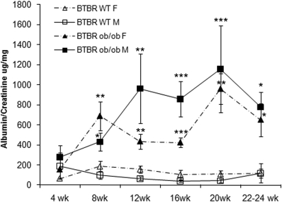
The BTBR ob/ob mice developed albuminuria, with increased albumin-creatinine ratios measured in spot urine samples (Table 1, Figure 1). Elevated albuminuria was detectable as early as 8 weeks of age (Figure 1) and remained elevated thereafter, achieving a 10-fold difference by age 20 weeks. When timed urine collections were obtained, there was also a >10-fold increase in albumin excretion in both male and female BTBR ob/ob mice when compared with littermate controls at 20 weeks (Table 1). Mice were studied up to the age of 22 to 24 weeks, because around this age and beyond, there was greatly increased mortality for reasons yet to be identified.
Figure 1.
BTBR ob/ob mice have markedly increased albuminuria. Albumin-creatinine ratios are significantly increased in BTBR ob/ob mice beginning at 8 weeks of age and remain elevated thereafter. There is a peak difference at 20 weeks, which becomes less marked at later time points as a result of increased mortality of the most severely affected mice, which can no longer be included in these measurements. ***P < 0.001, **P < 0.01, *P < 0.05 versus age- and gender-matched BTBR WT mice.
Renal Structural Alterations
Obese, diabetic BTBR ob/ob mice exhibited renal hypertrophy compared with their BTBR WT littermates (Supplemental Table 1). Compared with C57BL/6 ob/ob mice, which were similarly obese, BTBR ob/ob mice had increased kidney weight, as did BTBR WT mice compared with C57BL/6 WT mice (Supplemental Table 1).
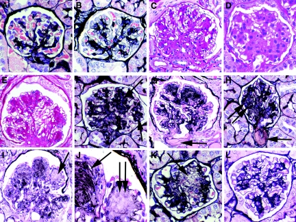
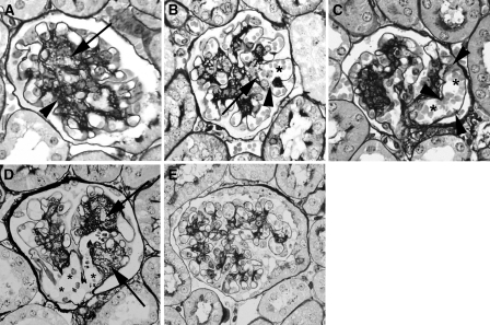
BTBR ob/ob mice develop renal lesions that consist of increasing glomerular mesangial matrix accumulation and are detectable histologically as early as 8 weeks of age (Figure 2). This mesangial matrix accumulation is either preceded or accompanied by episodes of mesangiolysis, because mesangiolysis can be detected in approximately 8% of glomeruli in tissue sections obtained from 8-week-old BTBR ob/ob mice (Figure 3, A and B). The degree of mesangiolysis increases with age, reaching an average of 24.5% of glomeruli exhibiting mesangiolysis at 16 weeks and 33.1% at 22 weeks of age in female mice (Figures 2K and 3, B through D). Male BTBR ob/ob mice had qualitatively similar degrees of mesangiolysis. In contrast, C57BL/6 ob/ob mice do not have markedly increased mesangial matrix or mesangiolysis (Figure 3E). Morphologically advanced glomerular abnormalities identified in both male and female BTBR ob/ob mice include diffuse and, rarely, nodular mesangial sclerosis (Figures 2, F and H, and 4), mesangiolysis (Figures 2K and 3, B through D), focal and mild interstitial fibrosis (Supplemental Figure 2), and, very focally, arteriolar hyalinosis (Figure 2H). Computer-aided morphometry, performed on collagen IV–immunostained tissue sections demonstrated significantly increased glomerular size and progressive accumulation of matrix in both male and female BTBR ob/ob mouse (Table 2, Supplemental Figure 3).
Figure 2.
The BTBR ob/ob mouse models human DN. Advanced DN in the BTBR ob/ob mouse with comparisons to human DN and other murine models of DN. The BTBR ob/ob mouse is a good model of human DN, revealed by the histologic appearances of representative glomeruli from mice and humans with diabetes. (A and B) Normal-appearing glomeruli from a control BTBR mouse (A) and BTBR ob/+ heterozygote mouse (B), both without diabetes. (C) Human DN demonstrating diffuse mesangial sclerosis. (D) Comparable appearance of BTBR ob/ob mouse at 20 weeks, showing diffuse mesangial sclerosis. (E) Human DN with nodular mesangial sclerosis. (F) Comparable appearance of BTBR ob/ob mouse at 20 weeks demonstrating progressive mesangial sclerosis approaching nodular mesangial sclerosis (arrows). (G) Human DN with characteristic hyalinosis of arterioles (arrow). (H) Comparable hyalinosis of arteriole in 21-week-old BTBR ob/ob mouse (arrow). The glomerulus shows diffuse and focally nodular (double arrow) mesangial sclerosis. (I) Human diabetic glomerulosclerosis with nodular mesangial sclerosis and focal lucency and dissolution of the normally compact mesangial matrix (arrow), indicative of mesangiolysis, compared with adjacent nodules composed of solidified matrix. (J) High-power view of H, demonstrating nodular glomerulosclerosis with laminated matrix indicative of repetitive episodes of mesangiolysis and repair (single arrow) and adjacent nodule undergoing mesangiolysis (double arrow). (K) Comparable focus of mesangiolysis with lucency and dissolution of the mesangial matrix in a 20-week-old BTBR ob/ob mouse (arrow). (L) Comparison of human DN in G, I, and J and comparable BTBR ob/ob changes in H and K with the limited mesangial change in similarly aged (22 weeks) leptin receptor–deficient db/db KS mice, the most widely used murine model of DN in type 2 diabetes, shown in L. A, B, and F through L, silver methenamine stain; C through E, periodic acid–Schiff stain.
Figure 3.
Mesangiolysis in BTBR ob/ob mice occurs at early and late stages in the evolution of DN. (A) Glomerulus from an 8-week-old BTBR ob/ob mouse shows characteristic, segmental dissolution (arrow) of normally compact silver staining matrix apparent in other mesangial regions (arrowhead). (B) Eight-week-old glomerulus demonstrates mesangiolysis (arrowhead) resulting in disrupted sites where capillary loops are anchored into the mesangium (arrow), leading to marked dilation/ballooning of the capillary loop (*). (C) A second example of mesangiolysis in 8-week-old BTBR ob/ob mice, with attenuation of mesangial matrix at the capillary luminal interface with resultant loss of anchoring sites of glomerular capillaries (arrowheads), leading to aneurysmal dilation of the capillary loop (*). (D) In older mice (22 weeks), glomeruli show pronounced areas of mesangial lucency (arrows) as well as marked dilation/ballooning of capillary loops (*). (E) C57BL/6 ob/ob mice do not have markedly expanded mesangial matrix or mesangiolysis.
Table 2.
BTBR ob/ob mice have significantly increased accumulations of mesangial matrix
| Age (weeks) | Fractional Volume of Mesangial Matrix in Glomerular Tuft | |||||
|---|---|---|---|---|---|---|
| BTBR WT | BTBR ob/ob | C57BL/6 WT | C57BL/6 ob/ob | C57BLKS/J db/+ | C57BLKS/J db/db | |
| 8 | 13.20±0.60 | 16.70±0.90a | ND | ND | ND | ND |
| 12 | 5.10±1.90 | 13.10±0.02a | ND | ND | ND | ND |
| 16 | 11.70±1.40 | 23.70±1.40b | ND | ND | ND | ND |
| 20 | 7.90±1.60 | 18.30±1.50b | ND | ND | ND | ND |
| 22 | 6.70±1.50 | 18.30±1.50a,c,d | 7.00±0.16 | 6.95±0.53 | 6.40±2.50 | 7.80±3.90 |
| 24 | 11.00±2.20 | 19.70±1.20e,f | ND | ND | 4.90±0.40 | 9.80±2.70 |
Measurement of glomerular capillary basement membrane thickness by electron microscopy showed an increase of 18% in BTBR ob/ob relative to BTBR WT at 20 weeks (181.4 ± 3.8 versus 152.9 ± 5.1 nm; n = 7; P < 0.001). Both electron microscopy and immunofluorescence confirmed the absence of immune deposits (Figure 4, Supplemental Figure 4).
Figure 4.
Ultrastructural changes in BTBR ob/ob mice resemble human DN. (A and B) Electron microscopy of glomeruli of 22-week-old BTBR ob/ob mice shows qualitatively good preservation of foot processes overall. There is increased mesangial matrix and evidence of mesangiolysis with fraying of the mesangial/capillary interface (arrows) in B. (C and D) Basement membranes are thickened and there is focal effacement of foot processes in BTBR ob/ob mice (C) when compared with BTBR WT mice (D). There is no evidence of immune deposits, confirmed by immunofluorescence studies (Supplemental Figure 4). (E) Advanced human DN, occurring after one or more decades of diabetes, also shows marked mesangial matrix accumulation, with similar fraying of the mesangial/capillary interface as seen in BTBR ob/ob mice (double arrows).
Immunostaining for α-smooth muscle actin (α-SMA), a marker of mesangial cell activation, and for Mac-2–positive monocyte/macrophages revealed that both were increased significantly in BTBR ob/ob mice compared with WT littermate controls. Increased Mac-2–positive cells were first detected in the glomerular capillaries of 12-week-old BTBR ob/ob mice, later than when both podocyte loss and mesangiolysis were first detected, and after changes of marked mesangial matrix expansion were already present (Table 3). Measurement of actin-positive mesangial cells within glomeruli was significantly increased in both male and female BTBR ob/ob mice at 22 weeks compared with WT littermates (3.70 ± 0.70 versus 0.17 ± 0.05% [P < 0.001] and 1.49 ± 0.46 versus 0.20 ± 0.06% [P < 0.05], respectively).
Table 3.
BTBR ob/ob mice have significantly more Mac-2–positive cells per glomerulus compared with WT littermates starting at approximately 12 weeks of age, after the time point when mesangiolysis is seen in a number of glomeruli
| Age (weeks) | Mac-2–Positive Monocyte/Macrophages per Glomerular Cross-Section (mean ± SEM) | |||
|---|---|---|---|---|
| Female | Male | |||
| BTBR WT | BTBR ob/ob | BTBR WT | BTBR ob/ob | |
| 4 | 0.62±0.17 | 0.99±0.21 | 0.74±0.26 | 0.48±0.08 |
| 8 | 0.77±0.10 | 1.41±0.17 | 0.68±0.11 | 0.42±0.03 |
| 12 | ND | ND | 0.89±0.07 | 1.63±0.24a |
| 20 | 0.77±0.11 | 1.40±0.28 | 0.82±0.19 | 2.20±0.22b |
| 22 | 0.75±0.06 | 4.30±0.79a | 1.14±0.17 | 10.5±3.70a |
Importantly, BTBR ob/ob mice do not develop atherosclerosis (data not shown), and they do not develop hypertension. In fact, BTBR ob/ob mice are hypotensive compared with BTBR WT and heterozygous controls (Supplemental Figure 5), likely a direct consequence of leptin deficiency.16, 17
Podocyte Number and Density
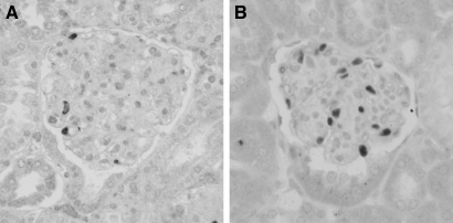
Podocyte number and density were measured at two time points, 8 and 20 weeks in BTBR WT and BTBR ob/ob mice and at 24 (n = 6) and 28 (n = 7) weeks in BTBR ob/ob mice only (Table 4). Podocyte number was reduced to an equivalent degree in BTBR ob/ob mice using both the Weibel and Sanden methods. Podocyte density was significantly reduced in BTBR ob/ob mice compared with BTBR WT mice at every time point studied (data using Sanden method illustrated in Table 4, Figure 5). The diminished density occurs in conjunction with increased glomerular volumes (0.530 ± 0.051 versus 0.270 ± 0.017 μ3 BTBR ob/ob versus BTBR WT at 20 weeks; P < 0.001). There was no increase in the number of apoptotic podocytes in the BTBR ob/ob mice at either early (8 weeks) or late (20 weeks) time points compared with WT littermates, and only extremely rarely were any terminal deoxynucleotidyl transferase–mediated digoxigenin-deoxyuridine nick-end labeling (TUNEL)-positive podocytes seen in any of the tissue sections examined (data not shown).
Table 4.
BTBR ob/ob mice have decreased podocyte number and podocyte density when compared to their WT littermates, apparent as early as 8 weeks of age, reaching statistical significance at 20 weeks of age
| Parameter | BTBR WT | BTBR ob/ob |
|---|---|---|
| Podocyte no. (per glomerular tuft) | ||
| 8 weeks | 85.44±12.61 | 69.57±6.50 |
| m20 weeks | 96.28±6.70 | 70.77±6.41a |
| Glomerular volume (×106 μm3) | ||
| 8 weeks | 0.210±0.014 | 0.290±0.023 |
| 20 weeks | 0.270±0.017 | 0.530±0.051b,c |
| Podocyte density (cell number/100 μm3 glomerular volume) | ||
| 8 weeks | 4.01±0.46 | 2.49±0.28a |
| 20 weeks | 3.57±0.33 | 1.35±0.10b |
| 24 weeks | ND | 1.20±0.15d |
| 28 weeks | ND | 0.99±0.13d |
Figure 5.
BTBR ob/ob mice have reduced podocyte number. (A and B) There is a reduction in podocyte number, assessed by WT-1 staining, in BTBR ob/ob (A) compared with BTBR WT (B) mice.
Interstitial Fibrosis
Diffuse interstitial fibrosis was not detected using conventional histologic stains in any cohort studied, although focal and mild fibrosis was present in ≥ 12-week-old BTBR ob/ob mice (Supplemental Figure 2). Computer-aided morphometry performed on picrosirius red–stained slides to measure the degree of interstitial collagen accumulation showed significantly increased collagen accumulation in BTBR ob/ob mice at 24 weeks when compared with BTBR WT littermates (0.01000 ± 0.00180 versus 0.00086 ± 0.00025%; P < 0.001, percentage of cortical interstitial area picrosirius red positive, excluding perivascular areas).
Comparisons with Controls
Comparisons of renal structural alterations achieved in BTBR and C57BL/6 mice treated with STZ (Supplemental Figure 6), C57BL/6 ob/ob mice (Figure 3E), and C57BLKS/J db/db (Figure 2L) with those in comparably aged BTBR ob/ob mice (Figure 2, D, F, and K) are illustrated in multiple figures. There was mildly to moderately increased mesangial matrix accumulation in STZ-treated BTBR WT mice at 38 weeks of age (30 weeks after STZ injection) compared with STZ-treated C57BL/6 mice and citrate buffer–treated control mice (BTBR WT and C57BL/6; Supplemental Figure 6). The generally mild and focally moderate mesangial matrix accumulation in STZ-treated mice was comparable to that in BTBR ob/ob mice at 8 weeks of age and exceeded that of 22-week-old C57BLKS db/db mice. The BTBR and C57BL/6 mice treated with STZ as well as the C57BL/6 ob/ob mice did not develop glomerular nodular lesions, arteriolar changes, or interstitial fibrosis. Supplemental Table 2 provides functional comparisons of blood glucose levels and albuminuria for these groups of mice. Together, these data demonstrate that hyperglycemia and BTBR strain or obesity are insufficient parameters for rapid development of the DN phenotype, but an interaction of BTBR strain and hyperglycemia with obesity and/or leptin deficiency also must be involved.
Discussion
What are the key criteria for an animal model of DN? The National Institutes of Health–funded Animal Models of Diabetic Complications Consortium (AMDCC) recently revisited this question and proposed as guidelines the following three criteria for an ideal mouse model: (1) Progressive renal insufficiency in the setting of hyperglycemia, more specifically characterized as >50% decline in GFR during the lifetime of the animal; (2) albuminuria (>10-fold increase compared with age-, gender-, and strain-matched controls); and (3) characteristic pathologic changes including basement membrane thickening by electron microscopy, advanced mesangial matrix expansion with or without mesangiolysis and nodular mesangial sclerosis, interstitial fibrosis, and any degree of arteriolar hyalinosis.5, 7 It was explicitly recognized by the consortium that it may not be possible to achieve all of these alterations in models that nonetheless remain useful.
The BTBR ob/ob mouse model of DN comes close to meeting all of the proposed criteria of the AMDCC (albuminuria, pathologic changes) and offers several important advantages compared with existing DN models. The most important of these is the degree to which it reproduces essential structural and functional features of human diabetic glomerular injury. Glomerular hypertrophy, marked expansion of mesangial matrix, mesangiolysis, capillary basement membrane thickening, and loss of podocytes each have been identified as characteristic features of diabetic glomerular injury in humans, and each is present in the BTBR ob/ob model. The functional consequence of these changes in humans—marked proteinuria—also is present in this mouse model with a 10-fold increase in urinary protein excretion compared with controls.
Second, the model is robust and progressive: BTBR ob/ob mice uniformly develop features of DN and do so in a predictable time course in which podocyte loss is already detectable by 8 weeks of age and persists throughout the disease. The basis for podocyte loss was not established in this study, although the lack of significant apoptosis detected by TUNEL staining suggests other means of cell death or detachment are likely important. Studies are under way to investigate the mechanism of podocyte loss in these mice. Significant proteinuria is detectable as early as 8 weeks of age, corresponding with detectable podocyte loss, although it can be detected in some mice at even earlier ages, albeit without achieving statistical significance, when comparing 4-week-old cohorts with controls. Mesangiolysis is also an early feature of the disease, detectable in approximately 10% of glomeruli at 8 weeks of age, and coincides with detectable expansion of the mesangial matrix. These mesangial alterations are progressive. Many murine models of DN, such as STZ-induced DN, develop mild to moderate mesangial expansion and hence are good models of lesions occurring early in the course of human DN; the BTBR ob/ob mouse is among the very few models in which pronounced mesangial expansion and mesangiolysis, modeling advanced human DN, predictably develops.
Third, DN develops more rapidly in BTBR ob/ob mice compared with models of leptin receptor deficiency (db/db mice) or most other mouse models currently used to study DN,7 which often require 30 to 50 weeks or more to develop relevant lesions. The relatively rapid onset allows opportunities for testing therapeutic strategies aimed at halting or ameliorating DN in a much shorter time span, especially important in the context of working with a model organism that under the best of circumstances has a lifespan of approximately 2 years.11, 18, 19
Fourth, there is increasing recognition of an inflammatory component in human and experimental DN, usually characterized by an influx of monocytes/macrophages. Progression of DN in the BTBR ob/ob mouse is also characterized by an influx of monocytes/macrophages. As in the human lesions, it is not clear whether subpopulations of these cells change during the course of the disease and whether subsets of monocytes/macrophages mediate specific injury processes such as mesangiolysis or contribute to progressive injury, repair, or both. Finally, the absence of hypertension in this model allows identification of mediators of DN independent of the confounding effects of coexisting hypertension as may occur in other models.
The BTBR ob/ob model compares favorably with other leading murine models of advanced DN—those based on endothelial nitric oxide synthase deficiency (eNOS−/−) in which diabetes is induced by STZ or by introducing the db/db mutation.20, 21 These models, unlike STZ-induced DN in other strains, have in common the features of mesangiolysis. We hypothesize, on the basis of this finding and the common finding of mesangiolysis in advanced human DN, that mesangiolysis is an essential injury for the development of advanced DN. Two key differences between the BTBR and eNOS−/− models are that the BTBR leptin-deficient model offers the potential of reversibility of established DN lesions with administration of leptin that is currently lacking in murine models in which eNOS is constitutively absent and that the BTBR model lacks the low level of confounding endothelial injury and tendency to thrombogenicity that has been reported in eNOS−/− mice.20 It has been reported that mesangiolysis and mesangial nodular lesions can be prevented by insulin or antihypertensive therapy in eNOS−/− mice made diabetic by STZ.22 The possibility that the unique susceptibility to DN in BTBR ob/ob mice may be due to endothelial dysfunction akin to the defects resulting from eNOS deficiency is being explored.
Like any animal model system, there also are limitations to the BTBR ob/ob model. Most important, this is a strain-dependent model system, as demonstrated by the striking differences between diabetic insulin-resistant BTBR ob/ob mice and similarly obese but nondiabetic C57BL/6 ob/ob mice. BTBR is not a strain familiar to many investigators; however, as differences in strain (and genetic background in humans) have become increasingly recognized as critical determinants of susceptibility to diabetes and nephropathy, this unique background may also be something of an advantage. It has been recognized that the most widely used mouse strain for chemical induction of diabetes by STZ, C57BL/6, is in fact poorly disposed to develop DN.7 A series of genetic studies of BTBR ob/ob mice have identified the genes responsible for insulin resistance and further have identified networks of gene expression in pancreas, adipose tissue, liver, skeletal muscle, and brain that mediate various metabolic abnormalities consequent to diabetes.23 These studies have the potential to provide a basis for understanding pathogenic events in the development of DN that may involve extrarenal sites and systemic perturbations consequent to diabetes and identify genetic differences between BTBR ob/ob mice and DN-resistant mice that may help elucidate critical mechanisms underlying DN. As an example, one gene identified by this analysis, SORCS1, has recently been identified as one of many genes where variants contribute to diabetes risk and glycemic control in humans, further demonstrating the utility of this model for understanding human disease.19, 24
A second potential limitation is that this is a mouse model dependent on both strain (BTBR) and leptin deficiency. Overt leptin deficiency is not a characteristic of human diabetes, although the obesity commonly encountered in patients with type 2 diabetes is associated with leptin resistance.25 This places limits on the overall similarity of leptin-deficient mouse models to the human condition. Nonetheless, the similarity of the diabetic complications that develop in this model to those in humans establish it as a valuable tool to investigate DN and other complications. A third important limitation is that although the glomerular alterations strongly model those of human DN, the degree of interstitial fibrosis achieved at the time points studied, although measurably different from controls, is histologically modest overall. Concomitant measures of serum BUN and creatinine have also failed to suggest significant renal insufficiency. A likely basis for this lack of progressive renal insufficiency in this model despite marked glomerular changes is that measurable loss of renal function and interstitial fibrosis are processes occurring later in the disease course than the glomerulopathy. Although fibrotic changes are present at 20 to 22 weeks, this is an insufficient period for these changes to become advanced or for the most advanced changes of global glomerulosclerosis to develop. We may also detect loss of renal function when we are able to use the more sensitive direct measure of GFR rather than rely on a relatively insensitive measure of serum creatinine for this purpose.
Despite the susceptibility to diabetes and development of DN in BTBR ob/ob mice, studies by the group of Attie et al.13 have shown that lean BTBR mice are normally insulin resistant (high circulating insulin levels) but normoglycemic. These mice are resistant to STZ-induced hyperglycemia, requiring much higher dosages to achieve similar blood glucose levels than STZ-treated C57BL/6 mice, a finding that was confirmed in this study. Hyperglycemic BTBR WT mice developed only modest manifestations of DN, after 30 weeks of hyperglycemia. The unique susceptibility of BTBR ob/ob mice toward developing diabetic complications requires both the BTBR genetic background and metabolic abnormalities conferred by the ob/ob mutation.
We anticipate the BTBR ob/ob mouse will prove an attractive model for study because unlike most other murine models of DN, not only do they develop lesions similar to human DN, but also there is preliminary evidence that complications in other organ systems that are typically encountered in humans with diabetes, such as cardiomyopathy26 and liver disease (data not shown), develop in this model. As a model of leptin deficiency (unlike the db/db leptin receptor–deficient mouse), these mice offer the potential for reversal of disease with leptin administration. Preliminary studies by our laboratory indicate regression of nephropathy can be achieved by this approach; these studies will be the subject of a separate report.
Concise Methods
Animals
The experimental protocol was reviewed and approved by the Animal Care Committee of the University of Washington in Seattle. The establishment of BTBR ob/ob mice has been previously described.11, 12 Breeding pairs of BTBR WT, BTBR/ob heterozygotes [BTBR_ob_+/−; BTBR.V(B6)-Lepob/WiscJ; stock no. 004824]), C57BLKS/J Leprdb (C57BLKS/J db/db), and C57BL/6J/ob mice were purchased from Jackson Laboratories (Bar Harbor, ME) and maintained in an specific pathogen–free facility with a 12-hour light cycle and with free access to standard diet and water. Male and female BTBR ob/ob, BTBR WT, C57BLKS/J db/db, and C57BLKS/J db/+ littermate mice were killed serially at 4, 8, 12, 16, 20, 22, and 24 weeks of age (n = 6 each group). A group of 28-week-old BTBR ob/ob mice (n = 7) were also studied. Male and female C57BL/6 ob/ob and WT littermate mice were killed at 22 and 24 weeks of age (n = 6).
As additional controls, male BTBR and C57BL/6 mice were made diabetic by five daily injections of STZ (80 and 50 mg/kg, respectively) beginning at 8 weeks of age and killed at 38 weeks of age (n = 8 BTBR) and 45 weeks of age (n = 8 C57BL/6), after 30 and 37 weeks of hyperglycemia, respectively. Control mice received five daily injections of citrate. The higher dosage of STZ used in BTBR mice was established in pilot studies conducted to determine the optimal dosage of STZ required to induce diabetes. BTBR WT mice received five daily STZ injections of 40, 50, 60, or 80 mg/kg (n = 3). The mice had an average starting blood glucose level of 130.3 ± 4.2 mg/dl, and this was not appreciably increased by the 40, 50, or 60 mg/kg STZ injections (average blood glucose levels of 100.7 ± 45.7, 130.7 ± 18.8, and 156.0 ± 3.8 mg/dl, respectively), whereas the 80-mg/kg dose resulted in an average blood glucose level of 287.7 ± 43.9 mg/dl after 3 weeks. C57 BL/6 mice treated with five daily doses of STZ at 50 mg/kg achieved blood glucose levels of 342.3 ± 22.3 mg/dl in the same pilot study.
Blood Chemistry
Blood samples were obtained by saphenous vein puncture and at the time when mice were killed. Blood glucose levels were monitored using a Freestyle Blood Glucose Monitor (Abbott Diabetes Care, Alameda, CA).
BUN was measured using the QuantiChrom Urea Assay Kit (BioAssay Systems, Hayward CA). Serum creatinine levels were measured by an HPLC-based method at the Yale University Mouse Metabolic Phenotyping Center metabolic testing core.
Urine Measurements
Timed (12 hour) urine and spot urine samples were collected from individual mice before being killed. Urine was collected during the evening dark cycle, with the mice having access to water but not food. Urinary albumin was measured using the Albuwell M Murine ELISA kit (Exocell, Philadelphia PA), and urinary creatinine was measured with the Creatinine Companion kit (Exocell). Total albumin measured in 12-hour samples was multiplied by 2 to obtain a 24-hour protein excretion rate.
BP Measurement
BPs were measured using the Coda-6 VPR tail-cuff system (Kent Scientific, Torrington, CT)27, 28 on conscious mice, as described previously,29 before mice were killed at the time points described already (n = 6 each group).
Histologic Analysis
Kidneys and other organs were obtained from BTBR WT, BTBR ob/ob, C57BL/6 ob/ob, C57BL/6 WT, C57BLKS/J db/db, C57BLKS/J db/+, and STZ-treated BTBR and C57BL/6 mice at each of the previously indicated time points, and portions were immersion-fixed in 10% neutral-buffered formalin and in methyl Carnoy fixative. Tissues were embedded in paraffin using standard methods; sectioned; and stained with silver methenamine, periodic acid–Schiff, hematoxylin and eosin, and picrosirius red reagents. Selected tissues fixed in ½ strength Karnovsky solution were processed, sectioned, and examined by electron microscopy according to standard protocols. In cases examined by this technique, a series of 10 photographs were taken at × 12,000 magnification, a grid was overlaid on the photograph and basement membrane thickness was measured at points where it intersected with the grid.
Immunohistochemistry and TUNEL
Four-micrometer sections of formalin or methyl Carnoy–fixed, paraffin-embedded tissue were immunostained as described previously.30, 31 The antibodies used were (1) rat anti–Mac-2 (Cedarlane; Hornby, Ontario, Canada32) to detect infiltrating monocytes/macrophages; (2) mouse anti–α-SMA, clone 1A4 (Sigma, St. Louis, MO); (3) rabbit anti–WT-1 (Santa Cruz Biotechnology, Santa Cruz, CA) to mark podocyte nuclei; (4) rat anti–Ki-67, clone Tec3 (Dako, Carpinteria, CA), as a measure of cell proliferation; and (5) rabbit anti–collagen IV (Southern Biotechnology, Birmingham, AL). Negative controls for immunohistochemistry included both substitution of the primary antibody with an isotype-matched irrelevant Ig or antisera from the same species and substitution with PBS. Immunofluorescence was performed on frozen tissue sections to detect the presence of IgG, IgA, IgM, and C3 as described previously.33 Apoptotic cells were detected using the ApopTag Plus kit (Chemicon Int., Temecula, CA) according to the manufacturer's instructions.
Quantitative Analysis of Glomerular and Interstitial Lesions
For each animal, 20 0.1-mm2 section areas were randomly photographed under ×400 magnification, and the glomerular cross-sectional area and degree of glomerular matrix accumulation (collagen IV expression and silver methenamine stain), mesangial actin expression, and area occupied by α-SMA–positive activated mesangial cells were quantified by computer image analysis (ImagePro Plus image analysis software) as described previously.30, 34 Mac-2–positive monocyte/macrophages within glomeruli were counted in a minimum of 50 glomerular cross-sections and expressed as average number of cells per glomerular area. Silver methenamine–stained histologic sections were examined in a blinded manner, and the number of glomeruli exhibiting mesangiolysis (defined as dissolution with areas of lucency of mesangial regions that normally exhibit compact silver staining matrix and/or marked microaneurysmal dilation of adjacent glomerular capillaries) in an entire cross-sectional kidney section was counted. Slides stained with picrosirius red were photographed under polarized light to achieve maximal brightness, and the percentage of positive interstitial staining was quantified using ImagePro Plus software.
Enumeration of Podocytes
Podocyte counting was performed on 3-μm sections of formalin-fixed tissue immunostained with a marker of podocyte nuclei (WT-1). Fifty stained glomerular sections were digitally photographed, and the images were imported into the ImagePro Plus software and analyzed morphometrically. The estimation of the average number of podocytes per glomerulus is then determined by the stereologic method published by Weibel,35 which is based on determining density of podocytes (identified by their WT-1 expression in nuclei) per glomerulus in histologic slides, and the multiplication of this density by the measured glomerular volume to obtain podocyte cell number, as used by others.36–38
Because of published concerns by others that this method may overestimate absolute podocyte numbers,39 a second approach to measure podocyte number and podocyte density was also used. We followed the method of Sanden et al.40 by using WT-1–stained nuclei to enumerate podocytes in kidney tissue sections of uneven thickness (3 and 9 μm). After glomerular volumes were calculated, the counted podocyte nuclei were used to determine podocyte density, a measure that overcomes the problem of cell counts in glomeruli of unequal sizes and that may be a better measure of podocyte integrity.7, 41
Statistical Analysis
All values are expressed as the mean ± SEM. Analysis was performed using InStat StatView for Windows (GraphPad Software, La Jolla, CA), using one-way ANOVA and the Tukey-Kramer Multiple Comparisons Test or the unpaired two-tailed t test to determine P values.
Disclosures
None.
Supplementary Material
[Supplemental Data]
Acknowledgments
This work was supported by an National Institute of Diabetes and Digestive and Kidney Diseases Mouse Metabolic Phenotypic Center's MICROMouse funding program (grant DK076169) and by National Institutes of Health grant DK076126 to the Seattle Mouse Metabolic Phenotyping Center, and by National Institutes of Health grants DK66369 and DK58037 to A.D.A. Measurement of creatinine by HPLC was performed by the Yale MMPC Analytic Core facility, supported in part by National Institute of Diabetes and Digestive and Kidney Diseases grant U24DK76169.
Portions of this work were presented at the annual meetings of the American Society of Nephrology, November 14 through 19, 2006, San Diego, CA; and November 5 through 9, 2008, Philadelphia, PA.
Footnotes
Published online ahead of print. Publication date available at www.jasn.org.
See related editorial, “Progress in Progression?” on pages 1414–1416.
Supplemental information for this article is available online at http://www.jasn.org/.
REFERENCES
- 1.US Renal Data System: USRDS 2004 Annual Data Report, Bethesda, National Institutes of Health, National Institute of Diabetes and Digestive and Kidney Diseases, 2004 [Google Scholar]
- 2.National Kidney Foundation: K/DOQI clinical practice guidelines for chronic kidney disease: Evaluation, classification, and stratification. Am J Kidney Dis 39[Suppl 1]: S1–S266, 2002 [PubMed] [Google Scholar]
- 3.Mohanram A, Toto RD:Outcome studies in diabetic nephropathy.Semin Nephrol 23: 255–271, 2003 [DOI] [PubMed] [Google Scholar]
- 4.Narayan KM, Boyle JP, Thompson TJ, Sorensen SW, Williamson DF: Lifetime risk for diabetes mellitus in the United States. JAMA 290: 1884–1890, 2003 [DOI] [PubMed] [Google Scholar]
- 5.Breyer MD, Bottinger E, Brosius FC, Coffman TM, Fogo A, Harris RC, Heilig CW, Sharma K: Diabetic nephropathy: Of mice and men. Adv Chronic Kidney Dis 12: 128–145, 2005 [DOI] [PubMed] [Google Scholar]
- 6.Breyer MD, Bottinger E, Brosius FC, 3rd, Coffman TM, Harris RC, Heilig CW, Sharma K: Mouse models of diabetic nephropathy. J Am Soc Nephrol 16: 27–45, 2005 [DOI] [PubMed] [Google Scholar]
- 7.Brosius FC, 3rd, Alpers CE, Bottinger EP, Breyer MD, Coffman TM, Gurley SB, Harris RC, Kakoki M, Kretzler M, Leiter EH, Levi M, McIndoe RA, Sharma K, Smithies O, Susztak K, Takahashi N, Takahashi T: Mouse models of diabetic nephropathy. J Am Soc Nephrol 20: 2503–2512, 2009 [DOI] [PMC free article] [PubMed] [Google Scholar]
- 8.Janssen U, Phillips AO, Floege J: Rodent models of nephropathy associated with type II diabetes. J Nephrol 12: 159–172, 1999 [PubMed] [Google Scholar]
- 9.Tesch GH, Nikolic-Paterson DJ: Recent insights into experimental mouse models of diabetic nephropathy. Nephron Exp Nephrol 104: e57–e62, 2006 [DOI] [PubMed] [Google Scholar]
- 10.Jefferson JA, Shankland SJ, Pichler RH: Proteinuria in diabetic kidney disease: A mechanistic viewpoint. Kidney Int 74: 22–36, 2008 [DOI] [PubMed] [Google Scholar]
- 11.Clee SM, Nadler ST, Attie AD: Genetic and genomic studies of the BTBR ob/ob mouse model of type 2 diabetes. Am J Ther 12: 491–498, 2005 [DOI] [PubMed] [Google Scholar]
- 12.Ranheim T, Dumke C, Schueler KL, Cartee GD, Attie AD: Interaction between BTBR and C57BL/6J genomes produces an insulin resistance syndrome in (BTBR x C57BL/6J) F1 mice. Arterioscler Thromb Vasc Biol 17: 3286–3293, 1997 [DOI] [PubMed] [Google Scholar]
- 13.Clee SM, Attie AD: The genetic landscape of type 2 diabetes in mice. Endocr Rev 28: 48–83, 2007 [DOI] [PubMed] [Google Scholar]
- 14.Stoehr JP, Byers JE, Clee SM, Lan H, Boronenkov IV, Schueler KL, Yandell BS, Attie AD: Identification of major quantitative trait loci controlling body weight variation in ob/ob mice. Diabetes 53: 245–249, 2004 [DOI] [PubMed] [Google Scholar]
- 15.Stoehr JP, Nadler ST, Schueler KL, Rabaglia ME, Yandell BS, Metz SA, Attie AD: Genetic obesity unmasks nonlinear interactions between murine type 2 diabetes susceptibility loci. Diabetes 49: 1946–1954, 2000 [DOI] [PubMed] [Google Scholar]
- 16.Aizawa-Abe M, Ogawa Y, Masuzaki H, Ebihara K, Satoh N, Iwai H, Matsuoka N, Hayashi T, Hosoda K, Inoue G, Yoshimasa Y, Nakao K: Pathophysiological role of leptin in obesity-related hypertension. J Clin Invest 105: 1243–1252, 2000 [DOI] [PMC free article] [PubMed] [Google Scholar]
- 17.Burgueno AL, Landa MS, Schuman ML, Alvarez AL, Carabelli J, Garcia SI, Pirola CJ: Association between diencephalic thyroliberin and arterial blood pressure in agouti-yellow and ob/ob mice may be mediated by leptin. Metabolism 56: 1439–1443, 2007 [DOI] [PubMed] [Google Scholar]
- 18.Clee SM, Yandell BS, Schueler KM, Rabaglia ME, Richards OC, Raines SM, Kabara EA, Klass DM, Mui ET, Stapleton DS, Gray-Keller MP, Young MB, Stoehr JP, Lan H, Boronenkov I, Raess PW, Flowers MT, Attie AD: Positional cloning of Sorcs1, a type 2 diabetes quantitative trait locus. Nat Genet 38: 688–693, 2006 [DOI] [PubMed] [Google Scholar]
- 19.Goodarzi MO, Lehman DM, Taylor KD, Guo X, Cui J, Quinones MJ, Clee SM, Yandell BS, Blangero J, Hsueh WA, Attie AD, Stern MP, Rotter JI: SORCS 1: A novel human type 2 diabetes susceptibility gene suggested by the mouse. Diabetes 56: 1922–1929, 2007 [DOI] [PubMed] [Google Scholar]
- 20.Nakagawa T, Sato W, Glushakova O, Heinig M, Clarke T, Campbell-Thompson M, Yuzawa Y, Atkinson MA, Johnson RJ, Croker B: Diabetic endothelial nitric oxide synthase knockout mice develop advanced diabetic nephropathy. J Am Soc Nephrol 18: 539–550, 2007 [DOI] [PubMed] [Google Scholar]
- 21.Zhao HJ, Wang S, Cheng H, Zhang MZ, Takahashi T, Fogo AB, Breyer MD, Harris RC: Endothelial nitric oxide synthase deficiency produces accelerated nephropathy in diabetic mice. J Am Soc Nephrol 17: 2664–2669, 2006 [DOI] [PMC free article] [PubMed] [Google Scholar]
- 22.Kosugi T, Heinig M, Nakayama T, Connor T, Yuzawa Y, Li Q, Hauswirth WW, Grant MB, Croker BP, Campbell-Thompson M, Zhang L, Atkinson MA, Segal MS, Nakagawa T: Lowering blood pressure blocks mesangiolysis and mesangial nodules, but not tubulointerstitial injury, in diabetic eNOS knockout mice. Am J Pathol 174: 1221–1229, 2009 [DOI] [PMC free article] [PubMed] [Google Scholar]
- 23.Keller MP, Choi Y, Wang P, Davis DB, Rabaglia ME, Oler AT, Stapleton DS, Argmann C, Schueler KL, Edwards S, Steinberg HA, Chaibub Neto E, Kleinhanz R, Turner S, Hellerstein MK, Schadt EE, Yandell BS, Kendziorski C, Attie AD: A gene expression network model of type 2 diabetes links cell cycle regulation in islets with diabetes susceptibility. Genome Res 18: 706–716, 2008 [DOI] [PMC free article] [PubMed] [Google Scholar]
- 24.Paterson AD, Waggott D, Boright AP, Hosseini SM, Shen E, Sylvestre MP, Wong I, Bharaj B, Cleary PA, Lachin JM, Below JE, Nicolae D, Cox NJ, Canty AJ, Sun L, Bull SB: A genome-wide association study identifies a novel major locus for glycemic control in type 1 diabetes, as measured by both HbA1c and glucose. Diabetes 59: 539–549, 2010 [DOI] [PMC free article] [PubMed] [Google Scholar]
- 25.Enriori PJ, Evans AE, Sinnayah P, Cowley MA: Leptin resistance and obesity. Obesity (Silver Spring) 14[Suppl 5]: 254S–258S, 2006 [DOI] [PubMed] [Google Scholar]
- 26.O’Brien K, Kim J, Wietecha T, Hudkins K, McDonald T, Minami E, Alpers C: A new murine model of diabetic cardiomyopathy: The female leptin-deficient, black and tan brachyuric mouse. Circulation 120: S490, 2009 [Google Scholar]
- 27.Whitesall SE, Hoff JB, Vollmer AP, D'Alecy LG: Comparison of simultaneous measurement of mouse systolic arterial blood pressure by radiotelemetry and tail-cuff methods. Am J Physiol Heart Circ Physiol 286: 2408–2415, 2004 [DOI] [PubMed] [Google Scholar]
- 28.Feng M, Whitesall S, Zhang Y, Beibel M, D'Alecy L, DiPetrillo K: Validation of volume-pressure recording tail-cuff blood pressure measurements. Am J Hypertens 21: 1288–1291, 2008 [DOI] [PubMed] [Google Scholar]
- 29.Guo S, Kowalewska J, Wietecha TA, Iyoda M, Wang L, Yi K, Spencer M, Banas M, Alexandrescu S, Hudkins KL, Alpers CE: Renin-angiotensin system blockade is renoprotective in immune complex-mediated glomerulonephritis. J Am Soc Nephrol 19: 1168–1176, 2008 [DOI] [PMC free article] [PubMed] [Google Scholar]
- 30.Taneda S, Hudkins KL, Cui Y, Farr AG, Alpers CE, Segerer S: Growth factor expression in a murine model of cryoglobulinemia. Kidney Int 63: 576–590, 2003 [DOI] [PubMed] [Google Scholar]
- 31.Alpers CE, Seifert RA, Hudkins KL, Johnson RJ, Bowen-Pope DF: Developmental patterns of PDGF B-chain, PDGF-receptor, and alpha-actin expression in human glomerulogenesis. Kidney Int 42: 390–399, 1992 [DOI] [PubMed] [Google Scholar]
- 32.Rosenberg I, Cherayil BJ, Isselbacher KJ, Pillai S: Mac-2-binding glycoproteins: Putative ligands for a cytosolic beta-galactoside lectin. J Biol Chem 266: 18731–18736, 1991 [PubMed] [Google Scholar]
- 33.Taneda S, Segerer S, Hudkins KL, Cui Y, Wen M, Segerer M, Wener MH, Khairallah CG, Farr AG, Alpers CE: Cryoglobulinemic glomerulonephritis in thymic stromal lymphopoietin transgenic mice. Am J Pathol 159: 2355–2369, 2001 [DOI] [PMC free article] [PubMed] [Google Scholar]
- 34.Taneda S, Pippin JW, Sage EH, Hudkins KL, Takeuchi Y, Couser WG, Alpers CE: Amelioration of diabetic nephropathy in SPARC-null mice. J Am Soc Nephrol 14: 968–980, 2003 [DOI] [PubMed] [Google Scholar]
- 35.Weibel ER: Practical Methods for Biological Morphometry, London, Academic Press, 1979, pp 40–116 [Google Scholar]
- 36.Pagtalunan ME, Miller PL, Jumping-Eagle S, Nelson RG, Myers BD, Rennke HG, Coplon NS, Sun L, Meyer TW: Podocyte loss and progressive glomerular injury in type II diabetes. J Clin Invest 99: 342–348, 1997 [DOI] [PMC free article] [PubMed] [Google Scholar]
- 37.Steffes MW, Schmidt D, McCrery R, Basgen JM: Glomerular cell number in normal subjects and in type 1 diabetic patients. Kidney Int 59: 2104–2113, 2001 [DOI] [PubMed] [Google Scholar]
- 38.Macconi D, Bonomelli M, Benigni A, Plati T, Sangalli F, Longaretti L, Conti S, Kawachi H, Hill P, Remuzzi G, Remuzzi A: Pathophysiologic implications of reduced podocyte number in a rat model of progressive glomerular injury. Am J Pathol 168: 42–54, 2006 [DOI] [PMC free article] [PubMed] [Google Scholar]
- 39.White KE, Bilous RW: Estimation of podocyte number: A comparison of methods. Kidney Int 66: 663–667, 2004 [DOI] [PubMed] [Google Scholar]
- 40.Sanden SK, Wiggins JE, Goyal M, Riggs LK, Wiggins RC: Evaluation of a thick and thin section method for estimation of podocyte number, glomerular volume, and glomerular volume per podocyte in rat kidney with Wilms’ tumor-1 protein used as a podocyte nuclear marker. J Am Soc Nephrol 14: 2484–2493, 2003 [DOI] [PubMed] [Google Scholar]
- 41.Siu B, Saha J, Smoyer WE, Sullivan KA, Brosius FC, 3rd: Reduction in podocyte density as a pathologic feature in early diabetic nephropathy in rodents: Prevention by lipoic acid treatment. BMC Nephrol 7: 6, 2006 [DOI] [PMC free article] [PubMed] [Google Scholar]
Associated Data
This section collects any data citations, data availability statements, or supplementary materials included in this article.
Supplementary Materials
[Supplemental Data]


