LTβR signaling in dendritic cells induces a type I IFN response that is required for optimal clonal expansion of CD8+ T cells (original) (raw)
Abstract
During an immune response, antigen-bearing dendritic cells (DCs) migrate to the local draining lymph node and present antigen to CD4+ helper T cells. Antigen-activated CD4+ T cells then up-regulate TNF superfamily members including CD40 ligand and lymphotoxin (LT)αβ. Although it is well-accepted that CD40 stimulation on DCs is required for DC licensing and cross-priming of CD8+ T-cell responses, it is likely that other signals are integrated into a comprehensive DC activation program. Here we show that a cognate interaction between LTαβ on CD4+ helper T cells and LTβ receptor on DCs results in unique signals that are necessary for optimal CD8+ T-cell expansion via a type I IFN-dependent mechanism. In contrast, CD40 signaling appears to be more critical for CD8+ T-cell IFNγ production. Therefore, different TNF family members provide integrative signals that shape the licensing potential of antigen-presenting DCs.
CD8+ T-cell responses are crucial for host responses to viral infection and are also involved in allograft rejection and tumor immunity. In some settings, provision of T-cell help is required to adequately license dendritic cells (DCs) for cross-priming of CD8+ T-cell responses (1). However, the nature of these help signals remains incompletely characterized, and it is unclear how these signals integrate into a program of DC maturation. Manipulation of such signals represents a promising therapeutic approach for promoting tumor immunity or for quieting autoimmune disease.
Tumor necrosis factor (TNF) family members including CD40 ligand (CD40L), RANK ligand, LIGHT, and lymphotoxin-αβ (LTαβ) are rapidly up-regulated on antigen (Ag)-activated CD4+ helper T cells (2, 3). During the immune response, DC/T-cell interactions result in the ligation of CD40, CD70, and RANK on DCs, and this has been shown to promote DC cross-priming capacity and survival (4–8). We have recently shown that LTαβ expression on Ag-specific CD4+ T cells is also critical for DC function in the context of protein Ag (3). These studies provoke the question of whether different TNF family pathways are redundant, or somehow act cooperatively in the context of DC maturation.
Previous studies have hinted at a role for the LT pathway in T-cell function (9). Focusing on cases where CD8+ T-cell responses rely on T-cell help, such as CD8+ T-cell responses to allo-Ag (10, 11) and tumor-Ag (12), LTβ-receptor (LTβR) signaling has a significant effect on CD8+ T-cell activation and clonal expansion. To resolve how this pathway may impact the maturation of a CD8+ T-cell response in vivo, we used different approaches whereby we selectively inhibited LTβR signaling on the hematopoietic compartment, or specifically on DCs, to evaluate effects of LTβR signaling on DC-mediated CD8+ T-cell cross-priming. Our data revealed that the LT pathway was important for CD8+ T-cell clonal expansion but not effector function, whereas the CD40 pathway was necessary for CD8+ T-cell function but was dispensable for T-cell expansion in response to cell-associated Ag. LTβR stimulation on DCs was found to provoke a type I interferon (IFN) response even in the absence of added Toll-like receptor (TLR) agonist, and exogenous IFN could recover CD8+ T-cell proliferation, suggesting a mechanism for the effects of this pathway on CD8+ T-cell priming. Therefore, the LT pathway provides necessary and nonredundant DC-intrinsic signals that provoke optimal CD8+ T-cell clonal expansion.
Results
LTβR Signaling Cooperates with CD40-Derived Signals for Priming of CD8+ T Cells in Vivo.
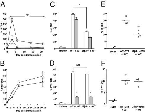
We previously demonstrated that the expression of LTαβ ligand on Ag-specific CD4+ T cells in vivo is required for DC function ex vivo (3). However, a hallmark of DC licensing is the ability to cross-prime a cytotoxic T-cell response. To ascertain the relevance of LTβR signaling on DC licensing, we assessed whether LTβR signaling was required for priming of a CD8+ T-cell response to cell-associated Ag in vivo. To achieve a relatively CD4+ T-cell help-dependent system, spleen and lymph node (LN) cells from bm1 mice were used as syngeneic vehicles for Ag (OVA) delivery so that bm1 cells could not directly present Ag to OTI T cells in bm1xB6 F1 recipient hosts (13). bm1 cells were hypotonically loaded with OVA protein (4) and were used to immunize mice that had received congenically labeled OTI T cells 1 d prior. We first assessed the consequences of global inhibition of LTβR signaling by treating recipient mice with a decoy fusion protein, LTβR-Ig, which was administered the day before immunization with bm1-OVA. We found that, compared with the control treatment group, OTI T-cell expansion was significantly impaired in LTβR-Ig–treated recipient mice (Fig. 1_A_). Reduced frequency of OTI T cells was observed throughout the immune response from day 3 to 21 in the spleen (see Fig. S1 for representative FACS) and the blood (Fig. 1_A_). However, despite this observed decrease in clonal expansion, OTI T cells exhibited no defect in IFNγ production at any time point, with equivalent proportions of IFNγ-producing OTI T cells in both control and LTβR-Ig–treated mice (Fig. 1_B_; see Fig. S1 for representative FACS). Because the homeostasis of DCs in naïve mice is perturbed in LTβR−/− animals (14–16), we confirmed that short-term treatment with the LTβR-Ig agent did not result in a reduction in DC numbers, an alteration in DC phenotype, or an inability to acquire and process OVA protein for presentation on MHC class I (Fig. S2). Therefore, LTβR-Ig treatment results in suboptimal clonal expansion, but not effector function, of CD8+ T cells in response to cell-associated Ag.
Fig. 1.
DC-derived LTβR and CD40 signals contribute distinctly to CD8+ T-cell cross-priming in vivo. (A and B) WT bm1xB6 F1 mice were given an A/T of responder CD45.1 or Thy1.1 OTI T cells, treated with either control human IgG (huIgG) or LTβR-Ig, and immunized the next day with OVA-loaded bm1 cells. At multiple days postimmunization, OTI expansion (A) and IFNγ production (B) were measured. Data are representative of six (A) or two (B) independent experiments (n = 3–6 per experiment). (C and D) WT→WT or LTβR−/−→WT chimeric mice were given an A/T of responder CD45.1 or Thy1.1 OTI T cells, treated with either control Ab or α-CD40L, and immunized with OVA-loaded bm1 cells, and OTI expansion (C) and IFNγ production (D) were measured at day 3. These experiments were performed four times with similar results, and results shown represent the average of four mice per group. NS, nonsignificant. (E and F) Mixed chimeric WT + CD11c-DTR→WT or LTβR−/− + CD11c-DTR→WT mice were given an A/T of responder CD45.1 OTI T cells, treated with diphtheria toxin, and immunized with OVA-loaded bm1 cells, and OTI expansion (E) and IFNγ production (F) were measured at day 3. Data are representative of two experiments (n = 4 per experiment). Empty circles, control Ig-treated mice; filled circles, LTβR-Ig–treated mice; white bars, control treated mice; gray bars, α-CD40L–treated mice. *P < 0.05, ***P < 0.0001 (two-way ANOVA for A).
CD40 signaling is a potent maturation cue during DC activation and can also induce CD86 expression on DCs (17). However, these data indicate that in the context of abrogated LTβR signaling, physiological CD40 signaling cannot compensate to maintain DC stimulatory function, and it is unclear whether signals derived from both CD40 and LTβR act additively or synergistically to license DCs for CD8+ T-cell cross-priming or alternatively whether these two pathways contribute something distinct to DC maturation. To resolve this, we generated WT→WT and LTβR−/−→WT chimeric mice, and at 8 wk postreconstitution these mice were injected with OTI T cells and immunized with cell-associated OVA as in Fig. 1_A_. At day 3, which in our system is the peak of the CD8 (OTI) response (see Fig. 1_A_ for kinetics), we observed robust expansion of OTI T cells in WT→WT chimeras; however, in mice lacking LTβR expression on Ag-presenting cells, we observed a statistically significant reduction in OTI T-cell accumulation (Fig. 1_C_, white bars). Not unlike in LTβR-Ig–treated mice, OTI T cells primed in LTβR−/−→WT mice exhibited no defect in IFNγ production, with equivalent proportions of IFNγ-producing OTI T cells in both groups (Fig. 1_D_, white bars). We also confirmed that DCs from LTβR−/− mice expressed normal levels of maturation markers and could acquire and process OVA protein for presentation on MHC class I in a manner that was comparable to DCs from WT mice (Fig. S3). Therefore, under circumstances where the organization of splenic stroma is normal (which depends on LTβR expression in radio-resistant host cells), expression of LTβR in radio-sensitive hematopoietic cells is required for full clonal expansion of OTI T cells.
To determine whether this CD8+ T-cell defect would be exacerbated by the additional absence of CD40 licensing cues, WT→WT and LTβR−/−→WT bone marrow (BM) chimeric mice were treated with α-CD40L-blocking Ab. In contrast to the LTβR−/−→WT BM chimeric mice, we observed no defect in OTI T-cell expansion in WT→WT mice in which CD40 signaling was prevented, and there was a minimal compound defect beyond the impaired OTI expansion in the absence of LTβR signaling when both CD40 and LTβR signaling were simultaneously abrogated (Fig. 1_C_, gray bars). Whereas secretion of IFNγ by OTI T cells was uncompromised in the absence of LTβR licensing, it was grossly impaired in α-CD40L–treated mice (Fig. 1_D_, gray bars). Together, these results identify unique roles for LTβR and CD40 signaling in promoting CD8+ T-cell responses to cell-associated Ag, the former in regulating CD8+ T-cell expansion and the latter in instructing CD8+ T-cell effector function.
DC-Intrinsic LTβR Signaling Is Required for Optimal CD8+ T-Cell Clonal Expansion.
We next addressed whether DCs, which express LTβR (15), are the relevant LTβR+ hematopoietically derived cell required for cross-priming CD8+ T cells. We therefore generated mixed BM chimeras using CD11c-DTR/GFP donor BM along with either WT or LTβR−/− donor BM transferred into lethally irradiated WT hosts, and reconstitution of BM-derived cells was confirmed using GFP and CD45 congenic markers. CD11c-DTR mice express a diphtheria toxin (DT) receptor (DTR) under the control of the CD11c promoter, and treatment of these mice with DT results in thorough but temporary depletion of DCs (18). Treatment of LTβR−/− + CD11c-DTR→WT chimeric mice with DT would therefore specifically deplete WT CD11c+ cells while preserving LTβR−/− DCs. At 12 wk postreconstitution, chimeric mice were given an adoptive transfer (A/T) of responder OTI T cells, and the following day were immunized with OVA-loaded bm1 splenocytes. Chimeric mice were treated with DT on day 0 and day 1 postimmunization, and depletion of CD11c-DTR+ (GFP+) DCs in the blood, spleen, and LN was confirmed. At the peak of the CD8+ response, expansion of OTI T cells was significantly impaired in LTβR−/− + CD11c-DTR chimeras in terms of frequency (P < 0.001; Fig. 1_E_) and also in the case of total numbers of OTI (P < 0.05; Fig. S4), whereas IFNγ production remained intact (Fig. 1_F_), recapitulating the defect observed in LTβR−/−→WT chimeras. Consistent with these data, mice that received LTβ-deficient helper T cells also exhibited a significant reduction in the peak expansion of splenic OTI CD8+ T cells (Fig. S5_A_) but not IFNγ production (Fig. S5_B_) following immunization with OVA-loaded bm1 cells, suggesting that cross-talk between LTαβ-expressing Ag-specific T cells and LTβR+ DCs is required for optimal clonal expansion of CD8+ T cells in response to cell-associated Ag. Collectively, these data identify DC-intrinsic LTβR signaling as a requirement for CD8+ T-cell clonal expansion but not for CD8+ T-cell-derived IFNγ production.
LTβR Signaling Is Required for Full Activation and Cell-Cycle Progression of Ag-Specific CD8+ T Cells.
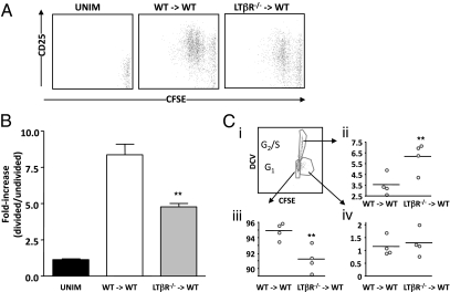
Given the reduction in the clonal burst of Ag-specific CD8+ T cells in immunized LT-inhibited mice, we asked whether the activation, proliferation, or persistence of OVA-specific CD8+ T cells was impaired. At 2 d postimmunization, we measured cell division/activation, and noted a lag in carboxyfluorescein succinimidyl ester (CFSE) dilution as well as a significant reduction in CD25 up-regulation on OTI T cells derived from LTβR−/−→WT mice compared with OTI T cells derived from WT→WT mice (Fig. 2 A and B). The reduction in CFSE dilution suggested that in the absence of LTβR-derived DC licensing, OTI T cells were either not dividing or were dividing but failing to survive. We therefore measured the cell-cycle status of CFSE-labeled OTI from OVA-bm1–immunized chimeric mice. Interestingly, a significant increase in the percent of cycling, CFSEint OTI in LTβR−/−→WT compared with WT→WT chimeric mice was observed (Fig. 2_C_; P < 0.01), and this was accompanied by a significant reduction in the percent of resting/G1 CFSEneg (fully divided) OTI in LTβR−/−→WT versus WT→WT chimeric mice (P < 0.005). Staining for cleaved caspase 3 leading up to and at the peak of the OTI response showed no differences in apoptotic OTI in WT→WT and LTβR−/−→WT mice; however, analysis of dying cells in vivo is unreliable because they are quickly phagocytosed, and although apoptotic demise is common, there exist additional death pathways that may not be captured by techniques designed to measure apoptotic cell death (19). It therefore seems likely that OTI T cells primed in LTβR−/−→WT mice are cycling in equivalent proportion to those primed in WT→WT mice, but are dying before reaching a terminally divided resting state.
Fig. 2.
LTβR signaling is required for normal CD8+ T-cell activation and cell-cycle completion. Chimeric mice (WT→WT or LTβR−/−→WT) were given an A/T of CFSE-labeled responder OTI T cells, and then were either left unimmunized or immunized 1 d later with OVA-loaded bm1 cells. At day 2 postimmunization, CFSE dilution and CD25 up-regulation were evaluated (A), and the extent of CD25 up-regulation on divided versus undivided OTI cells was measured (B). Data are representative of three independent experiments (n = 3–5 mice per experiment). **P < 0.01. (C) To assess cell cycling in the described in vivo experiment from A and B, CFSE-stained OTI T cells were gated and analyzed with DyeCycle Violet to measure cell-cycle status, with a representative FACS plot shown (i). Enumeration of OTI that had divided [CFSEmed (ii)], had “terminally divided” [CFSE− (iii)], or had remained undivided [CFSEhigh (iv)] was performed. Data are representative of two experiments (in one case performed at day 2, and another at day 3, n = 4 per experiment). Note that in the presence of DyeCycle Violet, CFSE intensity appears different from without DyeCycle Violet (A versus C). **P < 0.01.
Stimulation of LTβR on DCs Results in the Production of Type I IFN.
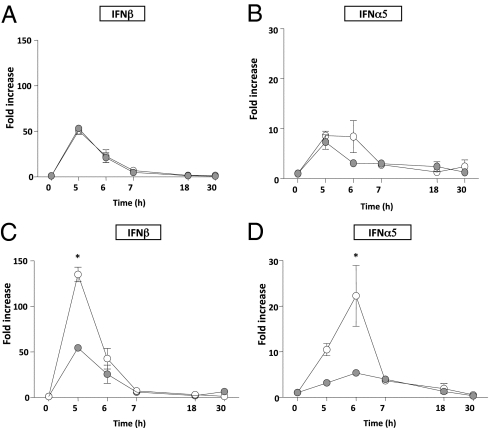
To determine the mechanism whereby DC-intrinsic LTβR signaling contributes to the clonal expansion of CD8+ T cells in vivo, we investigated the possibility that production of type I IFN was downstream of LTβR activation in DCs for several reasons. First, LTβR signaling has been shown to induce type I IFN in radio-resistant stromal cells independently of TLR-derived signals (20). Second, type I IFN has been shown to induce CD25 expression on T cells (21), is required for the optimal clonal expansion of CD8+ T cells (22, 23), and can exert these proproliferation effects independently of CD40/CD40L activity (24). Finally, type I IFN induces the expression of CD86 on DCs (25), and we have observed a transient decrease in CD86 expression on DCs post–OVA immunization in vivo that is recovered in the presence of WT DCs (Fig. S6), suggesting a factor that acts in trans can rescue CD86 expression. Therefore, we reasoned that the poor OTI expansion, the reduced expression of CD25 on OTI T cells, and the failure to up-regulate CD86 on LTβR−/− DCs could all reflect a defect in type I IFN production. To assess whether LTβR signaling induces type I IFN in conventional DCs, we generated bone marrow–derived DCs (BMDCs) and cocultured WT versus LTβR−/− BMDCs with OVA-specific CD4+ helper OTII T cells as a source of licensing signals (CD40L, LTαβ). These cocultures were then supplemented with either LPS alone or LPS + OVA323–339 to induce the expression of LTαβ/CD40L on OTII (3). Exposure to LPS alone induced a baseline level of IFNβ and IFNα5 mRNA, which did not differ between WT and LTβR−/− DCs (open versus closed circles, Fig. 3 A and B). However, the addition of the OTII cognate peptide OVA323–339 to the DC/T-cell cocultures, which activates OTII CD4+ T cells and stimulates them to express LTαβ/CD40L (3), resulted in a robust induction of both IFNβ and IFNα5 mRNA that was blunted in the absence of LTβR expression on DCs (open versus closed circles, Fig. 3 C and D). Indeed, the induction of IFNβ and IFNα5 mRNA from LTβR−/− DCs in response to LPS + OVA323–339 was not any different from what was observed with LPS alone. The reduction in IFNβ and IFNα5 mRNA from LTβR−/− DCs was not due to any developmentally associated DC-intrinsic defect because LTβR−/− BMDCs were similar to WT BMDCs in terms of DC surface marker expression and their capacity to make IL-12 (Fig. S7). Moreover, blockade of LTβR/LTαβ interactions between T cells and WT BMDCs with LTβR-Ig in vitro recapitulated the reduction in IFNβ and IFNα5 expression (Fig. S8). These data indicate that DC-intrinsic LTβR ligation induces a necessary and unique signal(s) that collaborates with TLR4 stimulation to provoke optimal induction of IFNβ and IFNα5 gene expression.
Fig. 3.
LTβR synergizes with TLR4 to maximize type I IFN production in BMDCs. WT (open circles) and LTβR−/− (gray circles) BMDCs were coincubated with OVA-specific CD4+ T cells (OTII) and LPS (A and B). In some cases, these cocultures were also supplemented with OVA323–339 (C and D). DCs were isolated at indicated time points and the expression of IFNβ (A and C) and IFNα5 (B and D) was measured by real-time RT-PCR. This experiment was performed three times with similar results. *P < 0.05.
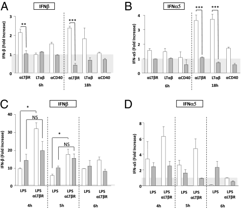
We next assessed whether LTβR signals could provoke type I IFN expression independently of TLR signaling by coculturing WT versus LTβR−/− BMDCs with recombinant LTαβ ligand or with agonist Abs directed at LTβR. Interestingly, in the absence of any added TLR signal, we detected a modest induction of IFNβ and IFNα5 mRNA in response to LTβR ligation in BMDCs (open versus closed bars, Fig. 4 A and B; P < 0.001 and P < 0.01 for IFNβ and IFNα5, respectively, between WT and LTβR−/− BMDCs). IFNβ and IFNα5 induction was specific to the LT pathway, as LTβR−/− BMDCs failed to induce IFNα/β under these conditions. Furthermore, IFNα/β was not significantly induced following stimulation with α-CD40 (Fig. 4 A and B), even though α-CD40 (but not α-LTβR) Abs readily provoked IL-12 secretion from BMDCs (Fig. S7).
Fig. 4.
LTβR signaling can mediate type I IFN production independently of TLR activation in BMDCs. BMDCs were stimulated with anti-LTβR, LTαβ, or anti-CD40 in the absence of any added LPS. Expression of IFNβ (A) and IFNα5 (B) was measured by real-time RT-PCR at 6 and 18 h poststimulation of WT (open bars) and LTβR−/− (gray bars) BMDCs. These experiments were performed at least three times with similar results. *P < 0.05, **P < 0.01, ***P < 0.001. BMDCs were also preincubated with LPS for 2 h, washed, and then stimulated with anti-LTβR. Expression of IFNβ (C) and IFNα5 (D) was measured by real-time RT-PCR at 4, 5, and 6 h poststimulation of WT (open bars) and LTβR−/− (gray bars) BMDCs. This experiment was performed three times with similar results. *P < 0.05 for data in (C).
Because the amount of IFNα/β expression was relatively modest in response to LTαβ ligand or agonist Abs directed at LTβR, we evaluated whether signals through TLR4 and LTβR could collaborate to increase type I IFN expression. Indeed, BMDCs pretreated with LPS showed evidence of further up-regulation of IFNα/β expression when subsequently treated with anti-LTβR agonist Ab, and this effect was not observed for LTβR−/− BMDCs (Fig. 4 C and D). Thus, we postulate that pathogen/danger-associated molecular patterns (PAMPs/DAMPs) precondition DCs to receive signals through the LTβR which further augment IFNα/β expression.
LTβR−/− DCs Fail to Support OTI Proliferation in Vitro but Proliferation Can Be Rescued by Exogenous IFNα.
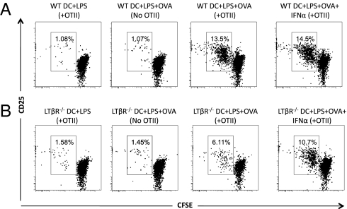
To determine whether reduced type I IFN production contributes to the failure of LTβR−/− DCs to induce full CD8+ T-cell activation and expansion in vivo, we established an in vitro system for measuring OTI T-cell proliferation. Given the flexible nature of such a system, we were able to carefully titrate the amount of LPS and the ratio of DCs to T cells such that OTI T-cell proliferation was rendered help-dependent and, indeed, in the absence of OTII CD4+ T cells, we observed minimal OTI proliferation which was equivalent to what was observed in the absence of any OVA Ag (Fig. 5_A_, left two panels). Although this in vitro system is an artificial system, we observed a similar defect in OTI proliferation when OVA-bm1 cells were introduced into the cultures (Fig. S9). However, because the OTI proliferation observed with bm1-OVA stimulation in vitro was less robust and somewhat delayed compared with LPS + OVA (Fig. 5_A_), we focused on the LPS version of the in vitro assay. Interestingly, using the LPS/OVA system, in the presence of OTII CD4+ T cells, we noted more than a twofold reduction in OTI CFSE dilution when OTI T cells were cultured with LTβR−/− versus WT DCs, confirming a requirement for DC-intrinsic LTβR signaling for optimal OTI expansion in vitro (Fig. 5_A_ versus Fig. 5_B_) and recapitulating the nature and magnitude of our OTI expansion defect that was observed in vivo (Fig. 1). Similar defects in CFSE dilution were observed with LTβR−/− BMDCs cocultured with bm1-OVA in vitro (Fig. S9), and also when WT OTII helper T cells were compared with LTβ−/− OTII CD4+ T cells (Fig. S5). These data demonstrate that LTαβ-LTβR DC licensing signals are required for stimulating OTI expansion in vitro.
Fig. 5.
LTβR−/− BMDCs do not support full OTI proliferation in vitro, and this can be rescued with type I IFN. WT (A) and LTβR−/− (B) BMDCs were preincubated with LPS and OVA protein for 18 h, washed, and then plated with CFSE-labeled OVA-specific CD8+ T cells (OTI) with or without OTII CD4+ T cells. Seventy-two hours later, OTI T cells were gated based on CD45.1 expression and/or CD8 expression, and then assessed for CD25 expression and CFSE dilution. In some cases, 25 U/mL of IFNα was added to the cultures at 48 h. The experiment is a representative example of three independent experiments.
To determine whether type I IFN could rescue the proliferation of OTI T cells primed by LTβR−/− DCs, we added IFNα into our WT or LTβR−/− DC-OTII cocultures and measured OTI proliferation by CFSE dilution. Exogenous IFNα restored proliferation of OTI T cells stimulated with LTβR−/− DCs, but had a minimal effect on proliferation of OTI stimulated by WT DCs (Fig. 5_A_ versus Fig. 5_B_), where maximal type I IFN induction would have been achieved through the combination of LPS and LTβR ligation. Furthermore, although we noted that CD25 levels on OTI T cells from LTβR−/− DC-OTII cocultures were reduced to 65% of the normal CD25 levels observed in WT DC-OTII cocultures, addition of IFNα restored CD25 levels on OTI T cells cocultured with LTβR−/− DCs to levels equivalent to those observed in WT DC-OTII cocultures (94% of normal). Therefore, production of type I IFN is a critical mediator downstream of LTβR signaling in DCs, and recovery of type I IFN levels can rescue the otherwise impaired proliferation of CD8+ T cells primed by LTβR−/− DCs. These data provide a mechanism for the unique function of LTβR in DC-mediated T-cell priming.
Discussion
T-cell help is required in many cases for optimal cross-priming of CD8+ T-cell responses to cell-associated Ag (1). The nature of these help signals, however, remains incompletely characterized. Here we show that DC-intrinsic LTβR signaling is required for ex vivo DC function and for optimal cross-priming of a CD8+ T-cell response in vivo. Our study identifies a nonredundant function for LTβR signaling alongside CD40 stimulation in promoting DC maturation and identifies LTβR-dependent type I IFN production as a unique contribution from this TNF family member in shaping an effective CD8+ T-cell response.
We have previously shown that LTαβ is rapidly up-regulated on OVA-specific CD4+ helper T cells in response to immunization with Ag, and the kinetics of LTαβ expression resemble those for CD69 expression (3). Such kinetics are very similar to the induction of CD40L on CD4+ helper T cells, and represents a form of “help” for DC conditioning in vivo via interaction with LTβR on DCs. Although our studies indicate that LTαβ on cells other than Ag-specific CD4+ T cells (B cells, for example) is insufficient to trigger LTβR on DCs to prime CD8+ T cells, it is possible that LIGHT expression on CD4+ helper T cells may also contribute to LTβR signaling in DCs to facilitate CD8+ T-cell responses to cell-associated Ag, and indeed the defect we observe with LTβR−/− DCs is typically larger than what is observed when we transfer LTβ−/− OTII (Fig. S5). Nevertheless, the expression of LTαβ on CD4+ helper T cells is likely strictly regulated because we observe very little expression on resting CD4+ helper T cells (3) and, indeed, the expression of LTαβ on Th1 and Th17 cells has been exploited for depletion strategies in the context of rodent models of autoimmunity (26).
One possible explanation for our findings is that DC-intrinsic LTβR signaling is required for their homeostatic maintenance in the spleen (14–16). However, we have confirmed that the reduction in OTI expansion is due to impaired DC function and not reduced DC numbers in three ways. First, in WT mice whose splenic DC populations are intact, the absence of LTβ expression on a small population of adoptively transferred helper CD4+ T cells recapitulates the defect in OTI clonal expansion observed in LTβR−/− settings. Second, treatment of WT mice with an LTβR signaling inhibitor, LTβR-Ig, 1 d before immunization again results in a twofold reduction in the peak OTI expansion without any impact on DC numbers. Finally, the defect of OTI expansion was confirmed using LTβR−/− BMDCs (as well as WT DC in the presence of LTβR-Ig) in vitro. Therefore, in scenarios in which DC numbers remain equivalent but the LTαβ-LTβR signal is absent, peak OTI expansion is significantly and comparably reduced. Thus, the LTβR licensing requirement for DC stimulatory function is independent of its role in DC homeostasis.
The discrepant capacity of CD40 and LTβR licensing signals to induce IFNα/β may explain, at least in part, the inability of physiological CD40 signals to compensate for the absence of DC-intrinsic LTβR signaling. The exception to this scenario is in cases where anti-CD40 agonist Abs are added in vivo, which we have previously shown can compensate for the absence of LTαβ on helper OTII T cells (3). However, anti-CD40 Abs have been shown to provoke robust and sustained up-regulation of LTαβ on B cells (up to 11 d) which could conceivably have overcome the absence of LTαβ on helper T cells under those circumstances (27). In any case, our finding that CD40 signaling was dispensable for CD8+ T-cell clonal expansion contradicts other reports that suggest the sufficiency of CD40 ligation for full DC licensing (4, 5) and for early CD8+ T-cell expansion (28–31). Indeed, the evidence for DC-derived CD40 signals supporting CD8+ T-cell expansion is mixed (32, 33). The variable requirement for CD40 in CD8+ T-cell cross-priming could be explained by the recent identification of CD40L expression on DCs which may drive CD8+ T-cell priming in more help-independent systems (34). Furthermore, CD70 has been shown to play a more predominant role in CD40-independent CD8+ T-cell responses (35). Given that the defects we observe in CD8+ T-cell clonal expansion are significant but not absolute (i.e., residual CD8+ T-cell proliferation is observed), a model of complex interplay of multiple TNF family receptors, rather than CD40 alone, in mediating DC licensing seems likely. In addition, the relative importance of each of these molecules will likely depend on the stimulation conditions.
We found that IFNα/β mRNA was synthesized in response to LTβR stimulation even in the absence of TLR costimulation (Figs. 3 and 4). Interestingly, IFNα/β is a potent inducer of costimulatory molecule expression on DCs, including CD86 (36, 37). Consistent with a role for IFNα/β in regulating CD86 expression, we observed a transient decrease in CD86 expression on DCs from LTβR−/− mice. Moreover, this defect was rescued by the presence of WT DCs, indicating that a soluble mediator such as IFNα/β may stimulate the expression of CD86 on LTβR−/− DCs in trans (Fig. S6). IFNα/β is also induced by TLR ligation, and additionally the IFNα/β receptor (IFNAR) has been shown to be critical for MyD88-independent DC maturation in response to Salmonella infection (37). Thus, it is likely that collaboration between PAMPs/DAMPs and LTβR signaling is required for optimal IFNα/β production, and indeed we found that treatment of LPS-stimulated DCs with anti-LTβR augmented type I IFN expression in vitro.
IFNAR expression on CD8+ T cells is critically required for CD8+ T-cell priming and expansion (24) and prolongs expression of genes involved in T-cell programming by modulating chromatin accessibility (38). As in the LT-deficient scenarios, Ag-specific IFNAR−/− CD8+ T cells fail to expand following priming, and overzealous cell cycling followed by defective persistence of IFNAR−/− CD8+ T cells has been reported (23), suggesting that normal proliferation and subsequent loss of CD8+ T cells in the absence of LTβR signaling may be the result of a suboptimal type I IFN response. Consistent with our finding that LT-derived signals are required for optimal CD25 expression, IFNα can also induce a dramatic up-regulation of CD25 on CD8+ T cells in vivo (21). Because add-back of IFNα to LTβR−/− BMDC cultures rescued poor CD8+ T-cell proliferation in vitro, such collaboration between innate signals and LTβR signals may be required for optimal CD8+ T-cell proliferation in vivo to cell-associated Ag such as auto-Ag or tumor-Ag.
Although the role of LTβR signaling in CD8+ responses during infectious disease has been mixed (9, 39, 40), this could reflect varying levels of help dependency in different systems where there may have been robust type I IFN production elicited by PAMPs/DAMPs. Our study focuses on the role of DC-intrinsic LTβR signaling in provoking a CD8+ T-cell response to cell-associated Ag, and this may have functional significance to help-dependent situations such as tumor eradication and graft rejections, scenarios where LTβR signaling has been implicated (10–12, 41). The evaluation of LTβR signaling in DCs during autoimmunity remains to be fully elucidated and would provide therapeutic insight into the potential value of LTβR inhibition for treatment of such chronic diseases.
Materials and Methods
In Vivo Help-Dependent CD8+ T-Cell Responses.
OTI (1 × 10⁶) T cells were purified (as above) and adoptively transferred into C57BL/6 mice treated with LTβR-Ig or control Ig or alternatively into WT→WT and LTβR−/−→WT BM chimeric mice. In some cases, 3 × 10⁶ of either WT OTII or LTβ−/− OTII cells were transferred into mice. On the following day, these mice were primed with 25 × 106 OVA-loaded bm1 splenocytes. OVA-loaded splenocytes were prepared by osmotic shock. Briefly, 20 × 107 bm1 splenocytes were resuspended in 1 mL of hypertonic solution (0.5 M sucrose, 10% polyethylene glycol 1000, and 10 mM Hepes in RPMI 1640) containing 10 mg/mL OVA protein for 10 min at 37 °C. Fourteen milliliters of prewarmed hypotonic solution (40% H2O, 60% RPMI 1640) was added, and the cells were incubated for an additional 2 min at 37 °C. The cells were spun immediately after the incubation, washed five times with PBS, and injected into mice. The OTI response was evaluated at day 3, 7, 14, and 21 in the blood and/or spleen.
In Vitro Help-Dependent CD8+ T-Cell Responses.
Day 10 BMDC cultures were treated with LPS (100 ng/mL) or LPS with OVA (50 μg/mL) overnight. OTI and OTII T cells were purified as described in SI Materials and Methods, and OTI T cells were CFSE-labeled (1 μM). Activated BMDCs were cocultured with OTII and OTI T cells at a ratio of 1:10:10. Cultures were treated with or without IFNα (25 U/mL) in rescue experiments. Similar results were obtained when OVA-loaded bm1 splenocytes were substituted for LPS/OVA.
In Vitro BMDC Stimulation.
Stimulations were performed in 96-well plates with cell density at 1 × 10⁶ cells/mL in complete RPMI medium 1640 (Sigma-Aldrich). Plates were coated for 1 h at 21 °C with anti-Armenian hamster antibody (BD Biosciences) (4 μg/mL) in 0.05 M NaHCO3 (pH 9.6), and agonistic anti-mLTβR antibody AFH6 (20 μg/mL), recombinant LTαβ (100 ng/mL), or anti-mCD40 antibody FGK4/5 (10 μg/mL) was added overnight in PBS at 4 °C. Supernatant was removed before adding DCs. In some cases, DCs were pretreated with 100 ng/mL LPS for 2 h, washed, and then transferred to plates coated with agonistic anti-mLTβR Ab AFH6. In coculture experiments, OVA-specific OTII CD4+ T cells were purified using negative bead isolation as above and then rested overnight. T cells were then incubated with BMDCs at a ratio of 2:1 and the mixed cultures were stimulated with either LPS alone (100 ng/mL) or LPS with OVA peptide 323–339 (5 μg/mL) for the indicated time points. In some cases, LPS/OVA was substituted with 50,000 bm1 cells hypotonically loaded with OVA. BMDCs were then isolated from T cells by CD11c-based positive selection as described in SI Materials and Methods. Isolated DC fractions were homogenized in TRIzol for cDNA preparation and quantitative PCR analysis.
More details can be found in SI Materials and Methods.
Supplementary Material
Supporting Information
Acknowledgments
We acknowledge Dr. Klaus Pfeffer for generating LTβR−/− mice and Dr. Rodney Newberry for shipping LTβR−/− mice, as well as Dr. Jeff Browning for provision of LTβR-Ig. We thank Dionne White, manager of the flow cytometry facility in the Faculty of Medicine, University of Toronto. We thank Dr. James Carlyle for B3Z cells and technical advice and Doug McCarthy, Dr. Pam Ohashi, and Dr. Jeff Browning for critical reading of the manuscript. This work was supported by a Multiple Sclerosis Society of Canada postdoctoral fellowship to Y.G., a Canadian Institutes of Health Research (CIHR) doctoral award to L.S.-d., a CIHR New Investigator award to J.L.G., and a CIHR operating grant to J.L.G. (MOP 67157).
Footnotes
The authors declare no conflict of interest.
*This Direct Submission article had a prearranged editor.
References
- 1.Bevan MJ. Helping the CD8(+) T-cell response. Nat Rev Immunol. 2004;4:595–602. doi: 10.1038/nri1413. [DOI] [PubMed] [Google Scholar]
- 2.Hochweller K, Anderton SM. Kinetics of costimulatory molecule expression by T cells and dendritic cells during the induction of tolerance versus immunity in vivo. Eur J Immunol. 2005;35:1086–1096. doi: 10.1002/eji.200425891. [DOI] [PubMed] [Google Scholar]
- 3.Summers-deLuca LE, et al. Expression of lymphotoxin-αβ on antigen-specific T cells is required for DC function. J Exp Med. 2007;204:1071–1081. doi: 10.1084/jem.20061968. [DOI] [PMC free article] [PubMed] [Google Scholar]
- 4.Bennett SR, et al. Help for cytotoxic-T-cell responses is mediated by CD40 signalling. Nature. 1998;393:478–480. doi: 10.1038/30996. [DOI] [PubMed] [Google Scholar]
- 5.Schoenberger SP, Toes RE, van der Voort EI, Offringa R, Melief CJ. T-cell help for cytotoxic T lymphocytes is mediated by CD40-CD40L interactions. Nature. 1998;393:480–483. doi: 10.1038/31002. [DOI] [PubMed] [Google Scholar]
- 6.Ridge JP, Di Rosa F, Matzinger P. A conditioned dendritic cell can be a temporal bridge between a CD4+ T-helper and a T-killer cell. Nature. 1998;393:474–478. doi: 10.1038/30989. [DOI] [PubMed] [Google Scholar]
- 7.Wong BR, Josien R, Choi Y. TRANCE is a TNF family member that regulates dendritic cell and osteoclast function. J Leukoc Biol. 1999;65:715–724. doi: 10.1002/jlb.65.6.715. [DOI] [PubMed] [Google Scholar]
- 8.Wong BR, et al. TRANCE (tumor necrosis factor [TNF]-related activation-induced cytokine), a new TNF family member predominantly expressed in T cells, is a dendritic cell-specific survival factor. J Exp Med. 1997;186:2075–2080. doi: 10.1084/jem.186.12.2075. [DOI] [PMC free article] [PubMed] [Google Scholar]
- 9.Puglielli MT, et al. Reversal of virus-induced systemic shock and respiratory failure by blockade of the lymphotoxin pathway. Nat Med. 1999;5:1370–1374. doi: 10.1038/70938. [DOI] [PubMed] [Google Scholar]
- 10.Guo Z, et al. Cutting edge: Membrane lymphotoxin regulates CD8(+) T cell-mediated intestinal allograft rejection. J Immunol. 2001;167:4796–4800. doi: 10.4049/jimmunol.167.9.4796. [DOI] [PubMed] [Google Scholar]
- 11.Tamada K, et al. Blockade of LIGHT/LTβ and CD40 signaling induces allospecific T cell anergy, preventing graft-versus-host disease. J Clin Invest. 2002;109:549–557. doi: 10.1172/JCI13604. [DOI] [PMC free article] [PubMed] [Google Scholar]
- 12.Kanodia S, et al. Expression of LIGHT/TNFSF14 combined with vaccination against human papillomavirus type 16 E7 induces significant tumor regression. Cancer Res. 2010;70:3955–3964. doi: 10.1158/0008-5472.CAN-09-3773. [DOI] [PMC free article] [PubMed] [Google Scholar]
- 13.Bennett SR, Carbone FR, Karamalis F, Miller JF, Heath WR. Induction of a CD8+ cytotoxic T lymphocyte response by cross-priming requires cognate CD4+ T cell help. J Exp Med. 1997;186:65–70. doi: 10.1084/jem.186.1.65. [DOI] [PMC free article] [PubMed] [Google Scholar]
- 14.De Trez C, et al. The inhibitory HVEM-BTLA pathway counter regulates lymphotoxin receptor signaling to achieve homeostasis of dendritic cells. J Immunol. 2008;180:238–248. doi: 10.4049/jimmunol.180.1.238. [DOI] [PMC free article] [PubMed] [Google Scholar]
- 15.Kabashima K, et al. Intrinsic lymphotoxin-β receptor requirement for homeostasis of lymphoid tissue dendritic cells. Immunity. 2005;22:439–450. doi: 10.1016/j.immuni.2005.02.007. [DOI] [PubMed] [Google Scholar]
- 16.Wang YG, Kim KD, Wang J, Yu P, Fu YX. Stimulating lymphotoxin β receptor on the dendritic cells is critical for their homeostasis and expansion. J Immunol. 2005;175:6997–7002. doi: 10.4049/jimmunol.175.10.6997. [DOI] [PubMed] [Google Scholar]
- 17.Caux C, et al. Activation of human dendritic cells through CD40 cross-linking. J Exp Med. 1994;180:1263–1272. doi: 10.1084/jem.180.4.1263. [DOI] [PMC free article] [PubMed] [Google Scholar]
- 18.Probst HC, et al. Histological analysis of CD11c-DTR/GFP mice after in vivo depletion of dendritic cells. Clin Exp Immunol. 2005;141:398–404. doi: 10.1111/j.1365-2249.2005.02868.x. [DOI] [PMC free article] [PubMed] [Google Scholar]
- 19.Driessens G, et al. Micronuclei to detect in vivo chemotherapy damage in a p53 mutated solid tumour. Br J Cancer. 2003;89:727–729. doi: 10.1038/sj.bjc.6601163. [DOI] [PMC free article] [PubMed] [Google Scholar]
- 20.Schneider K, et al. Lymphotoxin-mediated crosstalk between B cells and splenic stroma promotes the initial type I interferon response to cytomegalovirus. Cell Host Microbe. 2008;3:67–76. doi: 10.1016/j.chom.2007.12.008. [DOI] [PMC free article] [PubMed] [Google Scholar]
- 21.Le Bon A, et al. Direct stimulation of T cells by type I IFN enhances the CD8+ T cell response during cross-priming. J Immunol. 2006;176:4682–4689. doi: 10.4049/jimmunol.176.8.4682. [DOI] [PubMed] [Google Scholar]
- 22.Curtsinger JM, Valenzuela JO, Agarwal P, Lins D, Mescher MF. Type I IFNs provide a third signal to CD8 T cells to stimulate clonal expansion and differentiation. J Immunol. 2005;174:4465–4469. doi: 10.4049/jimmunol.174.8.4465. [DOI] [PubMed] [Google Scholar]
- 23.Kolumam GA, Thomas S, Thompson LJ, Sprent J, Murali-Krishna K. Type I interferons act directly on CD8 T cells to allow clonal expansion and memory formation in response to viral infection. J Exp Med. 2005;202:637–650. doi: 10.1084/jem.20050821. [DOI] [PMC free article] [PubMed] [Google Scholar]
- 24.Le Bon A, et al. Cross-priming of CD8+ T cells stimulated by virus-induced type I interferon. Nat Immunol. 2003;4:1009–1015. doi: 10.1038/ni978. [DOI] [PubMed] [Google Scholar]
- 25.Luft T, et al. Type I IFNs enhance the terminal differentiation of dendritic cells. J Immunol. 1998;161:1947–1953. [PubMed] [Google Scholar]
- 26.Chiang EY, et al. Targeted depletion of lymphotoxin-α-expressing TH1 and TH17 cells inhibits autoimmune disease. Nat Med. 2009;15:766–773. doi: 10.1038/nm.1984. [DOI] [PubMed] [Google Scholar]
- 27.Vu F, Dianzani U, Ware CF, Mak T, Gommerman JL. ICOS, CD40, and lymphotoxin β receptors signal sequentially and interdependently to initiate a germinal center reaction. J Immunol. 2008;180:2284–2293. doi: 10.4049/jimmunol.180.4.2284. [DOI] [PMC free article] [PubMed] [Google Scholar]
- 28.Ahonen CL, et al. Combined TLR and CD40 triggering induces potent CD8+ T cell expansion with variable dependence on type I IFN. J Exp Med. 2004;199:775–784. doi: 10.1084/jem.20031591. [DOI] [PMC free article] [PubMed] [Google Scholar]
- 29.Lefrançois L, Altman JD, Williams K, Olson S. Soluble antigen and CD40 triggering are sufficient to induce primary and memory cytotoxic T cells. J Immunol. 2000;164:725–732. doi: 10.4049/jimmunol.164.2.725. [DOI] [PubMed] [Google Scholar]
- 30.Matthews KE, et al. Increasing the survival of dendritic cells in vivo does not replace the requirement for CD4+ T cell help during primary CD8+ T cell responses. J Immunol. 2007;179:5738–5747. doi: 10.4049/jimmunol.179.9.5738. [DOI] [PubMed] [Google Scholar]
- 31.Maxwell JR, Ruby C, Kerkvliet NI, Vella AT. Contrasting the roles of costimulation and the natural adjuvant lipopolysaccharide during the induction of T cell immunity. J Immunol. 2002;168:4372–4381. doi: 10.4049/jimmunol.168.9.4372. [DOI] [PubMed] [Google Scholar]
- 32.Bachmann MF, et al. Cutting edge: Distinct roles for T help and CD40/CD40 ligand in regulating differentiation of proliferation-competent memory CD8+ T cells. J Immunol. 2004;173:2217–2221. doi: 10.4049/jimmunol.173.4.2217. [DOI] [PubMed] [Google Scholar]
- 33.Hernandez MG, Shen L, Rock KL. CD40 on APCs is needed for optimal programming, maintenance, and recall of CD8+ T cell memory even in the absence of CD4+ T cell help. J Immunol. 2008;180:4382–4390. doi: 10.4049/jimmunol.180.7.4382. [DOI] [PubMed] [Google Scholar]
- 34.Johnson S, et al. Selected Toll-like receptor ligands and viruses promote helper-independent cytotoxic T cell priming by upregulating CD40L on dendritic cells. Immunity. 2009;30:218–227. doi: 10.1016/j.immuni.2008.11.015. [DOI] [PMC free article] [PubMed] [Google Scholar]
- 35.Van Deusen KE, Rajapakse R, Bullock TN. CD70 expression by dendritic cells plays a critical role in the immunogenicity of CD40-independent, CD4+ T cell-dependent, licensed CD8+ T cell responses. J Leukoc Biol. 2010;87:477–485. doi: 10.1189/jlb.0809535. [DOI] [PMC free article] [PubMed] [Google Scholar]
- 36.Hoshino K, Kaisho T, Iwabe T, Takeuchi O, Akira S. Differential involvement of IFN-β in Toll-like receptor-stimulated dendritic cell activation. Int Immunol. 2002;14:1225–1231. doi: 10.1093/intimm/dxf089. [DOI] [PubMed] [Google Scholar]
- 37.Tam MA, Sundquist M, Wick MJ. MyD88 and IFN-αβ differentially control maturation of bystander but not Salmonella-associated dendritic cells or CD11cintCD11b+ cells during infection. Cell Microbiol. 2008;10:1517–1529. doi: 10.1111/j.1462-5822.2008.01144.x. [DOI] [PubMed] [Google Scholar]
- 38.Agarwal P, et al. Gene regulation and chromatin remodeling by IL-12 and type I IFN in programming for CD8 T cell effector function and memory. J Immunol. 2009;183:1695–1704. doi: 10.4049/jimmunol.0900592. [DOI] [PMC free article] [PubMed] [Google Scholar]
- 39.Kumaraguru U, Davis IA, Deshpande S, Tevethia SS, Rouse BT. Lymphotoxin α−/− mice develop functionally impaired CD8+ T cell responses and fail to contain virus infection of the central nervous system. J Immunol. 2001;166:1066–1074. doi: 10.4049/jimmunol.166.2.1066. [DOI] [PubMed] [Google Scholar]
- 40.Lund FE, et al. Lymphotoxin-α-deficient mice make delayed, but effective, T and B cell responses to influenza. J Immunol. 2002;169:5236–5243. doi: 10.4049/jimmunol.169.9.5236. [DOI] [PubMed] [Google Scholar]
- 41.Lukashev M, et al. Targeting the lymphotoxin-β receptor with agonist antibodies as a potential cancer therapy. Cancer Res. 2006;66:9617–9624. doi: 10.1158/0008-5472.CAN-06-0217. [DOI] [PubMed] [Google Scholar]
Associated Data
This section collects any data citations, data availability statements, or supplementary materials included in this article.
Supplementary Materials
Supporting Information
