IC261 induces cell cycle arrest and apoptosis of human cancer cells via CK1δ/ɛ and Wnt/β-catenin independent inhibition of mitotic spindle formation (original) (raw)
Abstract
Casein kinase 1 delta and epsilon (CK1δ/ɛ) are key regulators of diverse cellular growth and survival processes including Wnt signaling, DNA repair and circadian rhythms. Recent studies suggest that they have an important role in oncogenesis. RNA interference screens identified CK1ɛ as a pro-survival factor in cancer cells in vitro and the CK1δ/ɛ-specific inhibitor IC261 is remarkably effective at selective, synthetic lethal killing of cancer cells. The recent development of the nanomolar CK1δ/ɛ-selective inhibitor, PF670462 (PF670) and the CK1ɛ-selective inhibitor PF4800567 (PF480) offers an opportunity to further test the role of CK1δ/ɛ in cancer. Unexpectedly, and unlike IC261, PF670 and PF480 were unable to induce cancer cell death. PF670 is a potent inhibitor of CK1δ/ɛ in cells; nanomolar concentrations are sufficient to inhibit CK1δ/ɛ activity as measured by repression of intramolecular autophosphorylation, phosphorylation of disheveled2 proteins and Wnt/β-catenin signaling. Likewise, small interfering RNA knockdown of CK1δ and CK1ɛ reduced Wnt/β-catenin signaling without affecting cell viability, further suggesting that CK1δ/ɛ inhibition may not be relevant to the IC261-induced cell death. Thus, while PF670 is a potent inhibitor of Wnt signaling, it only modestly inhibits cell proliferation. In contrast, while sub-micromolar concentrations of IC261 neither inhibited CK1δ/ɛ kinase activity nor blocked Wnt/β-catenin signaling in cancer cells, it caused a rapid induction of prometaphase arrest and subsequent apoptosis in multiple cancer cell lines. In a stepwise transformation model, IC261-induced killing required both overactive Ras and inactive p53. IC261 binds to tubulin with an affinity similar to colchicine and is a potent inhibitor of microtubule polymerization. This activity accounts for many of the diverse biological effects of IC261 and, most importantly, for its selective cancer cell killing.
Keywords: casein kinase 1, Wnt, IC261, mitotic spindle, cancer, synthetic lethal
Introduction
Casein kinase 1 (CK1) is a family of ubiquitous serine/threonine-specific protein kinases that regulates diverse cellular processes, including Wnt signaling, circadian rhythms, cellular signaling, membrane trafficking, cytoskeleton maintenance, DNA replication, DNA damage and RNA metabolism (Vielhaber and Virshup, 2001; Knippschild et al., 2005; Price, 2006; Virshup et al., 2007). Small-molecule inhibitors that were developed to antagonize CK1 kinase activity have been valuable tools in dissecting the role of CK1 in these processes. However, one caveat with all small-molecule inhibitors (and indeed, with most perturbants) is that they may often antagonize the function of more than one cellular target (Hampton, 2004).
Deregulation of the Wnt signaling pathway has been implicated in a variety of human cancers. Targeting key mediators of the Wnt signaling pathway such as CK1δ/ɛ may inhibit tumor progression. Notably, several recent studies have highlighted the role of CK1δ and CK1ɛ in cell survival and cancer progression. Kinome RNA interference (RNAi) screens showed that knockdown of CK1ɛ slowed cancer cell growth (Grueneberg et al., 2008; Yang and Stockwell, 2008). CK1δ, a close homolog of CK1ɛ, has also been implicated in pancreatic cancer progression (Brockschmidt et al., 2008). In a number of these studies, data obtained from the RNAi knockdown of CK1δ/ɛ were corroborated with the use of a CK1δ/ɛ-specific inhibitor, IC261 (Brockschmidt et al., 2008; Yang and Stockwell, 2008). IC261 is a selective ATP-competitive inhibitor of CK1δ and CK1ɛ with less effect on CK1α (Behrend et al., 2000; Mashhoon et al., 2000). in vitro with purified protein it has a reported half-maximal inhibitory concentration (IC50) in the range of 1–25 μ (Behrend et al., 2000; Bain et al., 2007), but inhibition of CK1 in cultured cells most likely requires concentrations at the high end of this range. In a number of studies, cultured cells were treated with IC261 at concentrations of 25–100 μ. For instance, we demonstrated that 100 μ IC261 partially blocked the CK1δ/ɛ-dependent degradation of the circadian regulator Per2 (Eide et al., 2005), a finding consistent with genetic studies (Kloss et al., 1998; Price et al., 1998; Toh et al., 2001; Meng et al., 2008). Furthermore, inhibition of CK1δ/ɛ with 40 μ IC261 negatively modulates Wnt signaling by strengthening the interactions of proteins in the β-catenin destruction complex with both axin and β-catenin (Gao et al., 2002). However, IC261 has also been reported to induce cellular phenotypes at much lower doses. It has been reported that 0.4–1 μ IC261 causes cell cycle arrest, mitotic spindle defects and apoptosis, with the degree of cell killing dependent on p53 status (Behrend et al., 2000; Stöter et al., 2005). While these data appeared to be consistent with a key role for CK1 in chromosome segregation, subsequent CK1δ/ɛ-specific RNAi knockdown studies have not produced such dramatic effects on the cell cycle and cell survival (Brockschmidt et al., 2008; Yang and Stockwell, 2008).
Recently, new small-molecules that inhibit CK1δ/ɛ activity with greater potency have been reported (Badura et al., 2007; Walton et al., 2009). One of these compounds, PF670462 (herein referred to as PF670), with an IC50 of 80–130 n in vitro, was shown to alter CK1δ/ɛ-dependent processes in circadian rhythms in both cultured cells and in animals (Badura et al., 2007; Bryant et al., 2009; Sprouse et al., 2009). A second compound, PF4800567 (herein referred to as PF480) is selective for CK1ɛ, with an IC50 of 32 n for CK1ɛ and 711 n for CK1δ (Walton et al., 2009). In the present study, we compared the efficacy of PF670, PF480 and IC261 on the repression of CK1δ/ɛ-dependent Wnt/β-catenin signaling and inhibition of cancer cell growth. Consistent with its IC50, the dual kinase inhibitor PF670 is three orders of magnitude more potent than IC261 at inhibition of Wnt/β-catenin signaling. Notably, at sub-micromolar concentrations that effectively induced cell cycle arrest and apoptosis, IC261 failed to inhibit in vivo CK1δ/ɛ kinase activity. Instead, we find that IC261 blocks mitotic progression by direct inhibition of microtubule polymerization. Conversely, PF670 inhibited cellular CK1δ/ɛ but did not induce cell cycle arrest or apoptosis. Our findings have ramifications for studies employing sub-micromolar concentrations of IC261 as CK1δ/ɛ inhibitors and highlight its potential as a cancer selective drug that acts through inhibition of microtubule polymerization.
Results
IC261 selectively suppresses human cancer cell growth
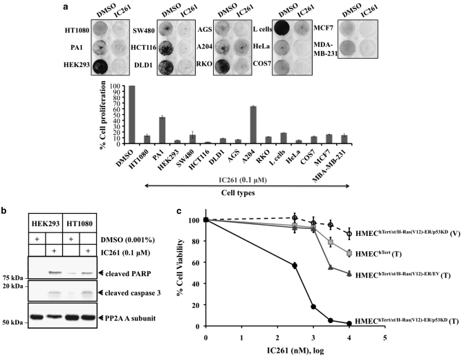
As CK1δ/ɛ activity has been reported to be essential for survival of cancer cell lines, including some that are dependent on β-catenin signaling (Grueneberg et al., 2008; Yang and Stockwell, 2008; Kim et al., 2010), we treated a panel of cancer cells with IC261 to assess its spectrum of activity. Consistent with previous reports, 100 n IC261 caused a near-total growth arrest of most of the cell lines that we screened (Figure 1a). In the two lines analyzed in more detail, the acute growth inhibition of HEK293 and HT1080 cells was characterized by caspase 3-dependent apoptosis (Figure 1b).
Figure 1.
IC261 selectively inhibits cancer cell proliferation. (a) IC261 inhibits proliferation of multiple cancer cell lines. A panel of transformed/cancer cell lines was cultured in 12-well plates and treated with either DMSO (0.001%) or IC261 (0.1 μ) for 5 days. Cell proliferation was measured by crystal violet staining (Materials and methods). Cell proliferation was calculated by OD595IC261/OD595DMSO of each respective cell lines and expressed as a percentage of proliferation in the DMSO control. Data were derived from three independent sets of experiments each performed in triplicate. (b) IC261 induces caspase activation. HEK293 and HT1080 cells were seeded at 80% confluence in 6-well plates and treated with DMSO (0.001%) and IC261 (0.1 μ), respectively for 48 h. Cells were lysed by 4% SDS and analyzed by SDS–PAGE/western blot. (c) IC261 is transformation selective. HMEC-derived cells (HMEChTert, HMEChTert/H−Ras(V12)−ER/EV and HMEChTert/H−Ras(V12)−ER/p53KD) were treated with methanol (0.005%) or 0.13 μ 4-OHT and the indicated concentrations of IC261 in triplicate for 72 h, before alamarBlue analyses. V, vehicle control (methanol); T, 4-OHT.
Several studies indicated that IC261 might selectively kill transformed cells (Behrend et al., 2000; Yang and Stockwell, 2008; Kim et al., 2010). To confirm and extend this finding, we utilized a series of human mammary epithelial cells (HMEC) either immortalized with hTert, partially transformed with hTert, small t antigen, and H-RasV12-ER, or fully transformed by the further addition of small hairpin RNA (shRNA) against p53 (Voorhoeve and Agami, 2003; Rangarajan et al., 2004). We exposed these HMEC-derived cells to dimethyl sulfoxide (DMSO)- or IC261-supplemented culture media with 0.13 μ 4-hydroxytamoxifen (4-OHT; to activate H-RasV12-ER) for 72 h. IC261 selectively suppressed the growth of fully transformed HMEC-derived cells, but not their non-transformed counterparts, at sub-micromolar concentrations (Figure 1c). The IC261-induced growth arrest of HMEC-derived cells appeared to require concurrent activation of the Ras signaling pathway and deregulation of the p53 pathway, as these cells remained viable in IC261-supplemented culture media without 4-OHT (Figure 1c). We observed similar results with a series of BJ-derived fibroblasts (Supplementary Figure S1). Taken together, our data confirm and extend prior reports that IC261 potently and selectively inhibits cancer cell growth.
PF670 is a weak inhibitor of proliferation
IC261 is widely used as a selective inhibitor of CK1δ/ɛ, albeit at micromolar concentrations, and the preceding data are consistent with the model that inhibition of CK1δ and/or CK1ɛ causes cancer cell death. It would be desirable to have additional small-molecule inhibitors of CK1δ/ɛ with higher affinities to further test this hypothesis. PF670, a recently developed CK1δ/ɛ inhibitor, inhibits CK1δ/ɛ at nanomolar concentrations in vitro. In addition, the compound is bioavailable, as it can induce a phase delay in circadian rhythms in cells, tissues, mice and monkeys (Badura et al., 2007; Sprouse et al., 2009). If inhibition of CK1δ/ɛ and Wnt/β-catenin is indeed responsible for the cytotoxic effects of IC261, PF670 should be an even better anti-cancer agent as it inhibits CK1δ/ɛ at concentrations that are three orders of magnitude lower.
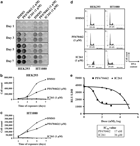
We therefore, tested PF670 on two representative cell lines that are sensitive to IC261. Unexpectedly, we found that 1 μ PF670 only modestly reduced the growth of HEK293 and HT1080 cells (Figures 2a, b and c). Furthermore, PF670 failed to induce either an acute cell cycle arrest at G2/M (in HEK293 cells) or a sub-G1 population indicative of apoptosis (HT1080 cells), as was observed within 24 h of exposure to IC261 (Figure 2a). In the same assays, 1 μ IC261 completely inhibited the growth of these cells, as demonstrated by both colony-forming (Figure 2a) and viable cell count (Figures 2b and c) analyses. These data suggest that either PF670 did not inhibit CK1δ/ɛ and subsequently β-catenin activity in these cell lines, or that CK1δ/ɛ and Wnt/β-catenin inhibition is irrelevant for the cytotoxic effects exhibited by IC261.
Figure 2.
PF670462 and IC261 have divergent effects on Wnt/β-catenin signaling and cellular growth. PF670462 is a weak inhibitor of transformed cell proliferation. HEK293 and HT1080 cells were seeded at 5% confluence in 6-well plates and treated with either DMSO (0.001%), PF670462 (1 μ) or IC261 (1 μ) over 7 days and assessed by (a) 0.5% crystal violet staining and (b, c) viable cell counts. (d) IC261, but not PF670462, causes cell cycle arrest. HEK293 and HT1080 cells were seeded at 80% confluence in 6-well plates and treated with DMSO (0.001%), PF670462 (1 μ) or IC261 (1 μ) for 24 h before DNA content analyses by flow cytometry. (e) PF670462 is a potent inhibitor of Wnt/β-catenin signaling. HEKSTF3A cells were treated with the indicated concentrations of PF670462 or IC261 for 16 h. DMSO (0.001%) was used as the vehicle control. Reporter activity was measured as relative light units (RLU) and normalized to lactate dehydrogenase (LDH) activity. PF670462 and IC261 dose–response curves were fitted using a non-linear regression model in GraphPad Prism 5.0c.
PF670 is a more potent inhibitor than IC261 of CK1δ/ɛ activity
To examine the first possibility, we tested the effects of PF670 on CK1δ/ɛ activity and the Wnt/β-catenin pathway. CK1δ/ɛ is required for high-level Wnt/β-catenin signaling (Peters et al., 1999; Sakanaka et al., 1999; Bryja et al., 2007). Using HEK293 cells expressing Wnt3A and containing an integrated luciferase gene driven by β-catenin responsive promoter (HEK293STF3A) as a Wnt/β-catenin reporter assay, the activities of IC261 and PF670 were compared. As shown in Figure 2e, PF670 inhibited Wnt/β-catenin signaling with an IC50 of ∼17 n, while IC261 had an IC50 of ∼36 μ, consistent with previously reported values for CK1 inhibition (Gao et al., 2002; Bain et al., 2007). Notably, PF670 gave near maximal inhibition of β-catenin activity at 1 μ (Figure 2e), but had only a small effect on proliferation at this concentration (Figures 2a, b and c), whereas IC261 did not significantly repress Wnt/β-catenin signaling at this concentration but had strong growth inhibitory activity toward HEK293 cells.
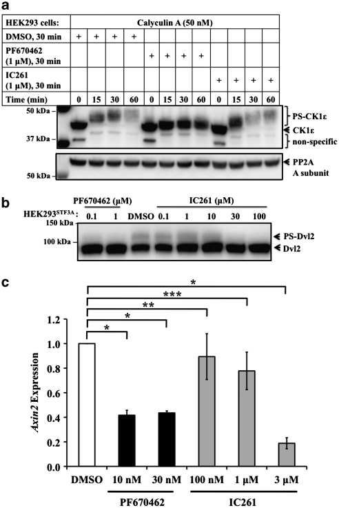
Thus far, our data suggest that PF670 represses Wnt/β-catenin signaling by inhibition of CK1δ/ɛ, while IC261 at 1 μ induces cancer cytotoxicity without inhibiting β-catenin activity. However, as transactivation of a β-catenin responsive promoter is an indirect measure of CK1δ/ɛ activity, we investigated whether phosphorylation of CK1δ/ɛ targets in vivo could also be inhibited by these drugs at 1 μ. To directly test CK1δ/ɛ activity we made use of the fact that the kinase autophosphorylates its carboxyl-terminus regulatory domain in an intramolecular reaction (Cegielska et al., 1998). This reaction is continuously reversed by cellular protein phosphatases that are sensitive to okadaic acid and calyculin A (Cegielska et al., 1998; Rivers et al., 1998), and addition of 50 n calyculin A consequently results in a rapid and distinct electrophoretic mobility shift of endogenous CK1δ and CK1ɛ due to unopposed phosphorylation. PF670 inhibited in vivo CK1ɛ and CK1δ carboxyl-terminal autophosphorylation completely at 1 μ (Figure 3a and Supplementary Figure S2). Notably, strong inhibition of in vivo CK1δ/ɛ autophosphorylation in this short-term assay was achieved with as low as 100 n PF670, consistent with its effect on Wnt/β-catenin signaling (data not shown). In contrast, 1 μ IC261 did not result in detectable inhibition of autophosphorylation by CK1, consistent with its failure to inhibit β-catenin activity at this concentration (Figure 2e). Although 1 μ IC261 can be cytotoxic to these cells, in the short time frame of this experiment (up to 60 min) no detrimental effect on cell viability or protein abundance was seen.
Figure 3.
IC261 and PF670462 have divergent effects in cells. (a) Cytotoxic concentrations of IC261 do not inhibit CK1δ/ɛ in cells. CK1ɛ intramolecular autophosphorylation in intact cells was unmasked as previously described (Rivers et al., 1998) (Materials and methods) by the addition of 50 n calyculin A in the absence or presence of the indicated concentrations of PF670462 or IC261. PP2A A subunit served as the loading control. PS denotes phosphorylated and shifted. The residual mobility shift of CK1ɛ in the presence of PF670462 is probably due to its phosphorylation by other cellular kinases (Clokie et al., 2009). The decreased immunoreactivity of the autophosphorylated CK1ɛ is due to phosphorylation-mediated masking of the Mab epitope. (b) PF670462, but not cytotoxic concentrations of IC261, inhibits Dvl2 phosphorylation. HEK293STF3A cells were treated with vehicle or the indicated concentration of PF670462 or IC261 for 30 min. Cells were then lysed and the Dvl2 mobility shift was assessed by SDS–PAGE and immunoblotting. (c) Dose-dependent inhibition of a β-catenin target gene by PF670462 and IC261. HEK293STF3A cells were treated with the indicated concentrations of compound for 6 h, followed by lysis and qPCR quantification of Axin2 mRNA. Data are representative of three independent sets of experiment each performed in triplicate. A Student's _t_-test was performed; *_P_-value <0.001; **_P_-value <0.01; ***_P_-value <0.05 for the comparison of PF670462- and IC261-treated samples with the DMSO control.
The inhibition of autophosphorylation by PF670 indicates the compound regulates intracellular CK1 activity. To examine the effect of the compounds on CK1 activity towards known Wnt pathway substrates, the phosphorylation of the Wnt regulator disheveled (Dvl) was examined. CK1ɛ phosphorylates Dvl proteins on multiple sites in response to Wnt signaling, inducing an electrophoretic mobility shift (Peters et al., 1999; Kishida et al., 2001). We exposed HEK293STF3A cells to PF670 or IC261 for 30 min and determined the effect on Dvl2 phosphorylation. Nanomolar concentrations of PF670 inhibited the electrophoretic mobility shift of Dvl2, while >10 μ IC261 was required to achieve the same effect (Figure 3b). In addition, we examined the differential activity of the CK1 inhibitors on the expression of Axin2, an endogenous target of Wnt/β-catenin signaling. Consistent with their effect on CK1 autophosphorylation and Dvl2 phosphorylation, after a 6-h incubation, nanomolar concentrations of PF670 but micromolar concentrations of IC261 were able to inhibit the expression of Axin2 (Figure 3c). Collectively, our findings demonstrate that pharmacological inhibition of CK1δ/ɛ, while capable of inhibiting Wnt/β-catenin signaling, does not block cell cycle progression or induce cell death.
Knockdown of CK1δ/ɛ phenocopies PF670 and not IC261
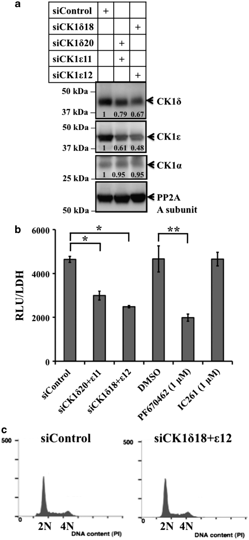
To test inhibition of CK1δ/ɛ activity via an independent approach, we performed RNAi knockdown of both CK1δ and CK1ɛ levels in the HEK293STF3A cells (Figure 4a). Consistent with previous reports, we observed significant repression of Wnt/β-catenin signaling upon reduction of CK1δ and CK1ɛ abundance (Figure 4b). However, the combined partial knockdown of CK1δ and CK1ɛ levels with small interfering RNA (siRNA) failed to induce cell cycle arrest and/or cell death (Figure 4c). Knockdown of CK1δ/ɛ gave similar results to those seen with treatment with 1 μ PF670, and distinct from those seen with 1 μ IC261.
Figure 4.
CK1δ/ɛ knockdown phenocopies PF670462 but not IC261. (a–c) RNAi knockdown of CK1δ/ɛ blocks Wnt/β-catenin signaling but has no effect on cell cycle progression. (a) RNAi-mediated knockdown of CK1δ/ɛ in HEK293STF3A cells. Two independent siRNA pools both produce partial knockdown of endogenous CK1δ and CK1ɛ in 72 h. (b) CK1 knockdown and PF670462, but not IC261, inhibits Wnt/β-catenin signaling in HEK293STF3A cells. Data are representative of three independent sets of experiment each performed in triplicate. A Student's _t_-test was performed; *_P_-value <0.001; **_P_-value <0.005 for the comparison of siCK1δ+ɛ- and PF670462-treated samples with their respective controls. (c) CK1δ/ɛ knockdown has no significant effect on cell cycle progression in HEK293 cells. Similar results were seen in three independent experiments.
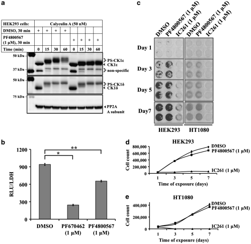
Several RNA interference studies have demonstrated that a number of shRNAs directed against CK1ɛ but not CK1δ can slow cancer cell growth, while both IC261 and PF670 inhibit both CKIδ and CK1ɛ. To investigate whether isolated CK1ɛ inhibition could be more toxic than combined CK1δ/ɛ inhibition, we tested a different, CK1ɛ-selective inhibitor, PF4800567 (PF480), that relatively spares CK1δ activity (Walton et al., 2009) (Figure 5). We confirmed that PF480 selectively inhibited CK1ɛ using the in vivo autophosphorylation assay (Figure 5a). PF480 was less effective than PF670 at inhibition of Wnt/β-catenin signaling (Figure 5b). These data suggest that CK1δ and CK1ɛ have additive roles in promoting Wnt/β-catenin signaling. The CK1ɛ-selective PF480 displayed no anti-proliferative effects at concentrations that completely inhibited CK1ɛ activity (Figures 5c and d). We conclude that combined inhibition of CK1δ and CK1ɛ is required for maximal inhibition of Wnt/β-catenin signaling and cell proliferation (Figures 2b, c and e), and that effective isolated pharmacologic inhibition of CK1ɛ does not significantly alter the proliferation of cells that are sensitive to IC261.
Figure 5.
PF4800567 is a potent inhibitor of CK1ɛ but not CK1δ. (a) PF4800567 inhibits CK1ɛ, but not CK1δ, autophosphorylation in cells. HEK293 cells were treated as described in Figure 4, but with 1 μ PF4800567. (b) Wnt/β-catenin signaling was assessed in HEK293STF3A cells after 16 h treatment with DMSO, 1 μ PF670462 or 1 μ PF4800567. Data were derived from a representative of three independent sets of experiment performed in triplicate. A Student's _t_-test was performed; *_P_-value <0.001; **_P_-value <0.005 for the comparison of PF670462- and PF4800567-treated samples with the DMSO control. (c, d) The CK1ɛ-selective inhibitor, PF4800567, does not inhibit cancer cell growth. The effect of IC261 and PF4800567 on HEK293 and HT1080 cell proliferation was compared as above.
PF670 and IC261 have divergent effects on gene expression
To assess whether PF670 and IC261 share a common mechanism of cancer cytotoxicity, we studied their effect on gene expression at near lethal doses. In the gastric cancer cell line AGS, the GI50 of IC261 was found to be 340 n, while PF670 failed to inhibit growth of AGS cells even at 10 μ (data not shown). Cells were treated with DMSO (<0.1%), 10 μ PF670 or 340 n IC261 for 6 h, respectively, before gene expression profiling. We analyzed the gene signature profiles by significance analysis of microarrays (with false discovery rate (FDR)⩽0.01%) and observed that only five genes were in common among the 186 and 86 genes differentially regulated by PF670 and IC261, respectively (Supplementary Figure S3 and Supplementary Table S1). Consistent with our quantitative PCR data (Figure 3c), the Wnt signaling-related genes Fzd6 and Axin2 were significantly downregulated by PF670 but not IC261 (Supplementary Table S1). Taken together, gene expression analysis supports the conclusion that PF670 and IC261 regulate distinctive intracellular pathways.
IC261 is an inhibitor of microtubule polymerization
The data indicate that IC261 induces both mitotic arrest and cytotoxicity independent of its inhibition of CK1δ/ɛ. Thus, there should be another widely expressed protein to which IC261 binds at nanomolar, rather than micromolar concentrations. To determine other potential cancer-associated cellular targets of IC261, we more closely examined the IC261 G2/M arrest phenotype. Time-lapse video microscopy was performed using asynchronous histone 2B-GFP-expressing HeLa cells. DMSO- or PF670 (1 μ)-treated HeLa cell progress normally through mitosis and give rise to a pair of daughter cells (Supplementary Video S1 and S2). However, 1 μ IC261 induced a prometaphase arrest of HeLa cells followed by apoptosis (Supplementary Video S3).
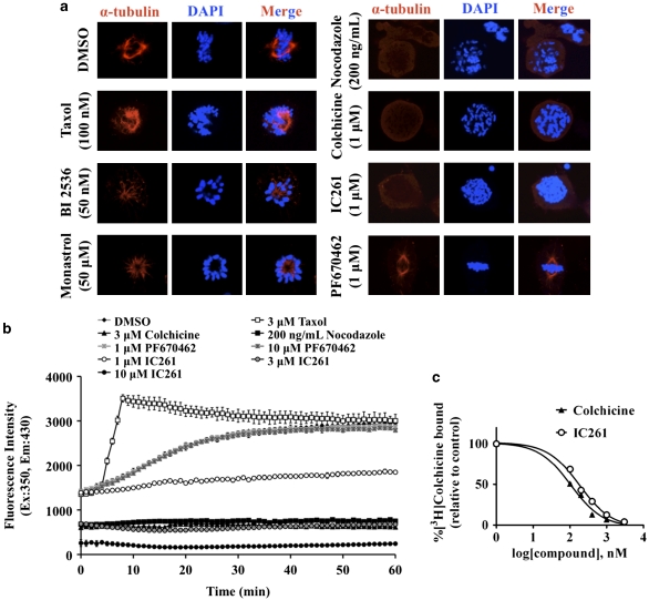
Prometaphase arrest is triggered by the spindle assembly checkpoint and can be caused by a number of compounds that block proper assembly and attachment of mitotic microtubules to kinetochores (Weaver and Cleveland, 2005). Different drugs produce distinctive effects on mitotic spindle formation that can readily be assessed. The effect of IC261 was therefore compared with other compounds that block the cell cycle at prometaphase, including inhibitors of Polo-like kinase 1 and the mitotic kinesin EG5, as well as the microtubule polymerization inhibitors nocodazole and colchicine, and the microtubule stabilizer taxol. As Figure 6a illustrates, 1 μ IC261 phenocopied the effect of nocodazole and colchicine, and blocked formation of mitotic spindles in the presence of condensed chromosomes.
Figure 6.
IC261 is an inhibitor of microtubule polymerization. (a) IC261 destabilizes microtubule network organization in vivo. HT1080 cells were grown to 50% confluence and treated with DMSO (0.02%), taxol, BI 2536, monastrol, nocodazole, colchicine, IC261 or PF670462 at the indicated concentrations for 18 h. Cells were then fixed, permeabilized and immunostained with α-tubulin antibody followed by Alexa Fluor 594-conjugated secondary antibody. Cellular DNA was counterstained with DAPI-VectorShield and visualized by a Carl Zeiss LSM710 laser scanning confocal microscope with a × 63 oil immersion lens. Images are representatives of three independent experiments. (b) IC261 inhibits in vitro tubulin polymerization in a dose-dependent manner. Tubulin (>99% pure) was exposed to DMSO (0.1%), taxol, nocodazole, colchicine, IC261 and PF670462 at the indicated concentrations in a black multi-well plate and incubated at 37 °C. Fluorescence intensity was recorded at a 1-min interval for 1 h (excitation wavelength (Ex): 350 nm; emission wavelength (Em): 430 nm). (c) IC261 binds to the colchicine-binding site of tubulin. Tubulin (>99% pure) was incubated with 100 n [3H]colchicine and the indicated concentrations of competitor colchicine or IC261 at 37 °C for 1 h. The amount of [3H]colchicine bound to tubulin was determined by a Beckman liquid scintillation counter. Assays were performed in triplicate and repeated.
To test directly whether IC261 is a bona fide microtubule polymerization inhibitor, the effect of IC261 on microtubule assembly was determined in vitro. IC261 directly inhibited the polymerization of purified porcine brain tubulin with equimolar potency to the microtubule binding agents, colchicine and nocodazole, while PF670 had no effect on microtubule formation (Figure 6b). There are two well-characterized sites on tubulin that destabilize polymerization, the vinca site and the colchicine site (reviewed in Gigant et al., 2009). We find that IC261 competes with [3H]colchicine for binding to tubulin (Figure 6c). Taken together, while the biological effects of PF670 appear to be explained by inhibition of CK1δ/ɛ, the cytotoxicity of IC261 is a direct consequence of its ability to bind to the colchicine site on tubulin and thereby inhibit microtubule polymerization.
Discussion
Genetic ablation and pharmacological intervention represent two complementary approaches that are widely used to dissect the role(s) of druggable cellular targets such as protein kinases in tumorigenesis. The ability to cross-validate RNA knockdown and drug studies can provide valuable confirmation of appropriate targets and may help exclude off target effects. A number of recent studies have found that shRNAs that knockdown CK1ɛ slowed cancer cell growth. Additionally, these and other studies found that the CK1δ/ɛ-selective inhibitor IC261 is a potent transformation-selective anti-cancer agent. Here, we independently validated the potency of IC261 as an anti-cancer drug and confirmed that it induces cell cycle arrest and apoptosis in a wide range of transformed cells at nanomolar concentration. We find that IC261 acts by a CK1δ/ɛ- and Wnt/β-catenin-independent induction of the spindle assembly checkpoint. This prometaphase arrest initially suggested IC261 might be inhibiting a mitotic kinase. However, in prior surveys, IC261 did not inhibit a number of mitotic kinases, including Polo-like kinase 1, Aurora B and C, and Chk1 and Chk2 (Bain et al., 2007; Fedorov et al., 2007). Instead, our data indicate that IC261 inhibits microtubule polymerization at doses far below those needed to inhibit CK1, a finding consistent with induction of prometaphase arrest. The effect is due to direct binding of IC261 to the colchicine-binding site on β-tubulin (Figure 6c). Intriguingly, several other trimethoxy phenolic compounds that are chemically similar to IC261 have been reported to be microtubule polymerization inhibitors with potent anti-proliferative and anti-tumor activity (Kuo et al., 2004; Arora et al., 2009). This suggests that the trimethoxy phenol moiety of these anti-mitotic compounds is critical for their shared microtubule depolymerization property.
IC261 selectively suppressed growth of both transformed fibroblasts and HMEC-derived cells, but not their non-transformed counterparts, consistent with reports indicating that IC261 may be transformation selective (Behrend et al., 2000; Yang and Stockwell, 2008). This highlights the selective sensitivity of transformed over non-transformed cells to microtubule poisons that trigger spindle assembly checkpoints. We confirmed that activation of RAS combined with inactivation of p53 was required to fully sensitize cells to killing by IC261. This effect was seen in both fibroblasts and HMEC. The mechanism underlying this sensitization is not well understood. Spindle poisons can lead to damaged DNA, an effect that may be more pronounced in transformed than in non-transformed cells (Dalton et al., 2007). Similarly, RAS and p53 mutations can cooperate to cause chromosome breaks (Agapova et al., 1999). Microtubule poison-induced DNA damage has been postulated to lead to caspase activation (Gascoigne and Taylor, 2008). One speculative model is therefore, that chromosome damage occurs in response to prolonged exposure to IC261 and other microtubule poisons and RAS activation and p53 loss potentiates this damage. Excessive damage may lead to caspase activation and cell death. Further studies will be required to fully understand this synthetic lethality.
The anti-cancer activity of IC261 is largely independent of its described inhibitory effects on CK1δ/ɛ activity. We confirmed the new compound PF670 as a potent inhibitor of CK1δ/ɛ activity and CK1δ/ɛ-dependent processes in vivo, including Wnt/β-catenin signaling. At concentrations that effectively inhibit in vivo CK1δ/ɛ kinase activity, PF670 did not cause cytotoxicity or cell cycle arrest. In contrast, at concentrations that effectively killed human transformed/cancer cells, IC261 did not inhibit CK1δ/ɛ. Consistent with this data, Kim et al. (2010) recently demonstrated that PF670 preferentially inhibits proliferation of β-catenin-positive MCF7 cells as compared to β-catenin-negative MDA-MB-453 cells, albeit 12-fold less effectively than IC261. Taken together, we conclude that effective inhibition of CK1δ/ɛ by PF670 or RNAi impairs Wnt/β-catenin signaling and Dvl phosphorylation but does not lead to significant cell cycle arrest or cytotoxicity.
Why do multiple shRNA screens find an anti-proliferative effect of CK1ɛ knockdown, while we see different results with siRNA and perhaps with PF480 and PF670? Knockdown by shRNA can be more durable than knockdown by siRNA, and more effective knockdown or inhibition of CK1δ/ɛ does in fact impair cell growth, albeit via a different mechanism than IC261. The shRNA-mediated growth inhibition reported by others is similar to the ∼50% maximal growth inhibition we see in cells treated with PF670 (Figures 2b and c), and supports a positive but not essential role for CK1δ/ɛ in proliferation, perhaps via their function in the Wnt/β-catenin pathway. Interestingly, we did not see significant growth inhibition by the CK1ɛ-selective agent, PF480. This suggests either the growth effect is via inhibition of CK1δ, or redundancy of CK1ɛ with CK1δ, but fails to explain why shRNA against CK1ɛ was so effective in slowing cell growth in other reports. We note that CK1ɛ could also have non-catalytic functions in cell proliferation, as a structural role in circadian rhythms has been reported for the Drosophila ortholog double-time (Yu et al., 2009). A scaffolding role for CK1ɛ might account for some of the differences between the shRNA and PF480 drug studies.
PF670, more so than PF480, was effective at inhibiting Wnt/β-catenin signaling, and this could contribute to its ability to slow cancer cell proliferation. Notably, PF670 only produced sub-maximal inhibition of β-catenin signaling even at levels that fully inhibit CK1δ/ɛ. This is likely to be attributable to a basal level of Wnt/β-catenin signaling that does not require CK1δ/ɛ activity, as has been previously reported (Bryja et al., 2007). A number of cell lines have aberrant activation of β-catenin due to diverse mechanisms including autocrine Wnt loops, mutation of APC and mutation of β-catenin. CK1δ/ɛ can act both at the level of the disheveled/axin complex and downstream as well at the level of the translational co-repressor Tcf3 (Lee et al., 2001). Pharmacologic inhibition of CK1δ/ɛ can therefore diminish β-catenin signaling via several routes. However, the sub-maximal inhibition may only slow, rather than fully block, proliferation. Inhibition of β-catenin signaling via targeting of CK1δ/ɛ may therefore not be an effective way to halt proliferation of many cancer cell lines.
Finally, we conclude that previous studies where the biological effect of low micromolar concentrations of IC261 in transformed cells was attributed to CK1 inhibition should be interpreted with caution.
Materials and methods
Plasmid DNA, cell culture and other reagents
The use of pCL-eco, pBabehyg-hTERT, pBabepuro-H-RASV12ER, pMSCV-st-GFP, pMSCV-Blast and pMSCV-Blast-p53KD expression constructs have been previously described (Voorhoeve and Agami, 2003); KD, knockdown. Other cell lines were purchased from American Type Culture Collection (Manassas, VA, USA). Ecopack-2 and Phoenix-Ampho cells were purchased from Clontech (Mountain View, CA, USA). Cell culture conditions and other reagents are described in Supplementary Information.
Crystal violet staining
Cells were then fixed with methanol for 10 min and stained with 0.5% crystal violet dye (in methanol:deionized water, 1:5) for 10 min. Excess crystal violet dye was removed by five washes of deionized water on a shaker (10 min for each wash) and the culture plates were dried overnight. The crystal violet dye was released from cells by incubation with 1% sodium dodecyl sulfate (SDS) for 6 h before optical density (OD)595 nm measurement.
Denaturing sodium dodecyl sulfate–polyacrylamide gel electrophoresis (SDS–PAGE) and western blot analysis
Cells were lysed by 4% SDS and total protein content was measured using the bicinchoninic acid protein assay kit (Thermo Scientific, Rockford, IL, USA). Proteins from whole cell extracts were resolved using denaturing SDS–PAGE and analyzed by western blot. Antibodies used in western blot analyses include anti-cleaved poly ADP ribose polymerase (PARP, ab32561, abcam), anti-cleaved caspase 3 (#9664, Cell Signaling Technology, Danvers, MA, USA), anti-CK1δ (128A, Eli Lilly, Indianapolis, IN, USA), anti-CK1ɛ (610445, BD Biosciences, San Jose, CA, USA), anti-Dvl2 (sc-13974, Santa Cruz Biotechnology, Santa Cruz, CA, USA) and anti-PP2A A subunit (JH242) (McCright and Virshup, 1995).
AlamarBlue cell viability assay
HMEC-derived cells (5000 cells/well) were plated in 96-well tissue culture plates (pre-treated with poly--lysine) and then treated with 4-OHT and DMSO (vehicle control) or the indicated concentrations of IC261 in triplicate for 72 h. Cell number and viability were assayed after drug treatment by the reduction of alamarBlue (DAL1025, Invitrogen, Carlsbad, CA, USA), a measure of mitochondrial fitness that provides a surrogate endpoint for cell number, as previously described (Nakayama et al., 1997).
Flow cytometric DNA content analysis
HEK293 and HT1080 cells were grown to 50% confluence on 6-well plates 24 h before incubation with DMSO (0.001%), PF670462 (1 μ) and IC261 (1 μ), respectively for an additional 24 h. Cells were trypsinized, washed with ice-cold 1 × phosphate buffered saline (PBS) and fixed overnight with 70% ethanol/30% 1 × PBS. Cells were then washed once in ice-cold 1 × PBS and stained with 50 mg/ml propidium iodide (P4170, Sigma-Aldrich, St Louis, MO, USA) in 1 × PBS solution that was supplemented with 0.1 mg/ml RNase A (R6513, Sigma-Aldrich) and 0.05% (v/v) Triton X-100 (T8787, Sigma-Aldrich). Flow cytometric DNA content analysis (excitation wavelength: 488 nm; emission wavelength: 620 nm) was performed using the Beckman Coulter FC500 flow cytometer (Brea, CA, USA).
Super TOPFlash luciferase reporter assay
The Super TOPFlash-Wnt3A (STF3A) reporter cell line and luciferase and lactate dehydrogenase assays were performed as described (Coombs et al., 2010). All assays were verified in three independent sets of experiment with triplicate.
CK1δ/ɛ autophosphorylation assay
HEK293 cells were seeded at 80% confluence in 6-well plates and treated with DMSO (0.001%), PF670462 (1 μ), IC261 (1 μ) or PF4800567 (1 μ) for 30 min before addition of 50 n calyculin A to the culture media.
qPCR analysis
Total RNA was isolated from DMSO- or drug-treated HEK293STF3A cells using the Qiagen RNeasy Mini Kit (74106, Valencia, CA, USA). Total RNA (2 μg) was reverse transcribed by the iScript Select cDNA Synthesis Kit (170–8896, Bio-Rad, Hercules, CA, USA) in accordance to the manufacturer's instructions. Using 1/10 of the cDNA sample and _Axin2_-specific primers, qPCR were performed by the SsoFast EvaGreen Supermix Kit (172–5200, Bio-Rad) in accordance to the manufacturer's instructions. The _Axin2_-specific primer sequences are available upon request.
RNAi of human CK1δ and CK1ɛ expression
In all, 2 × 10⁵ HEK293 or HEK293STF3A cells were plated in 6-well plates and transfected with 100 n ON TARGET_plus_ non-targeting siRNA (siControl; D-001810-0X) or two different combinations of human CK1δ- and CK1ɛ-specific ON TARGET_plus_ siRNAs (in 1:1 ratio), siCK1δ18+siCK1ɛ12 and siCK1δ20+siCK1ɛ11 (Dharmacon RNAi Technologies, Lafayette, CO, USA) using Dharmafect Transfection Reagent, in accordance with the manufacturer's instructions, for 72 h. The target sequences of human CK1δ- and CK1ɛ-specific ON TARGET_plus_ siRNAs are siCK1δ18 (J-0034788-18): CGACCUCACAGGCCGACAA, siCK1δ20 (J-003478-20): AGGCUACCCUUCCGAAUUU, siCK1ɛ11 (J-003479-11): CCUCCGAAUUCUCAACAUA and siCK1ɛ12 (J-003479-12): CGACUACUCUUACCUACGU.
Immunofluorescence staining
HT1080 cells were grown to 50% confluence on glass coverslips in a 12-well tissue culture plate, and were subsequently treated with test compounds at the indicated concentrations for 18 h. They were washed three times with 1 × PBS, fixed in pre-chilled 4% paraformaldehyde for 20 min, permeabilized in 0.1% Triton X-100 for 10 min and blocked with 3% bovine serum albumin for 1 h. Primary immunostaining with α-tubulin antibody (T5168, Sigma-Aldrich) was performed at room temperature for 1 h, followed by immunostaining with Alexa Fluor 594-conjugated secondary antibody (Invitrogen). Cellular DNA was subsequently counterstained with 4′,6-diamidino-2-phenylindole (DAPI)-VectorShield (H-1200, Vector Laboratories, Inc, Brulingame, CA, USA). Staining was visualized and photographed using a LSM710 laser scanning confocal microscope with a × 63 oil immersion lens (Carl Zeiss Microimaging, Thornwood, NY, USA).
Fluorescence-based in vitro tubulin polymerization assay
in vitro tubulin polymerization assays were performed using a fluorescence-based in vitro tubulin polymerization assay kit (BK011, Cytoskeleton, Inc, Denver, CO, USA) in accordance with the manufacturer's instructions.
Tubulin competitive binding assay
Tubulin competitive binding assays were performed as described (Arora et al., 2009). Assays were performed in triplicate and repeated.
Acknowledgments
We thank Velani Utomo, Ha Yin Lee, Simran Kaur and Qin Luo (Duke-NUS) for their excellent technical and computational support. We are also grateful to our Duke and Duke-NUS colleagues Tim Haystead, Sin Tiong Ong, Shirish Shenolikar, Patrick Casey and members of the Virshup laboratory for their helpful discussions. This work was supported by the Duke-NUS Signature Research Program funded by the Agency for Science, Technology and Research, Singapore, and the Ministry of Health, Singapore, and a Singapore Translational Research Investigator Award to DMV funded by the National Research Foundation and the National Medical Research Council. JKC was supported by a Singapore Millennium Foundation (SMF) Postdoctoral Fellowship.
The authors declare no conflict of interest.
Footnotes
Supplementary Information accompanies the paper on the Oncogene website (http://www.nature.com/onc)
Supplementary Material
Supplementary Figure S1
Supplementary Figure S2
Supplementary Figure S3
Supplementary Table S1
Supplementary Video S1
Supplementary Video S2
Supplementary Video S3
Supplementary Information
References
- Agapova LS, Ivanov AV, Sablina AA, Kopnin PB, Sokova OI, Chumakov PM, et al. P53-dependent effects of RAS oncogene on chromosome stability and cell cycle checkpoints. Oncogene. 1999;18:3135–3142. doi: 10.1038/sj.onc.1202386. [DOI] [PubMed] [Google Scholar]
- Arora S, Wang XI, Keenan SM, Andaya C, Zhang Q, Peng Y, et al. Novel microtubule polymerization inhibitor with potent antiproliferative and antitumor activity. Cancer Res. 2009;69:1910–1915. doi: 10.1158/0008-5472.CAN-08-0877. [DOI] [PMC free article] [PubMed] [Google Scholar]
- Badura L, Swanson T, Adamowicz W, Adams J, Cianfrogna J, Fisher K, et al. An inhibitor of casein kinase I epsilon induces phase delays in circadian rhythms under free-running and entrained conditions. J Pharmacol Exp Ther. 2007;322:730–738. doi: 10.1124/jpet.107.122846. [DOI] [PubMed] [Google Scholar]
- Bain J, Plater L, Elliott M, Shpiro N, Hastie CJ, McLauchlan H, et al. The selectivity of protein kinase inhibitors: a further update. Biochem J. 2007;408:297–315. doi: 10.1042/BJ20070797. [DOI] [PMC free article] [PubMed] [Google Scholar]
- Behrend L, Milne DM, Stöter M, Deppert W, Campbell LE, Meek DW, et al. IC261, a specific inhibitor of the protein kinases casein kinase 1-delta and -epsilon, triggers the mitotic checkpoint and induces p53-dependent postmitotic effects. Oncogene. 2000;19:5303–5313. doi: 10.1038/sj.onc.1203939. [DOI] [PubMed] [Google Scholar]
- Brockschmidt C, Hirner H, Huber N, Eismann T, Hillenbrand A, Giamas G, et al. Anti-apoptotic and growth-stimulatory functions of CK1 delta and epsilon in ductal adenocarcinoma of the pancreas are inhibited by IC261 in vitro and in vivo. Gut. 2008;57:799–806. doi: 10.1136/gut.2007.123695. [DOI] [PubMed] [Google Scholar]
- Bryant CD, Graham ME, Distler MG, Munoz MB, Li D, Vezina P, et al. A role for casein kinase 1 epsilon in the locomotor stimulant response to methamphetamine. Psychopharmacology (Berl) 2009;203:703–711. doi: 10.1007/s00213-008-1417-z. [DOI] [PMC free article] [PubMed] [Google Scholar]
- Bryja V, Schulte G, Arenas E. Wnt-3a utilizes a novel low dose and rapid pathway that does not require casein kinase 1-mediated phosphorylation of Dvl to activate beta-catenin. Cell Signal. 2007;19:610–616. doi: 10.1016/j.cellsig.2006.08.011. [DOI] [PubMed] [Google Scholar]
- Cegielska A, Gietzen KF, Rivers A, Virshup DM. Autoinhibition of casein kinase I epsilon (CKI epsilon) is relieved by protein phosphatases and limited proteolysis. J Biol Chem. 1998;273:1357–1364. doi: 10.1074/jbc.273.3.1357. [DOI] [PubMed] [Google Scholar]
- Clokie S, Falconer H, Mackie S, Dubois T, Aitken A. The interaction between casein kinase Ialpha and 14-3-3 is phosphorylation dependent. FEBS J. 2009;276:6971–6984. doi: 10.1111/j.1742-4658.2009.07405.x. [DOI] [PubMed] [Google Scholar]
- Coombs GS, Yu J, Canning CA, Veltri CA, Covey TM, Cheong JK, et al. WLS-dependent secretion of WNT3A requires Ser209 acylation and vacuolar acidification. J Cell Sci. 2010;123:3357–3367. doi: 10.1242/jcs.072132. [DOI] [PMC free article] [PubMed] [Google Scholar]
- Dalton WB, Nandan MO, Moore RT, Yang VW. Human cancer cells commonly acquire DNA damage during mitotic arrest. Cancer Res. 2007;67:11487–11492. doi: 10.1158/0008-5472.CAN-07-5162. [DOI] [PMC free article] [PubMed] [Google Scholar]
- Eide EJ, Woolf MF, Kang H, Woolf P, Hurst W, Camacho F, et al. Control of mammalian circadian rhythm by CKIepsilon-regulated proteasome-mediated PER2 degradation. Mol Cell Biol. 2005;25:2795–2807. doi: 10.1128/MCB.25.7.2795-2807.2005. [DOI] [PMC free article] [PubMed] [Google Scholar]
- Eisen MB, Spellman PT, Brown PO, Botstein D. Cluster analysis and display of genome-wide expression patterns. Proc Natl Acad Sci 282165703USA. 1998;95:14863–14868. doi: 10.1073/pnas.95.25.14863. [DOI] [PMC free article] [PubMed] [Google Scholar]
- Fedorov O, Marsden B, Pogacic V, Rellos P, Muller S, Bullock AN, et al. A systematic interaction map of validated kinase inhibitors with Ser/Thr kinases. Proc Natl Acad Sci USA. 2007;104:20523–20528. doi: 10.1073/pnas.0708800104. [DOI] [PMC free article] [PubMed] [Google Scholar]
- Gao Z-H, Seeling JM, Hill V, Yochum A, Virshup DM. Casein kinase I phosphorylates and destabilizes the beta-catenin degradation complex. Proc Natl Acad Sci USA. 2002;99:1182–1187. doi: 10.1073/pnas.032468199. [DOI] [PMC free article] [PubMed] [Google Scholar]
- Gascoigne KE, Taylor SS. Cancer cells display profound intra- and interline variation following prolonged exposure to antimitotic drugs. Cancer Cell. 2008;14:111–122. doi: 10.1016/j.ccr.2008.07.002. [DOI] [PubMed] [Google Scholar]
- Gigant B, Cormier A, Dorleans A, Ravelli R, Knossow M. Microtubule-destabilizing agents: structural and mechanistic insights from the interaction of colchicine and vinblastine with tubulin. Top Curr Chem. 2009;286:259–278. doi: 10.1007/128_2008_11. [DOI] [PubMed] [Google Scholar]
- Grueneberg DA, Degot S, Pearlberg J, Li W, Davies JE, Baldwin A, et al. Kinase requirements in human cells: I. Comparing kinase requirements across various cell types. Proc Natl Acad Sci USA. 2008;105:16472–16477. doi: 10.1073/pnas.0808019105. [DOI] [PMC free article] [PubMed] [Google Scholar]
- Hampton T. ‘Promiscuous' anticancer drugs that hit multiple targets may thwart resistance. JAMA. 2004;292:419–422. doi: 10.1001/jama.292.4.419. [DOI] [PubMed] [Google Scholar]
- Kim SY, Dunn IF, Firestein R, Gupta P, Wardwell L, Repich K, et al. CK1epsilon is required for breast cancers dependent on beta-catenin activity. PLoS One. 2010;5:e8979. doi: 10.1371/journal.pone.0008979. [DOI] [PMC free article] [PubMed] [Google Scholar]
- Kishida M, Si H, Michiue T, Yamamoto H, Kishida S, Fukui A, et al. Synergistic activation of the Wnt signaling pathway by Dvl and casein kinase Iepsilon. J Biol Chem. 2001;276:33147–33155. doi: 10.1074/jbc.M103555200. [DOI] [PubMed] [Google Scholar]
- Kloss B, Price JL, Saez L, Blau J, Rothenfluh A, Wesley CS, et al. The Drosophila clock gene double-time encodes a protein closely related to human casein kinase Iepsilon. Cell. 1998;94:97–107. doi: 10.1016/s0092-8674(00)81225-8. [DOI] [PubMed] [Google Scholar]
- Knippschild U, Wolff S, Giamas G, Brockschmidt C, Wittau M, Wurl PU, et al. The role of the casein kinase 1 (CK1) family in different signaling pathways linked to cancer development. Onkologie. 2005;28:508–514. doi: 10.1159/000087137. [DOI] [PubMed] [Google Scholar]
- Kuo CC, Hsieh HP, Pan WY, Chen CP, Liou JP, Lee SJ, et al. BPR0L075, a novel synthetic indole compound with antimitotic activity in human cancer cells, exerts effective antitumoral activity in vivo. Cancer Res. 2004;64:4621–4628. doi: 10.1158/0008-5472.CAN-03-3474. [DOI] [PubMed] [Google Scholar]
- Lee E, Salic A, Kirschner MW. Physiological regulation of [beta]-catenin stability by Tcf3 and CK1epsilon. J Cell Biol. 2001;154:983–993. doi: 10.1083/jcb.200102074. [DOI] [PMC free article] [PubMed] [Google Scholar]
- Mashhoon N, DeMaggio AJ, Tereshko V, Bergmeier SC, Egli M, Hoekstra MF, et al. Crystal structure of a conformation-selective casein kinase-1 inhibitor. J Biol Chem. 2000;275:20052–20060. doi: 10.1074/jbc.M001713200. [DOI] [PubMed] [Google Scholar]
- McCright B, Virshup DM. Identification of a new family of protein phosphatase 2A regulatory subunits. J Biol Chem. 1995;270:26123–26128. doi: 10.1074/jbc.270.44.26123. [DOI] [PubMed] [Google Scholar]
- Meng Q-J, Logunova L, Maywood ES, Gallego M, Lebiecki J, Brown TM, et al. Setting clock speed in mammals: the CK1 epsilon tau mutation in mice accelerates circadian pacemakers by selectively destabilizing PERIOD proteins. Neuron. 2008;58:78–88. doi: 10.1016/j.neuron.2008.01.019. [DOI] [PMC free article] [PubMed] [Google Scholar]
- Nakayama GR, Caton MC, Nova MP, Parandoosh Z. Assessment of the Alamar Blue assay for cellular growth and viability in vitro. J Immunol Methods. 1997;204:205–208. doi: 10.1016/s0022-1759(97)00043-4. [DOI] [PubMed] [Google Scholar]
- Peters JM, McKay RM, McKay JP, Graff JM. Casein kinase I transduces Wnt signals. Nature. 1999;401:345–350. doi: 10.1038/43830. [DOI] [PubMed] [Google Scholar]
- Price JL, Blau J, Rothenfluh A, Abodeely M, Kloss B, Young MW. double-time is a novel Drosophila clock gene that regulates PERIOD protein accumulation. Cell. 1998;94:83–95. doi: 10.1016/s0092-8674(00)81224-6. [DOI] [PubMed] [Google Scholar]
- Price MA. CKI, there's more than one: casein kinase I family members in Wnt and Hedgehog signaling. Genes Dev. 2006;20:399–410. doi: 10.1101/gad.1394306. [DOI] [PubMed] [Google Scholar]
- Rangarajan A, Hong SJ, Gifford A, Weinberg RA. Species- and cell type-specific requirements for cellular transformation. Cancer Cell. 2004;6:171–183. doi: 10.1016/j.ccr.2004.07.009. [DOI] [PubMed] [Google Scholar]
- Rivers A, Gietzen KF, Vielhaber E, Virshup DM. Regulation of casein kinase I epsilon and casein kinase I delta by an in vivo futile phosphorylation cycle. J Biol Chem. 1998;273:15980–15984. doi: 10.1074/jbc.273.26.15980. [DOI] [PubMed] [Google Scholar]
- Sakanaka C, Leong P, Xu L, Harrison SD, Williams LT. Casein kinase iepsilon in the wnt pathway: regulation of beta-catenin function. Proc Natl Acad Sci USA. 1999;96:12548–12552. doi: 10.1073/pnas.96.22.12548. [DOI] [PMC free article] [PubMed] [Google Scholar]
- Sprouse J, Reynolds L, Swanson TA, Engwall M. Inhibition of casein kinase I epsilon/delta produces phase shifts in the circadian rhythms of Cynomolgus monkeys. Psychopharmacology (Berl) 2009;204:735–742. doi: 10.1007/s00213-009-1503-x. [DOI] [PubMed] [Google Scholar]
- Stöter M, Bamberger A-M, Aslan B, Kurth M, Speidel D, Löning T, et al. Inhibition of casein kinase I delta alters mitotic spindle formation and induces apoptosis in trophoblast cells. Oncogene. 2005;24:7964–7975. doi: 10.1038/sj.onc.1208941. [DOI] [PubMed] [Google Scholar]
- Toh KL, Jones CR, He Y, Eide EJ, Hinz WA, Virshup DM, et al. An hPer2 phosphorylation site mutation in familial advanced sleep phase syndrome. Science. 2001;291:1040–1043. doi: 10.1126/science.1057499. [DOI] [PubMed] [Google Scholar]
- Tusher VG, Tibshirani R, Chu G. Significance analysis of microarrays applied to the ionizing radiation response. Proc Natl Acad Sci 282165720USA. 2001;98:5116–5121. doi: 10.1073/pnas.091062498. [DOI] [PMC free article] [PubMed] [Google Scholar]
- Vielhaber E, Virshup DM. Casein kinase I: from obscurity to center stage. IUBMB Life. 2001;51:73–78. doi: 10.1080/15216540117461. [DOI] [PubMed] [Google Scholar]
- Virshup DM, Eide EJ, Forger DB, Gallego M, Harnish EV. Reversible protein phosphorylation regulates circadian rhythms. Cold Spring Harb Symp Quant Biol. 2007;72:413–420. doi: 10.1101/sqb.2007.72.048. [DOI] [PubMed] [Google Scholar]
- Voorhoeve PM, Agami R. The tumor-suppressive functions of the human INK4A locus. Cancer Cell. 2003;4:311–319. doi: 10.1016/s1535-6108(03)00223-x. [DOI] [PubMed] [Google Scholar]
- Walton K, Fisher K, Rubitski D, Marconi M, Meng Q, Sladek M, et al. Selective inhibition of casein kinase 1 epsilon minimally alters circadian clock period. J Pharmacol Exp Ther. 2009;330:430–439. doi: 10.1124/jpet.109.151415. [DOI] [PubMed] [Google Scholar]
- Weaver BA, Cleveland DW. Decoding the links between mitosis, cancer, and chemotherapy: the mitotic checkpoint, adaptation, and cell death. Cancer Cell. 2005;8:7–12. doi: 10.1016/j.ccr.2005.06.011. [DOI] [PubMed] [Google Scholar]
- Yang WS, Stockwell BR. Inhibition of casein kinase 1-epsilon induces cancer-cell-selective, PERIOD2-dependent growth arrest. Genome Biol. 2008;9:R92. doi: 10.1186/gb-2008-9-6-r92. [DOI] [PMC free article] [PubMed] [Google Scholar]
- Yu W, Zheng H, Price JL, Hardin PE. DOUBLETIME plays a noncatalytic role to mediate CLOCK phosphorylation and repress CLOCK-dependent transcription within the Drosophila circadian clock. Mol Cell Biol. 2009;29:1452–1458. doi: 10.1128/MCB.01777-08. [DOI] [PMC free article] [PubMed] [Google Scholar]
Associated Data
This section collects any data citations, data availability statements, or supplementary materials included in this article.
Supplementary Materials
Supplementary Figure S1
Supplementary Figure S2
Supplementary Figure S3
Supplementary Table S1
Supplementary Video S1
Supplementary Video S2
Supplementary Video S3
Supplementary Information
