TRPC1 proteins confer PKC and phosphoinositol activation on native heteromeric TRPC1/C5 channels in vascular smooth muscle: comparative study of wild-type and TRPC1−/− mice (original) (raw)
Abstract
Ca2+-permeable cation channels consisting of canonical transient receptor potential 1 (TRPC1) proteins mediate Ca2+ influx pathways in vascular smooth muscle cells (VSMCs), which regulate physiological and pathological functions. We investigated properties conferred by TRPC1 proteins to native single TRPC channels in acutely isolated mesenteric artery VSMCs from wild-type (WT) and TRPC1-deficient (TRPC1−/−) mice using patch-clamp techniques. In WT VSMCs, the intracellular Ca2+ store-depleting agents cyclopiazonic acid (CPA) and 1,2-_bis_-(2-aminophenoxy)ethane-N,N,_N_′,_N_′-tetraacetic acid (BAPTA-AM) both evoked channel currents, which had unitary conductances of ∼2 pS. In TRPC1−/− VSMCs, CPA-induced channel currents had 3 subconductance states of 14, 32, and 53 pS. Passive depletion of intracellular Ca2+ stores activated whole-cell cation currents in WT but not TRPC1−/− VSMCs. Differential blocking actions of anti-TRPC antibodies and coimmunoprecipitation studies revealed that CPA induced heteromeric TRPC1/C5 channels in WT VSMCs and TRPC5 channels in TRPC1−/− VSMCs. CPA-evoked TRPC1/C5 channel activity was prevented by the protein kinase C (PKC) inhibitor chelerythrine. In addition, the PKC activator phorbol 12,13-dibutyrate (PDBu), a PKC catalytic subunit, and phosphatidylinositol-4,5-bisphosphate (PIP2) and phosphatidylinositol-3,4,5-trisphosphate (PIP3) activated TRPC1/C5 channel activity, which was prevented by chelerythrine. In contrast, CPA-evoked TRPC5 channel activity was potentiated by chelerythrine, and inhibited by PDBu, PIP2, and PIP3. TRPC5 channels in TRPC1−/− VSMCs were activated by increasing intracellular Ca2+ concentrations ([Ca2+]i), whereas increasing [Ca2+]i had no effect in WT VSMCs. We conclude that agents that deplete intracellular Ca2+ stores activate native heteromeric TRPC1/C5 channels in VSMCs, and that TRPC1 subunits are important in determining unitary conductance and conferring channel activation by PKC, PIP2, and PIP3.—Shi, J., Ju, M., Abramowitz, J., Large, W. A., Birnbaumer, L., Albert, A. P. TRPC1 proteins confer PKC and phosphoinositol activation on native heteromeric TRPC1/C5 channels in vascular smooth muscle: comparative study of wild-type and TRPC1−/− mice.
Keywords: Ca2+ signaling, hypertension, transgenic
Changes in cytosolic Ca2+ concentration ([Ca2+]i) trigger signaling events in contractile vascular smooth muscle cells (VSMCs), which control many physiological functions, including vasoconstriction (1–3). Abnormal changes in [Ca2+]i initiate excess vasoconstriction and hypertension, and also induce secretion of proinflammatory factors, and VSMC growth, proliferation, and migration, which are key features of VSMCs switching from contractile to synthetic phenotypes that contribute to vascular diseases, such as atherosclerosis (3). Molecules involved in determining [Ca2+]i in VSMCs are therapeutic targets for prevention and treatment of vascular disease. Canonical transient receptor potential (TRPC) channel proteins represent such molecular targets, and there is considerable interest in expression, activation mechanisms and functions of native TRPC channels in VSMCs.
The TRPC family of channel proteins is composed of 7 members (TRPC1–C7, with TRPC2, a pseudogene in humans), which associate with each other to form homomeric and heteromeric nonselective Ca2+-permeable cation channels (1–5). Opening of TRPC channels mediates Ca2+ influx pathways through 3 main mechanisms: inducing membrane depolarization and stimulation of voltage-dependent Ca2+ channels (VDCCs), increasing intracellular Na+ levels and activating Na+/Ca2+ exchangers in reverse mode, and direct Ca2+ influx (1–5).
TRPC proteins are expressed throughout the vasculature, and they are implicated in controlling vascular physiology and pathology (1–3). Physiological activation of TRPC channels by vasoconstrictors involves stimulation of Gαq/11-protein-coupled receptors linked to the phospholipase C (PLC) signaling cascade, which leads to phosphatidylinositol 4,5-bisphosphate (PIP2) hydrolysis and generation of diacylglycerol (DAG) and inositol 1,4,5-trisphosphate (IP3) (1–5). In addition, constitutively active Gαi/o-protein-mediated stimulation of phospholipase D (PLD) and generation of DAG also activates TRPC channels in VSMCs (4, 5). The molecular composition of TRPC channels is an important factor in determining channel activation mechanisms. Homomeric and heteromeric TRPC channels composed of TRPC3, TRPC6, and TRPC7 subunits are often termed classical receptor-operated channels (ROCs), as they are activated by DAG through a protein kinase C (PKC)-independent mechanism that does not involve intracellular Ca2+ stores (1–5). TRPC channels containing TRPC1 subunits (termed TRPC1 channels herein) are gated by DAG via a PKC-dependent action, and also by agents that deplete intracellular Ca2+ stores (4–8). Vasoconstrictors activate both TRPC3/C6/C7 and TRPC1 channels at low concentrations, but only TRPC1 channels at higher concentrations (4, 5). Concentration-dependent effects of vasoconstrictors on TRPC channels are proposed to be caused by TRPC1-mediated Ca2+ influx coupled to PKC-dependent phosphorylation and inhibition of TRPC3/C6/C7 channels (9). In summary, vasoconstrictor-evoked TRPC1 channels represent both ROCs (e.g., DAG production) and store-operated channels (SOCs; e.g., IP3-mediated release of intracellular Ca2+ stores) (4, 5).
There is evidence of a pivotal role for endogenous phosphoinositols (PIs), phosphatidylinositol 4,5-bisphosphate (PIP2) and phosphatidylinositol 3,4,5-trisphosphate (PIP3), in activating TRPC1 channels (4, 5, 9, 10). This hypothesis states that vasoconstrictor stimulation causes PKC-mediated phosphorylation of TRPC1 subunits through both receptor- and store-operated pathways, which increases affinity for endogenous PIP2 (or PIP3) to associate with TRPC1 proteins, leading to channel opening (4, 5). The present work investigates molecular evidence for this proposal of TRPC1 gating, by comparing properties and activation mechanisms of cation channel activity in VSMCs from wild-type (WT) and TRPC1-deficient (TRPC1−/−) mice. We used agents that deplete intracellular Ca2+ stores instead of vasoconstrictors to evoke channel activity, as the former chemicals only activate TRPC1 channels, whereas the later agents have complex actions on both TRPC1 and TRPC3/C6/C7 channels in VSMCs (see above). Thus, agents that deplete Ca2+ stores enable selective investigation of TRPC1 channel properties, which is the focus of the present work.
The present work concludes that agents that deplete intracellular Ca2+ stores activate native heteromeric TRPC1/C5 channels in freshly isolated, contractile mesenteric artery VSMCs, and that TRPC1 subunits are important for conferring unitary conductance and activation by PKC, PIP2 and PIP3. Moreover, TRPC1−/− VSMCs express homomeric TRPC5 channels, which are activated by a rise in [Ca2+]i, and are inhibited by PKC, PIP2, and PIP3.
MATERIALS AND METHODS
Cell isolation
WT and TRPC1−/− mice were sacrificed by cervical dislocation in accordance with the UK Animals Scientific Procedures Act, 1986. As described previously, TRPC1−/− mice were viable and fertile with normal littler sizes compared to WT mice, and appeared healthy (12). All animal protocols were carried out as specified by the St. George's, University of London, animal welfare committee. Single cells were prepared from mouse mesenteric arteries (first to third order) in physiological salt solution containing (mM): 126 NaCl, 6 KCl, 10 glucose, 11 HEPES, 1.2 MgCl2, and 1.5 CaCl2, adjusted to pH 7.2 with 10 M NaOH. Enzymatic digestion and VSMC isolation were subsequently carried out using methods previously described (9).
Electrophysiology
Single-channel cation currents, using cell-attached, inside-out and outside-out patch configurations, and whole-cell recordings were made with an AXOpatch 200B amplifier (Axon Instruments, Union City, CA, USA) at room temperature (20–23°C), as described previously (9). Single-channel I/V relationships were obtained by manually altering the holding potential of −70 mV between −120 and +120 mV. In WT VSMCs, TRPC1/C5 single-channel current records were filtered between 0.1 and 0.5 kHz (−3 dB, low-pass 8-pole Bessel filter; Frequency Devices model LP02; Scensys, Aylesbury, UK) and acquired at 1–5 kHz (Digidata 1322A and pCLAMP 9.0 software; Molecular Devices, Sunnydale, CA, USA), whereas TRPC5 channel currents expressed in TRPC1−/− VSMCs were filtered between 0.5 and 1 kHz and sampled between 5 and 10 kHz. Single-channel current amplitudes were calculated from idealized traces of ≥30 s in duration using the 50% threshold method and analyzed using pCLAMP 9.0 software. TRPC1/C5 events lasting for <1.32–6.64 ms and TRPC5 events lasting for <0.64–1.32 ms (×2 rise time of filtering used, see above) were excluded during creation of idealized traces, to maximize the number of channel openings reaching their full current amplitude. Open probability (NPO) was used as a measure of channel activity and was calculated automatically by pCLAMP 9. Single-channel current amplitude histograms were plotted from the event data of the idealized traces, using bin widths appropriate for the unitary amplitudes, e.g., 0.01 pA for TRPC1/C5 and 0.05 pA for TRPC5 channels. Amplitude histograms were fitted using gaussian curves, with peak values corresponding to channel open levels. Mean channel amplitudes at different membrane potential were plotted, and current-voltage relationships curves were fitted by linear regression with the gradient determining conductances values.
Whole-cell currents were filtered at 1 kHz and sampled at 2 kHz. To evaluate I/V relationships of whole-cell currents, voltage ramps from +100 to −150 mV (750 ms duration) were applied every 30 s from a holding potential of 0 mV.
Figure preparation was carried out using MicroCal Origin 6.0 software (MicroCal Software, Northampton, MA, USA), where inward single-cation channel openings are shown as downward deflections.
Immunoprecipitation and Western blot analysis
Dissected tissues were flash-frozen and stored in 10 mM Tris-HCl (pH 7.4) at −80°C for subsequent use. Briefly (13), proteins from total cell lysate were extracted and then immunoprecipitated using antibodies raised against TRPC1 or TRPC5 proteins with a Millipore Catch and Release kit (Millipore, Billerica, MA, USA) followed by 1-dimensional protein gel electrophoresis (10–20 μg total protein/lane). Separated proteins were transferred onto PVDF membranes, and then membranes were incubated with anti-TRPC1 or -TRPC5 antibodies (1 μg/ml) at 4°C overnight. Visualization was carried out using a horseradish peroxidase-conjugated secondary antibody (80 ng/ml) and ECL chemiluminescence reagents (Pierce, Rockford, IL, USA) for 1 min and exposure to photographic films. Data shown represent n values of ≥3 separate experiments.
Solutions and drugs
In cell-attached patch experiments, the membrane potential was set to ∼0 mV by perfusing cells in a KCl external solution containing (mM): 126 KCl, 1.5 CaCl2, 10 HEPES, and 11 glucose, adjusted to pH 7.2 with 10 M KOH. Nicardipine (5 μM) was also included to prevent smooth muscle cell contraction by blocking Ca2+ entry through voltage-dependent Ca2+ channels. The pipette solutions used in cell-attached and inside-out patch experiments contained (mM) 126 NaCl, 1.5 CaCl2, 10 HEPES, and 11 glucose, adjusted to pH 7.2 with 10 M NaOH. Nicardipine (5 μM), 100 μM DIDS, and 100 μM niflumic acid were included to block voltage-dependent Ca2+ channels (VDCCs) and Ca2+-activated and swell-activated Cl− conductances. The external solution used in inside-out patches and for the patch pipette solution used in outside-out patches were both K+-free and contained (mM) 18 CsCl, 108 cesium aspartate, 1.2 MgCl2, 10 HEPES, 11 glucose, 1 Na2ATP, and 0.2 NaGTP, adjusted to pH 7.2 with Tris. Standard free [Ca2+]i was set at 100 nM by adding 0.48 mM CaCl2 + 1 mM 1,2-_bis_-(2-aminophenoxy)ethane-N,N,_N_′,_N_′-tetraacetic acid (BAPTA) and was increased further by adding CaCl2 (according to EQCAL software; Biosoft, Cambridge, UK). The external solution used in outside-out patches was K+ free and contained (mM) 126 NaCl, 1.5 CaCl2, 10 HEPES, and 11 glucose, adjusted to pH 7.2 with 10 M NaOH.
In whole-cell recording experiments, the external solution was composed of (mM) 135 Na-methanesulfonate, 10 CsCl, 1.2 MgSO4, 10 HEPES, 20 CaCl2, 10 glucose, 0.005 nicardipine, 0.1 DIDS, and 0.1 niflumic acid, adjusted to pH 7.4 with NaOH. The patch pipette solution contained (mM) 145 Cs-methanesulfonate, 20 BAPTA, 8 MgCl2, and 10 HEPES, adjusted to pH 7.2 with CsOH.
TRPC1, TRPC3, TRPC4, TRPC5, TRPC6, and TRPC7 antibodies were generated (Genscript, Piscataway, NJ, USA) using peptide sequences from previously characterized putative intracellular regions of TRPC channels (14). Anti-TRPC1 and -TRPC5 antibodies were also obtained from Santa Cruz Biotechnology (Santa Cruz, CA, USA). All drugs were purchased from Calbiochem (Nottingham, UK), Sigma (Poole, UK) or Tocris (Bristol, UK), and agents were dissolved in distilled H2O or DMSO (0.1%). DMSO alone had no effect on channel activity. Data values are means ± se of n cells. Statistical analysis was carried out using paired (comparing effects of agents on the same cell) or unpaired (comparing effects of agents between cells) Student's t test with the level of significance set at P < 0.05.
RESULTS
Biophysical properties conferred by TRPC1 to cyclopiazonic acid (CPA)-evoked cation channel activity in VSMCs
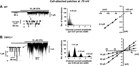
We initially investigated biophysical properties conferred by TRPC1 to native cation channel currents activated by agents known to deplete organelle Ca2+ stores in acutely isolated mesenteric artery VSMCs from WT and TRPC1−/− mice. We used CPA, which depletes Ca2+ stores by inhibiting sarcoplasmic recticulum (SR) Ca2+-ATPases, and also BAPTA-AM, a cell-permeable Ca2+ chelator, which causes passive depletion of Ca2+ stores. In WT and TRPC1−/− VSMCs, bath application of 10 μM CPA induced channel activity in cell-attached patches held at −70 mV, which had similar mean NPO values of 0.62 ± 0.02 (_n_=22) and 0.88 ± 0.04 (_n_=27), respectively. However, CPA-evoked native cation channel currents had much smaller single-channel conductance in WT compared to TRPC1−/− VSMCs.
In WT VSMCs, CPA-induced channel currents had a mean unitary amplitude of −0.21 ± 0.01 pA at −70 mV (_n_=22), which represented an unitary channel conductance of 2.2 pS that had an extrapolated reversal potential (_E_r) of about +20 mV (Fig. 1A). In contrast, TRPC1−/− VSMCs expressed CPA-induced channel currents, which had 3 distinct open levels between −1 and −4 pA at −70 mV that related to conductance values of 14, 32, and 53 pS (Fig. 1B). In addition, all three subconductance states had values of _E_r ∼ 0 mV (Fig. 1B).
Figure 1.
CPA activates distinct cation channel currents in WT and TRPC1−/− VSMCs. A) Representative trace showing that CPA activates cation channel currents in WT VSMCs (left panel), which have an unitary amplitude of −0.2 pA at −70 mV (middle panel), that correspond to a unitary conductance of 2.2 pS between −120 and −50 mV, with an extrapolated _E_r of about +20 mV (right panel). Multiple peaks of the amplitude histogram represent more than one channel in the patch (middle panel). B) CPA induced cation channel currents in TRPC1−/− VSMCs (left panel), which had 3 open amplitudes of −1.01, −2.10, and −3.63 pA (middle panel), that relate to 3 subconductance levels of 14, 32, and 53 pS, with all conductances levels having an _E_r of ∼0 mV (right panel).
In WT VSMCs, bath application of 50 μM BAPTA-AM-induced channel currents with a mean NPO of 0.58 ± 0.07 (_n_=6) in cell-attached patches held at −70 mV, which had a unitary conductance of 2.1 pS and an extrapolated _E_r of about +23 mV (Fig. 2A). An interesting finding was that BAPTA-AM did not activate channel activity in TRPC1−/− VSMCs (_n_=7, Fig. 2B).
Figure 2.
BAPTA-AM activates cation channel activity in WT but not TRPC1−/− VSMCs. A) In WT VSMCs, BAPTA-AM activated cation channel currents (left panel), which had an unitary amplitude of −0.2 pA at −70 mV (middle panel), that related to a conductance of 2.1 pS between −120 and −50 mV, and an extrapolated _E_r of about +20 mV (right panel). B) BAPTA-AM had no effect on quiescent cell-attached patches from TRPC1−/− VSMCs.
TRPC1 contributes to whole-cell currents evoked by passive depletion of intracellular Ca2+ stores in VSMCs
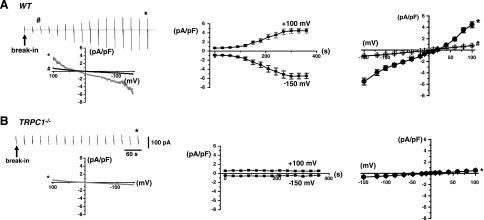
To investigate whether TRPC1 channels mediate genuine SOCs, defined as channels evoked by depletion of Ca2+ levels within intracellular Ca2+ stores and not a rise in [Ca2+]i, we compared macroscopic whole-cell currents evoked by passive Ca2+ store depletion in VSMCs from WT and TRPC1−/− mice. The archetypical and most characterized SOC is the calcium-release-activated channel (_I_crac), which is mediated by Orai1 proteins (15, 16). Therefore, we used established experimental procedures for activating _I_crac, with a patch pipette solution containing 20 mM BAPTA with no added Ca2+ to induce passive depletion of intracellular Ca2+ stores without inducing a rise in [Ca2+]i, an external solution containing 20 mm CaCl2, and voltage ramps applied from +100 to −150 mV to minimize any potential Ca2+-dependent inactivation (see Materials and Methods and refs. 15, 16). In WT VSMCs, whole-cell currents slowly developed following patch rupture into whole-cell configurations, which reached a mean peak current density of −2.02 ± 0.21 pA/pF (_n_=9) at −80 mV after 5 min (Fig. 3A). Whole-cell currents evoked by passive depletion exhibited a mean current-voltage relationship, which displayed relative S-shaped rectification and an _E_r of about +20 mV (Fig. 1A). In contrast, passive store depletion failed to activate whole-cell currents in TRPC1−/− VSMCs (_n_=9, Fig. 3B).
Figure 3.
TRPC1 contributes to whole-cell currents evoked by passive Ca2+ store depletion in VSMCs. A) Left panel: in WT VSMCs, a whole-cell current slowly developed following break-in into the whole-cell configuration using a patch pipette solution containing 20 mM BAPTA with no added Ca2+. Vertical deflections represent currents evoked by a voltage ramps from +100 to −150 mV (750 ms duration) every 30 s. Inset: individual current recordings after 1 min (#) and 5 min (*). Middle panel: time course of slowly developing current densities at +100 and −150 mV. Right panel: passive depletion activated whole-cell currents, which exhibited a mean I/V relationship with S-shaped rectification and an _E_r of about +20 mV. B) In TRPC1−/− VSMCs, inclusion of 20 mM BAPTA with no added Ca2+ in the patch pipette solution failed to activate any whole-cell currents.
CPA activates native heteromeric TRPC1/C5 channels in WT and homomeric TRPC5 channels in TRPC1−/− VSMCs
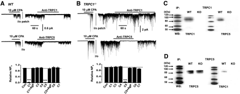
The distinct conductance values of CPA-evoked cation channels in WT and TRPC1−/− VSMCs indicate that these two channels have distinct molecular compositions. We used anti-TRPC antibodies as blocking agents of channel activity and coimmunoprecipitation (17) to provide evidence that native CPA-induced channel activity is composed of heteromeric TRPC1/C5 channel structures in WT VSMCs and homomeric TRPC5 channels in TRPC1−/− VSMCs. Channel activities were initially activated by 10 μM CPA in cell-attached patches, and then patches were excised into the inside-out configuration, in which channel activity was maintained (up to 30 min), allowing anti-TRPC antibodies (1:200) raised against putative intracellular epitopes to be applied to the cytosolic surface of patches. In WT VSMCs, anti-TRPC1 and anti-TRPC5 antibodies produced >90% inhibition of CPA-induced channel activity (Fig. 4A). Anti-TRPC1 and anti-TRPC5 antibodies preincubated with their antigenic peptides, and anti-TRPC3, anti-TRPC4, anti-TRPC6, and anti-TRPC7 antibodies had no effect on channel activity (Fig. 4A). In contrast, CPA-induced channel activity expressed in TRPC1−/− VSMCs was only inhibited by anti-TRPC5 antibodies, and this inhibition was prevented by incubating the anti-TRPC5 antibody with its antigenic peptide (Fig. 4B). In WT mesenteric artery tissue lysates, immunoprecipitation with anti-TRPC1 (Fig. 4C) or anti-TRPC5 (Fig. 4D) followed by immunoblotting with anti-TRPC1 or anti-TRPC5 revealed expression and association between TRPC1 and TRPC5 proteins. TRPC1−/− tissue lysates also expressed TRPC5 proteins (Fig. 4D), but as expected, these lysates did not express TRPC1 proteins (Fig. 4C, D).
Figure 4.
Native heteromeric TRPC1/C5 channel structures are expressed in WT VSMCs. A) In WT VSMCs, CPA-evoked channel activity was inhibited by anti-TRPC1 (top panel) and anti-TRPC5 antibodies (middle panel) by >90% (bottom panel). Preincubation of anti-TRPC1 and anti-TRPC5 antibodies with their antigenic peptide (AgP) prevents channel inhibition (bottom panel). B) In TRPC1−/− VSMCs, CPA-evoked channel activity was not affected by anti-TRPC1 (top panel) but was inhibited by anti-TRPC5 antibodies (middle panel) by >90% (bottom panel). C) Coimmunoprecipitation (IP) with anti-TRPC1 followed by Western blotting (WB) with anti-TRPC1 or anti-TRPC5 shows expression of TRPC1 and association between TRPC1 and TRPC5 in WT tissue lysates. There is no expression of TRPC1 or association between TRPC1 and TRPC5 in TRPC1−/− tissue lysates. D) IP with anti-TRPC5 followed by WB with anti-TRPC1 or anti-TRPC5 illustrates that TRPC5 is expressed in WT and TRPC1−/− tissue lysates, but association between TRPC1 and TRPC5 is only present in WT.
PKC dependence of channel activation by store-depleting agents is conferred by TRPC1 proteins
Studies have shown that native TRPC channels evoked by agents that deplete Ca2+ stores in VSMCs require an obligatory role for PKC (4–8, 10, 11). Here, we provide molecular evidence that TRPC1 confers this obligatory role for PKC in activation of native TRPC channel by CPA. In WT VSMCs, the mean NPO of CPA-induced TRPC1/C5 channel activity was significantly reduced from 0.63 ± 0.05 to 0.03 ± 0.01 by chelerythrine (3 μM), a PKC inhibitor, in cell-attached patches held at −70 mV (_n_=5, P < 0.01, Fig. 5A). Consistent with these results, bath application of the PKC activator, phorbol 12,13-dibutyrate (PDBu; 1 μM) activated 2-pS channels with a mean NPO of 0.47 ± 0.05, which was significantly inhibited to 0.03 ± 0.01 by 3 μM chelerythrine in cell-attached patches held at −70 mV (_n_=5, P<0.01, Fig. 5A). Moreover, a PKC catalytic subunit (0.01 U/ml) also activated 2-pS channels with a mean NPO of 0.21 ± 0.04, which was significantly attenuated to 0.02 ± 0.01 by 3 μM chelerythrine in inside-out patches held at −70 mV (_n_=7, P<0.05, Fig. 5A).
Figure 5.
TRPC1 confers obligatory action of PKC in CPA-evoked TRPC1/C5 channel activity in WT VSMCs. A) In WT VSMCs, CPA-evoked (left panel), PDBu-evoked (middle panel), and PKC catalytic subunit-evoked TRPC1/C5 channel activities (right panel) were inhibited by chelerythrine. B) In comparison, CPA-evoked TRPC5 channel activity in TRPC1−/− VSMCs was potentiated by chelerythrine (left panel) and attenuated by PBDu (middle panel); a PKC catalytic subunit had no effect on quiescent inside-out patches from TRPC1−/− VSMCs (right panel).
In contrast to the excitatory actions of PKC on TRPC1/C5 channels in WT VSMCs, PKC had inhibitory effects on TRPC5 channel activity in TRPC1−/− VSMCs. The mean NPO of CPA-induced TRPC5 channel activity in TRPC1−/− VSMCs was significantly increased from 0.78 ± 0.06 to 2.22 ± 0.15 by chelerythrine (3 μM) in cell-attached patches held at −70 mV (_n_=7, P<0.05, Fig. 5B). In addition, the mean NPO of CPA-evoked TRPC5 channel activity was significantly inhibited from 0.76 ± 0.17 to 0.02 ± 0.02 by PDBu (1 μM) in cell-attached patches held at −70 mV (_n_=6, P<0.01, Fig. 5B). PDBu (1 μM, _n_=6), or the PKC catalytic subunit (0.01 U/ml, _n_=7, Fig. 5B) did not activate channel activity in quiescent patches from TRPC1−/− VSMCs.
TRPC1 proteins confer channel activation by PIP2 and PIP3
Endogenous PIP2 and PIP3 are also reported to have essential roles in activation of native TRPC channels consisting of TRPC1 in VSMCs (4, 5, 10–13). Here, we show that TRPC1 confers excitatory actions of PIP2 and PIP3 on native TRPC1/C5 channels. Figure 6A shows that in WT VSMCs, bath application of the water-soluble analogues, 10 μM diC8-PIP2 and 10 μM diC8-PIP3, to the cytosolic surface of inside-out patches activated 2-pS channels, which had mean NPO values that were significantly inhibited from 0.45 ± 0.03 to 0.03 ± 0.01 (_n_=8, P<0.01) and 0.51 ± 0.05 to 0.05 ± 0.01, respectively, at −70 mV (_n_=6, P<0.01) by chelerythrine (3 μM).
Figure 6.
TRPC1 is responsible for stimulatory action of PIP2 and PIP3 on TRPC1/C5 channels in WT VSMCs. A) In WT VSMCs, diC8-PIP2 (left panel) and diC8-PIP3 (right panel) activated TRPC1/C5 channel activities, which were inhibited by chelerythrine in inside-out patches held at −70 mV. B) In TRPC1−/− VSMCs, diC8-PIP2 (left panel) and diC8-PIP3 (right panel) inhibited TRPC5 channel activity in inside-out patches (i/o) previously induced by CPA in cell-attached patches.
In contrast, in TRPC1−/− VSMCs, 10 μM diC8-PIP2 (_n_=6) and 10 μM diC8-PIP3 (_n_=6) did not activate channel activity in quiescent patches (data not shown), but these PIs did inhibit CPA-induced TRPC5 channel activity. In these experiments, channels were initially activated by 10 μM CPA in cell-attached patches, and then patches were excised into the inside-out configuration, in which channel activity was maintained (up to 30 min), allowing PIs to be applied to the cytosolic surface of patches. The mean NPO of CPA-evoked TRPC5 channel activity in TRPC1−/− VSMCs was greatly attenuated from 0.89 ± 0.11 to 0.13 ± 0.03 (_n_=8, P<0.01) by 10 μM diC8-PIP2 and from 0.91 ± 0.14 to 0.05 ± 0.02 by 10 μM diC8-PIP3 at −70 mV (_n_=7, P<0.01, Fig. 6B).
Low La3+ concentrations activate cation channel currents with similar conductances to CPA-evoked channels in native WT and TRPC1−/− VSMCs
The present study indicates that WT VSMCs express native heteromeric TRPC1/TRPC5 channels, whereas TRPC1−/− VSMCs contain homomeric TRPC5 channels. We provided further evidence for TRPC5 subunits contributing to both these channel structures by studying the effect of low external concentrations of La3+, known to activate expressed TRPC5 channels (18, 19) on VSMCs from WT and TRPC1−/− mice. Bath application of 10 μM La3+ activated channel activity in outside-out patches from WT and TRPC1−/− VSMCs, which had a unitary conductance of 2.3 pS and subconductance states of 14, 33, and 54 pS at positive membrane potentials (Fig. 7), and a slightly lower conductance value of 1.5 pS and subconductance values of 9, 21, and 38 pS at negative membrane potentials (Fig. 7). The similarity between the conductance values of La3+-evoked channel current compared to CPA-evoked channel currents in VSMCs from WT and TRPC1−/− mice provide further support for involvement of TRPC5 subunits in the composition of both these CPA-evoked channels.
Figure 7.
Low concentrations of La3+ activate cation channel currents with similar properties to TRPC1/C5 in WT and TRPC5 channels in TRPC1−/− VSMCs. A) Representative trace showing La3+-activated cation channel currents in outside-out patches held at −70 mV from WT VSMCs (left panel), which had a 1.5-pS conductance at negative membrane potentials and a 2.3 pS conductance at positive potentials, that had an _E_r of about +20 mV (right panel). B) In TRPC1−/− VSMCs, LA3+-activated cation channel currents (left panel) with 3 subconductances between 9–38 pS at negative membrane potentials, and 14–54 pS at positive potentials, which all had _E_r of ∼0 mV (right panel).
Native homomeric TRPC5 channels are activated by a rise in [Ca2+]i
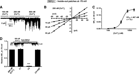
There is currently no information on the properties or activation mechanisms of native TRPC5 channels. Expressed TRPC5 channels are activated or potentiated by a rise in [Ca2+]i, which may involve calmodulin (CaM)-dependent processes (19–22), and we show that [Ca2+]i activates TRPC5 channels in TRPC1−/− VSMCs. Bath application of an intracellular solution containing 100 nM Ca2+ to the cytosolic surface of inside-out patches did not activate channel activity (Fig. 8A), but increasing the concentration of Ca2+ from 100 to 200 nM activated cation channel currents with subconductance states of 14, 30, and 55 pS (Fig. 8A, B). The concentration-dependent increase in channel activity produced by [Ca2+]i had an effective half-maximal concentration (EC50) of 467 nM (Fig. 8C). Channel activity activated by 800 nM Ca2+ was significantly inhibited by anti-TRPC5 but not by anti-TRPC5 antibodies preincubated with their antigenic peptide or anti-TRPC1 antibodies (Fig. 8D). It should be noted that increasing [Ca2+]i to 1.6 μM did not activate any channel activity in WT VSMCs.
Figure 8.
Increasing [Ca2+]i activates homomeric native TRPC5 channels in TRPC1−/− VSMCs. A) Representative trace showing that increasing [Ca2+]i activates channel activity in inside-out patches held at −70 mV. B) Channel currents induced by 200 nM [Ca2+]i had 3 subconductance states of 14, 30, and 55 pS, which all had _E_r of ∼0 mV. C) Concentration-response curve of channel activity induced by increasing [Ca2+]i, which had an effective concentration at 50% maximum activity of 467 nM. D) Channel activity activated by 800 nM [Ca2+]i was inhibited by anti-TRPC5 (inset) but not by anti-TRPC5 antibodies preincubated with its antigenic peptide (AgP) or anti-TRPC1 antibodies.
DISCUSSION
The present work investigates the role of TRPC1 proteins in native single TRPC channels activated by agents that deplete intracellular Ca2+ stores in freshly isolated, contractile VSMCs, which were acutely isolated from mesenteric arteries of WT and TRPC1−/− mice. We provide conclusive evidence that these agents activate heteromeric TRPC1/C5 channels in WT VSMCs and that TRPC1 subunits are important in determining unitary conductance and conferring channel activation by PKC, PIP2, and PIP3. In contrast, TRPC1−/− VSMCs express homomeric TRPC5 channels, which are activated by increasing [Ca2+]i, and inhibited by PKC, PIP2, and PIP3.
TRPC1 is a determinant of biophysical properties of native TRPC channels in VSMCs
VSMCs express two distinct TRPC channel groups, TRPC1 and TRPC3/C6/C7, which are both activated by vasoconstrictors, with low concentrations activating both TRPC1 and TRPC3/C6/C7 channels and high concentrations only inducing TRPC1 channels (4, 5). Concentration-dependent actions of vasoconstrictors on different TRPC channel groups are, in part, mediated by TRPC1-mediated Ca2+ influx, causing PKC-dependent phosphorylation and inhibition of TRPC3/C6/C7 channels (9). In contrast, agents that deplete intracellular Ca2+ stores only activate TRPC1 channels (4, 5); therefore, we used agents that deplete Ca2+ stores to allow selective investigation of TRPC1 channel properties. In WT VSMCs, CPA activated cation channel currents with a unitary conductance of ∼2 pS and an _E_r of about +20 mV. In contrast, TRPC1−/− VSMCs exhibited CPA-evoked cation channel currents with 3 subconductance states of 13, 32, and 53 pS and _E_r of 0 mV. Inclusion of 20 mM BAPTA with no added Ca2+ in the patch pipette solution to induce passive depletion of intracellular Ca2+ stores activated whole-cell currents, which displayed S-shaped I/V relationships that had _E_r of about +20 mV, in WT VSMCs but failed to induce any currents in TRPC1−/− VSMCs. These findings demonstrate that TRPC1 is pivotal for conferring unitary conductance to native TRPC channels. The positive _E_r values of single-channel and whole-cell currents in WT compared to those in TRPC1−/− VSMCs also suggest that TRPC1 confers significant Ca2+ permeability to native TRPC channels. Contribution of TRPC1 to channel composition will, therefore, have important consequences for the physiological role of native TRPC channels in VSMCs.
Openings of nonselective cation channels are considered to initiate excitation of VSMCs and vasoconstriction, although recently TRPC1 channels have also been adjudged to contribute to vessel relaxation. Activation of TRPC1 channels are proposed to induce spatially restricted increases in [Ca2+]I, which stimulate Ca2+-activated K+ channels (BKCa) that lead to relaxation of rat aorta (23). Furthermore, TRPC1-mediated relaxations may also be caused by negative interactions between TRPC1 and TRPC3/C6/C7 channels (9). However, TRPC1 has been shown to contribute to depolarizations associated with vasoconstriction and vasomotion (24). The present data support the ideas that TRPC1 contributes to TRPC channels in VSMCs, which are involved in producing direct excitatory actions through increasing membrane permeability to Na+ and Ca2+, and also indirect inhibitory effects through simultaneous inhibition of TRPC3/C6/C7 channels, and activation of BKCa.
Native heteromeric TRPC1/C5 channels are expressed in WT VSMCs
The vastly different conductance values of CPA-evoked channel currents in WT and TRPC1−/− VSMCs provides evidence that these channels have distinct molecular identities from each other. We investigated the molecular identity of these channels by using antibodies raised against TRPC subtypes as blocking agents, which has previously been used as an effective method of investigating native channel composition in other vascular preparations (17). In WT VSMCs, CPA-evoked channel activity was inhibited by anti-TRPC1 and anti-TRPC5 antibodies, whereas CPA-induced channel activity in TRPC1−/− VSMCs was only prevented by anti-TRPC5 antibodies. Antibodies for TRPC3, TRPC4, TRPC6, and TRPC7 had no effect on channel activity in WT or TRPC1−/− VSMCs, which indicates that these TRPC subunits are not involved in the composition of these CPA-evoked channels. Coimmunoprecipitation experiments reveal that TRPC1 and TRPC5 proteins are both expressed and coassociate with each other in WT mesenteric artery tissue lysates, whereas only TRPC5 proteins are expressed in TRPC1−/− lysates. The inhibition of CPA-evoked channel activity and identification of a single protein band in VSMCs and tissue lysates from WT but not TRPC1−/− mice indicates that the anti-TRPC1 antibody used in the present study had high selectivity.
Previous work on native and expressed TRPC channels support our evidence that native CPA-evoked cation channels in WT VSMCs are mediated by heteromeric TRPC1/C5 channels with a conductance of ∼2 pS, and that TRPC1−/− VSMCs express homomeric TRPC5 channels with subconductance states between 15 and 55 pS. The 2-pS TRPC1/C5 channels present in WT VSMCs have similar biophysical properties to previously described channel activity evoked by agents that deplete Ca2+ stores in rabbit vascular preparations (17, 25, 26). Studies concluded that heteromeric TRPC1/C5 structures also mediated these native TRPC channels in rabbit mesenteric artery VSMCs (7, 17). Moreover, it is proposed that TRPC1 and TRPC5 provide a molecular template with additional TRPC6 or TRPC7 subunits for composition of native channels in rabbit coronary artery and portal vein VSMCs (7, 17), respectively. Coexpression of TRPC1 and TRPC5 in cell lines produces functional heteromeric channels with conductances of 5 pS, whereas expressed homomeric TRPC5 channels have conductances between 38 and 63 pS (19, 27, 28), which are similar to the conductances of the channels recorded in the present study.
TRPC1 confers an obligatory role for PKC in activation of CPA-evoked TRPC1/C5 channels, and is also responsible for activation of these channels by PIP2 and PIP3
The present data provides evidence that PKC is obligatory for activation of native TRPC1/C5 channels by agents that deplete Ca2+ stores in WT VSMCs. In addition, PIP2 and PIP3 also activate native TRPC1/C5 channels. Furthermore, our data indicate that TRPC1 confers these excitatory actions of PKC, and PIP2 and PIP3 on TRPC1/C5 channels. CPA- and BAPTA-AM-evoked TRPC1/C5 channel activity were inhibited by the PKC inhibitor chelerythrine, and the PKC activator, PDBu, plus a PKC catalytic subunit activated 2 pS channels, which were inhibited by chelerythrine. PIP2 and PIP3 applied to the intracellular surface of quiescent isolated patches evoked channel activity with conductances of ∼2 pS, which were prevented by chelerythrine. These data indicate that PKC activity is also important for activation of TRPC1/C5 channels by PIP2 and PIP3. As expected for actions conferred by TRPC1, the excitatory effects of PKC, PIP2, and PIP3 on TRPC1/C5 channel activities were absent in TRPC1−/− VSMCs.
These data are consistent with findings that show that interactions between PKC and endogenous PIP2/PIP2 govern activation of native channels containing TRPC1 subunits in VSMCs (4, 5, 10, 11). The hypothesis is that CPA and PDBu activate channel activity by inducing PKC-dependent phosphorylation of TRPC1 proteins, which increases the affinity of endogenous PIP2 and PIP3 tethered to TRPC1 proteins to act as activating ligands.
Consequently, channel activation by PIP2 and PIP3 is also prevented by PKC inhibition, as PKC is obligatory for binding of PIs to TRPC1 proteins. PKC has also been proposed to be important for store-operated TRPC1 activation in cultured human endothelial cells (29) and in cultured human glomerular mesangial cells, which are thought to be composed of TRPC1 and TRPC4 subunits (30–32). To date, the hypothesis that PIs are essential components in activating channel-containing TRPC1 subunits is a novel discovery.
Homomeric TRPC5 channels are expressed in TRPC1−/− VSMCs
CPA stimulates channel activity in TRPC1−/− VSMCs, which has three conductance states of 14 pS, 32 pS, and 53 pS. These conductance levels are likely to represent subconductance states as the conductance values are not multiples of the lowest conductance level, the three open levels are observed in all patches and at low open probabilities, all three conductance levels had similar _E_r, and multiple subconductance states seem to be a characteristic of native TRPC channels in VSMCs, which do not contain TRPC1 (8, 33, 34).
Several lines of evidence indicate that CPA activates homomeric TRPC5 channels in TRPC1−/− VSMCs. CPA-evoked native channels in TRPC1−/− VSMCs and expressed TRPC5 proteins have similar conductances of 15–55 and 38–55 pS, respectively (19, 27). CPA-evoked channel activity in TRPC1−/− VSMCs was only inhibited by anti-TRPC5 antibodies. Low concentrations of external La3+, which selectively activates expressed TRPC5 channels (18, 19), induced native cation channel activities that had similar conductances to CPA-evoked channels in WT and TRPC1−/− VSMCs. The slightly lower conductance values of La3+-activated channel currents compared to CPA-evoked channel currents at negative membrane potentials in both WT and TRPC1−/− VSMCs probably reflects simultaneous channel block and channel activation by La3+, as observed with expressed TRPC5 channels in cell lines (18).
We believe this is the first description of a native homomeric TRPC5 channel in any cell type, and accordingly, activation mechanisms of native TRPC5 channels are not known. CPA inhibits SR Ca2+-ATPases and prevent reuptake of Ca2+ into the SR, which deplete Ca2+ stores and induces an associated rise in [Ca2+]i, whereas BAPTA-AM chelates Ca2+ leaking from Ca2+ stores to passively deplete Ca2+ stores with a reduction in [Ca2+]i. The findings that CPA, but not BAPTA-AM, activates TRPC5 channel activity, and also that passive depletion did not activate a whole-cell current in TRPC1−/− VSMCs indicates that a rise in [Ca2+]i is an important trigger for native TRPC5 channel activation. Increasing [Ca2+]i at the cytosolic surface of quiescent isolated inside-out patches also activated TRPC5 channel activity with an EC50 of ∼450 nM. This present work agrees with studies that have shown activation or potentiation of expressed TRPC5 channel activity by increasing [Ca2+]i (20–22). Our data also show that TRPC5 channel activity activated by CPA in TRPC1−/− VSMCs is inhibited by PKC, PIP2, and PIP3. Previous data have shown that PKC inhibits expressed TRPC5 channel activity, probably via a mechanism involving phosphorylation of serine-712 (35, 36). In addition, PIP2 has both excitatory and inhibitory actions on expressed TRPC5 channels (37, 38). The present work is the first study to show that PIP3 also regulates TRPC5 channel activation.
The opposing excitatory actions of PKC and PIP2 and PIP3 on TRPC1/C5 channels (see above) vs. their inhibitory effects on TRPC5 channels is an intriguing observation, as it suggests that properties conferred by TRPC1 dominants over characteristics of TRPC5 subunits in a native heteromeric TRPC1/C5 channel structure. Why this is the case is not known; it may be a consequence of TRPC1:TRPC5 subunit stoichiometry within the putative tetrameric structure. However, these findings strongly impress the importance of studying properties of native TRPC channels in their physiological environment, and not just investigating properties of individual TRPC subunits using expression systems.
Approaches used to study native single TRPC channels
Investigations into the properties of TRPC1 channels in VSMCs have generally manipulated TRPC1 protein expression using antisense (AS) or small interference RNA (siRNA) technologies, and then measured macroscopic signals, such as whole-cell conductances and Ca2+ signals (17). AS or siRNA studies have provided important information, but as they require 48–72 h to down-regulate channel proteins, these procedures are more appropriate for studying properties of synthetic VSMCs than contractile VSMCs. In addition, AS and siRNA may be susceptible to artifacts, and nonspecific effects (39), and macroscopic signals may contain multiple molecular mechanisms. In an attempt to circumvent some of these issues to enable us to measure single molecular mechanisms in contractile VSMCs, we recorded single native TRPC channel activity in acutely isolated VSMCs from WT and TRPC1−/− mice, and used anti-TRPC antibodies as blocking agents (17). It must also be acknowledged that there may also be issues with this experimental approach, such as the selectivity of antibodies and pharmacological agents used and that transgenic mice may contain compensatory changes in expression levels of other TRPC subtype genes (40). However, compensatory changes in TRPC subtype genes were not previously observed in cerebral artery and thoracic aorta from TRPC1−/− mice (12). We believe that an important outcome of the present work is the successful use of complementary molecular and pharmacological approaches to investigate properties conferred by a TRPC subunit which contributes to a native single TRPC channel structure in contractile VSMCs.
Comparison of the present work with an earlier study (12) reveals interesting differences in macroscopic signals purported to involve TRPC conductances. Our work clearly demonstrates that passive depletion, a defining activation procedure for genuine SOCs, activates a whole-cell cation conductance in WT but not TRPC1−/− VSMCs, which indicates that TRPC1 contributes to SOC in mesenteric artery VSMCs. However, Dietrich et al. (12) showed that current densities and current-voltage relationships of CPA-evoked whole-cell cation conductances in cerebral artery VSMCs were similar in WT and TRPC1−/− mice, even though functional TRPC1 and TRPC5 proteins are expressed in cerebral artery VSMCs (41–44). Reasons for these differences in the involvement of TRPC1 are not known, but they may involve differences between vascular beds and experimental protocols. Also, other TRPC subtypes or other TRP/Orai channel proteins may underlie native CPA-induced whole-cell currents in cerebral artery VSMCs. Functional Orai1 proteins are expressed in synthetic VSMCs but are not reported to be present in freshly isolated VSMCs with contractile phenotypes, as used in the present study (16, 45–47). It should be also noted that passive depletion did not activate whole-cell currents with properties resembling those of Icrac or Orai1 currents (15, 16) in contractile TRPC1−/− VSMCs.
In summary, the present study provides molecular evidence that TRPC1 forms native heteromeric TRPC1/C5 channels in VSMCs with a contractile phenotype, which are activated by agents that deplete Ca2+ stores. Moreover, TRPC1 confers activation by PKC, PIP2, and PIP3 on these native channels.
Acknowledgments
This work was supported by the British Heart Foundation.
REFERENCES
- 1.Abramowitz J., Birnbaumer L. (2009) Physiology and pathophysiology of canonical transient receptor potential channels. FASEB J. 23, 297–328 [DOI] [PMC free article] [PubMed] [Google Scholar]
- 2.Birnbaumer L. (2009) The TRPC class of ion channels: a critical review of their roles in slow, sustained increases in intracellular Ca2+ concentrations. Annu. Rev. Pharmacol. Toxicol. 49, 395–426 [DOI] [PubMed] [Google Scholar]
- 3.Dietrich A., Kalwa H., Gudermann T. (2010) TRPC channels in vascular function. Thromb. Haemost. 103, 262–270 [DOI] [PubMed] [Google Scholar]
- 4.Albert A. P. (2011) Gating mechanisms of canonical transient receptor potential channel proteins: role of phosphoinositols and diacylglycerol. Adv. Exp. Med. Biol. 704, 391–411 [DOI] [PubMed] [Google Scholar]
- 5.Large W. A., Saleh S. N., Albert A. P. (2009) Role of phosphoinositol 4,5-bisphosphate and diacylglycerol in regulating native TRPC channel proteins in vascular smooth muscle. Cell Calcium 45, 574–582 [DOI] [PubMed] [Google Scholar]
- 6.Albert A. P., Saleh S. N., Peppiatt-Wildman C. M., Large W. A. (2007) Multiple activation mechanisms of store-operated TRPC channels in smooth muscle cells. J. Physiol. 583, 25–36 [DOI] [PMC free article] [PubMed] [Google Scholar]
- 7.Saleh S. N., Albert A. P., Peppiatt-Wildman C. M., Large W. A. (2008) Diverse properties of store-operated TRPC channels activated by protein kinase C in vascular myocytes. J. Physiol. 586, 2463–2476 [DOI] [PMC free article] [PubMed] [Google Scholar]
- 8.Saleh S. N., Albert A. P., Peppiatt C. M., Large W. A. (2006) Angiotensin II activates two cation conductances with distinct TRPC1 and TRPC6 channel properties in rabbit mesenteric artery myocytes. J. Physiol. 577, 479–495 [DOI] [PMC free article] [PubMed] [Google Scholar]
- 9.Shi J., Ju M., Saleh S. N., Albert A. P., Large W. A. (2010) TRPC6 channels stimulated by angiotensin II are inhibited by TRPC1/C5 channel activity through a Ca2+- and PKC-dependent mechanism in native vascular myocytes. J. Physiol. 588, 3671–3682 [DOI] [PMC free article] [PubMed] [Google Scholar]
- 10.Saleh S. N., Albert A. P., Large W. A. (2009) Obligatory role for phosphatidylinositol 4,5-bisphosphate in activation of native TRPC1 store-operated channels in vascular myocytes. J. Physiol. 587, 531–540 [DOI] [PMC free article] [PubMed] [Google Scholar]
- 11.Saleh S. N., Albert A. P., Large W. A. (2009) Activation of native TRPC1/C5/C6 channels by endothelin-1 is mediated by both PIP3 and PIP2 in rabbit coronary artery myocytes. J. Physiol. 587, 5361–5375 [DOI] [PMC free article] [PubMed] [Google Scholar]
- 12.Dietrich A., Kalwa H., Storch U., Mederos Y., Schnitzler M., Salanova B., Pinkenburg O., Dubrovska. G., Essin K., Gollasch M., Birnbaumer L., Gudermann T. (2007) Pressure-induced and store-operated cation influx in vascular smooth muscle cells is independent of TRPC1. Pflügers Arch. 455, 465–477 [DOI] [PubMed] [Google Scholar]
- 13.Ju M., Shi J., Saleh S. N., Albert A. P., Large W. A. (2010) Ins(1,4,5)P3 interacts with PIP2 to regulate activation of TRPC6/C7 channels by diacylglycerol in native vascular myocytes. J. Physiol. 588, 1419–1433 [DOI] [PMC free article] [PubMed] [Google Scholar]
- 14.Goel M., Sinkins W. G., Schilling W. P. (2002) Selective association of TRPC channel subunits in rat brain synaptosomes. J. Biol. Chem. 277, 48303–48310 [DOI] [PubMed] [Google Scholar]
- 15.Bird G. S., DeHaven W. I., Smyth J. T., Putney J.W., Jr. (2008) Methods for studying store-operated calcium entry. Methods 46, 204–212 [DOI] [PMC free article] [PubMed] [Google Scholar]
- 16.Zhang W., Halligan K. E., Zhang X., Bisaillon J. M., Gonzalez-Cobos J. C., Motiani R. K., Hu G., Vincent P. A., Zhou J., Barroso M., Singer H. A., Matrougui K., Trebak M. (2011) Orai1-mediated ICRAC is essential for neointima formation after vascular injury. Circ. Res. 109, 534–542 [DOI] [PMC free article] [PubMed] [Google Scholar]
- 17.Albert A. P., Saleh S. N., Large W. A. (2009) Identification of canonical transient receptor potential (TRPC) channel proteins in native vascular smooth muscle cells. Curr. Med. Chem. 16, 1158–1165 [DOI] [PubMed] [Google Scholar]
- 18.Jung S., Mühle A., Schaefer M., Strotmann R., Schultz G., Plant T. D. (2003) Lanthanides potentiate TRPC5 currents by an action at extracellular sites close to the pore mouth. J. Biol. Chem. 278, 3562–3571 [DOI] [PubMed] [Google Scholar]
- 19.Plant T. D., Schaefer M. (2005) Receptor-operated cation channels formed by TRPC4 and TRPC5. Naunyn Schmiedebergs Arch. Pharmacol. 371, 266–276 [DOI] [PubMed] [Google Scholar]
- 20.Ordaz B., Tang J., Xiao R., Salgado A., Sampieri A., Zhu M. X., Vaca L. (2005) Calmodulin and calcium interplay in the modulation of TRPC5 channel activity. Identification of a novel C-terminal domain for calcium/calmodulin-mediated facilitation. J. Biol. Chem. 280, 30788–30796 [DOI] [PubMed] [Google Scholar]
- 21.Blair N. T., Kaczmarek J. S., Clapham D. E. (2009) Intracellular calcium strongly potentiates agonist-activated TRPC5 channels. J. Gen. Physiol. 133, 525–546 [DOI] [PMC free article] [PubMed] [Google Scholar]
- 22.Gross S. A., Guzmán G. A., Wissenbach U., Philipp S. E., Zhu M. X., Bruns D., Cavalié A. (2009) TRPC5 is a Ca2+-activated channel functionally coupled to Ca2+-selective ion channels. J. Biol. Chem. 284, 34423–34432 [DOI] [PMC free article] [PubMed] [Google Scholar]
- 23.Kwan H. Y., Shen B., Ma X., Kwok Y. C., Huang Y., Man Y. B., Yu S., Yao X. (2009) TRPC1 associates with BK(Ca) channel to form a signal complex in vascular smooth muscle cells. Circ. Res. 104, 670–678 [DOI] [PubMed] [Google Scholar]
- 24.Wölfle S. E., Navarro-Gonzalez M. F., Grayson T. H., Stricker C., Hill C. E. (2010) Involvement of non-selective cation channels in the depolarisation initiating vasomotion. Clin. Exp. Pharmacol. Physiol. 37, 536–543 [DOI] [PubMed] [Google Scholar]
- 25.Trepakova E. S., Gericke M., Hirakawa Y., Weisbrod R. M., Cohen R. A., Bolotina V. M. (2001) Properties of a native cation channel activated by Ca2+ store depletion in vascular smooth muscle cells. J. Biol. Chem. 276, 7782–7790 [DOI] [PubMed] [Google Scholar]
- 26.Albert A. P., Large W. A. (2002) A Ca2+-permeable non-selective cation channel activated by depletion of internal Ca2+ stores in single rabbit portal vein myocytes. J. Physiol. 538, 717–728 [DOI] [PMC free article] [PubMed] [Google Scholar]
- 27.Strubing C., Krapivinsky G., Krapivinsky L., Clapham D. E. (2001) TRPC1 and TRPC5 form a novel cation channel in mammalian brain. Neuron 29, 645–655 [DOI] [PubMed] [Google Scholar]
- 28.Alfonso S., Benito O., Alicia S., Angélica Z., Patricia G., Diana K., Vaca L. (2008) Regulation of the cellular localization and function of human transient receptor potential channel 1 by other members of the TRPC family. Cell Calcium 43, 375–387 [DOI] [PubMed] [Google Scholar]
- 29.Ahmmed G. U., Mehta D., Vogel S., Holinstat M., Paria B. C., Tiruppathu C., Malik A. B. (2004) Protein kinase C alpha phosphorylates the TRPC1 channel and regulates store-operated Ca2+ entry in endothelial cells. J. Biol. Chem. 279, 20941–20949 [DOI] [PubMed] [Google Scholar]
- 30.Ma R., Pluznick J., Kudlacek P. E., Sansom S. C. (2001) Protein kinase C activates store-operated Ca2+ channels in human glomerular mesangial cells. J. Biol. Chem. 276, 25759–25765 [DOI] [PubMed] [Google Scholar]
- 31.Ma R., Kudlacek P. E., Sansom S. C. (2002) Protein kinase Calpha participates in activation of store-operated Ca2+ channels in human glomerular mesangial cells. Am. J. Physiol. Cell Physiol. 283, C1390–C1398 [DOI] [PubMed] [Google Scholar]
- 32.Sours-Brothers S., Ding M., Graham S., Ma R. (2009) Interaction between TRPC1/TRPC4 assembly and STIM1 contributes to store-operated Ca2+ entry in mesangial cells. Exp. Biol. Med. 234, 673–682 [DOI] [PubMed] [Google Scholar]
- 33.Albert A. P., Pucovsky V., Prestwich S. A., Large W. A. (2006) TRPC3 properties of a native constitutively active Ca2+-permeable cation channel in rabbit ear artery myocytes. J. Physiol. 571, 361–369 [DOI] [PMC free article] [PubMed] [Google Scholar]
- 34.Peppiatt-Wildman C. M., Albert A. P., Saleh S. N., Large W. A. (2007) Endothelin-1 activates a Ca2+-permeable cation channel with TRPC3 and TRPC7 properties in rabbit coronary artery myocytes. J. Physiol. 580, 755–764 [DOI] [PMC free article] [PubMed] [Google Scholar]
- 35.Trebak M., Hempel M., Wedel B. J., Smyth J. T., Bird G. J., Putney J.W., Jr. (2005) Negative regulation of TRPC3 channels by protein kinase C-mediated phosphorylation of serine 712. Mol. Pharmacol. 67, 558–563 [DOI] [PubMed] [Google Scholar]
- 36.Zhu M. H., Chae M., Kim H. J., Lee Y. M., Kim M. J., Jin N. G., Yang D. K., So I., Kim K. W. (2005) Densensitization of canonical transient receptor potential channel 5 by protein kinase C. Am. J. Physiol. Cell Physiol. 289, C591–C600 [DOI] [PubMed] [Google Scholar]
- 37.Kim B. J., Kim M. T., Jeon J. H., Kim S. J., So I. (2008) Involvement of phosphatidylinositol 4,5-bisphosphate in the desensitization of canonical transient receptor potential 5. Biol. Pharm. Bull. 31, 1733–1738 [DOI] [PubMed] [Google Scholar]
- 38.Trebak M., Lemonnier L., Dehaven W. I., Wedel B. J., Bird G. S., Putney J.W., Jr. (2008) Complex functions of phosphatidylinositol-4, 5-bisphosphate on regulation of TRPC5 cation channels. Pflügers Arch. 457, 757–769 [DOI] [PMC free article] [PubMed] [Google Scholar]
- 39.Desai B. N., Clapham D. E. (2005) TRP channels and mice deficient in TRP channels. Pflügers Arch. 451, 11–18 [DOI] [PubMed] [Google Scholar]
- 40.Dietrich A., Mederos Y., Schnitzler M., Gollasch M., Gross V., Storch U., Dubrovska G., Obst M., Yildirim E., Salanova B., Kalwa H., Essin K., Pinkenburg O., Luft F. C., Gudermann T., Birnbaumer L. (2005) Increased vascular smooth muscle contractility in TRPC6−/− mice. Mol. Cell. Biol. 25, 6980–6989 [DOI] [PMC free article] [PubMed] [Google Scholar]
- 41.Xu S. Z., Beech D. J. (2000) TrpC1 is a membrane-spanning subunit of store-operated Ca2+ channels in native vascular smooth muscle cells. Circ. Res. 88, 84–87 [DOI] [PubMed] [Google Scholar]
- 42.Bergdahl A., Gomez M. F., Wihlborg A. K., Erlinge D., Eyjolfson A., Xu S. Z., Beech D. J., Dreja K., Hellstrand P. (2005) Plasticity of TRPC expression in arterial smooth muscle: correlation with store-operated Ca2+ entry. Am. J. Physiol. Cell Physiol. 288, C872–C880 [DOI] [PubMed] [Google Scholar]
- 43.Xu S. Z., Boulay G., Flemming R., Beech D. J. (2006) E3-targeted anti-TRPC5 antibody inhibits store-operated calcium entry in freshly isolated pial arterioles. Am. J. Physiol. Heart Circ. Physiol. 291, H2653–H2659 [DOI] [PubMed] [Google Scholar]
- 44.Xie A., Aihara Y., Bouryi V. A., Nikitina E., Jahromi B. S., Zhang Z. D., Takahashi M., Macdonald R. L. (2007) Novel mechanism of endothelin-1-induced vasospasm after subarachnoid haemorrhage. J. Cereb. Blood Flow Metab. 27, 1692–1701 [DOI] [PubMed] [Google Scholar]
- 45.Berra-Romani R., Mazzocco-Spezzia A., Pulina M. V., Golovina V. A. (2008) Ca2+ handling is altered when arterial myocytes progress from a contractile to a proliferative phenotype in culture. Am. J. Physiol. 295, C779–C790 [DOI] [PMC free article] [PubMed] [Google Scholar]
- 46.Potier M., Gonzalez J. C., Motiani R. K., Abdullaev I. F., Bisaillion J. M., Singer H. A., Trebak M. (2009) Evidence for STIM1- and Orai1-dependent store-operated calcium influx through ICRAC in vascular smooth muscle cells: role in proliferation and migration. FASEB J. 23, 2425–2437 [DOI] [PMC free article] [PubMed] [Google Scholar]
- 47.Bisaillion J. M., Motiani R. K., Gonzalez-Cobos J. C., Potier M., Alzawahra W. F., Barroso M., Singer H. A., Jourd'heuil D., Trebak M. (2009) Essential role for STIM1/Orai1-mediated calcium influx in PDGF-induced smooth muscle migration. Am. J. Physiol. Cell Physiol. 298, C993–C1005 [DOI] [PMC free article] [PubMed] [Google Scholar]


